Article

Vasoprotection by Dietary Nitrate in Rats with Vitamin D3-Induced Vascular Calcification

Department of Pathological and Molecular Pharmacology, Faculty of Pharmacy, Osaka Medical and Pharmaceutical University, Takatsuki 569-1094, Japan
*
Author to whom correspondence should be addressed.
J. Vasc. Dis. 2026, 5(1), 3; https://doi.org/10.3390/jvd5010003
Submission received: 15 December 2025 / Revised: 9 January 2026 / Accepted: 16 January 2026 / Published: 20 January 2026
(This article belongs to the Section Cardiovascular Diseases)

Abstract

Background/Objectives: Vascular calcification is a major contributor to cardiovascular mortality and disability. Here, we investigated whether dietary nitrate, an exogenous source of nitric oxide (NO), could inhibit vascular calcification in a rat model induced by excess vitamin D3. Methods: The rats were injected subcutaneously with phosphate-buffered saline or 200,000 IU/kg cholecalciferol and the abdominal aorta was isolated 7 and 14 d after injection. Results: Von Kossa staining revealed mild vascular calcification 7 d after injection, with the positive area expanding by 14 d. Vasorelaxation induced by the NO donor sodium nitroprusside was normal 7 d after injection but weakened 14 d after injection. In a separate experiment, sodium nitrate (3 or 10 mM in drinking water) was administered for the last 7 and 13 d, prior to sacrifice, 14 d after cholecalciferol injection. Von Kossa staining-positive areas and calcium content in the abdominal aortas did not decrease with short-term administration of sodium nitrate but decreased with long-term administration; no difference in effect based on dosage was observed in either short-term or long-term administration. Low-dose sodium nitrate tended to increase plasma nitrite and nitrate levels, which are indicators of NO bioavailability, similar to both short- and long-term administration, which increased significantly at higher doses. Conclusions: These findings suggest that NO homeostasis in blood vessels deteriorates with the progression of vascular calcification, and that dietary nitrate may be a useful therapeutic approach.

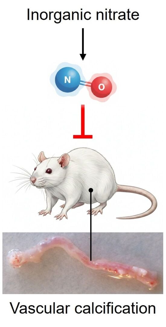
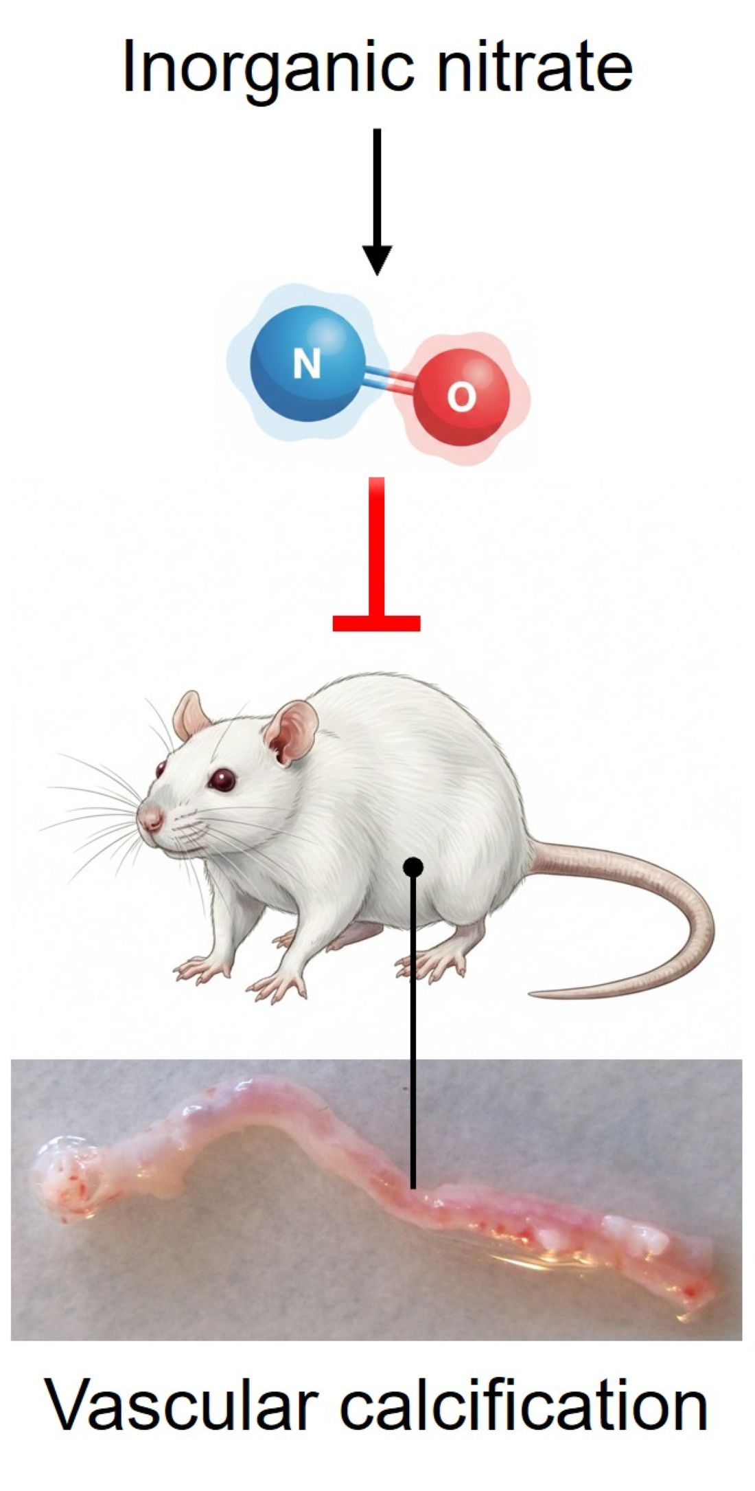
Graphical Abstract

1. Introduction

Vascular calcification, a common complication of aging, chronic kidney disease, diabetes, and hypertension, is a pathological process in which calcium accumulates in the walls of blood vessels, mainly in the large elastic and muscular arteries [1]. When calcification occurs in blood vessels, they lose flexibility and elasticity, leading to a significant decrease in compliance [2]. Vascular calcification has been reported to be present in 24% of asymptomatic individuals [3] and 46% of individuals with diverse backgrounds [4], suggesting that the total number of individuals with vascular calcification is likely quite high. Of clinical importance is that vascular calcification is independently associated with an increased risk for cardiovascular events and mortality [5,6]. However, there is an unmet need for an effective therapy [1].
Nitric oxide (NO), a trigger molecule for the activation of soluble guanylate cyclase (sGC) and the subsequent formation of cGMP [7], is closely related to the development and progression of vascular calcification. For example, the induction of vascular calcification causes diminished release of NO [8,9], and exogenously applied NO prevents vascular calcification [10,11]. In recent years, dietary inorganic nitrates have attracted attention as potential sources of NO. Oral bacteria can reduce nitrate to nitrite, which enters the circulation where it is converted to NO using mammalian nitrite reductases [12]. As the expression and activity of these reductases increase under pathological conditions, inorganic nitrate may hold significant potential as a therapeutic agent for diseases [13]. However, whether inorganic nitrates inhibit vascular calcification remains unclear. Here, we examined this issue using a rat model of vascular calcification induced by vitamin D3 overdose.

2. Materials and Methods

2.1. Animals

All animal experiments were conducted in accordance with the Guide for the Care and Use of Laboratory Animals (8th Edition, 2011). Eight-week-old male Sprague Dawley rats were purchased from Jackson Laboratory Japan (Kanagawa, Japan) and paired-housed (two rats per cage) under the following conditions: a 12 h/12 h light/dark photocycle, 45% to 65% humidity, and free access to food and water. This animal study was approved by the Animal Research Committee of Kanazawa Medical University (Permit number 2020-29, approved 1 April 2020); and M.T. was previously affiliated with Kanazawa Medical University.

2.2. Experimental Design

The rats were administered a single subcutaneous injection of phosphate-buffered saline (PBS) or 200,000 IU/kg cholecalciferol. In the first experiment, 14 d after PBS injection (referred to as “Cont”) or 7 and 14 d after cholecalciferol injection (referred to as “VD7” and “VD14”, respectively), each rat was anesthetized with an intraperitoneal injection of sodium pentobarbital (50 mg/kg) and subcutaneously injected with meloxicam (1.0 mg/kg). Rats were euthanized by bleeding after an intravenous injection of heparin (500 U/kg). Abdominal aortas were isolated for histological analysis and vascular reactivity measurements. The present study was performed on the abdominal aorta because it is relatively prone to vascular calcification [14].
In the second experiment, rats were injected with PBS or cholecalciferol as described above, and 7 d later, cholecalciferol-injected rats were assigned to receive either drinking water (referred to as “VD”), low-dose sodium nitrate (3 mM, referred to as “S-LN”), or high-dose sodium nitrate (10 mM, referred to as “S-HN”); the concentrations were determined based on previous studies [15,16]. PBS-injected rats were given drinking water (referred to as “Cont”). The solutions were changed every 1–2 d, and supplementation was maintained for 7 d; the weight of the water bottle was measured during replacement to calculate the water intake every 1–2 d. Rat body weight was measured at the start of the intervention and at the endpoint, and the average of these measurements was considered the body weight for that period. Assuming that each rat in the cage received an equal amount of water, the average nitrate intake per day was calculated by dividing the average water intake during that period by the body weight. Blood samples were collected from the inferior vena cava under deep anesthesia 7 d after the initiation of supplementation, as described above, and abdominal aortas were collected for histological and biochemical analyses.
In the third experiment, the duration of supplementation was 13 d, from 1 to 14 d after the PBS or cholecalciferol injection. The grouping was the same as the second experiment; the low-dose sodium nitrate and high-dose sodium nitrate groups were referred to as “L-LN” and “L-HN”, respectively. Daily nitrate intake was calculated every 6–7 d as described above, and the values obtained were averaged over the two periods and per group. Blood and tissue samples were collected 13 d after the initiation of supplementation.

2.3. Von Kossa Staining

Abdominal aortas were fixed in 10% phosphate-buffered formalin and embedded in paraffin. The samples were sectioned into 5 µm thickness and stained with von Kossa, according to standard procedures. Briefly, the slides were deparaffinized, hydrated with distilled water, and immersed in 5% silver nitrate solution for 1 h under intense sunlight. The stained sections were then washed three times with deionized water, and a 5% thiosulfate solution was added for 10 min to remove un-reacted silver. Slides were photographed (cellSens, Olympus Co., Ltd., Tokyo, Japan) and black-stained areas were quantified using ImageJ version 2.14.0/1.54f (NIH) and expressed as percentages of the total cross-sectional area of the vessel.

2.4. Organ Bath Experiments

Aortic rings with endothelium 3–5 mm in length were prepared and used in organ bath experiments as previously described [17]. Briefly, rings, with resting tension adjusted to 1.5 g, were partially contracted with endothelin-1 (1 nmol/L). Once contraction plateaued, concentration–response curves for sodium nitroprusside (SNP; NO donor), BAY 60-2770 (sGC activator), and 8-Br-cGMP (cGMP mimetic) were obtained following the stepwise and cumulative addition of the drugs. At the end of each experiment, papaverine (100 µmol/L) was added to induce maximal relaxation, considered 100% relaxation induced by the agonists.

2.5. Calcium Content Analysis

Calcium content in the abdominal aorta was determined using the chlorophosphonazo-III chromogenic method (metalloassay calcium assay LS kit; Metallogenics Co., Ltd., Chiba, Japan). Tissues were heated to constant dry weight for 18 h at 55 °C. Dry tissue samples were weighed and decalcified with 0.6 M HCl (10 mg/mL) and left overnight at room temperature. The samples were then centrifuged at 10,000 rpm for 15 min, and the supernatant was used for calcium content measurements. Calcium content was normalized to the dry tissue weight and expressed in milligrams of calcium per gram of tissue.

2.6. Nitrite and Nitrate Measurement

Plasma was prepared by centrifugation of heparinized blood samples at 3000 rpm for 10 min at 4 °C. Plasma nitrite and nitrate levels were measured using an ENO-20 NOx Analyzer HPLC system (Eicom, Kyoto, Japan), as previously described [17]. The levels were calculated by comparison with values obtained from a standard solution (NO-STD, Eicom).

2.7. Drugs

The following drugs were used: cholecalciferol and sodium pentobarbital (Kyoritsu Seiyaku Co., Tokyo, Japan); meloxicam (Virbac Japan Co., Ltd., Osaka, Japan); heparin (Mitsubishi Tanabe Pharma Co., Osaka, Japan); sodium nitrate and SNP (Nacalai Tesque, Kyoto, Japan); endothelin-1 (Peptide Institute Inc., Osaka, Japan); BAY 60-2770 (provided by Dr. Peter Sandner, Bayer AG, Pharmaceuticals R&D, Pharma Research Center, Wuppertal, Germany); 8-Br-cGMP (Sigma-Aldrich Co. LLC, St. Louis, MO, USA); papaverine hydrochloride (Dainippon-Sumitomo Pharma Co., Osaka, Japan). Dimethyl sulfoxide was used as the solvent to prepare stock solutions of BAY 60-2770. This solvent did not significantly affect the vascular response at the concentrations used in the present study. Distilled water was used to dissolve all other drugs and prepare serial dilutions, as required, from the stocks on the day of the experiment.

2.8. Statistical Analysis

All values are expressed as the mean ± standard error of the mean (SEM). Statistical analyses were performed using the Graph Pad Prism 7.0 software (GraphPad Software Inc., San Diego, CA, USA). Concentration–response curves were analyzed using two-way repeated-measures analysis of variance (ANOVA) and Bonferroni post hoc tests. All other data were compared using one-way ANOVA and Bonferroni post hoc tests. Statistical significance was set at p < 0.05.

3. Results

3.1. Influence of Vascular Calcification on the Responsiveness to Relaxant Agonists of the NO Pathway

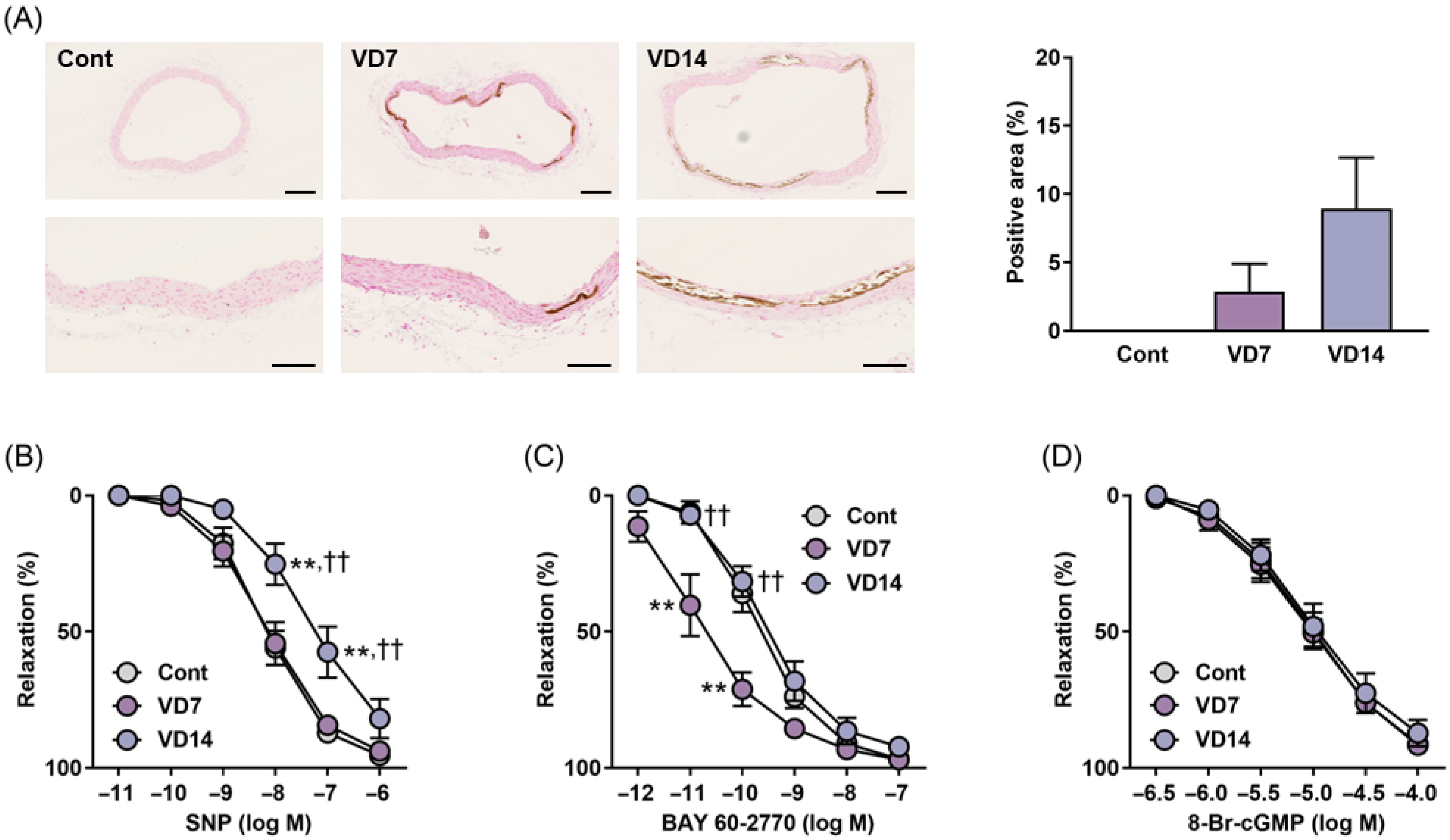
The Cont group was tested negative on von Kossa staining. Some rats in the VD7 group showed partial staining of the tunica media of the abdominal aortas by von Kossa staining, whereas vascular calcification was more pronounced in the VD14 group than in the VD7 group (Figure 1A).
SNP concentrations ranging 10 pM–1 µM evoked concentration-dependent relaxation in the Cont group, and the response was not different in the VD7 group but attenuated in the VD14 group (Figure 1B). BAY 60-2770 also relaxed the aortas in a concentration-dependent manner; however, in contrast to the SNP, the response was significantly stronger in the VD7 group than in the Cont group. Such augmentation was not observed in the VD14 group (Figure 1C). The concentration–response curve of 8-Br-cGMP did not differ among the three groups (Figure 1D).

3.2. Effects of Short-Term Supplementation with Sodium Nitrate on Vascular Calcification

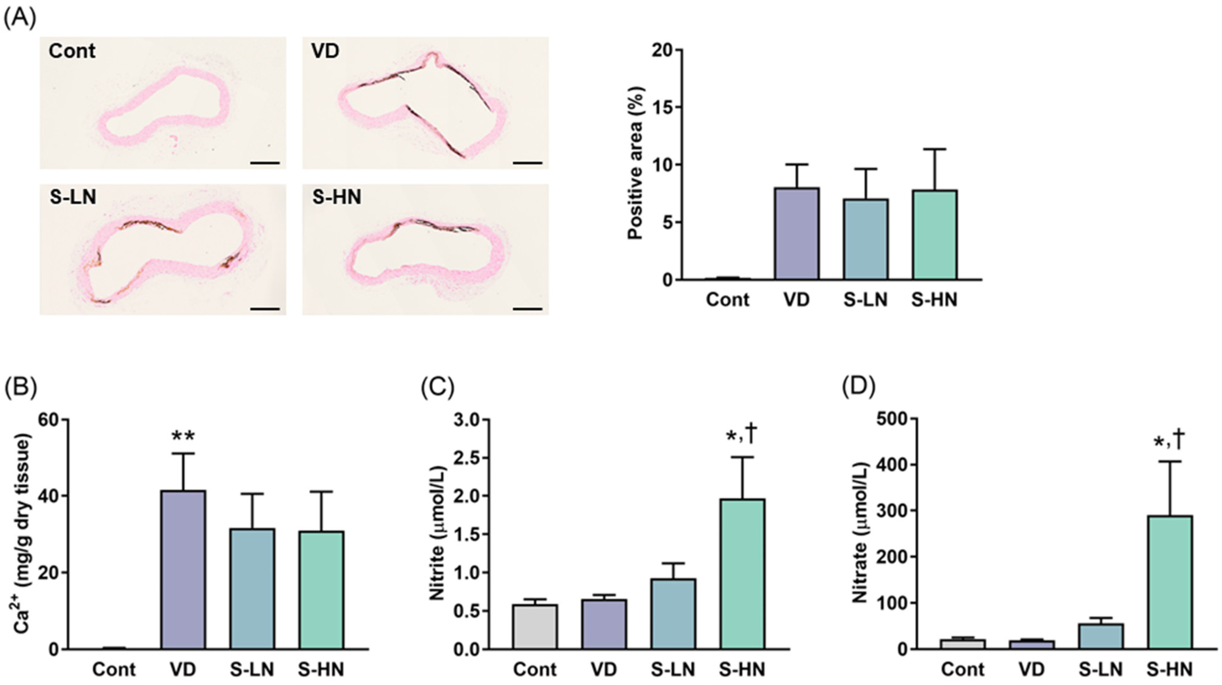
The nitrate intake in the S-LN and S-HN groups were 0.54 ± 0.02 and 1.85 ± 0.19 mmol/kg/day, respectively. No significant difference was observed in the von Kossa-positive areas between the VD, S-LN, and S-HN groups (Figure 2A). Similarly, the calcium content was not significantly reduced in either the S-LN or S-HN groups (Figure 2B). Plasma nitrite and nitrate levels did not differ between the Cont and VD groups; however, these levels tended to increase in the S-LN group and were significantly higher in the S-HN group than in the VD group (Figure 2C,D).

3.3. Effects of Long-Term Supplementation with Sodium Nitrate on Vascular Calcification

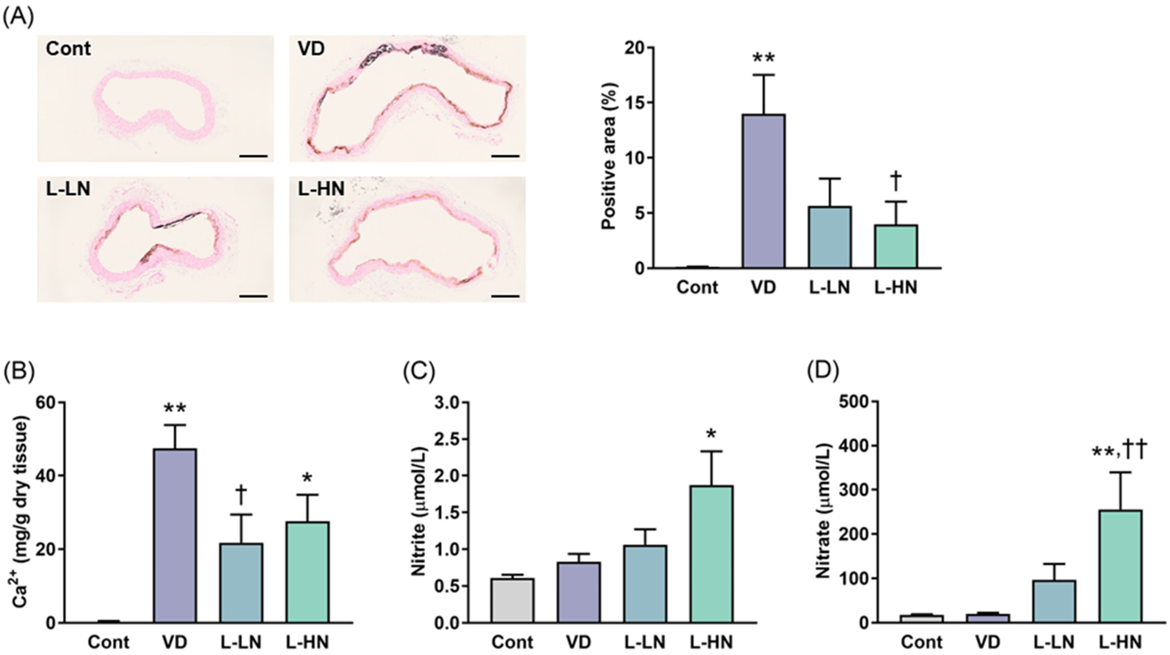
The nitrate intake in the L-LN and L-HN groups were 0.47 ± 0.04 and 1.23 ± 0.05 mmol/kg/day, respectively. The areas positive for von Kossa staining in the L-LN and L-HN groups were reduced compared to the areas in the VD group, with the difference being significant in the latter (Figure 3A). Calcium content also decreased with sodium nitrate supplementation, and this decrease was significant in the L-LN group (Figure 3B). Plasma nitrite and nitrate levels increased in a concentration-dependent manner with sodium nitrate supplementation, similar to the short-term supplementation (Figure 3C,D).

4. Discussion

A close relationship exists between vascular calcification and disruption of NO homeostasis [8,9]. This study showed the appearance of vascular calcification 7 d after cholecalciferol administration, which is consistent with previous reports that detected an increase in the calcium content of the aortic tissue 5 or 8 d after administration [18,19]. At this point, vascular responsiveness to SNP and 8-Br-cGMP was normal, suggesting that the NO pathway in the vascular smooth muscle was not impaired in the early stages of vascular calcification. In contrast, responsiveness to BAY 60-2770 was enhanced; it acts as an agonist for the oxidized form of sGC, which differs from the reduced form of sGC, where NO acts [20]. Therefore, the oxidized form of sGC may have increased. As an aside, because the relaxant response to acetylcholine (ACh) had already diminished (Figure S1), NO homeostasis in the vascular endothelium was disrupted at this point. The relaxant response to SNP was attenuated 14 d after cholecalciferol administration, although no change was observed in the response to 8-Br-cGMP. This reveals that as vascular calcification progresses, not only is NO homeostasis in the vascular endothelium disrupted, but also that in the vascular smooth muscle is compromised. Previous studies have also reported that NO homeostasis in blood vessels eventually fails in a vitamin D3-induced vascular calcification model [21,22].
Vascular calcification had already occurred before the SNP-induced vasorelaxation was attenuated, suggesting that the impairment of the NO pathway in vascular smooth muscle was induced as a consequence of structural changes associated with vascular calcification. However, it is likely that this disruption of NO homeostasis in the vascular smooth muscle formed a vicious cycle in which vascular calcification progressed further. On the other hand, since diminished ACh-induced vasorelaxation, defined as endothelial dysfunction, was already observed at the early stages of vascular calcification, it is difficult to determine which is the cause and which is the effect. In general, endothelial dysfunction is well known to precede structural remodeling in many vascular diseases [23]. Therefore, endothelial dysfunction, characterized by decreased NO bioavailability, may function as a trigger/inducer of vitamin D3-induced vascular calcification.
Since the NO pathway in the vascular smooth muscle functioned normally 7 d after cholecalciferol administration, we hypothesized that inorganic nitrate supplementation from this point onward might reduce the degree of vascular calcification. However, sodium nitrate supplementation, regardless of low or high dosage, did not sufficiently reduce the indicators of vascular calcification, including von Kossa staining-positive areas and calcium content. We believe that NO was supplied sufficiently, at least at high doses, as nitrite and nitrate levels increased. Vascular calcification had already begun to appear 7 d after cholecalciferol administration, and it is highly likely that the switch for osteoblast-like transformation of vascular smooth muscle cells had been activated. Supporting this notion, the expression of osteoblast markers increases in blood vessels 4 d after excess vitamin D3 administration [24]. Therefore, even if NO is supplied after the transformation switch is turned on, it may not be possible to prevent vascular calcification. A previous report showed that supplying NO before the onset of vascular calcification can mitigate its severity [11]. Consequently, we attempted to initiate intervention at an earlier stage. The results showed that sodium nitrate supplementation starting 1 d after cholecalciferol administration inhibited vascular calcification. In addition, no significant difference in the inhibitory effect was observed between low- and high-dose supplementation. Although the nitrite and nitrate levels differed between the two groups, the low dose may have provided sufficient NO to the blood vessels. Altogether, this is the first study to demonstrate that inorganic nitrate intake starting from the early stages of vascular calcification can suppress its progression.
Von Kossa staining-positive areas showed slight differences between the VD groups in the short- and long-term intervention experiments. Von Kossa staining can only detect calcium in a cross section of blood vessels, making it insufficient for quantifying vascular calcification on its own. In contrast, calcium content, which reflects calcium deposits throughout the entire tissue, was similar between the two VD groups. Therefore, we believe that there was no difference in the degree of vascular calcification between the two groups, and the evaluation of the sodium nitrate intervention and interpretation of the results were conducted appropriately.
No dose-dependence was observed in the degree of inhibition of vascular calcification by sodium nitrate supplementation. Plasma nitrite/nitrate levels increased in a dose-dependent manner with sodium nitrate, but it is unclear whether levels in vascular tissue exhibited similar behavior. Interestingly, there is a report indicating that the dose-related increase pattern in circulating nitrite/nitrate levels following nitrate intake does not necessarily correlate with that in tissue nitrite/nitrate levels [25]. Considering this, it cannot be ruled out that nitrite/nitrate levels at the vascular levels did not differ between the low-dose sodium nitrate and high-dose sodium nitrate groups. Nevertheless, it remains impossible to definitively state why there was no dose-dependence in the inhibitory effect on vascular calcification.
This study demonstrated that sodium nitrate supplementation is useful for inhibiting vascular calcification; however, it did not confirm whether this intervention improves endothelial dysfunction. In hypertensive patients with high augmentation index (AIx), an indicator of vascular calcification, a 4-wk dietary nitrate consumption has been reported to increase flow-mediated dilation (FMD), an index of vascular endothelial function [26]. In addition, there is a report that a 12-wk dietary nitrate intake improved FMD in postmenopausal women with elevated AIx [27]. Findings regarding whether nitrate intake improves endothelial dysfunction in calcified vessels remain insufficient, and further accumulation of evidence is necessary.
The progression of vascular calcification is influenced by various factors. No differences in body weight changes were observed with or without sodium nitrate supplementation (Figure S2), suggesting that body weight is not a confounder for the effect of nitrate intake on vascular calcification. However, since renal function and calcium/phosphate levels were not evaluated in this study, it is impossible to determine whether these were confounders. Therefore, it is necessary to keep in mind the existence of possible confounders.
Inorganic nitrates are found in foods such as leafy green vegetables and beetroots and can be consumed through diet [28]. Therefore, habitual consumption of these foods has the potential to prevent the onset of vascular calcification. Interestingly, a clinical trial revealed an inverse correlation between the intake of cruciferous vegetables and the degree of vascular calcification [29]. Although the evidence remains insufficient, dietary intake of inorganic nitrate could be a simple self-care strategy to prevent vascular calcification. This will be a topic for future research.
The nitrate intake was approximately 0.5 mmol/kg/day in the low-dose sodium nitrate group and 1.5 mmol/kg/day in the high-dose sodium nitrate group. These values correspond to a daily nitrate intake of 35 mmol (2170 mg) to 105 mmol (6510 mg) for a 70 kg adult male. The estimated dietary nitrate requirement for cardiovascular health improvement is several hundred mg per day [30,31,32]. Compared to that value, the intake levels in this study were exceptionally high. However, it is well known that because the kinetic of nitrate (entero-salivary circulation) differs between humans and rats, higher doses are required to achieve effects in rats equivalent to those in humans [33]. In short, when extrapolating the results obtained in this study to humans, it is essential to remember that the intake (dose) must be adjusted.
Numerous studies have demonstrated that NO, the bioactive form of nitrate, suppresses the expression of osteogenic markers, including alkaline phosphatase, osteocalcin, osteopontin, type I collagen, and Runx2 [10,11,34,35]. For example, Kanno et al. reported important findings that NO prevents differentiation of vascular smooth muscle cells into osteoblastic cells by inhibiting the transforming growth factor (TGF)-β/Smad signaling pathway, a key regulator of Runx2 expression/activity. Furthermore, this report also clarified that the inhibitory effect of NO on vascular calcification occurs via the cGMP/protein kinase G pathway [10]. Although the target organ was the heart rather than blood vessels, dietary intake of inorganic nitrate has been also reported to suppress TGF-β-mediated Smad activation [36]. Majumdar et al. found that NO activates the NOTCH1 pathway in aortic valvular cells, thereby decreasing Runx2 expression and suppress calcification [34]. In this context, there is a report that nitrate-rich beetroot juice intake upregulates NOTCH1 expression in the heart [37]. As several molecules can be targets of nitrate or nitrate-derived NO, future research should focus on how inorganic nitrate suppresses the progression of vascular calcification.
There are several limitations in this study. One of them is that we used only male rats, and it is unclear whether female rats exhibit the same response to the sodium nitrate intervention as male rats. Another is that we studied using only a vitamin D3-induced vascular calcification model. As vascular calcification is induced through various mechanisms, it is important to determine whether dietary nitrate also exhibits therapeutic effects in other models. Addressing these issues would lead to a deeper understanding of the therapeutic effect of dietary nitrate on vascular calcification.

5. Conclusions

In conclusion, this study demonstrated that sodium nitrate can exert a protective effect against vascular calcification in rats. Moreover, the protective effect of sodium nitrate was likely mediated by the enhanced bioavailability of NO. The dietary intake of inorganic nitrate (or nitrate-rich foods) may be a simple strategy for preventing vascular calcification.

Supplementary Materials

The following supporting information can be downloaded at: https://www.mdpi.com/article/10.3390/jvd5010003/s1, Figure S1: Concentration–response curves of abdominal aortas to acetylcholine. Figure S2: Body weight changes in the short- and long-term intervention experiments.

Author Contributions

Conceptualization, M.T.; investigation, M.T.; data curation, M.T.; writing—original draft preparation, M.T.; writing—review and editing, K.N. and M.O.; supervision, M.O. All authors have read and agreed to the published version of the manuscript.

Funding

This research was funded by Grants-in-Aid for the Scientific Research Program from the Japan Society for the Promotion of Science, grant number 22K15299 to M.T., and a Grant for Promoted Research from Kanazawa Medical University, grant number S2020-1 to M.T.

Institutional Review Board Statement

The animal study protocol was approved by the Animal Research Committee of Kanazawa Medical University (Permit number 2020-29, approved 1 April 2020).

Informed Consent Statement

Not applicable.

Data Availability Statement

The original contributions presented in this study are included in the article/Supplementary Material. Further inquiries can be directed to the corresponding author(s).

Acknowledgments

The authors thank the staff of the Translational Research Program, Osaka Medical and Pharmaceutical University for their support.

Conflicts of Interest

The authors declare no conflicts of interest.

Abbreviations

The following abbreviations are used in this manuscript:
AChAcetylcholine
AIxAugmentation index
FMDFlow-mediated dilation
NONitric oxide
sGCSoluble guanylate cyclase
PBSPhosphate-buffered saline
SNPSodium nitroprusside
SEMStandard error of the mean
ANOVAAnalysis of variance
TGFTransforming growth factor

References

  1. Lanzer, P.; Hannan, F.M.; Lanzer, J.D.; Janzen, J.; Raggi, P.; Furniss, D.; Schuchardt, M.; Thakker, R.; Fok, P.W.; Saez-Rodriguez, J.; et al. Medial Arterial Calcification: JACC State-of-the-Art Review. J. Am. Coll. Cardiol. 2021, 78, 1145–1165. [Google Scholar] [CrossRef] [Scilit]
  2. Chen, Y.; Zhao, X.; Wu, H. Arterial Stiffness: A Focus on Vascular Calcification and Its Link to Bone Mineralization. Arterioscler. Thromb. Vasc. Biol. 2020, 40, 1078–1093. [Google Scholar] [CrossRef] [Scilit]
  3. Chen, S.; Li, N.; Gao, Y.; Jiang, H.; Shen, Y. Prevalence and risk factors for vascular calcification based on the ankle-brachial index in the general population: A cross-sectional study. BMC Cardiovasc. Disord. 2022, 22, 227. [Google Scholar] [CrossRef] [Scilit]
  4. Kamenskiy, A.; Poulson, W.; Sim, S.; Reilly, A.; Luo, J.; MacTaggart, J. Prevalence of Calcification in Human Femoropopliteal Arteries and its Association with Demographics, Risk Factors, and Arterial Stiffness. Arterioscler. Thromb. Vasc. Biol. 2018, 38, e48–e57. [Google Scholar] [CrossRef] [Scilit]
  5. Leow, K.; Szulc, P.; Schousboe, J.T.; Kiel, D.P.; Teixeira-Pinto, A.; Shaikh, H.; Sawang, M.; Sim, M.; Bondonno, N.; Hodgson, J.M.; et al. Prognostic Value of Abdominal Aortic Calcification: A Systematic Review and Meta-Analysis of Observational Studies. J. Am. Heart Assoc. 2021, 10, e017205. [Google Scholar] [CrossRef] [Scilit] [PubMed]
  6. Sheng, C.; Cai, Z.; Yang, P. Association of the abdominal aortic calcification with all-cause and cardiovascular disease-specific mortality: Prospective cohort study. PLoS ONE 2025, 20, e0314776. [Google Scholar] [CrossRef] [Scilit]
  7. Carlström, M.; Weitzberg, E.; Lundberg, J.O. Nitric Oxide Signaling and Regulation in the Cardiovascular System: Recent Advances. Pharmacol. Rev. 2024, 76, 1038–1062. [Google Scholar] [CrossRef] [Scilit] [PubMed]
  8. Cao, X.; Li, H.; Tao, H.; Wu, N.; Yu, L.; Zhang, D.; Lu, X.; Zhu, J.; Lu, Z.; Zhu, Q. Metformin inhibits vascular calcification in female rat aortic smooth muscle cells via the AMPK-eNOS-NO pathway. Endocrinology 2013, 154, 3680–3689. [Google Scholar] [CrossRef] [Scilit]
  9. Freise, C.; Querfeld, U. Inhibition of vascular calcification by block of intermediate conductance calcium-activated potassium channels with TRAM-34. Pharmacol. Res. 2014, 85, 6–14. [Google Scholar] [CrossRef] [Scilit] [PubMed]
  10. Kanno, Y.; Into, T.; Lowenstein, C.J.; Matsushita, K. Nitric oxide regulates vascular calcification by interfering with TGF-β signalling. Cardiovasc. Res. 2008, 77, 221–230. [Google Scholar] [CrossRef] [Scilit]
  11. Wang, F.; Qin, K.; Wang, K.; Wang, H.; Liu, Q.; Qian, M.; Chen, S.; Sun, Y.; Hou, J.; Wei, Y.; et al. Nitric oxide improves regeneration and prevents calcification in bio-hybrid vascular grafts via regulation of vascular stem/progenitor cells. Cell Rep. 2022, 39, 110981. [Google Scholar] [CrossRef] [Scilit]
  12. Kapil, V.; Khambata, R.S.; Jones, D.A.; Rathod, K.; Primus, C.; Massimo, G.; Fukuto, J.M.; Ahluwalia, A. The Noncanonical Pathway for In Vivo Nitric Oxide Generation: The Nitrate-Nitrite-Nitric Oxide Pathway. Pharmacol. Rev. 2020, 72, 692–766. [Google Scholar] [CrossRef] [Scilit]
  13. Khatri, J.; Mills, C.E.; Maskell, P.; Odongerel, C.; Webb, A.J. It is rocket science—Why dietary nitrate is hard to ‘beet’! Part I: Twists and turns in the realization of the nitrate-nitrite-NO pathway. Br. J. Clin. Pharmacol. 2017, 83, 129–139. [Google Scholar] [CrossRef] [Scilit]
  14. Muyor, K.; Laget, J.; Cortijo, I.; Duranton, F.; Jover, B.; Argilés, À.; Gayrard, N. Vascular calcification in different arterial beds in ex vivo ring culture and in vivo rat model. Sci. Rep. 2022, 12, 11861. [Google Scholar] [CrossRef] [Scilit] [PubMed]
  15. Allan, P.D.; Tzeng, Y.C.; Gowing, E.K.; Clarkson, A.N.; Fan, J.L. Dietary nitrate supplementation reduces low frequency blood pressure fluctuations in rats following distal middle cerebral artery occlusion. J. Appl. Physiol. 2018, 125, 862–869. [Google Scholar] [CrossRef] [Scilit]
  16. Guimarães, D.D.; Cruz, J.C.; Carvalho-Galvão, A.; Zhuge, Z.; Marques, S.M.; Naves, L.M.; Persson, A.E.G.; Weitzberg, E.; Lundberg, J.O.; Balarini, C.M.; et al. Dietary Nitrate Reduces Blood Pressure in Rats with Angiotensin II-Induced Hypertension via Mechanisms That Involve Reduction of Sympathetic Hyperactivity. Hypertension 2019, 73, 839–848. [Google Scholar] [CrossRef] [Scilit] [PubMed]
  17. Tawa, M.; Abe, S.; Fujita, A.; Ando, R.; Yoshinari, H.; Nakagawa, K.; Ohkita, M. Beneficial effects of magnesium nitrate on disease severity in male rats with monocrotaline-induced pulmonary hypertension. Physiol. Rep. 2025, 13, e70416. [Google Scholar] [CrossRef] [Scilit]
  18. Qualglia, N.B.; Hofer, C.G.; Torres, A.M. Pharmacokinetics of organic anions in rats with arterial calcinosis. Clin. Exp. Pharmacol. Physiol. 2002, 29, 48–52. [Google Scholar] [CrossRef] [Scilit] [PubMed]
  19. Wang, Y.; Wen, Y.; Fang, Y.; Pang, H.; Guo, Z.; Shi, L.; Li, J. Experimental vasoprotection by a novel erythrocyte-derived depressing factor in rats with arterial calcinosis. Vascul. Pharmacol. 2009, 50, 65–70. [Google Scholar] [CrossRef] [Scilit]
  20. Tawa, M.; Okamura, T. Factors influencing the soluble guanylate cyclase heme redox state in blood vessels. Vascul. Pharmacol. 2022, 145, 107023. [Google Scholar] [CrossRef] [Scilit]
  21. Takeo, S.; Anan, M.; Fujioka, K.; Kajihara, T.; Hiraga, S.; Miyake, K.; Tanonaka, K.; Minematsu, R.; Mori, H.; Taniguchi, Y. Functional changes of aorta with massive accumulation of calcium. Atherosclerosis 1989, 77, 175–181. [Google Scholar] [CrossRef] [Scilit]
  22. Henrion, D.; Chillon, J.M.; Godeau, G.; Muller, F.; Capdeville-Atkinson, C.; Hoffman, M.; Atkinson, J. The consequences of aortic calcium overload following vitamin D3 plus nicotine treatment in young rats. J. Hypertens. 1991, 9, 919–926. [Google Scholar] [CrossRef] [Scilit]
  23. Tesauro, M.; Mauriello, A.; Rovella, V.; Annicchiarico-Petruzzelli, M.; Cardillo, C.; Melino, G.; Di Daniele, N. Arterial ageing: From endothelial dysfunction to vascular calcification. J. Intern. Med. 2017, 281, 471–482. [Google Scholar] [CrossRef] [Scilit]
  24. Han, M.S.; Che, X.; Cho, G.H.; Park, H.R.; Lim, K.E.; Park, N.R.; Jin, J.S.; Jung, Y.K.; Jeong, J.H.; Lee, I.K.; et al. Functional cooperation between vitamin D receptor and Runx2 in vitamin D-induced vascular calcification. PLoS ONE 2013, 8, e83584. [Google Scholar] [CrossRef] [Scilit] [PubMed]
  25. Wei, C.; Vanhatalo, A.; Black, M.I.; Rajaram, R.; Massey, G.; Jones, A.M. Dose-response relationship between dietary nitrate intake and nitric oxide congeners in various blood compartments and skeletal muscle: Differential effects on skeletal muscle torque and velocity. Free Radic. Biol. Med. 2025, 229, 520–533. [Google Scholar] [CrossRef] [Scilit] [PubMed]
  26. Kapil, V.; Khambata, R.S.; Robertson, A.; Caulfield, M.J.; Ahluwalia, A. Dietary nitrate provides sustained blood pressure lowering in hypertensive patients: A randomized, phase 2, double-blind, placebo-controlled study. Hypertension 2015, 65, 320–327. [Google Scholar] [CrossRef] [Scilit] [PubMed]
  27. Alvares, T.S.; Pinheiro, V.; Proctor, D.N.; Junior, C.A.C.; Soares, R.N. Twelve-week nitrate-rich beetroot extract supplementation improves lower limb vascular function and serum angiogenic potential in postmenopausal women. Am. J. Physiol. Heart Circ. Physiol. 2025, 329, H1047–H1054. [Google Scholar] [CrossRef] [Scilit]
  28. Lidder, S.; Webb, A.J. Vascular effects of dietary nitrate (as found in green leafy vegetables and beetroot) via the nitrate-nitrite-nitric oxide pathway. Br. J. Clin. Pharmacol. 2013, 75, 677–696. [Google Scholar] [CrossRef] [Scilit]
  29. Blekkenhorst, L.C.; Sim, M.; Radavelli-Bagatini, S.; Bondonno, N.P.; Bondonno, C.P.; Devine, A.; Schousboe, J.T.; Lim, W.H.; Kiel, D.P.; Woodman, R.J.; et al. Cruciferous vegetable intake is inversely associated with extensive abdominal aortic calcification in elderly women: A cross-sectional study. Br. J. Nutr. 2021, 125, 337–345. [Google Scholar] [CrossRef] [Scilit]
  30. Grönroos, R.; Eggertsen, R.; Bernhardsson, S.; Björk, M.P. Effects of beetroot juice on blood pressure in hypertension according to European Society of Hypertension Guidelines: A systematic review and meta-analysis. Nutr. Metab. Cardiovasc. Dis. 2024, 34, 2240–2256. [Google Scholar] [CrossRef] [Scilit]
  31. Norouzzadeh, M.; Rashedi, M.H.; Ghaemi, S.; Saber, N.; Harijani, A.M.; Habibi, H.; Mostafavi, S.; Sarv, F.; Farhadnejad, H.; Teymoori, F.; et al. Plasma nitrate, dietary nitrate, blood pressure, and vascular health biomarkers: A GRADE-Assessed systematic review and dose-response meta-analysis of randomized controlled trials. Nutr. J. 2025, 24, 47. [Google Scholar] [CrossRef] [Scilit]
  32. Celik, B.; Muriuki, E.; Kuhnle, G.G.C.; Spencer, J.P.E.; Mills, C.E. The Impact of Inorganic Nitrate on Endothelial Function: A Systematic Review of Randomized Controlled Trials and Meta-analysis. Nutr. Rev. 2026, 84, 36–46. [Google Scholar] [CrossRef] [Scilit]
  33. Montenegro, M.F.; Sundqvist, M.L.; Nihlén, C.; Hezel, M.; Carlström, M.; Weitzberg, E.; Lundberg, J.O. Profound differences between humans and rodents in the ability to concentrate salivary nitrate: Implications for translational research. Redox Biol. 2016, 10, 206–210. [Google Scholar] [CrossRef] [Scilit]
  34. Majumdar, U.; Manivannan, S.; Basu, M.; Ueyama, Y.; Blaser, M.C.; Cameron, E.; McDermott, M.R.; Lincoln, J.; Cole, S.E.; Wood, S.; et al. Nitric oxide prevents aortic valve calcification by S-nitrosylation of USP9X to activate NOTCH signaling. Sci. Adv. 2021, 7, eabe3706. [Google Scholar] [CrossRef] [Scilit]
  35. Richards, J.; El-Hamamsy, I.; Chen, S.; Sarang, Z.; Sarathchandra, P.; Yacoub, M.H.; Chester, A.H.; Butcher, J.T. Side-specific endothelial-dependent regulation of aortic valve calcification: Interplay of hemodynamics and nitric oxide signaling. Am. J. Pathol. 2013, 182, 1922–1931. [Google Scholar] [CrossRef] [Scilit] [PubMed]
  36. Gee, L.C.; Massimo, G.; Lau, C.; Primus, C.; Fernandes, D.; Chen, J.; Rathod, K.S.; Hamers, A.J.P.; Filomena, F.; Nuredini, G.; et al. Inorganic nitrate attenuates cardiac dysfunction: Roles for xanthine oxidoreductase and nitric oxide. Br. J. Pharmacol. 2022, 179, 4757–4777. [Google Scholar] [CrossRef] [Scilit] [PubMed]
  37. Yin, C.; Rehman, S.; Kukreja, R.C.; Xi, L. Upregulation of Cardiac MicroRNA 34a, Sirt1, and Notch1 Following Oral Ingestion of Beetroot Juice and Doxorubicin Treatment: A Novel Protective Signaling Against Cardiotoxicity. Circulation 2014, 130, A12237. [Google Scholar]
Figure 1. Representative images (left panel) and quantification (right panel) of von Kossa staining in abdominal aorta sections (A). Scale bars, 250 µm (upper panels) and 100 μm (bottom panels). Concentration–response curves of abdominal aortas to SNP (B), BAY 60-2770 (C), and 8-Br-cGMP (D). Each column/point and bar represent the mean ± SEM. PBS-injected rats 14 d after injection (Cont, n = 6), cholecalciferol-injected rats 7 d after injection (VD7, n = 6), and cholecalciferol-injected rats 14 d after injection (VD14, n = 6). ** p < 0.01, compared with the Cont group; †† p < 0.01, compared with the VD7 group. Statistical analysis was performed using one-way ANOVA with Bonferroni post hoc test (A) or two-way repeated measures ANOVA with Bonferroni post hoc test (BD).
Jvd 05 00003 g001
Figure 2. Representative images (left panel) and quantification (right panel) of von Kossa staining in abdominal aorta sections (A). Scale bars, 250 μm. Calcium contents in abdominal aortas (B). Plasma nitrite (C) and nitrate (D) levels. Each column and bar represent the mean ± SEM. PBS-injected rats (Cont, n = 8), cholecalciferol-injected rats (VD, n = 8), cholecalciferol-injected rats receiving short-term and low-dose sodium nitrate treatment (S-LN, n = 8), and cholecalciferol-injected rats receiving short-term and high-dose sodium nitrate treatment (S-HN, n = 8). * p < 0.05 and ** p < 0.01, compared with the Cont group; † p < 0.05, compared with the VD group. Statistical analysis was performed using one-way ANOVA with Bonferroni post hoc test.
Jvd 05 00003 g002
Figure 3. Representative images (left panel) and quantification (right panel) of von Kossa staining in abdominal aorta sections (A). Scale bars, 250 μm. Calcium contents in abdominal aortas (B). Plasma nitrite (C) and nitrate levels (D). Each column and bar represent the mean ± SEM. PBS-injected rats (Cont, n = 6), cholecalciferol-injected rats (VD, n = 6), cholecalciferol-injected rats receiving long-term and low-dose sodium nitrate treatment (L-LN, n = 6), and cholecalciferol-injected rats receiving long-term and high-dose sodium nitrate treatment (L-HN, n = 6). * p < 0.05 and ** p < 0.01, compared with the Cont group; † p < 0.05 and †† p < 0.01, compared with the VD group. Statistical analysis was performed using one-way ANOVA with Bonferroni post hoc test.
Jvd 05 00003 g003
Disclaimer/Publisher’s Note: The statements, opinions and data contained in all publications are solely those of the individual author(s) and contributor(s) and not of MDPI and/or the editor(s). MDPI and/or the editor(s) disclaim responsibility for any injury to people or property resulting from any ideas, methods, instructions or products referred to in the content.