Review

Extended Color Doppler Ultrasound in the Diagnosis of Giant Cell Arteritis: Clinical Insights and Literature Review with Emphasis on Posterior Circulation Involvement

1
Department of Clinical and Experimental Medicine, University of Catania, 95123 Catania, Italy
2
Angiology Unit, San Marco Hospital, 95123 Catania, Italy
3
Geriatrics Unit, Garibaldi Hospital, 95123 Catania, Italy
*
Authors to whom correspondence should be addressed.
J. Vasc. Dis. 2025, 4(3), 37; https://doi.org/10.3390/jvd4030037
Submission received: 18 July 2025 / Revised: 11 August 2025 / Accepted: 14 September 2025 / Published: 18 September 2025
(This article belongs to the Section Peripheral Vascular Diseases)

Abstract

Background: Giant cell arteritis (GCA) is a systemic granulomatous vasculitis affecting large and medium-sized arteries, predominantly in individuals over 50 years. While it traditionally involves cranial branches of the external carotid artery, particularly the temporal arteries, growing evidence underscores frequent extracranial involvement, especially in the supra-aortic trunks. Objective: We aimed to critically review the diagnostic utility of extended Color Doppler Ultrasound (CDUS) in GCA, with a focus on vertebrobasilar involvement and current international imaging guidelines. Methods: Taking inspiration from a representative case of extracranial GCA with vertebrobasilar ischemic events, the current literature and international recommendations (e.g., EULAR, ACR, BSR and SIR) were reviewed. Results: Diagnostic accuracy significantly improves when CDUS is extended to include carotid, vertebral, subclavian and axillary arteries. Elevated inflammatory markers such as erythrocyte sedimentation rate (ESR) and C-reactive protein (CRP) correlate with large-vessel involvement and support the use of extended scanning protocols. International guidelines vary in their emphasis on extended CDUS, but consensus is growing toward ultrasound imaging-first strategies in expert hands. Conclusion: Extended CDUS is a sensitive, non-invasive first-line diagnostic tool for GCA. In patients with symptoms of the posterior cerebral circulation and elevated inflammatory indices, early comprehensive vascular imaging reduces diagnostic delay and may obviate the need for temporal artery biopsy.

1. Introduction

In 1932, Bayard T. Horton, a neurologist at the Mayo Clinic, described a puzzling condition affecting elderly women who presented with new-onset temporal headache, low-grade fever and sudden, episodic vision loss. Temporal artery biopsy revealed granulomatous inflammation with multinucleated giant cells, which became known as “temporal arteritis” [1].
Giant cell arteritis (GCA) is the most common systemic vasculitis in adults over 50 years of age. It is histologically characterized by granulomatous inflammation with lymphomonocytic infiltrates, multinucleated giant cells, internal elastic lamina fragmentation and concentric intimal hyperplasia leading to luminal stenosis and subsequent ischemia [2,3].
Clinically, GCA presents in two major phenotypes:
  • Cranial GCA (C-GCA) affects the superficial temporal arteries, presenting with classical symptoms such as new-onset headache, scalp tenderness, jaw claudication and vision loss.
  • Large-vessel GCA (LV-GCA or extracranial GCA) involving the aorta and major branches, including the subclavian, axillary, carotid, and vertebral arteries [4,5].
Recent studies using PET-CT and high-resolution ultrasound have demonstrated that extracranial involvement is present in up to 60–70% of GCA cases, often in the absence of cranial symptoms. Vertebrobasilar ischemia, in particular, may be under-recognized. Elevated ESR and CRP are frequently the only early laboratory clues [6,7,8].
This narrative review based on a representative clinical case stems from our renewed interest in GCA. We present the case of a 74-year-old woman who presented with transient ischemic episodes in the cerebral vertebrobasilar territory over a 3-month period. Initially misdiagnosed with atherosclerotic disease and possible vertebral dissection, her condition deteriorated despite vascular risk factor management.
Brain MRI revealed multifocal ischemic lesions in the posterior circulation and bilateral vertebral artery occlusion. Inflammatory markers were significantly elevated (ESR 120 mm/h, CRP 4.2 mg/dL). Autoimmune screening was negative except for anti-parietal cell antibodies.
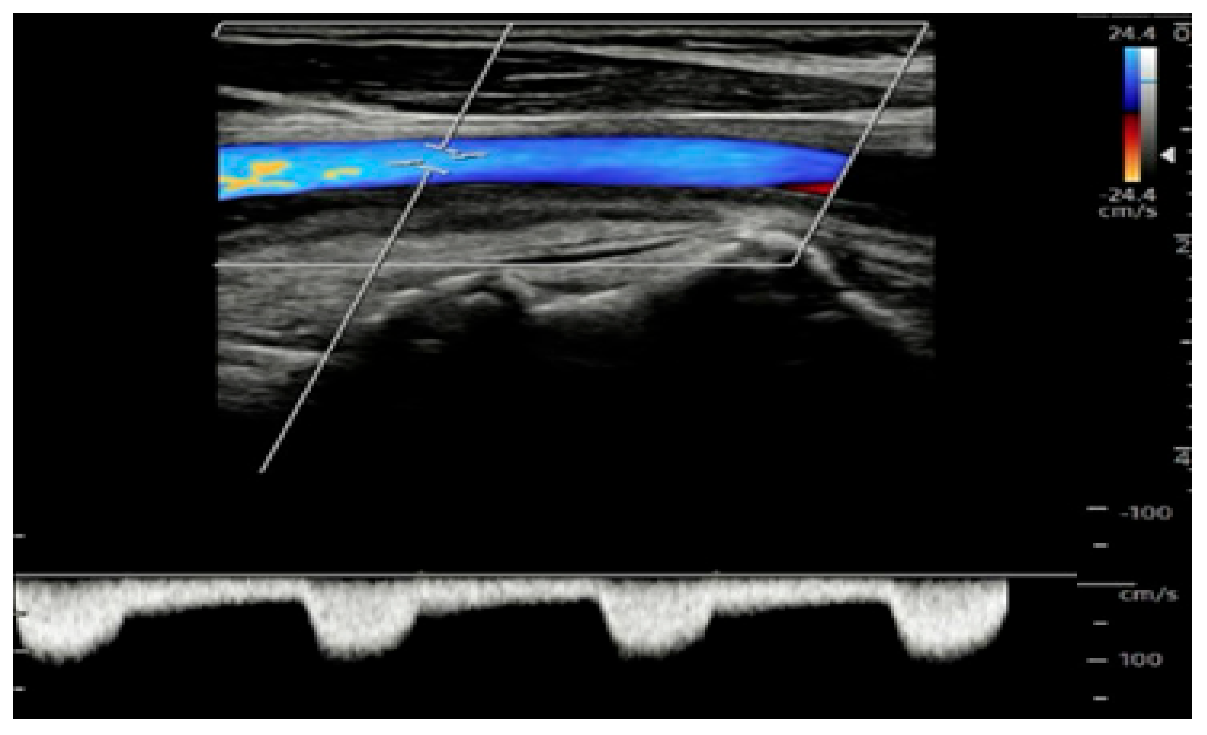
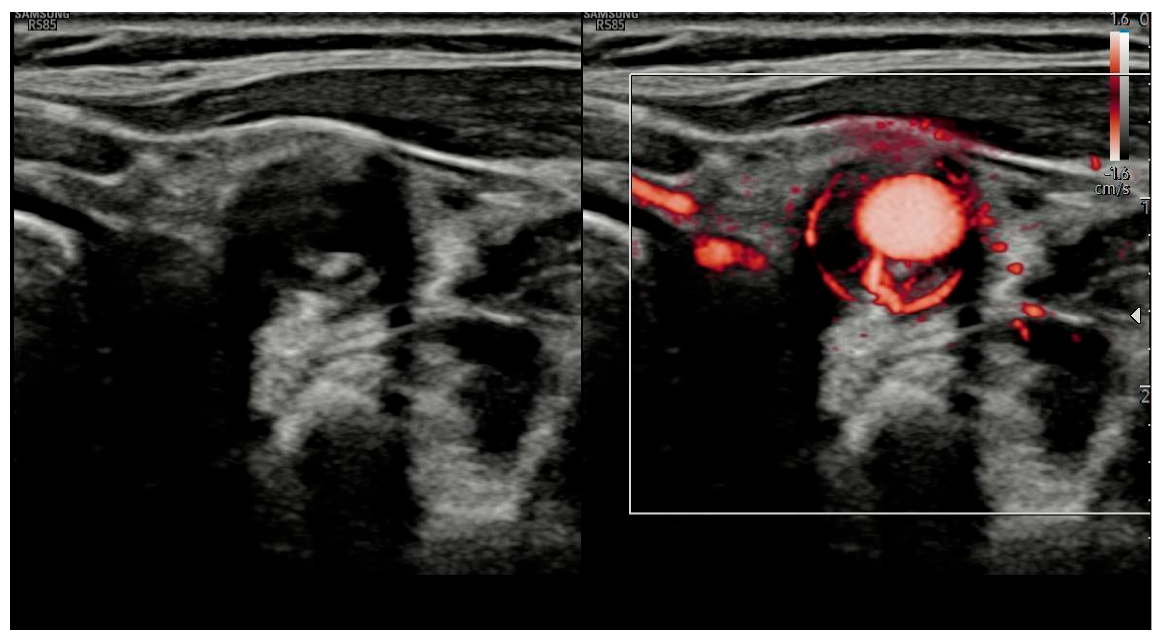
Extended CDUS (Figure 1, Figure 2 and Figure 3) revealed a bilateral halo sign of temporal arteries, hypoechoic circumferential thickening of the subclavian and common carotid arteries, increased intima-media thickness (IMT) and non-compressibility of arteries. Growing evidence confirms that extended CDUS protocols, including extracranial vessels, significantly increase diagnostic yield in patients with suspected LV-GCA [9].
A subsequent 18F-FDG PET/CT scan confirmed systemic vasculitic involvement of the temporal, vertebral and subclavian arteries. The patient began high-dose corticosteroid therapy with rapid clinical and biochemical improvement.
This case would support the importance of considering extracranial GCA in patients with posterior stroke syndromes and reinforces the use of extended CDUS protocols [9,10].

2. Methods

A systematic literature review was conducted to evaluate the diagnostic utility of extended color Doppler ultrasound (CDUS) in giant cell arteritis (GCA), with particular emphasis on the detection of posterior circulation involvement.
The search was performed in PubMed, Cochrane Library and Google Scholar from January 2010 to August 2025, using the following keywords: “Giant Cell Arteritis”, “Color Doppler Vltrasound”, “Extended ultrasound”, “Large Vessel Vasculitis”, “Vertebral Artery”, “Posterior Circulation Diagnosis” and related synonyms.
Inclusion criteria were as follows:
  • Original studies, systematic reviews and meta-analyses published in English or Italian from 2010 onward.
  • Adult patients (age > 50 years) with suspected or confirmed GCA.
  • The use of extended CDUS protocols, including assessment of temporal, axillary, subclavian, carotid and vertebral arteries.
  • Outcome reporting diagnostic sensitivity, specificity, accuracy, or clinical impact (e.g., association with ischemic complications, especially posterior circulation stroke).
  • Studies comparing CDUS with other imaging modalities (PET/CT, MRI) or with clinical reference standards.
Exclusion criteria were as follows:
  • Studies on pediatric populations or vasculitides other than GCA.
  • Studies lacking specific data on CDUS or not distinguishing between limited and extended protocols.
  • Articles without relevant diagnostic or clinical outcome data.
Study selection: The search identified 23 records (Pubmed: 5; Cochrane Library: 1; Google Scholar additional relevant studies: 17). All 23 records underwent title/abstract screening, with 8 exclusions (not related to GCA, pre-2010, or no ultrasound data). Full-text review was performed on 15 articles; 5 were excluded due to the absence of data on extended CDUS or lack of diagnostic/clinical outcomes. A total of 10 studies met the inclusion criteria and were included in the qualitative synthesis.

3. Results

As shown in Table 1, CDUS is a first-line diagnostic imaging tool in the diagnosis of suspected GCA due to its high sensitivity, specificity, repeatability and non-invasiveness. While initial protocols focused on temporal arteries, modern evidence supports the extension of CDUS to include supra-aortic trunks.
The ultrasound examination involves a ≥15 MHz linear probe for cranial vessels and ≥10 MHz transducer with power Doppler settings for peripheral arteries. These technical recommendations are consistent with current expert consensus, which also emphasizes the importance of probe frequency, Doppler settings, and operator training to avoid artifacts and optimize diagnostic accuracy [11]. Imaging pathological signs include the following: (a) A halo sign: Characterized by a hypoechoic (dark) thickening of the vessel wall. The halo sign is sensitive to change and typically disappears within 2–4 weeks of initiating glucocorticoid therapy, supporting its value in both diagnosis and early monitoring [12]. (b) Compression sign: Defined as visibility of the vessel wall upon transducer-imposed compression of the artery. (c) Intima-media thickness (IMT) > 1 mm [10,13,14,15].
Recent EULAR-endorsed cut-off values for intima-media thickness (IMT) include ≥0.42 mm for temporal arteries and ≥1.0 mm for axillary arteries [11].
Current international guidelines differ in their degree of endorsement of extended CDUS protocols, as shown in the comparative summary in Table 2.

4. Discussion

Despite advances in awareness and imaging technology, the early diagnosis of giant cell arteritis (GCA) remains challenging due to its heterogeneous clinical presentation symptom and overlap with other vascular disorders. Over the past decade, international guidelines have progressively incorporated color Doppler ultrasonography (CDUS) into diagnostic pathways, though variations persist regarding vascular territories to be assessed and its role relative to temporal artery biopsy (TAB), MRI or PET/CT.
Comparative studies confirm the diagnostic accuracy of CDUS. Hansen et al. reported that CDUS, particularly the compression sign, achieved a sensitivity of 63% and specificity of 79% compared to 69% sensitivity and 100% specificity for TAB, with CDUS identifying more overall positive cases [19].
Extended CDUS protocols further improve diagnostic yield, especially for large-vessels involvement. Pacheco et al. recently demonstrated that the simultaneous assessment of the superficial temporal and axillary arteries delivers robust performance and is feasible in fast-track settings [20]. Similarly Skoog et al. observed that sensitivity increased from 86% with temporary assessment alone to 95% when extracranial vessels were included, with 10% of cases presenting isolated extracranial disease that would have been otherwise missed [21,22].
Guideline recommendations vary. EULAR (2018, 2023) and SIR advocate extended vascular assessment even in cranial presentations, supporting CDUS as a first-line modality that may replace biopsy in expert hands [14,17,23]. The 2024 EULAR-endorsed “Points to Consider” recommend the systematic inclusion of carotid and vertebral arteries, when clinically indicated, and emphasize CDUS as a tool for both diagnosis and monitoring [11].
In contrast ACR/Vasculitis Foundation Guidelines (2021) maintain TAB (temporal artery biopsy) as the diagnostic gold standard, reserving imaging for atypical cases or biopsy-negative cases [16], while the British Society for Rheumatology (BSR) 2020 guidelines support CDUS in fast-track clinics but retain biopsy in low-ultrasound-expertise settings [18].
Posterior circulation involvement, particularly vertebral artery vasculitis, is under-recognized despite being associated with cerebrovascular events in up to 10% of GCA cases [6,8,9,18,24].
Prünte et al. emphasized the need to evaluate vertebral arteries in atypical presentations, as isolated extracranial disease can occur without cranial symptoms [24]. In such cases, extended CDUS facilitates earlier diagnosis and prompt treatment.
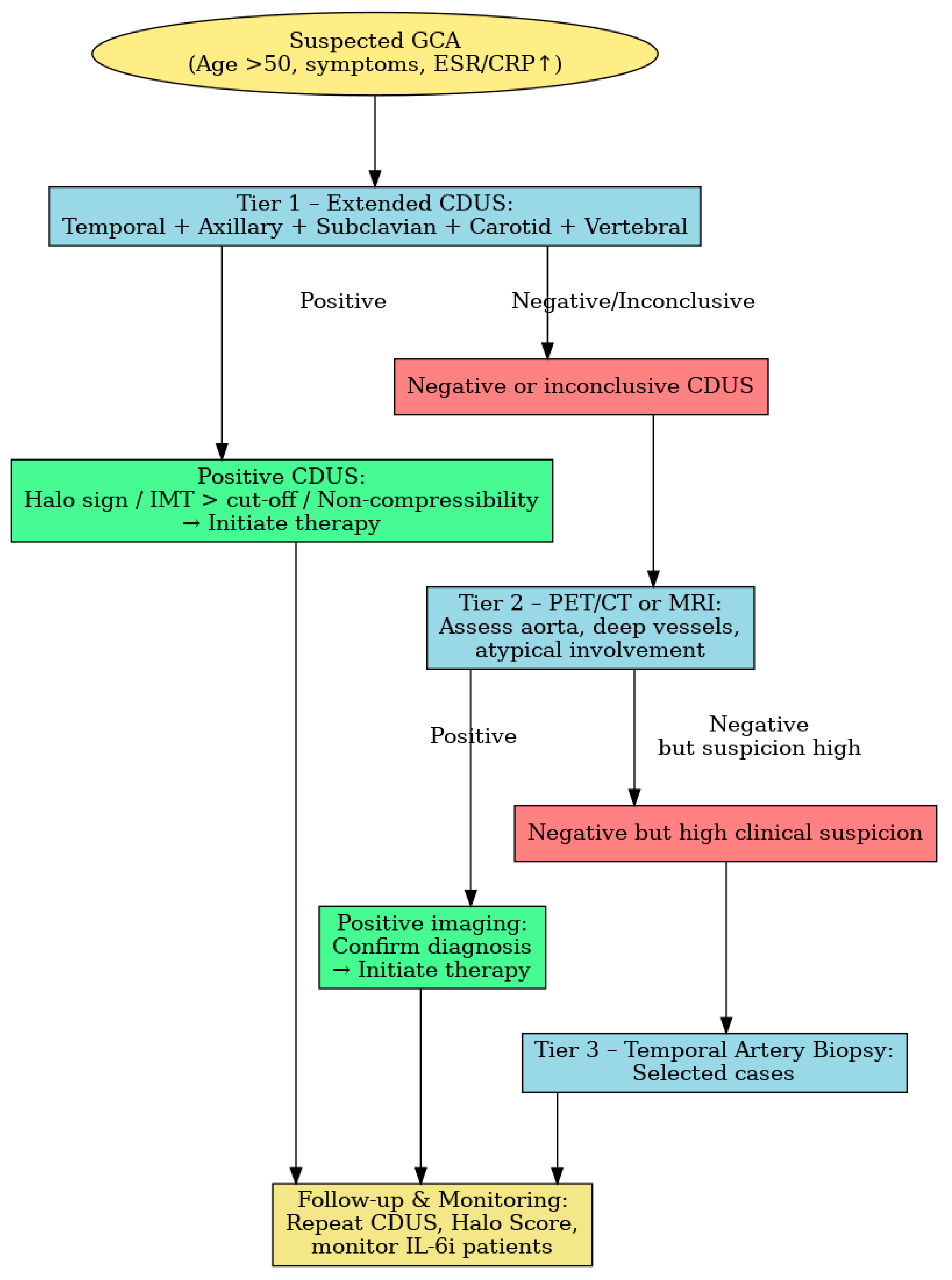
We propose a tiered diagnostic approach Figure 4:
  • Tier 1: A CDUS of temporal, axillary and subclavian arteries and extension to carotid and vertebral vessels in high-risk patients.
  • Tier 2: MRI or PET/CT, when CDUS is negative or inconclusive. Van Nieuwland et al. found these modalities to have comparable overall diagnostic accuracy, with differing artery-specific performance [25].
  • Tier 3: Temporal artery biopsy in selected cases in the context of persistently high clinical suspicion despite negative imaging findings.
Beyond diagnosis, CDUS also supports disease monitoring, particularly in patients receiving IL-6 inhibitors such as tocilizumab, where CRP (C-reactive protein) may be suppressed [15,16,26]. Persistent wall changes can be detected despite clinical remission, serving as structural correlates of subclinical disease. Quantitative scores such as the Halo Score may standardize follow-up and relapse prediction.
Overall, extended CDUS, when performed by trained operators following standardized protocols, aligns with a modern paradigm prioritizing imaging as a first-line diagnostic tool. It enables faster diagnosis, reduces reliance on invasive biopsy, and supports longitudinal disease assessment, particularly in large-vessel involvement and posterior circulation disease.

5. Conclusions

This review highlights the pivotal role of extended color Doppler ultrasound (CDUS) in the diagnostic work-up of giant cell arteritis (GCA). By systematically evaluating not only the temporal arteries but also extracranial vessels such as the axillary, subclavian, carotid, and vertebral arteries, extended CDUS markedly increases the diagnostic yield [8,9,15,21,22,25,27,28]. This broader approach enables the detection of both cranial and extracranial disease phenotypes, allowing an earlier recognition of large-vessel involvement and posterior circulation disease [8,9,21,27]. Such findings are clinically relevant, as posterior circulation involvement is often underdiagnosed and may precede or occur in the absence of cranial symptoms, potentially leading to disabling or fatal ischemic events [8,9,15]. Compared with other imaging modalities such as PET/CT and MRI, extended CDUS is rapid, non-invasive, widely available, and cost-effective, making it an ideal first-line technique, especially in fast-track clinics [21,25,27].
Nevertheless, despite the growing evidence base, some challenges remain. Differences in operator expertise, equipment settings and diagnostic thresholds for intima-media thickness (IMT) continue to limit the generalizability of results [15,22].
Furthermore, while several studies support the inclusion of vertebral arteries in the scanning protocol, standardized acquisition and interpretation criteria are still lacking [15,22].
In conclusion, extended CDUS represents a powerful, first-line imaging tool for the early diagnosis of GCA [28], with the potential to improve patient outcomes through the timely recognition of extracranial and posterior circulation involvement. The standardization of protocols and broader dissemination of expertise will be crucial to consolidating its role in routine clinical practice.

Author Contributions

Conceptualization: I.P., L.C., P.M.S.L.; Methodology: M.R., R.R.; Software: I.P.; Validation: P.M.S.L., M.R.; Investigation: L.C.; Resources: M.R., S.P.; Data Curation: I.P.; Writing—Review and Editing: I.P.; Visualization: P.M.S.L., I.P.; Supervision: M.R.; Project Administration: M.R., S.P.; Funding Acquisition: M.R. All authors have read and agreed to the published version of the manuscript.

Funding

This research received no external funding.

Data Availability Statement

No new data were created or analyzed in this study.

Conflicts of Interest

The authors declare no conflict of interest.

References

  1. Horton, B.T.; Magath, T.B.; Brown, G.E. An undescribed form of arteritis of temporal vessels. Mayo. Clin. Proc. 1932, 7, 700–701. [Google Scholar]
  2. Weyand, C.M.; Goronzy, J.J. Giant-cell arteritis and polymyalgia rheumatica. N. Engl. J. Med. 2014, 371, 50–57. [Google Scholar] [CrossRef]
  3. Schimdt, W.A.; Nielsen, B.D. Imaging in large vessel vasculitis. Best Pract. Res. Clin. Rheumatol. 2020, 34, 101589. [Google Scholar] [CrossRef]
  4. De Mornac, D.; Espitia, O.; Néel, A.; Connault, J.; Masseau, A.; Espitia-Thibault, A.; Artifoni, M.; Achille, A.; Wahbi, A.; Lacou, M.; et al. Large-vessel involvement is predictive of multiple relapses in giant cell arteritis. Ther. Adv. Musculoskelet. Dis. 2021, 13, 1759720X211009029. [Google Scholar] [CrossRef]
  5. Schmidt, W.A.; Kraft, H.E.; Völker, L.; Vorpahl, K.; Gromnica-Ihle, E.J. Color duplex ultrasonography in the diagnosis of temporal arteritis. N. Engl. J. Med. 1997, 337, 1336–1342. [Google Scholar] [CrossRef] [PubMed]
  6. Schmidt, W.A.; Halloush, T.; Dejaco, C. Diagnosis vasculitis with ultrasound: Findings and pitfalls. Ther. Adv. Musculoskelet. Dis. 2024, 16, 1759720X241251742. [Google Scholar] [CrossRef] [PubMed]
  7. Bosch, P.; Bond, M.; Dejaco, C.; Ponte, C.; Mackie, S.L.; Falzon, L.; Schmdit, W.A.; Ramiro, S. Imaging in diagnosis, monitoring and outcome prediction of large vessel vasculitis: A systematic literature review and meta-analysis informing the 2023 update of the EULAR recommendations. RMD Open 2023, 9, e003379. [Google Scholar] [CrossRef] [PubMed]
  8. Kargiotis, O.; Psychogios, K.; Safouris, A.; Bakila, E.; Andreadou, E.; Karapanayiotides, T.; Finitsis, S.; Palaiodimou, L.; Giannopoulos, S.; Magoufis, G.; et al. Cervical duplex ultrasound for the diagnosis of giant cell arteritis with vertebral artery involvement. J. Neuroimaging 2021, 31, 656–664. [Google Scholar] [CrossRef]
  9. Burr-Haaversen, A.C.; Brekke, L.K.; Kermani, T.A.; Molberg, Ø.; Diamantopoulos, A.P. Extended ultrasound examination identifies more large vessel involvement in patients with giant cell arteritis. Rheumatology 2023, 62, 1887–1894. [Google Scholar] [CrossRef] [PubMed]
  10. van der Geest, K.S.; Sandovici, M.; Bley, T.A.B.; Stone, J.R.; Slart, R.H.; Brouwer, E. Large vessel giant cell arteritis. Lancet Rheumatol. 2023, 6, e397–e408. [Google Scholar] [CrossRef] [PubMed]
  11. Seitz, P.; Lötscher, F.; Bucher, S.; Bütikofer, L.; Maurer, B.; Hakim, A.; Seitz, L. Ultrasound intima-media thickness cut-off values for the diagnosis of giant cell arteritis using a dual clinical and MRI reference standard and cardiovascular risk stratification. Front. Med. 2024, 11, 1389655. [Google Scholar] [CrossRef] [PubMed]
  12. Coath, F.L.; Mukhthyar, C. Ultrasonography in the diagnosis and follow-up of giant cell arteritis. Rheumatology 2021, 60, 2528–2536. [Google Scholar] [CrossRef]
  13. Nakajima, E.; Moon, F.H.; Junior, N.C.; Macedo, C.R.; de Souza, A.W.S.; Iared, W. Accuracy of Doppler ultrasound in the diagnosis of giant cell arteritis: A systematic review and meta-analysis. Adv. Rheumatol. 2023, 8, 5. [Google Scholar] [CrossRef]
  14. Dejaco, C.; Ramiro, S.; Duftner, C.; Besson, F.L.; Bley, T.A.; Blockmans, D.; Brouwer, E.; Cimmino, M.A.; Clark, E.; Dasgupta, B.; et al. EULAR recommendations for the use of imaging in large vessel vasculitis in clinical practice. Ann. Rheum. Dis. 2018, 77, 636–643. [Google Scholar] [CrossRef]
  15. Yip, A.; Jernberg, E.T.; Bardi, M.; Geiger, J.; Lohne, F.; Schmidt, W.A.; Mykebust, G.; Diamantopoulos, A.P. Magnetic resonance imaging compared to ultrasonography in giant cell arteritis: A cross-sectional study. Arthritis Res. Ther. 2020, 22, 247. [Google Scholar] [CrossRef]
  16. Maz, M.; Chung, S.A.; Abril, A.; Langford, C.A.; Gorelik, M.; Guyatt, G.; Archer, A.M.; Conn, D.L.; Full, K.A.; Grayson, P.C.; et al. 2021 American College of Rheumatology/Vasculitis Foundation Guideline for the Management of Giant Cell Arteritis and Takayasu Arteritis. Arthritis Rheumatol. 2021, 73, 1349–1365. [Google Scholar] [CrossRef] [PubMed]
  17. Società Italiana di Reumatologia (SIR). Linee Guida Per la Diagnosi e il Trattamento Delle Vasculitis; SIR: Milano, Italy, 2020; Available online: https://www.reumatologia.it/obj/files/lineeguida/1.VASCULITI.pdf (accessed on 11 August 2025).
  18. Mackie, S.L.; Dejaco, C.; Appenzeller, S.; Camellino, D.; Duftner, C.; Gonzalez-Chiappe, S.; Mahr, A.; Mukhtyar, C.; Reynolds, G.; de Souza, A.W.S.; et al. British Society for Rheumatology guideline on diagnosis and treatment of giant cell arteritis. Rheumatology 2020, 59, e1–e23. [Google Scholar] [CrossRef] [PubMed]
  19. Hansen, M.S.; Terslev, L.; Jensen, M.R.; Brittain, J.M.; Døhn, U.M.; Faber, C.; Heegaard, S.; Klefter, O.N.; Kønig, E.B.; Subhi, Y.; et al. Comparison of temporal artery ultrasound versus biopsy in the diagnosis of giant cell arteritis. Eye 2023, 37, 344–349. [Google Scholar] [CrossRef]
  20. Pacheco, M.; Sà Costa, R.; Soares, C.; Costa, A.; Azevedo, E. Ultrasonography of the superficial temporal and Axillary arteries in giant cell arteritis diagnosis. J. Stroke Cerebrovasc. Dis. 2024, 33, 107845. [Google Scholar] [CrossRef] [PubMed]
  21. Skoog, J.; Svensson, C.; Eriksson, P.; Sjöwall, C.; Zachrisson, H. The diagnostic performance of an extended ultrasound protocol in patients with clinically suspected giant cell arteritis. Front. Med. 2022, 8, 807996. [Google Scholar] [CrossRef] [PubMed]
  22. Monti, S.; Floris, A.; Ponte, C.; Schmidt, W.A.; Diamantopoulos, A.P.; Pereira, C.; Piper, J.; Luqmani, R. The use of ultrasound to assess giant cell arteritis: Review of the current evidence and practical guide for the rheumatologist. Rheumatology 2018, 57, 227–235. [Google Scholar] [CrossRef]
  23. Dejaco, C.; Ramiro, S.; Bond, M.; Bosch, P.; Ponte, C.; Mackie, S.L.; Bley, T.A.; Blockmans, D.; Brolin, S.; Bolek, E.C.; et al. EULAR recommendations for the use of imaging in large vessel vasculitis in clinical practice: 2023 update. Ann. Rheum. Dis. 2024, 83, 741–751. [Google Scholar] [CrossRef] [PubMed]
  24. Prünte, M.K.R.; Naumann, A.; Christ, M.; Naumann, M.; Bayas, A. Giant cell arteritis with vertebral artery involvement—Baseline characteristics and follow-up of a monocentric patient cohort. Front. Neurol. 2023, 14, 1188073. [Google Scholar] [CrossRef] [PubMed]
  25. Van Nieuwland, M.; Nienhuis, P.H.; Haagsma, C.; van der Geest, K.S.; Wagenaar, N.R.; Appelman, A.P.; Vijbrief, O.D.; van Bon, L.; Colin, E.M.; Brouwer, E.; et al. An in-depth comparison of vascular inflammation on ultrasound, FDG-PET/CT and MRI in patients with suspected giant cell arteritis. Eur. J. Nucl. Med. Mol. Imaging 2025, 52, 2491–2501. [Google Scholar] [CrossRef] [PubMed]
  26. Bond, M.; Tomelleri, A.; Buttgereit, F.; Matteson, E.L.; Dejaco, C. Therapeutic Advances in Muscoloskeletal Disease. Sage J. 2022, 14, 1759720X221096366. [Google Scholar] [CrossRef]
  27. Chrysidis, S.; Døhn, U.M.; Terslev, L.; Fredberg, U.; Lorenzen, T.; Christensen, R.; Larsen, K.; Diamantopoulos, A.P. Diagnostic accuracy of vascular ultrasound in patients with suspected giant cell arteritis (EUREKA): A prospective, multicentre, non-interventional, cohort study. Lancet Rheumatol. 2021, 3, e865–e873. [Google Scholar] [CrossRef]
  28. van Nieuwland, M.; Colin, E.M.; Vermeer, M.; Wagenaar, N.R.; Vijlbrief, O.D.; van Zandwijk, J.K.; Slart, R.H.; Koffijberg, H.; Jebbink, E.G.; van der Geest, K.S.; et al. A direct comparison in diagnostic performance of CDUS, FDG-PET/CT and MRI in patients suspected of giant cell arteritis. Rheumatology 2025, 64, 1392–1399. [Google Scholar] [CrossRef] [PubMed]
Figure 1. Increased intima-media thickness (IMT) and non-compressibility of temporal arteries.
Jvd 04 00037 g001
Figure 2. Increased intima-media thickness (IMT) and non-compressibility of carotid arteries.
Jvd 04 00037 g002
Figure 3. Increased intima-media thickness (IMT) and non-compressibility of the common carotid artery evaluated with next-generation M-Flow imaging.
Jvd 04 00037 g003
Figure 4. The proposed tiered diagnostic algorithm for suspected giant cell arteritis (GCA). Tier 1 includes extended color Doppler ultrasound (CDUS) of temporal axillary, subclavian, carotid and vertebral arteries. Tier 2 involves FDG-PET/TC or MRI for an assessment of deep or atypical vessel involvement when CDUS is inconclusive. Tier 3 consists of temporal artery biopsy in selected high suspicion cases with negative imaging. The follow-up phase includes repeat CDUS, Halo Score assessment and monitoring patients on IL-6 inhibitors. Adapted from current evidence [7,11,14,20,25].
Jvd 04 00037 g004
Table 1. Summary of diagnostic value of extended CDUS.
Study/ProtocolScope of ExtensionDiagnostic ImpactReference
Oxford A2 Protocol (2023)Carotid, subclavian, axillary, vertebral arteriesSensitivity increased from 42.1% to 69.9%[9]
van der Geest et al. (2022)Carotid and axillary arteries in addition to temporal arteriesSensitivity rose to 95%, specificity maintained at 98%[10]
Bosch et al., RMD Open (2023)Vertebral and subclavian extensionPredicts higher relapse and steroid dependence[7]
Table 2. Comparative summary of CDUS protocols.
GuidelinesMain Recommendations
EULAR (2018 and 2023)
[7,14]
-CDUS of temporal ± axillary arteries as initial test
-Can replace biopsy if positive
-Supports extended protocol
-Diagnosis possible without biopsy
EULAR Points to Consider
(2024) [11]
-Endorses extended CDUS including carotid and vertebral arteries
-Supports biopsy sparing approach in expert centers
-CDUS recommended for diagnosis, monitoring and relapse prediction
ACR/Vasculitis Foundation (2021)
[16]
-CDUS as non-invasive first-line tool only in expert centers
-Still prioritizes temporal artery biopsy
-Additional imaging (PET/CT, MRA) after confirmation
SIR (Italian Society of Rheumatology)
[17]
-Aligned with EULAR recommendations
-CDUS as a first-line diagnostic tool
-Often replaces biopsy
-Supports fast-track pathways and vascular extension
BSR 2020 (British Society for Rheumatology)
[18]
-CDUS preferred in fast-track clinics
-TAB still considered where ultrasound expertise is lacking
Key Differences-CDUS replacing biopsy
-EULAR, SIR, BSR: supportive
-ACR: cautious, prioritizes biopsy
Overall Consensus-Extended CDUS is crucial in GCA diagnosis when performed by trained operators with proper protocols
Disclaimer/Publisher’s Note: The statements, opinions and data contained in all publications are solely those of the individual author(s) and contributor(s) and not of MDPI and/or the editor(s). MDPI and/or the editor(s) disclaim responsibility for any injury to people or property resulting from any ideas, methods, instructions or products referred to in the content.