Article

Vertebral Artery Sacrifice After Balloon Test Occlusion in Endovascular Repair of Subclavian Artery Aneurysm

by
Carlo Coscarella
1,
Rocco Giudice
1,
Marta Minucci
2,
Adelaide Borlizzi
3,
Federico Francisco Pennetta
3,*,
Bernardo Orellana Davila
3 and
Ciro Ferrer
1
1
Vascular Surgery Unit, San Giovanni-Addolorata Hospital, Via dell’Amba Aradam, 8, 00184 Roma, Italy
2
Vascular and Endovascular Surgery Unit, Policlinico Universitario A. Gemelli, Largo Agostino Gemelli 8, 00168 Rome, Italy
3
Department of Vascular and Endovascular Surgery, University of Rome Tor Vergata, Via Cracovia 90, 00133 Rome, Italy
*
Author to whom correspondence should be addressed.
J. Vasc. Dis. 2025, 4(3), 35; https://doi.org/10.3390/jvd4030035
Submission received: 9 July 2025 / Revised: 24 July 2025 / Accepted: 9 September 2025 / Published: 11 September 2025
(This article belongs to the Section Peripheral Vascular Diseases)

Abstract

Introduction: Isolated true subclavian artery aneurysm (SAA) is a rare form of peripheral arterial aneurysm that poses significant anatomical challenges to endovascular repair, especially in cases requiring planned vertebral artery (VA) sacrifice. The Balloon Occlusion Test (BOT) is a critical preoperative tool for evaluating collateral circulation before VA embolization. Case Report: A 74-year-old male was admitted with a pulsatile mass in the left supraclavicular fossa, and a 65 mm aneurysm of the intrathoracic segment of the left subclavian artery (LSA) involving critical arterial branches was diagnosed by computed tomography angiography. Due to his comorbidities, the patient was judged unfit for an open surgical repair of the aneurysm, and a two-stage endovascular subclavian aneurysm repair (EVSAR) was planned. The first step included embolization of the internal mammary artery and thyrocervical trunk, followed by BOT of the left VA, which confirmed an adequate perfusion of the posterior cerebral and cerebellar circulation that allowed safe VA embolization. The second step included zone 2 thoracic endograft placement (TEVAR) with LSA coverage and vascular plug occlusion of the proximal segment of the LSA and the axillary artery. Postoperative monitoring revealed no neurological deficit, and the patient was discharged home without complications. Follow-up imaging up to 24 months confirmed complete aneurysm exclusion and significant sac shrinkage. Conclusions: EVSAR with thoracic endograft and VA sacrifice, preceded by BOT, may be a safe and effective minimally invasive approach for the treatment of intrathoracic SAA.

1. Introduction

Subclavian artery aneurysms (SAAs) represent a rare clinical condition, accounting for approximately 1% of all peripheral artery aneurysms [1,2]. They are classified as intrathoracic or extrathoracic according to their relationship to the superior thoracic outlet [3].
SAAs can pose a significant treatment challenge due to their location and the complexity of surrounding anatomy.
Open surgical repair has been the traditional approach for SAAs, but it is burdened by high rates of perioperative and postoperative complications, mostly due to sternal and clavicular resection and to injury to adjacent vessels or brachial plexus that may occur during subclavian artery manipulation [4].
In recent years, the advent of endovascular techniques has transformed the management of aortic and peripheral aneurysms. In this context, endovascular subclavian aneurysm repair (EVSAR) has been increasingly used to manage SAAs, with lower mortality and complication rates when compared to open surgery [4,5]. EVSAR consists of the endovascular exclusion of the aneurysm through the placement of a stent graft inside the subclavian artery, thereby preventing rupture or distal embolization and maintaining vessel patency [6]. However, adequate proximal and distal sealing zones are often lacking. Indeed, when the aneurysm involves the proximal segment of the left subclavian artery (LSA), thoracic aortic endografting to occlude the LSA ostium may be required [3]. Conversely, the proximity of the aneurysm to the origin of the vertebral artery (VA) may necessitate VA sacrifice to achieve an adequate sealing zone for the subclavian stent graft, as well as to reduce the risk of retrograde perfusion of the aneurysm. In such cases, a preoperative Balloon Occlusion Test (BOT) to predict tolerance after permanent VA occlusion may be necessary.
We report the case of a patient with a large true LSA aneurysm successfully treated with EVSAR preceded by preoperative BOT. Written informed consent was obtained from the patient for publication of this case report and accompanying images.

2. Case Report

A 74-year-old male was admitted with a pulsatile mass in the left supraclavicular fossa and venous distention of the left shoulder. Computed tomography angiography (CTA) revealed a 65 mm aneurysm of the intrathoracic segment of the LSA, from which arose the vertebral, internal mammary, and thyrocervical arteries (Figure 1).
The patient presented common cardiovascular risks factors such as active smoking, poorly managed hypertension, and chronic obstructive pulmonary disease. Instrumental exams did not show aneurysmal dilatation in other body districts. The patient had no history of trauma or TOS, nor any risk factors for connective tissue disorders. Daily antiplatelet therapy (acetylsalicylic acid 100 mg) and statin therapy were initiated upon admission.
Given the patient’s operative risk, the intrathoracic localization of the aneurysm, which would mandate sternotomy and clavicle resection for open surgical repair, and in accordance with the patient’s preference, we opted for an endovascular intervention with a two-stage EVSAR.
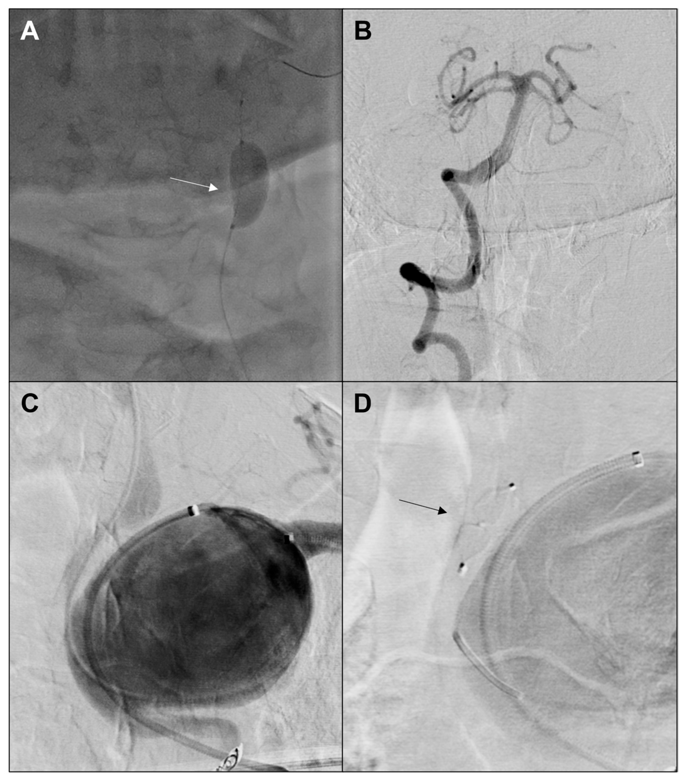
The first stage of the procedure, performed under local anesthesia through a percutaneous left brachial and right femoral approach, consisted of the left internal mammary artery and thyrocervical trunk embolization with 4 mm and 6 mm Ruby Coils (Penumbra Inc., Alameda, CA, USA) (Figure 2). Subsequently, BOT of the left VA was performed with an 8 × 20 mm balloon catheter inflated for 30 min with continuous neurological monitoring. The patient was observed for any symptoms of vertebrobasilar ischemia including dizziness or neurological deficits. Simultaneous angiography from the contralateral VA was conducted to demonstrate complete perfusion of the posterior circulation and the posterior inferior cerebellar artery, and retrograde flow into the occluded left vertebral artery, which was found to be adequate (Figure 3). Thus, the left VA was safely occluded with an 8 mm Amplatzer Vascular Plug IV (Abbott Vascular, Abbott Park, IL, USA).
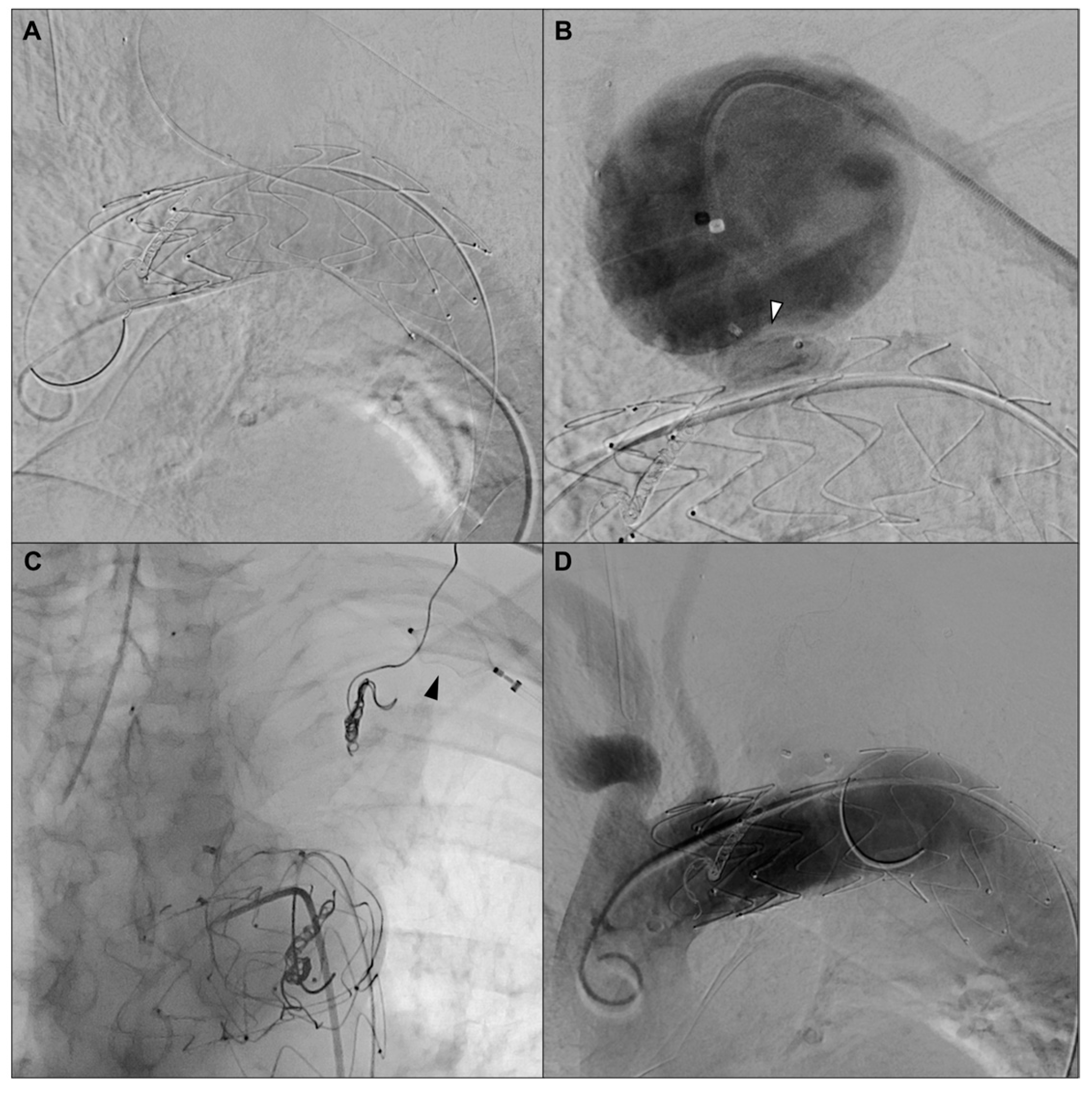
Three days later, the second procedural step was performed. EVSAR was achieved via zone 2 thoracic endograft placement (TEVAR) with a Cook Alpha 38 × 38 × 117 mm thoracic endograft (Cook Inc., Bloomington, IN, USA) and intentional coverage of the ostium of the LSA. Then, 16 mm and 14 mm Amplatzer Vascular Plugs (Abbott Vascular, Abbott Park, IL, USA) were used to seal the proximal segment of the LSA and distally occlude the axillary artery, respectively. The procedure was carried out under local anesthesia via percutaneous bilateral femoral and left brachial access. The final intraoperative angiography showed complete exclusion of the aneurysm without any signs of endoleak (Figure 4). Postoperative duplex ultrasound confirmed patency of the left brachial, radial, and ulnar arteries.
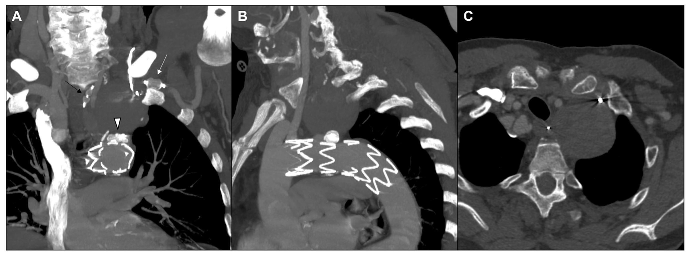
The postoperative course was uneventful. The patient was discharged on the third postoperative day on dual antiplatelet therapy. CTA was performed at 3, 6, and 12 months after surgery, showing the complete exclusion of the aneurysm without endoleak or other major complications (Figure 5).
After 1 month, the patient reported a moderate effort-induced pain in the left arm, which was successfully managed with the addition of Cilostazol 100 mg daily for 3 months, leading to progressive improvement of local symptoms. Subsequent follow-up revealed complete resolution of arm symptoms, likely due to the development of collateral circulation.
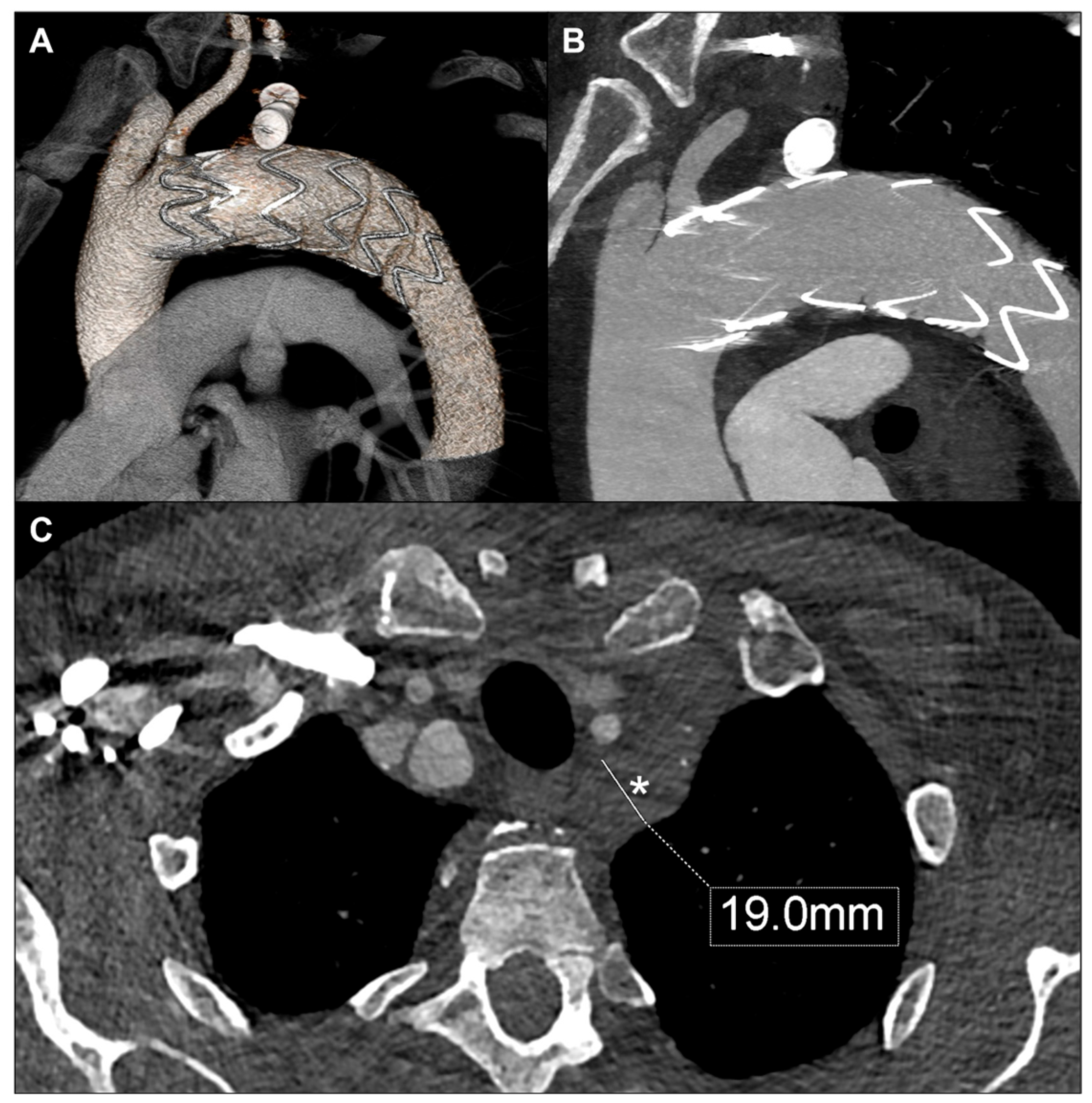
The 24-month CTA confirmed complete exclusion of the aneurysm, with significant aneurysmal sac shrinkage down to 19 mm (Figure 6).

3. Discussion

We report the case of a high-risk patient with a large intrathoracic subclavian artery aneurysm successfully treated with endovascular repair. A preoperative Balloon Occlusion Test guided safe vertebral artery sacrifice, ensuring adequate collateral perfusion. This case highlights the value of BOT in extending the indications for minimally invasive treatment in anatomically complex SAAs.
Subclavian artery aneurysms (SAAs) are uncommon vascular abnormalities, comprising less than 1% of all reported peripheral artery aneurysms [1,2]. The rarity of these cases has led to a lack of consensus regarding the optimal management strategy, particularly concerning size thresholds for intervention. Some experts advocate proactive repair in both symptomatic and asymptomatic patients, citing the potential for aneurysm growth and associated complications such as compression, embolization, rupture, and thrombosis, which may result in upper limb ischemia [7]. Conversely, other specialists recommend intervention only in cases of symptomatic aneurysms, rapid growth, or rupture [8].
Historically, open surgical repair has been the standard treatment for SAAs. This approach typically involves aneurysm resection and reconstruction with either direct end-to-end anastomosis or interposition grafting. However, open surgical repair often requires invasive procedures such as sternotomy or thoracotomy, which are associated with significant morbidity and mortality. A meta-analysis conducted by Vierhout et al. reported an 8% mortality rate and a 26% complication rate for open surgical repairs [1]. These statistics highlight the substantial risks associated with traditional surgical approaches, particularly in high-risk patients. In recent years, hybrid and total endovascular repair techniques have emerged as promising alternatives, offering minimally invasive options with reduced mortality and morbidity rates, especially for high-risk patients. EVSAR typically involves deploying a covered stent across the affected artery’s segment, with self-expanding stents preferred due to their adaptability in tortuous and dynamic arterial segments. However, in cases where standard endovascular approaches are not feasible due to unsuitable landing zones for stent graft sealing, more complex techniques may be required. These can include TEVAR with LSA coverage, vascular plug occlusion of the aneurysm outflow, and coil embolization of collateral branches originating from the aneurysm, including VA.
In our case, both a vascular plug and a thoracic endograft were used to ensure complete aneurysm exclusion. While the plug occludes distal flow, it does not prevent retrograde perfusion or fully depressurize the sac. The stent graft excludes the aneurysm from antegrade inflow, reduces sac pressure, and promotes thrombosis. This combined approach minimizes the risk of endoleak and should enhance long-term durability.
While EVSAR offers a less invasive alternative to open surgery, it is not without potential complications. Risks include endoleak, stent graft migration or occlusion, access-site complications, and the need for reintervention. These risks may be heightened in cases with challenging anatomy, limited landing zones, or extensive collateral circulation. Careful preoperative planning and patient selection remain essential.
Iatrogenic VA occlusion carries a low, yet clinically relevant, risk of posterior circulation stroke, potentially leading to severe neurological sequelae. Posterior circulation ischemia, resulting from impaired perfusion of the brainstem, cerebellum, thalamus, or occipitoparietal lobe, accounts for about 20% of ischemic strokes, and it is an important cause of disability and mortality [9,10]. However, this risk is mitigated by the basilar artery’s dual blood supply from both VAs. The collateral circulation from the contralateral VA, the internal carotid arteries, and a complete Circle of Willis likely explains the high tolerance to occlusion of surgically revascularized VAs reported in the literature [11]. Moreover, VA sacrifice is a therapeutic option employed to address issues such as penetrating VA injuries requiring embolization and cervical spine tumor requiring en-bloc tumoral mass and VA resection, without significant stroke or complication rates [12,13,14].
Ultimately, surgical revascularization of VA via a proximal VA-to-carotid artery transposition entails substantial risk of complications, such as recurrent laryngeal nerve palsy leading to hoarseness, Horner’s syndrome, posterior circulation transient ischemic attack or stroke, thoracic duct injury leading to chylothorax, and restenosis of the transposed artery. A recent comprehensive review of the literature on VA interventions reported a non-negligible complication rate for VA surgical revascularization, such as transient Horner syndrome (21.6%), transient vocal cord palsy (6.1%), stroke (4.1%), and death (9.4%) [15].
Considering the aforementioned aspects, we deemed it reasonable to perform VA sacrifice to achieve SAA exclusion, preceded by a BOT to demonstrate an adequate collateral flow. The latter allows for the simultaneous monitoring of the patient’s neurological status and the assessment of the retrograde ipsilateral vertebral filling. Although a comprehensive and large-scale validation of BOT in the vertebrobasilar circulation is lacking, this test remains a recognized clinical tool for tolerance prediction in patient undergoing therapeutic sacrifice of supra-aortic vessels [12,16].
BOT provides functional assessment of the posterior circulation, which static imaging techniques such as CT angiography or digital subtraction angiography cannot fully capture. While imaging may demonstrate vertebral artery patency or symmetry, it cannot predict real-time neurological tolerance to occlusion. This is especially relevant when both vertebral arteries are of comparable size, as anatomical symmetry does not always correlate with hemodynamic dominance. In such cases, BOT helps guide clinical decision-making and avoid both unnecessary revascularization and unexpected ischemic complications.

4. Conclusions

EVSAR, combined with preoperative BOT, may represent a minimally invasive and effective treatment option for managing complex SAAs in high-risk patients, particularly when the intrathoracic segment of the subclavian artery is affected. Preoperative BOT appears to be a valuable tool in assessing the safety of VA sacrifice, helping tailor treatment to individual anatomical and clinical contexts. Additionally, complete embolization of all arterial outlets is advisable to minimize the risk of type II endoleak and ensure durable aneurysm exclusion.

Author Contributions

Conceptualization, C.C. and C.F.; methodology, F.F.P. and M.M.; validation, R.G., C.C., and C.F. writing—original draft preparation, A.B. and F.F.P.; writing—review and editing, A.B. and F.F.P.; visualization, B.O.D.; supervision R.G., C.C. and C.F. All authors have read and agreed to the published version of the manuscript.

Funding

This research received no external funding.

Institutional Review Board Statement

Written informed consent was obtained from the patient for publication of this case report and accompanying images. No identifiable patient information is disclosed in this report. The study, due to its nature, did not require ethics committee approval.

Informed Consent Statement

Written informed consent was obtained from the patient for publication of this case report and accompanying images.

Conflicts of Interest

There are no conflicts of interest to disclose.

References

  1. Vierhout, B.P.; Zeebregts, C.J.; van den Dungen, J.J.; Reijnen, M.M. Changing profiles of diagnostic and treatment options in subclavian artery aneurysms. Eur. J. Vasc. Endovasc. Surg. 2010, 40, 27–34. [Google Scholar] [CrossRef] [PubMed]
  2. Davidovic, L.B.; Zlatanovic, P.; Ducic, S.; Koncar, I.; Cvetic, V.; Kuzmanovic, I. Single center experience in the management of a case series of subclavian artery aneurysms. Asian J. Surg. 2020, 43, 139–147. [Google Scholar] [CrossRef] [PubMed]
  3. Andersen, N.D.; Barfield, M.E.; Hanna, J.M.; Shah, A.A.; Shortell, C.K.; McCann, R.L.; Hughes, G.C. Intrathoracic subclavian artery aneurysm repair in the thoracic endovascular aortic repair era. J. Vasc. Surg. 2013, 57, 915–925. [Google Scholar] [CrossRef] [PubMed]
  4. Lee, K.S.; Jung, Y.; Jeong, I.S.; Song, S.Y.; Na, K.J.; Oh, S.G. Open surgical treatment of subclavian artery pseudoaneurysm after endovascular repair: A case report. J. Cardiothorac. Surg. 2022, 17, 25. [Google Scholar] [CrossRef] [PubMed]
  5. Marjanović, I.; Tomić, A.; Marić, N.; Pecarski, D.; Sarac, M.; Paunovic, D.; Rusovic, S. Endovascular treatment of the subclavian artery aneurysm in high-risk patient—A single-center experience. Vojnosanit. Pregl. 2016, 73, 941–944. [Google Scholar] [CrossRef] [PubMed]
  6. Maskanakis, A.; Patelis, N.; Moris, D.; Tsilimigras, D.I.; Schizas, D.; Diakomi, M.; Bakoyiannis, C.; Georgopoulos, S.; Klonaris, C.; Liakakos, T. Stenting of Subclavian Artery True and False Aneurysms: A Systematic Review. Ann. Vasc. Surg. 2018, 47, 291–304. [Google Scholar] [CrossRef] [PubMed]
  7. Leverich, M.; Nazzal, M.; Osman, M. Single-stage hybrid repair of a right subclavian artery aneurysm involving the origin of the right vertebral artery. Ann. Vasc. Surg. Brief. Rep. Innov. 2023, 3, 100173. [Google Scholar] [CrossRef]
  8. French, B.L.; Menghini, A.M.; Burke, C.R.; Byers, P.H.; Shalhub, S. Operative repair of right intrathoracic subclavian artery aneurysms in patients with genetic arteriopathy. J. Vasc. Surg. Cases Innov. Tech. 2022, 9, 101081. [Google Scholar] [CrossRef] [PubMed] [PubMed Central]
  9. Schneider, A.M.; Neuhaus, A.A.; Hadley, G.; Balami, J.S.; Harston, G.W.; DeLuca, G.C.; Buchan, A.M. Posterior circulation ischaemic stroke diagnosis and management. Clin. Med. 2023, 23, 219–227. [Google Scholar] [CrossRef] [PubMed] [PubMed Central]
  10. Zhang, C.C.; Li, L.Q.; Guo, L.R.; Qi, L.X.; Guo, J.M.; Gu, Y.Q. Endovascular and hybrid treatments for subclavian artery aneurysms. Ann. Ital. Chir. 2021, 92, 709–714. [Google Scholar] [PubMed]
  11. Shergill, E.S.; Udwadia, F.R.; Grubisic, M.; Salata, K.; Misskey, J.; Faulds, J. Comparative study of left vertebral artery revascularization in patients with and without aberrant left vertebral anatomy. J. Vasc. Surg. 2024, 79, 991–996. [Google Scholar] [CrossRef] [PubMed]
  12. Sorteberg, A.; Bakke, S.J.; Boysen, M.; Sorteberg, W. Angiographic balloon test occlusion and therapeutic sacrifice of major arteries to the brain. Neurosurgery 2008, 63, 651–660, dicussion 660-1. [Google Scholar] [CrossRef] [PubMed]
  13. Westbroek, E.M.; Pennington, Z.; Ehresman, J.; Ahmed, A.K.; Gailloud, P.; Sciubba, D.M. Vertebral Artery Sacrifice versus Skeletonization in the Setting of Cervical Spine Tumor Resection: Case Series. World Neurosurg. 2020, 139, e601–e607. [Google Scholar] [CrossRef] [PubMed]
  14. Peeters, J.B.; Dessesard Olijnyk, L.; Janelle, F.; Shedid, D.; Bojanowski, M.W.; Labidi, M. Surgical management of tumors of the cervical spine and craniovertebral junction involving the vertebral artery: A narrative review. Neurochirurgie 2024, 70, 101550. [Google Scholar] [CrossRef] [PubMed]
  15. Maitas, O.; Bob-Manuel, T.; Price, J.; Noor, A.; Obi, K.; Okoh, N.; Garikapati, K.; Kim, J.; Jahan, S.; Jenkins, J.S. Vertebral Artery Interventions: A Comprehensive Updated Review. Curr. Cardiol. Rev. 2023, 19, e170322202296. [Google Scholar] [CrossRef] [PubMed] [PubMed Central]
  16. Tansavatdi, K.; Dublin, A.B.; Donald, P.J.; Dahlin, B. Combined Balloon Test Occlusion and SPECT Analysis for Carotid Sacrifice: Angiographic Predictors for Success or Failure? J. Neurol. Surg. B Skull Base 2015, 76, 249–251. [Google Scholar] [CrossRef] [PubMed] [PubMed Central]
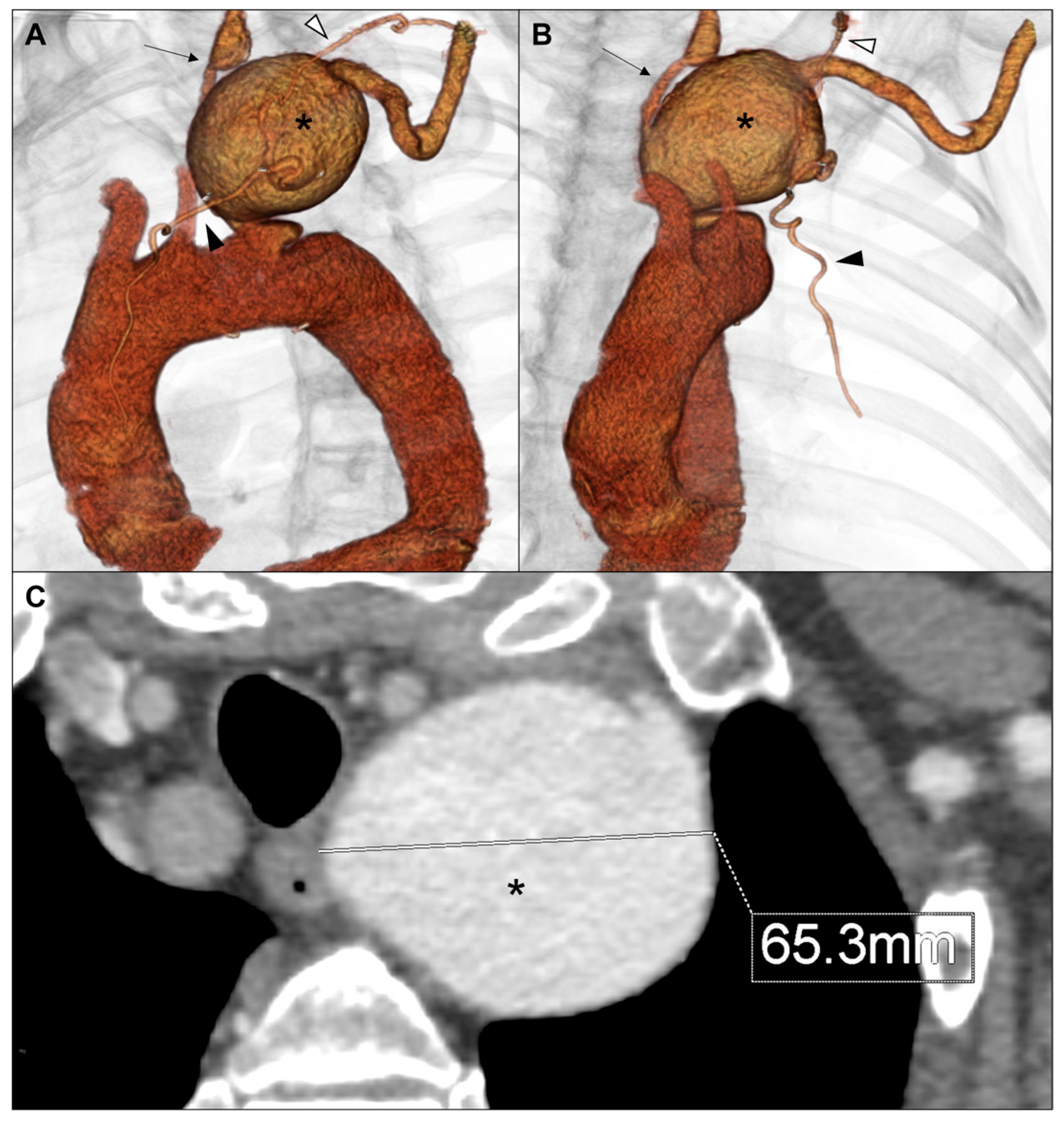
Figure 1. Volume rendering of preoperative computed tomography angiography in lateral (A) and anterior (B) view showing left subclavian artery aneurysm (*), left vertebral artery (black arrow), left internal mammary artery (black arrowhead), and left thyrocervical trunk (white arrowhead). Axial view showing aneurysm’s maximum diameter (C).
Jvd 04 00035 g001
Figure 2. Intraoperative details showing selective angiography (A,C) and coil placement (B,D) of the left internal mammary artery (black arrow) and left thyrocervical trunk (white arrow), respectively.
Jvd 04 00035 g002
Figure 3. Balloon Occlusion Test (A) with an 8 × 20 mm balloon catheter (white arrow) during concomitant contralateral vertebral artery selective angiography (B), followed by selective angiography of the left vertebral artery (C) and embolization (D) by the placement of an 8 mm vascular plug (black arrow).
Jvd 04 00035 g003
Figure 4. Catheterization of the proximal left subclavian artery from left brachial access (A) and embolization (B) by the placement of a 16 mm vascular plug (white arrowhead). Occlusion of the axillary artery (C) by the placement of a 14 mm vascular plug (black arrowhead), and final angiography showing the aneurysm’s exclusion (D).
Jvd 04 00035 g004
Figure 5. Maximum intensity projection in anterior view (A) of the 3-month computed tomography angiography highlighting the vascular plug in the left vertebral artery (black arrow), left axillary artery (white triangle), and proximal portion of the left subclavian artery (white arrow). Lateral (B) and axial view (C) showing the complete exclusion of the aneurysm.
Jvd 04 00035 g005
Figure 6. Volume rendering (A), maximum intensity projection (B), and axial view (C) of the 24-month computed tomography angiography showing significant shrinkage of the aneurysm (*).
Jvd 04 00035 g006
Disclaimer/Publisher’s Note: The statements, opinions and data contained in all publications are solely those of the individual author(s) and contributor(s) and not of MDPI and/or the editor(s). MDPI and/or the editor(s) disclaim responsibility for any injury to people or property resulting from any ideas, methods, instructions or products referred to in the content.