Review

Biochemical Insights and Clinical Applications of Ischemia-Modified Albumin in Ischemic Conditions

by
Nimesha N. Senadeera
1,*,
Chathuranga B. Ranaweera
2,*,
Inoka C. Perera
1 and
Darshana U. Kottahachchi
2
1
Department of Zoology and Environment Sciences, Faculty of Science, University of Colombo, Colombo 00300, Sri Lanka
2
Department of Medical Laboratory Sciences, Faculty of Allied Health Sciences, General Sir John Kotelawala Defence University, Werahera 10290, Sri Lanka
*
Authors to whom correspondence should be addressed.
J. Vasc. Dis. 2024, 3(3), 245-266; https://doi.org/10.3390/jvd3030020
Submission received: 26 April 2024 / Revised: 9 June 2024 / Accepted: 27 June 2024 / Published: 8 July 2024
(This article belongs to the Section Cardiovascular Diseases)

Abstract

:
Atherosclerotic coronary artery disease is a significant global health threat, impacting millions annually. Over time, plaque buildup narrows the coronary arteries, reducing blood flow to the heart muscle and resulting in myocardial ischemia. Timely diagnosis and intervention are crucial for restoring the blood flow to the heart muscle and preventing myocardial infarction. Given the limited availability of screening and diagnostic tests, the early diagnosis of myocardial ischemia remains challenging. While cardiac troponin is considered the gold standard for detecting myocardial injury, its effectiveness in identifying myocardial ischemia is limited. Ischemia-modified albumin (IMA) is a modified albumin variant that serves as a sensitive and early marker for ischemia. Despite extensive research on diagnostic applications of IMA as a biomarker for ischemia, significant gaps remain in understanding its formation, sensitive and specific detection, and precise clinical utility. This review aims to address these gaps by compiling literature on IMA discussing the latest findings on structure and formation, and detection methods. Further research is required to enhance understanding of the structure and formation of IMA, aiming to develop novel detection techniques or improve existing ones. However, currently, available sophisticated methods are associated with higher expenses and require specialized equipment and qualified personnel.

1. Introduction

Atherosclerotic coronary artery disease (ACAD) is a condition in which fatty plaques accumulate in the cells lining the arteries that supply blood to the heart (coronary arteries) [1]. Plaque formation causes the arteries to narrow over time, obstructing blood flow partially or entirely. Lack of blood flow to the heart muscle can cause myocardial ischemia. This reduces the ability of the cardiac muscle to pump blood and causes chest pain, leading to angina [2]. Sudden, severe obstruction of one of the coronary arteries can lead to myocardial infarction [3]. According to the American Heart Association (AHA), the prevalence of coronary artery disease (CAD) rises with age [4]. CAD affects around 315 million people [5]. In the year 2021, the total number of deaths globally amounted to almost 20.5 million, which is nearly one-third of all recorded deaths [6]. In Sri Lanka, the rate of CAD has doubled over the last two decades, establishing it as the primary leading cause of death in the country [7,8]. There is a lack of recent prevalence data available for Sri Lanka after 2020. However, the World Health Organization (WHO) reported that in Sri Lanka in 2021, cardiovascular diseases (CVDs) were responsible for 40% of all fatalities [9]. Stroke and ischemic heart disease were shown to be the significant causes of death, accounting for 10% and 20% of all fatalities, respectively [10].
CAD is initially diagnosed based on a comprehensive assessment that includes evaluating the patient’s symptoms, medical and family history, and risk factors. Various tests and procedures, such as blood tests, electrocardiograms (EKG or ECG), exercise stress tests, and cardiac imaging, contribute to diagnosing CAD [11]. During angina or acute myocardial infarction episodes, cardiac biomarkers like troponins, ECG, and cardiac imaging are employed to make accurate diagnoses [12].
The identification of cardiac ischemia, especially in emergency cases, poses challenges. Although cardiac troponin is a sensitive marker for myocardial injury, it may not elevate in cases of myocardial ischemia [13]. Recent research has indicated that Ischemia Modified Albumin (IMA) serves as a sensitive and early marker for ischemia, detectable within 6–10 min of a cardiac ischemic event and remaining increased for 12 h [14,15]. The current detection method, albumin cobalt binding (ACB) assay, offers rapid results but has limitations such as limited reproducibility and accuracy, along with potential false negatives due to denaturation of albumin during the process [16].
Most studies to date have focused on the diagnostic utility of IMA, often overlooking its formation and structural modifications, which has become a significant obstacle to developing novel detection methods or improving the sensitivity and specificity of existing detection methods. To address this gap, this review aims to compile existing literature on IMA, discussing the latest findings, exploring the structural modifications and current hypotheses on the formation of IMA and the existing detection methods, along with their principles, advantages, and limitations. To conduct a thorough literature analysis, we employed internet search engines like Google, as well as academic databases such as Google Scholar, PubMed, ScienceDirect, and MEDLINE. These methods enabled us to collect important studies, peer-reviewed articles, and other scholarly materials related to our research topic.

2. Atherosclerotic Coronary Artery Disease (ACAD)

ACAD, also known as coronary artery disease, arises when coronary arteries become constricted or blocked over time by cholesterol-containing deposits (plaques) and inflammation. It is also known as ischemic heart disease (IHD) or ACAD [1]. The coronary arteries supply blood to the heart, oxygen, and nourishment [17]. Endothelial dysfunction in the coronary arteries is one of the earliest observable alterations and the first step to atherosclerosis. It allows lipid material to flow into the subintimal region of the arteries, where macrophages and other cells trigger a potent inflammatory response. Due to prolonged inflammation, cell debris and cholesterol crystal build-up eventually lead to plaque formation and advancement. Plaque formation can narrow these arteries, reducing blood flow to the heart. Reduced blood flow may lead to chest pain (angina), shortness of breath, and other signs and symptoms of CAD [3]. There are two main types of risk factors for CAD. Risk factors that can be controlled and that are beyond our control. Controllable risk factors are smoking, obesity, diabetes mellitus, hypertension, abnormal cholesterol levels in the blood, lack of exercise, excessive stress and depression, etc. These are also known as modifiable risk factors. Uncontrollable risk factors include age, with age, the risk of heart disease increases; gender, men over the age of 55 and women following menopause are at risk; heredity and ethnicity, people of African or Asian ethnicities are more likely to acquire heart disease than other ethnicities [18,19].

2.1. Global Prevalence

CAD affects around 315 million people [1]. The age-standardized prevalence of CAD in 2022 was 3605 per 100,000, and Central Europe, Eastern Europe, and Central Asia had the highest age-standardized prevalence of the condition, with a rate of 8019 per 100,000. On the other hand, South Asia had the lowest prevalence, with a rate of 2393 per 100,000 [5]. The disease first appeared in the mid 1940s and has become more common as people age—adults under the age of 65 account for around 2 out of every 10 fatalities due to CAD. The present prevalence rate of 1655 per 100,000 people is predicted to rise to 1845 by 2030, according to the calculations [17]. The AHA (2021) stated that the prevalence of CAD increases with age. In the 20–39-year age group, the prevalence is 0.6% in both sexes, while in the age group of 40–59 years, both sexes have a greater frequency. In the 60–79-year age group, 20% of males and 9.7% of females, and the 80+ year age group, 32% of males and 19% of females have been reported. According to the data, males were more affected than females [19,20,21].

2.2. Local Prevalence

CVDs are the main leading cause of death in Sri Lanka, accounting for 34% of all deaths [22]. Notably, there is a lack of recent prevalence data on CAD for Sri Lanka after 2020. However, the WHO reported that in Sri Lanka, in 2021, CVDs were responsible for 40% of all fatalities [9]. Stroke and ischemic heart disease were shown to be the significant causes of death, accounting for 10% and 20% of all fatalities, respectively [10]. The incidence of CAD ranged from 455.4 in 2011 to 667.2 in 2019 per 100,000 population [8]. During the last 10 years, CAD was ranked as the number one leading cause of hospital deaths in the country, accounting for about 15.1% of all hospital deaths, with 37.2 deaths per 100,000 population. However, CAD’s net case fatality rate decreased from 5.8% in 2014 to 5.6% in 2019. Jayawardena et al. conducted a study with adults aged 30 years and above, 579 females and 421 males in the Bope-Poddala Medical Officer of Health (MOH) area. The prevalence of coronary artery disease was 6.9%, and silent ischemia was identified as 2.2%. Their study revealed that males and those with a history of non-communicable diseases linked to cardiovascular risk were more likely to develop CAD. [4].

2.3. Pathophysiology of CAD

This process begins with endothelial dysfunction (Figure 1), caused by factors such as hypertension, hyperlipidemia, smoking, and diabetes. During the quiescent or resting state of the endothelium, cells inhibit platelet activation, blood clotting, and leukocyte adherence through the secretion of chemicals such as nitric oxide (NO), prostacyclin, tissue plasminogen activator, and antithrombin III. Mechanical factors, such as turbulent flow and shear stress, exert on endothelial cells and influence the alterations in cellular morphology, intracellular signaling, and gene expression. Inflammation enhances the expression of adhesion molecules, facilitating the infiltration of leukocytes into the endothelium and the oxidative modification of lipoproteins. Endothelial dysfunction is characterized by changes in the production and availability of endothelial-derived NO, prostacyclin, and endothelin, impacting vascular reactivity. As a result, reactive oxygen species (ROS) are able to reach regulatory molecules, which then activate cells and create an environment that constricts blood vessels and promotes blood clotting. The media compromises the endothelium’s structural integrity, increasing activity and buildup within the artery wall. Low-density lipoprotein (LDL) cholesterol penetrates the damaged endothelium and accumulates in the arterial wall. Modified lipids activate inflammatory cells, producing chemokines and cytokines that activate other cells and adhesion molecules. These cells recruit inflammatory cells. Macrophages absorb cholesterol-rich lipoproteins from LDL and secrete pro-oxidant substances like ROS and RNS. This can lead to lipid peroxidation and LDL oxidation. Oxidized phospholipids trigger inflammation. Oxidized LDL-engulfed macrophages form foam cells, which aggregate and form fatty streaks. This is the earliest visible sign of atherosclerosis. As the fatty streaks evolve, smooth muscle cells migrate from the arterial media to the intima, proliferating and secreting extracellular matrix. This forms a fibrous cap over the lipid core, creating a mature atherosclerotic plaque. Advanced plaques may become unstable due to a thin fibrous cap and a large lipid core, making them prone to rupture. Plaque rupture exposes the underlying pro-thrombotic materials, leading to platelet aggregation and thrombus formation. The thrombus can partially or entirely occlude the coronary artery, reducing or halting blood flow to the myocardium. This results in ischemia and, if prolonged, leads to myocardial infarction (heart attack) [23,24].
Coronary atherosclerosis develops into two clinicopathological conditions: angina pectoris and acute coronary syndrome. Stable angina is caused by plaque development in vessel lumens, resulting in ischemia when cardiac oxygen demand increases. This condition is reversible and can cause exercise-induced pain. The acute coronary syndrome is a fast-progressing condition characterized by local thrombosis, a rapid decrease in lumen diameter, and a thrombus when the luminal side of an atherosclerotic plaque ruptures, exposing thrombogenic material to blood. The extent of thrombosis, vasospasm, and occlusion duration determine whether the event causes unstable angina or myocardial infarction [19,25].

2.4. Diagnostic Tests of CAD

There are two types of diagnostic tests: non-invasive and invasive. Non-invasive diagnostic testing involves cardiac imaging, while a tube, device, or scope can be inserted in invasive tests [20]. Invasive tests are blood tests used to evaluate the blood levels of cholesterol, glucose, lipoproteins, inflammatory markers, cardiac catheterization, transesophageal echocardiography (TEE), electrophysiologic tests, etc. Non-invasive tests are electrocardiograms (EKG or ECG) to detect whether the heart’s rhythm is regular or irregular, exercise stress tests to see how the heart responds to physical activity, echocardiogram, magnetic resonance imaging (MRI), chest X-rays, and computed tomographic (CT) scans of the heart and coronary arteries to detect tissue damage or difficulties with blood flow, coronary angiography used to see the inside of the coronary arteries, and coronary calcium scans to determine how much calcium is present in the walls of coronary arteries. During symptoms of angina or acute myocardial infarction, cardiac biomarkers, ECG, and cardiac imaging are used in diagnosis [11].

2.5. Use of Biomarkers in the Diagnosis of CAD

During these different stages of CAD, biomarkers are released. For example, biomarkers of myocardial necrosis, cardiac troponins; biomarkers of myocardial ischemia, ischemia-modified albumin (IMA); biomarkers of hemodynamic stress, natriuretic peptides (NPs); inflammatory markers, C-reactive protein (CRP). The identification and quantification of these biomarkers are useful in the early detection and staging of CAD [14]. Biomarkers of myocardial ischemia, hemodynamic stress, and inflammatory markers can be detected during the early stages of CAD. Ischemia occurs when blood flow to the heart muscle is diminished, preventing the heart muscle from receiving adequate oxygen and nutrition to pump blood [17] The changes in the dynamics of blood flow, like increased blood flow into the ventricles, create a pressure that stretches the ventricular wall and induces the release of molecules like natriuretic peptides, which are biomarkers of hemodynamic stress [26]. Inflammation is detected by circulating chemicals and molecules produced as a cause or consequence of an inflammatory reaction. These markers are not specific to a particular disease and are primarily used in disease prognosis and follow-ups rather than screening. Cardiac troponins increase where myocardial injury and cell necrosis occur [27].
Table S1 tabulates key biomarkers of diagnosis and management of cardiovascular diseases (see Supplementary Materials).

3. Ischemia Modified Albumin (IMA) as a Biomarker of Myocardial Ischemia

Presently, choline, free fatty acids, and IMA are identified as biomarkers of cardiac ischemia [14]. IMA is a modified form of HSA that serves as an early indicator of ischemic events. The rationale behind using IMA lies in its sensitivity to ischemic conditions. When ischemia occurs, the biochemical environment changes, causing alterations in the structure of albumin [28]. IMA was found to have a reduced capacity to bind transition metals like cobalt and copper in the N-terminal region, resulting in a measurable decrease in its ability to bind these metals [29,30]. The mechanism underlying the modification of albumin in ischemic conditions is multifaceted and will be discussed in detail in the subsequent sections. Several risk factors contribute to IMA formation. These include traditional cardiovascular risk factors and ischemia risk factors. Age, gender, and ethnicity are examples of non-modifiable risk factors, while modifiable risk factors include hypertension, diabetes mellitus, hyperlipidemia, dyslipidemia, and smoking [31,32]. Conditions that increase oxidative stress, such as obesity, systemic inflammatory conditions, and metabolic syndrome, also elevate IMA levels [33,34,35].
The clinical significance of ischemia-modified albumin lies in its potential as a diagnostic marker for ischemic conditions, especially acute coronary syndromes, which encompass conditions like unstable angina and myocardial infarction. This modification occurs rapidly during ischemic events, elevating the levels of IMA within 6–10 min and remaining elevated for 12 h [14,15]. Furthermore, ischemia-modified albumin has been investigated in various other clinical contexts, including acute cerebrovascular diseases, peripheral artery disease, and preeclampsia, where early detection and intervention are crucial for patient management and prognosis [36,37,38,39,40,41]. Given that IMA levels can be affected by conditions other than cardiac ischemic events, it is critical to use specific detection methods and conduct extensive examinations of structural alterations associated with each ischemic event. This is essential for creating accurate procedures and protocols for various ischemic conditions and setting reference ranges accordingly. Studies have demonstrated associations between elevated levels of IMA and the severity, extent, and prognosis of ischemic events, suggesting its potential utility as a prognostic marker in addition to its diagnostic role. The measurement of IMA has been evaluated using the Albumin Cobalt Binding (ACB) assay. It is now available as a Food and Drug Administration, United States of America (FDA-USA)-approved diagnostic for regular clinical use [29,42,43]. India [44,45], the USA [43], and China [46] have used this assay in emergency departments for rapid assessment of patients with chest pain. ACB assay is made based on the reduced metal binding capacity due to structural modification of HSA. Despite the promise of ischemia-modified albumin as a biomarker, challenges remain in standardizing assays, establishing reference ranges, and elucidating its precise role in different disease states. Further research is warranted to validate its clinical utility, refine assay techniques, and explore its potential as a therapeutic target in ischemic conditions.

3.1. Human Serum Albumin

HSA and IMA share a common structural framework, with IMA being a modified form of HSA. Therefore, discussing HSA allows us to understand the implications of ischemia-induced modifications on its three-dimensional conformation, ligand-binding properties, and biological functions. Human Serum Albumin is more than 60% of human blood plasma proteins. It is a monomer, globular protein, heart-shaped with alpha helix, beta turns, and loops. It is found in the circulation and acts as a transporter protein in plasma. HSA is a water-soluble protein, with blood concentrations ranging from 30 to 50 g/L in a pH range of 7.35–7.4 [47]. The ALB (Albumin) gene encodes HSA. Pre-pro albumin, the first translation product, has a molecular weight of 69.3 kDa and 609 amino acids (AA). It includes an N-terminal endoplasmic reticulum (ER) import signal sequence. This signal sequence facilitates the co-translational translocation of the nascent protein into the ER lumen, where the signal sequence is cleaved by signal peptidase. This targeting allows the protein to undergo proper folding and post-translational modifications, and it plays a role in the ER’s quality control processes, ensuring proteins reach their correct destinations and function properly. Pre-pro albumin is made in the liver and then modified post-translationally in the Golgi apparatus. Before the mature HSA protein is released into the circulation, the first 24 amino acids from the N-terminus (1–18 signal peptide and 19–24 pro-peptide) are cleaved. The mature HSA protein consists of 585 amino acids and has a molecular weight of 66.5 kDa (Figure 2) [48,49].
The three-dimensional structure of HSA provides detailed information about its binding sites and interaction domains. This structural knowledge is essential for understanding how albumin interacts with various ligands and divalent metal ions. This is crucial because understanding the modifications in certain protein regions, such as the N-terminus, can alter its binding affinity and specificity. The first three-dimensional structure (Figure 2) of HSA was determined by X-ray crystallography in 1992 (Protein Data Bank (PDB) id: 1UOR) [50]. Since then, about 100 stationary structures of HSA and HSA with bound ligands have been determined, primarily using X-ray crystallography, and have been deposited in the PDB. Matured and active HSA contains 17 disulfide bridges that shape the protein fold (Figure 3) and one free cysteine at position 34 (Cys 34). It is made up of three topologically identical and structurally similar domains: IA (AA 5–107), IB (AA 108–196), IIA domain (AA 197–297), IIB (AA 298–383), and domain IIIA (AA 384–497), IIIB (AA 498–585) [51]. Each domain is made up of one antiparallel six-helix (sub-domain A) and one four-helix (sub-domain B) motif (Figure 3). It has several binding sites for fatty acids, heme, ligands, and metal ions. Four metal binding sites include sites A, site B, N-terminal site (NTS), and Cys34. Site A can bind metals such as Zn2+, Cd2+, Ni2+, Co2+, and Cu2+. Cadmium ions have a high affinity for Site B. Cobalt, copper, and nickel ions bind to the NTS, while gold and platinum have the highest affinity for Cys34 [27,28,47,52].
With its inherent flexibility, this N-terminal motif was never observed in the experimental X-ray structures. Typically, the defatted HSA structure lacks the first 1–5 amino acid residues, but the fatty acid-containing structure lacks only the first two amino acids of the N-terminus. N-terminal residues, indicating an allosteric action and fatty acid-driven modulation/regulation of the HSA’s metal ion coordination capacities. This highlights the need for high-resolution, detailed investigations of the molecular dynamics of the protein, as well as information on binding affinity to ligands and metal ions [47,48].
Amino acid sequence alignment provides valuable insights into structural and functional similarities. By comparing the sequences of different species, researchers can identify conserved regions and the degree of conservation reflecting evolutionary significance. Amino acid sequence HSA aligned with serum albumin of mouse, rat, rabbit, bovine, and pigs revealed that it is conserved more than 70% among these species (Supplementary Data provided in Supplementary Materials).

3.2. Formation of IMA

The mechanism of IMA formation was not well defined. The literature provides a few hypotheses and models to explain what is observed.

3.2.1. First Model/Hypothesis—Auto Degradation of NTS

The first model is the auto degradation of the N-terminus. Chan et al. studied the nature of this auto degradation using HSA (synthetic and isolated from clinical specimens). In their experiment, different HSA solutions (synthetic, clinically isolated, and purified) were just incubated at different temperatures (4 °C, 30 °C, 37 °C, 57 °C) for varying periods. With increased temperature and time of incubation, auto degradation was evident. Two amino acids were lacking when the N-terminal was sequenced (Aspartate-Alanine). These two amino acids were found to be released as intact dipeptides rather than individual amino acids. The results implied that this would occur in vivo during the lifetime of the HSA molecule (the half-life is approximately 21 days). Dipeptide that was released at 37 °C incubation (for eight weeks) was difficult to detect by electrospray mass spectrometry (ES-MS) analysis due to high background signals at low mass ranges. This same experiment was conducted for the non-human albumin samples, and no N-terminal degradation was evident as HSA. This indicates that auto degradation was highly specific for the HSA amino acid sequence. Further, the dipeptides that were released from synthetic HSA and purified HSA from clinical specimens were different in structure (observed in nuclear magnetic resonance (NMR) results) [53].
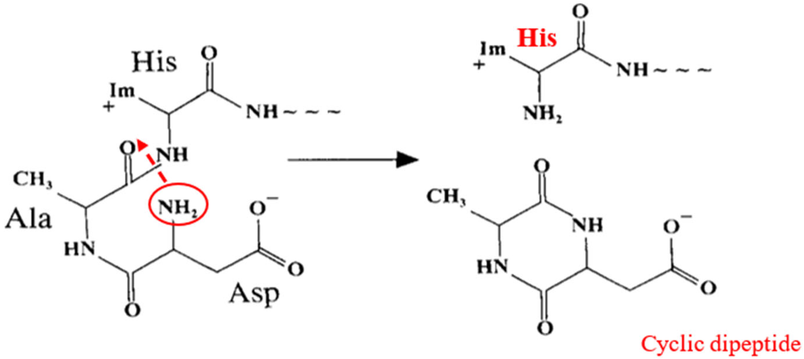
As shown in Figure 4, The Asp1 alpha-amino group has nucleophilic properties (nucleophilicity of the alpha-amino group of Asp1 increased due to proton withdrawal by the Aspl carboxyl group, and electrophilicity of the carbonyl group of Ala2 may increase due to proton donation from imidazole of His3). Amine nitrogen exerts a nucleophilic attack on the carbonyl of the peptide bond between Ala2 and His3. This cleaves the peptide bond between alanine and histidine and releases a cyclic dipeptide. Additionally, it was reported that this degradation is not due to oxidative damage by metal ions or protease action. Both these situations have been experimentally evaluated and eliminated. Furthermore, the authors of the study suggested performing further investigations on the detection and role of this dipeptide since dipeptides are known to be active neuropeptides in the central nervous system [53].

3.2.2. The Second Model—N-Terminal Site Modification by Free Radicals

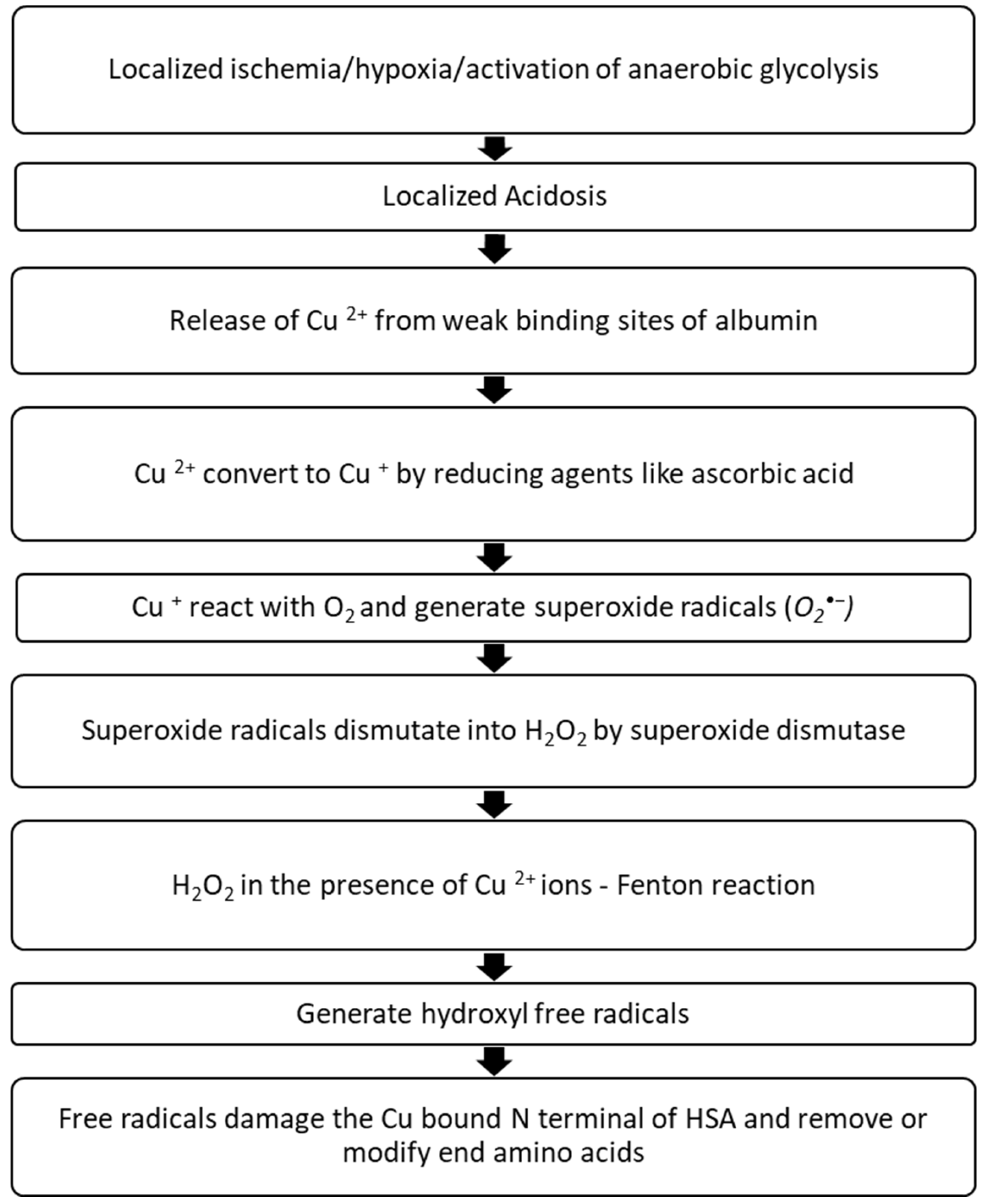
This postulated mechanism describes the generation of IMA as radical damage to the HSA N-terminal. As shown in Figure 5, localized ischemia, hypoxia, and activation of anaerobic glycolysis result in acidosis. Cu2+ ions are released from their weak binding sites in a localized acidic environment. In the presence of a reducing agent such as ascorbic acid, free Cu2+ is converted to Cu+ ions. These Cu+ ions react with oxygen to form Cu2+, generating superoxide free radicals. This reaction between Cu2+/Fe3+ and ascorbate generates free radicals, which can modify proteins in various ways. Normally, these superoxide radicals are converted to hydrogen peroxide (H2O2) by superoxide dismutase, and H2O2 is degraded to oxygen and water by catalase. Cu2+ ions (which were formed during Cu+ oxidation) immediately scavenge by HSA and tightly bind with N-terminal (Cu2+ have the highest affinity towards the N-terminal site; Dissociation constant (KD) = 6.61 × 10 −17 M−1). H2O2 in the presence of Cu2+/Fe3+ ions undergo Fenton reaction and generate hydroxyl free radicals. These hydroxyl radicals damage the copper-bound N-terminus of HSA, causing the last three amino acids to cleave and release Cu2+ ions. The process will repeat in a chain reaction [27].
This site-specific modification was investigated by Marx and Chevion using a reaction with Cu2+ and ascorbate. These modifications can be measured in a few ways, such as the detection of the NH3 released during the modifications, decrease in fluorescence, loss of enzyme activity, protein coagulation, and changes in amino acid sequences. They experimentally demonstrated that the reaction between Cu2+ and ascorbate leads to radical formation, and site-specific modifications were identified. To detect the modifications, they used decreased fluorescence signals (in the emission fluorescence spectrum) and Sodium Dodecyl Sulfate/Polyacrylamide gel electrophoresis to visualize the cleaved fragments. The authors also revealed that this site-specific degradation did not occur in the absence of Cu2+ ions or metal-chelating agents like Ethylene Diamine Tetra Acetic acid [54].
Roy et al. researched to model the formation of IMA in vitro using chemically generated radicals. Serum aliquots were randomly chosen. Group 1 was peroxide-treated, group 2 was superoxide-treated, group 3 was hydroxyl-treated, and group 4 was a technical control in which aliquots of serum were incubated CuSO4 as a technical control for group 3. Group 5 was treated with hydroxyl and mercapto propionyl glycine (MPG) (hydroxyl radical scavenger) serum, and Group 6 was treated with control sera, incubated for 15 min without any reactants. After the specific treatments, IMA was measured using an ACB assay. IMA concentrations in the control serum (group 6) did not change considerably at any time. Compared to the control, neither group 1 nor group 2 exhibited significant changes in IMA concentration. Nevertheless, group 3 was linked with a rapid rise in IMA concentration. The hydroxyl scavenger MPG (group 5) was added to the Fenton reaction mixture, which reduced the generation of IMA. The findings of this study revealed that the hypothesis of IMA formation by ROS, specifically hydroxyl radicals, can chemically modify Human Serum Albumin, leading to the generation of IMA [55].

3.2.3. 3rd Hypothesis—Acetylation of the N-Terminus

Bar-or et al. (the first authors who introduced the ACB assay) investigated exogenous Co2+ and Ni2+ binding to the N-terminal of HSA using synthetic peptides. This experiment used the synthesized peptide N-acetyl DAHKSEVAHRFK (acetylated amino acids including the N-terminus of HSA). Then, CoCl2 was mixed with this dodecapeptide and analyzed by 1H NMR and spectrophotometric cobalt binding methods. The results revealed that the acetylation of the N-terminus of this dodecapeptide inhibited cobalt binding [56].

3.2.4. Other Hypotheses—Inhibition of Cobalt Binding to HSA by Fatty Acids

Another hypothesis was suggested based on the relationship between increased free fatty acids (FFA) and IMA levels during myocardial ischemia. This hypothesis is more connected to the outcomes of the ACB assay rather than to IMA formation. In 2012, Jin et al. investigated the relationship between high levels of FFA and the reduction of cobalt binding to HSA by isothermal titration calorimetry (ITC), NMR spectroscopy, and ACB assays. Bovine Serum Albumin (BSA), metal ions (Co2+, Cu2+, Zn2+, Cd2+), and fatty acids were mixed and analyzed using the above-mentioned test methods. The presence of FFA mainly affected Zn and Cd, with almost no binding observed. ITC data revealed reduced cobalt binding affinity. Furthermore, the results revealed site A of HSA was mostly affected by FFA rather than N-terminus. The authors suggested that considering the FFA/albumin ratio when interpreting the IMA values can improve the prognostic value of IMA and will aid in optimizing ACB assay [57].

4. Detection Methods of IMA

4.1. Albumin Cobalt Binding (ACB) Assay

The ACB test was first introduced by Bar-Or et al. (2000) as a novel assay for detecting cobalt-albumin binding. This assay is a rapid colorimetric test for assessing ischemia-induced changes in albumin and its ability to bind to exogenous cobalt. In this study, the authors collected serum samples from two groups of human populations. Group 1 consisted of emergency department patients with symptoms of myocardial ischemia and acute coronary syndrome, and group 2 was a control group with no evidence of myocardial ischemia. In the test procedure, to facilitate albumin-cobalt binding, cobalt chloride (about 1.5 equivalents per albumin molecule) was added to a serum sample, gently mixed, and then incubated. As a colorizing agent, dithiothreitol (DTT) was added. Then, the reaction was quenched by adding sodium chloride (NaCl) after 2 min, and a brown color generated by DTT-cobalt chelation was assessed using a spectrophotometer at 470 nm. A serum-cobalt blank with no DTT was utilized for comparison, and the findings were expressed in absorbance units (ABSU). ABSU values higher than 0.400 ABSU were considered positive for myocardial ischemia (reduced cobalt binding ability), and values equal to or less than 0.400 ABSU were considered negative for myocardial ischemia. In group 1, 96% of the population had more than 0.400 ABSU values; in group 2, 92.5% had values less than 0.400 ABSU. The ABSU results represent the amount of unbound cobalt left. As a result of the lower cobalt-albumin binding, more free, unbound cobalt was present, resulting in higher test values. Using radioactive 57Co, the ACB assay was verified by the first authors of this assay [58].

4.1.1. Further Investigations on ACB Assay

In 2001, Bar-Or et al. conducted another study to investigate the N-terminal modification of human serum albumin further. Different N-terminal peptide regions were synthesized, and a cobalt binding assay was performed using those peptides. Furthermore, the binding of cobalt ions to these peptides was separately analyzed using liquid chromatography-mass spectrometry (LC-MS). Results revealed that the Aspartate-Alanine-Histidine-Lysine region of the N terminal is essential for strongly binding transitional metal ions such as Co2+, Ni2+, and Cu2+ ions.
Then, the peptides (octapeptide DAHKSEVA) were enzymatically hydrolyzed using endoprotease Lys-C, and the ACB assay was subsequently performed. High-performance liquid chromatography (HPLC) and LC-MS results revealed two peaks for the enzyme digest peptide mixture. One major UV absorbing peak and a broader peak. This was explained as Lys-C hydrolyzed the octapeptide (DAHKSEVA) into two tetra peptides (DAHK and SEVA). The first major peak corresponded to the N terminal tetra peptide (DAHK), and the second peak corresponded to the SEVA part. After adding cobalt, the first major peak disappeared and was replaced by a stronger UV-absorbing peak. This loss of the first peak and its replacement was due to cobalt-N-terminal complex formation. The second peak was unchanged, indicating no complex formation with cobalt. These results confirmed that the N-terminal region of HSA binds to transitional metal ions like Co2+.
The essentiality of DAH tripeptide and Histidine at position 3 of the N-terminal was also investigated. DAH tripeptide (without Lysine at position 4) was mixed in equal amounts of CoCl2 and analyzed by LC-MS. All the tripeptides were converted to cobalt complexes. Thus, Lysine at position 4 of the N-terminal did not appear to be required for cobalt binding. Apart from deletions of the amino acids, acetylation of the N-terminus was another hypothesis of IMA formation. It was tested using dodecapeptide (DAHKSEVAHRKF). The acetylated peptide was used in a spectrophotometric cobalt-binding assay and analyzed by 1H NMR. Results revealed that cobalt binding was reduced. Furthermore, there was no indication of cobalt binding after removing either the N-terminus aspartate residue or the N-terminus aspartate and the adjacent alanine residue from the dodecapeptide. In this study, the authors used only a range of N-terminus peptides from two to twelve and transitional metal ions Co2+ and Ni2+ [56].

4.1.2. Automation of ACB Assay

IMA detection using ACB assay was later developed for automated systems. CoCl2 and DTT solutions were mixed in various concentrations, and the optimal concentrations were found to give the maximum absorbance. Using spectral scanning, the max was found. The main advantage of using automation is the shorter turn-around time (around 10 min), which aids in early diagnosis and allows treatment to begin immediately [59].

4.2. Cobalt Albumin Binding (CAB) Assay

In 2013, another test called the cobalt albumin binding assay was developed as an improved version of the ACB assay. To overcome the following limitations, Lee et al. designed this test. The limitations are that the main binding site of cobalt ions is not the N-terminus, accuracy is not assured due to the lack of an available HSA standard (majority of HSA on sale are denatured or in modified forms), lack of consideration of pH changes in the assay, lack of sensitivity, and use of a large sample volume. To overcome the effect of pH, the ACB assay was performed with buffers with different pH conditions (ranging from 2.1 to 8). It was found that when pH was decreased, the cobalt-DTT binding also decreased. They recommended NaH2PO4 buffer with pH 7.4 as the optimum. Also, they attempted to optimize each step in the ACB assay by changing different conditions/parameters. In brief, the steps of this assay were similar to those of the ACB assay, with the following changes. The specimen volume and reagent volumes were reduced, and the final reaction volume was made to 200 µL (7.7 µL of 0.1% CoCl2; then, 30.8 µL of the specimen was mixed and incubated, followed by the addition of 7.7 µL of 1.5 mg/mL DTT). The reaction was quenched by adding 153.8 µL of 0.9% NaCl, and to maintain the pH, H3PO4 was used. The results of this assay indicated that using a reduced sample and reagent volume would still allow for adequate IMA detection. Finally, they mentioned some limitations and issues that are still unsolved, such as the unavailability of a standard HSA and the possibility of HSA changing its conformation according to the environment [42].

The Cobalt (II) Principal Binding Site Is Not the N-Terminus?

HSA has four different metal binding sites. The first is the N-terminus, where Co2+, Ni2+, and Cu2+ metal ions can bind in square planar geometry. Albumin binds to these metal ions and acts as a transporter. Second is the Cys34 binding site for Au+, which binds with thiolate sulfur. The other two sites are sites A and B. Cd2+ can bind to sites A and B, and Zn2+ can bind to sites A. Co2+ is also known to bind with sites A and B because, according to the coordination chemistry of Co2+, it prefers octahedral, Penta, or tetrahedral geometries over square planar geometry, which it forms at the N-terminal site. Since the ACB assay uses the reduced cobalt ion binding capability to the N-terminus to evaluate IMA, the cobalt binding site and capacity are very important. To investigate the principal binding site of cobalt in HSA, Mothes et al. performed a study by mixing differently concentrated metal ion solutions (CoCl2, CdCl2, CuCl2 in H2O) with HSA and N-terminal tetra peptide (DAHK). UV-visible spectra showed that Co2+ first binds with sites A and B. The third preferred site was the N-terminus. These findings suggest another deficiency in ACB assay. If N-terminus is not the principal binding site, then the specific modification at N-terminus cannot be measured using ACB assay. In addition, they confirmed that Co2+ binds with the tetra peptide of the N-terminus as expected [60].
A detailed investigation was performed on the thermodynamics of Co2+ binding to HSA using UV-visible spectroscopy, circular dichroism spectroscopy, and isothermal titration calorimetry. According to the binding affinities, Co2+ bonded strongly with site B > site A > N-terminal of HSA, respectively. The dissociation equilibrium constants of Co2+ with these three sites were as follows. For site B, 1 × 10⁻⁵ M; for site A, 9 × 10⁻⁵ M; and for N-terminal 1 × 10⁻⁴ M (at pH 7.4) [61,62].

4.3. Albumin Copper Binding (ACuB) Assay

It has been previously mentioned that Cu2+ and Ni2+ bind efficiently to the N-terminus with the dissociation equilibrium constants 6.7 × 10⁻¹⁷ M and 2.5 × 10⁻¹⁰ M, respectively. According to these values, Cu2+ has the highest binding affinity towards the N-terminus. It was apparent that the ACB test needed to be improved with different metal ions for enhanced sensitivity. In 2014, a novel test was introduced using copper binding characteristics of HSA for detecting IMA. This was named Albumin Copper Binding Assay (ACuB). In this assay, HSA is mixed with CuCl2 and incubated. As the colorizing and copper selective agent lucifer yellow (LY) carbohydrazide was added, fluorescence was measured. Using LY is a major advantage compared to DTT in ACB assay because DTT can denature the protein structures and release metal ions from binding sites. The final reaction volume was kept at 200 µL. NaH2PO4 (pH 7.4) was used to maintain the pH as described by Lee et al. [42]. They performed the ACuB assay for the tetrapeptide and later tested it for stroke-induced rat models. ACB test was also performed to compare the results. The results of the assay suggested that the ACuB assay is a reliable and sensitive approach for determining IMA levels in normal versus stroke rat serum and determining the ischemia status [63].

4.4. Nickel-Albumin Binding Assay

Copper and nickel binding with HAS were also investigated. Nickel-Albumin Binding Assay (NABA) was developed to access the ischemia-induced changes in HSA using the binding ability of the N-terminus to Ni2+ ions. The assay procedure is similar to the ACB assay. First, 50 µL of 0.1% nickel sulfate was mixed with 100 µL of patient serum and incubated. Then, 100 µL of DTT was added as the colorizing agent, and after 2 min, the reaction was quenched using 0.9% NaCl. At 470 nm, absorbance was measured against a blank. ACB assay was also performed to compare. The assay results indicated that patients with positive cTnI and ECG changes (MI group) have less nickel and cobalt binding to HSA. Also, they suggested that there is a significant correlation between the ACB assay and NABA [64]. Later, the authors reproduced the same results by testing a larger sample population [65].

4.5. Quantum Dot Coupled X-ray Fluorescence Spectroscopy (Q-XFR)

In addition to the colorimetric assays, IMA was detected using quantum dot-coupled X-ray fluorescence spectroscopy (Q-XRF). This is a rapid and simple method where a primary X-ray irradiates the target element, and that interaction emits a characteristic secondary X-ray, which is detected by an X-ray fluorescence detector. This characteristic secondary X-ray is unique to a particular element of interest. The intensity of the XRF peak is related to the amount of the element. This method is mostly used for trace metallic element detection. This method was explored for detecting nucleic acids and proteins in clinical samples using metallic probes [66,67].
A study performed by Luo et al. determined the true IMA value by calculating the difference between total HSA and intact HSA levels. First, HSA molecules were captured onto the microplate well surface using anti-HSA antibodies. Then, CoCl2 was added and incubated. After washing steps, intact HSA was determined using XRF spectroscopy by identifying the Co2+ ions bound to intact HSA. This detection was different from the ACB assay detection method. In the ACB assay, free or unbound Co2+ ions were measured and indirectly measured the IMA. Subsequently, total HSA was measured using QD coupled sandwich immunoassay, and lastly, the difference between the total HSA and intact HSA was calculated to obtain the IMA level. The interferences from excessively high or low albumin levels were successfully removed by this two-step strategy. Therefore, this method has less/no interferences, rapid analysis, and is specific and accurate. The assay was performed for ischemic and non-ischemic patient samples along with the ACB assay, and it found that XRF spectroscopy detection had higher specificity (95.9%) than ACB assay (82.9%). Yet, both assays had around the same sensitivity (78.1%). Although there were other metal ions like Cu2+, Ni2+, and Fe2+, the authors suggested using cobalt because it has less toxicity and a characteristic peak in XRF spectroscopy. Additionally, they suggest combining the total HSA and intact HSA analysis into a single auto analyzer can boost productivity and reduce the turnaround time of the Q-XRF assay [68].

4.6. Immunological Methods

Enzyme-Linked Immunosorbent Assay (ELISA)

The role of Enzyme-Linked Immunosorbent Assay (ELISA) as a diagnostic tool for the detection of N-terminal modified HSA was investigated. Oh et al. investigated the correlation between ACB assay results and ELISA test results in patients presented to the emergency department with chest pain. Two patient groups were studied in their study: the acute coronary syndrome (ACS) patient group (diagnosed with ECG, clinical symptoms, and positive cTnI) and the non-ischemic chest pain group. In both groups, IMA was measured using ACB assay and ELISA. Regarding ELISA, they used pre-coated plates with biotin-conjugated polyclonal ab specific to N-terminal modified HSA. Then, the diluted sera were added following avidin horseradish peroxidase conjugate. After incubating 3,3′,5,5′-tetramethylbenzidine (TMB) solution was added for color development. Sulfuric acid was added as the stop solution, and color change was measured at 450 nm. The amount of IMA was measured in ng/mL. The ACS group showed high cTnI and ACB assay results for IMA compared to the non-ischemic chest pain group. However, there was no difference between the levels of IMA measured using ELISA in the two groups. However, in the ACS group, samples collected at the late phase of the chest pain have given significantly lower results for ELISA. The authors concluded that exogenous cobalt’s binding to other metal binding sites, not just to NTS, may cause a negative correlation between the ACB assay and ELISA. Furthermore, they stated that the N-terminal site may have a confounding effect on the ACB assay in discriminating patients with chest pain because of the time-dependent change they observed in ELISA [69].
ELISA for the detection of IMA was used in another study done by Ahmad et al. to evaluate IMA and some other markers of vascular injury in diabetic nephropathy (DN) patients [70]. In diabetes mellitus, free radicals are generated due to hyperglycemia through different pathways [71]. Increased free radicals could damage the proteins, affect renal cellular functions, and lead to DN. ROS generation, hyperglycemia, and protein damage are all interconnected [72,73]. In this study, IMA was measured in diabetic patients (with varying degrees of proteinuria) and a healthy control group, along with kidney function and diabetes tests. IMA was measured using the solid-phase double-sandwich ELISA method, similar to the previous study [69]. The results revealed that IMA was significantly increased in early DN patients (154 ng/mL) and diabetic patients with no microalbuminuria compared to the control. Also, IMA increased markedly with the level of albuminuria. Results suggest that IMA can be used as an early marker for DN even before microalbuminuria, and its high negative predictive value aids in excluding DN from other diseases. In conclusion, IMA can be used along with other renal markers and diabetic markers for the diagnosis of early DN [70].
Another aspect of IMA and its association with early pregnancy loss was evaluated by Hasan et al. Since IMA is strongly associated with ischemia and oxidative stress, it can correlate with many diseases/conditions. One such condition is early pregnancy loss. Although the actual causes of pregnancy loss are frequently unclear, there is evidence that it can be due to trophoblastic oxidative damage and placental degeneration. Identification of these conditions early is vital to minimize complications. To evaluate the association, IMA was measured in pregnant women in first trimester of pregnancy. The first group consisted of women with uncomplicated pregnancies (control group), and the second group consisted of women diagnosed with missed miscarriages (silent miscarriage) at the time of administration. IMA was measured using the quantitative double sandwich ELISA method, and results were expressed in ng/mL along with serum albumin levels. IMA values were adjusted according to the serum albumin levels using the formula [36,74,75].
A d j u s t e d   I M A = I n d i v i d u a l   s e r u m   a l b u m i n   c o n c e n t r a t i o n m e d i a n   a l b u m i n   c o n c e n t r a t i o n × I M A   v a l u e
This way, false-negative and false-positive results caused by serum albumin can be eliminated. According to the study results, there was no significant difference between serum albumin levels of the two groups, but IMA and adjusted IMA values were significantly higher in the early pregnancy loss group (second group) [74].
A similar study by Cengiz et al., which evaluated the association between early pregnancy loss and IMA elevation, agreed with the results of the Hasan et al. study. Both studies have the same study objectives and methodology. The study population of Cengiz et al. study consisted of three groups. A healthy pregnant women group, an early pregnancy loss group, and a non-pregnant healthy women group. Results indicated that IMA and adjusted IMA values were higher in the early pregnancy loss group than in the other two groups [36].
The detection of IMA using ELISA, d-dimer, and monocyte chemoattractant protein-1 (MCP-1) for diagnosing acute myocardial infarction has been carried out. Serum IMA levels measured by ELISA were significantly higher in the AMI group than in the control group (patients without coronary artery disease). It was found that high IMA values were also associated with a poor prognosis of AMI [76].

4.7. Liquid Crystal Biosensor for IMA

Another method for detecting IMA was investigated utilizing liquid crystal biosensors (LC). LC biosensors are a novel technique with many advantages. They have high sensitivity and specificity, low cost, and simple operation. Liquid crystals are phase transition metals that exist between liquid and crystal states. They have both liquid and crystal properties. They respond readily to external stimuli such as changes in surface properties (binding specific biomolecules onto the surface), temperature, pressure, and the electromagnetic field. These stimuli will alter the orientation of the LC and change the color/brightness of the sensor. Using this property, LC displays and LC biosensors have been developed. LC biosensors have been used to detect proteins (including albumin), antibodies, and cells [77]. In 2019, this method was used to detect HSA [78]. Later, based on that study, an LC biosensor was developed to detect IMA. In this method, the substrate was first prepared on a glass slide, followed by the immobilization of anti-IMA antibodies. Then, IMA was applied, and antigen-antibody binding was facilitated. This antigen-antibody binding altered the topography of the LC and resulted in optical signal changes observed through a polarizing microscope. IMA concentration was varied, and a series of IMA solutions were tested. Increased IMA concentrations have given higher results. The lowest detection concentration was identified as 50 µg/mL. This method is very important in developing point-of-care tests because the results can be observed rapidly, onsite, with high sensitivity and specificity [79].

4.8. Surface Plasmon Resonance (SPR) Immunosensor for IMA

A novel sensitive method using surface plasmon resonance (SPR) immunosensor has been used to detect IMA. SPR is an optical-electrical phenomenon that occurs when a photon of incoming light strikes a metal surface. A portion of the light energy interacts with the metal surface, and the electrons in the metal surface move due to excitation. Electron movements are known as plasmons, traveling parallel to the metal surface. The plasmon oscillation, in turn, generates an electric field from the metal surface. This allows for more precise measurements of molecule adsorption on the metal surface and subsequent interactions with certain ligands. SPR sensors are used to study protein and nucleic acid interactions. As for the conventional metal surface, gold nanoparticles have been widely used. This enables signal amplification, biocompatibility, rapid and easy synthesis, and efficient surface modifications. Nanoparticles can be utilized in either colloidal form or as a surface [80]. A study performed by Li et al. utilized SPR immunosensors with and without colloidal gold nanoparticles for the detection of IMA. The carboxymethylated dextran (CM5) chip was cleaned, and anti-IMA antibodies were immobilized. IMA solutions were prepared and applied to the chip first without adding gold nanoparticles. Later, IMA-colloidal gold nanoparticle complexes were introduced onto the chip, and signals were measured. The limit of detection (LOD) of direct binding SPR without gold nanoparticles was around 100 ng/L, whereas, with gold nanoparticles, the LOD was improved to 10 ng/L. No interference substances were identified for this assay. This simple, label-free, rapid, real-time in situ SPR immunosensor is valuable for detecting IMA. Low LOD value will aid in patients who have low serum albumin levels (<34 g/L) where an ACB assay will be unworkable [81]. Table 1 Summarize the detection methods for measuring IMA.

5. Conclusions

This review summarizes current knowledge on the formation of IMA, methods of detection of IMA, how each method improved the existing method or added a novelty to the detection, and its significance as an ischemic biomarker. Currently, several methods are available for detecting IMA. While traditional methods like the ACB test are more accessible and cost-effective, they often lack the sensitivity and specificity required for the early and accurate detection of IMA. Newer techniques such as ELISA, quantum dot coupled X-ray fluorescence spectroscopy, liquid crystal biosensor, and SPR offer significant improvements in these areas, providing more reliable and precise measurements. However, these advanced methods come with higher costs and the need for specialized equipment and trained personnel, which may limit their widespread use in routine clinical practice. There is a lack of knowledge on the mechanisms of formation and modification of IMA. IMA is associated with ischemia and oxidative stress, resulting in structural alterations that reduce metal binding affinity. Evaluation of the IMA concentration in blood would provide useful information regarding the duration of ischemia and its complications. IMA is an early biomarker of ischemia, which is a valuable tool for early detection of the disease to initiate treatment for a better prognosis.

Supplementary Materials

The following supporting information can be downloaded at: https://www.mdpi.com/article/10.3390/jvd3030020/s1, Table S1: Key biomarkers of diagnosis and management of cardiovascular diseases. Figure S1: Multiple sequence alignments of HSA with mouse, rat, rabbit, bovine, and pig serum albumin are provided as Supplementary Data. Alignment was done using multiple sequence comparison by log- expectation (MUSCLE), EMBL EBI.

Author Contributions

This work was carried out in collaboration among all authors. Authors N.N.S. and C.B.R. developed the conceptualization of the current work. All authors contributed to the manuscript’s writing, reviewing, and editing. All authors have read and agreed to the published version of the manuscript.

Funding

This research received no external funding.

Acknowledgments

We acknowledge the University of Colombo and General Sir John Kotelawala Defence University for providing support and facilities for this research.

Conflicts of Interest

The authors declare no conflicts of interest.

References

  1. CDC. Centers for Disease Control and Prevention. Heart Disease Facts|cdc.gov. 2022. Available online: https://www.cdc.gov/heartdisease/facts.htm (accessed on 21 February 2023).
  2. Overview—Cardiovascular Disability—NCBI Bookshelf. Available online: https://www.ncbi.nlm.nih.gov/books/NBK209971/#ddd00024 (accessed on 8 April 2023).
  3. Nelson, A.J.; Ardissino, M.; Psaltis, P.J. Current approach to the diagnosis of atherosclerotic coronary artery disease: More questions than answers. Ther. Adv. Chronic Dis. 2019, 10, 2040622319884819. [Google Scholar] [CrossRef] [PubMed]
  4. Jayawardena, J.B.; Samarutilake, G.D.N.; Zacky, M.H.M.; De Silva, P.V.; Karunanayake, A.; Weerasooriya, M.A. Prevalence of coronary artery disease in a semi urban population in Southern Sri Lanka. Ceylon Med. J. 2017, 62, 34. [Google Scholar] [CrossRef]
  5. Stark, B.; Johnson, C.; Roth, G.A. Global prevalence of coronary artery disease: An update from the global burden of disease study. J. Am. Coll. Cardiol. 2024, 83, 2320. [Google Scholar] [CrossRef]
  6. World Heart Federation, World Heart Report 2023: Full Report. Available online: https://world-heart-federation.org/resource/world-heart-report-2023/ (accessed on 1 June 2024).
  7. Ralapanawa, U.; Sivakanesan, R. Epidemiology and the Magnitude of Coronary Artery Disease and Acute Coronary Syndrome: A Narrative Review. J. Epidemiol. Glob. Health 2021, 11, 169–177. [Google Scholar] [CrossRef]
  8. Ministry of Health MS Unit. Annual Health Statistics 2019 Sri Lanka. 2021. Available online: https://www.health.gov.lk/wp-content/uploads/2022/12/AHB-2019-1.pdf (accessed on 9 June 2024).
  9. Cardiovascular Disease Burden in Sri Lanka|Cardiovascular|RemediumOne. Available online: https://www.remediumone.com/therapeutic-expertise/therapeutic-areas/cardiovascular/cardiovascular-disease-burden-in-sri-lanka/ (accessed on 1 June 2024).
  10. Senaviratna, N. Cardiovascular disease burden in a country: In the context of Sri Lanka. Eur. J. Med. Health Res. 2023, 1, 55–58. [Google Scholar] [CrossRef] [PubMed]
  11. Coronary Heart Disease—What is Coronary Heart Disease?|NHLBI, NIH. 2022. Available online: https://www.nhlbi.nih.gov/health/coronary-heart-disease (accessed on 21 February 2023).
  12. Hamm, C.W. Cardiac Biomarkers for Rapid Evaluation of Chest Pain. Circulation 2001, 104, 1454–1456. [Google Scholar] [CrossRef] [PubMed]
  13. Sinha, M.K.; Roy, D.; Gaze, D.C.; Collinson, P.O.; Kaski, J.-C. Role of “Ischemia Modified Albumin”, a new biochemical marker of myocardial ischaemia, in the early diagnosis of acute coronary syndromes. Emerg. Med. J. 2004, 21, 29–34. [Google Scholar] [CrossRef] [PubMed]
  14. Jacob, R.; Khan, M. Cardiac Biomarkers: What Is and What Can Be. Indian J. Cardiovasc. Dis. Women WINCARS 2018, 3, 240–244. [Google Scholar] [CrossRef]
  15. Mishra, B.; Pandey, S.; Niraula, S.R.; Rai, B.K.; Karki, P.; Baral, N.; Lamsal, M. Utility of Ischemia Modified Albumin as an Early Marker for Diagnosis of Acute Coronary Syndrome. J. Nepal Health Res. Counc. 2018, 16, 16–21. [Google Scholar] [CrossRef]
  16. Oran, I.; Oran, B. Ischemia-Modified Albumin as a Marker of Acute Coronary Syndrome: The Case for Revising the Concept of “N-Terminal Modification” to “Fatty Acid Occupation” of Albumin. Dis. Markers 2017, 2017, 5692583. [Google Scholar] [CrossRef]
  17. Coronary Artery Disease—Symptoms and Causes—Mayo Clinic. Available online: https://www.mayoclinic.org/diseases-conditions/coronary-artery-disease/symptoms-causes/syc-20350613 (accessed on 8 April 2023).
  18. University of Ottawa Heart Institute. Coronary Artery Disease (Atherosclerosis). Available online: https://www.ottawaheart.ca/heart-condition/coronary-artery-disease-atherosclerosis (accessed on 8 April 2023).
  19. WHO 2021 Cardiovascular Diseases (CVDs). Available online: https://www.who.int/news-room/fact-sheets/detail/cardiovascular-diseases-(cvds) (accessed on 6 March 2023).
  20. Virani, S.S.; Alonso, A.; Aparicio, H.J.; Benjamin, E.J.; Bittencourt, M.S.; Callaway, C.W.; Carson, A.P.; Chamberlain, A.M.; Cheng, S.; Delling, F.N.; et al. Heart Disease and Stroke Statistics-2021 Update: A Report From the American Heart Association. Circulation 2021, 143, e254–e743. [Google Scholar] [CrossRef]
  21. World Health Organization. Global Status Report on Noncommunicable Diseases 2014; World Health Organization: Geneva, Switzerland, 2014; 280p, Available online: https://apps.who.int/iris/handle/10665/148114 (accessed on 1 June 2024).
  22. Ministry of Health Sri Lanka. Performance and Progress Report—Ministry of Health Sri Lanka. p. 104. Available online: https://www.health.gov.lk (accessed on 1 June 2024).
  23. Frąk, W.; Wojtasińska, A.; Lisińska, W.; Młynarska, E.; Franczyk, B.; Rysz, J. Pathophysiology of Cardiovascular Diseases: New Insights into Molecular Mechanisms of Atherosclerosis, Arterial Hypertension, and Coronary Artery Disease. Biomedicines 2022, 10, 1938. [Google Scholar] [CrossRef]
  24. Shahjehan, R.D.; Bhutta, B.S. Coronary Artery Disease. In StatPearls; StatPearls Publishing: Treasure Island, FL, USA, 2024. Available online: http://www.ncbi.nlm.nih.gov/books/NBK564304/ (accessed on 3 June 2024).
  25. Bhakthavatsala Reddy, C.; Cyriac, C.; Desle, H.B. Role of “Ischemia Modified Albumin” (IMA) in acute coronary syndromes. Indian Heart J. 2014, 66, 656–662. [Google Scholar] [CrossRef] [PubMed]
  26. Volpe, M.; Battistoni, A.; Rubattu, S. Natriuretic peptides in heart failure: Current achievements and future perspectives. Int. J. Cardiol. 2019, 281, 186–189. [Google Scholar] [CrossRef] [PubMed]
  27. Gaze, D.C. Ischemia Modified Albumin: A Novel Biomarker for the Detection of Cardiac Ischemia. Drug Metab. Pharmacokinet. 2009, 24, 333–341. [Google Scholar] [CrossRef]
  28. Shevtsova, A.; Gordiienko, I.; Tkachenko, V.; Ushakova, G. Ischemia-Modified Albumin: Origins and Clinical Implications. Dis. Markers 2021, 2021, 9945424. [Google Scholar] [CrossRef] [PubMed]
  29. Bar-Or, D.; Lau, E.; Rao, N.; Bampos, N.; Winkler, J.; Curtis, C. Reduction in the cobalt binding capacity of human albumin with myocardial ischemia. Ann. Emerg. Med. 1999, 34, S56. [Google Scholar] [CrossRef]
  30. Yücel, D. Ischemia—Modified albumin by albumin cobalt binding test: A false myth or reality. Turk. J. Biochem. 2023, 48, 1–4. [Google Scholar] [CrossRef]
  31. Palomeras Soler, E.; Casado Ruiz, V. Epidemiology and Risk Factors of Cerebral Ischemia and Ischemic Heart Diseases: Similarities and Differences. Curr. Cardiol. Rev. 2010, 6, 138–149. [Google Scholar] [CrossRef]
  32. George, M.G. Risk Factors for Ischemic Stroke in Younger Adults. Stroke 2020, 51, 729–735. [Google Scholar] [CrossRef]
  33. Silveira Rossi, J.L.; Barbalho, S.M.; Reverete de Araujo, R.; Bechara, M.D.; Sloan, K.P.; Sloan, L.A. Metabolic syndrome and cardiovascular diseases: Going beyond traditional risk factors. Diabetes Metab. Res. Rev. 2022, 38, e3502. [Google Scholar] [CrossRef] [PubMed]
  34. Daiber, A.; Chlopicki, S. Revisiting pharmacology of oxidative stress and endothelial dysfunction in cardiovascular disease: Evidence for redox-based therapies. Free Radic. Biol. Med. 2020, 157, 15–37. [Google Scholar] [CrossRef] [PubMed]
  35. Zhang, Z.; Zhao, L.; Zhou, X.; Meng, X.; Zhou, X. Role of inflammation, immunity, and oxidative stress in hypertension: New insights and potential therapeutic targets. Front. Immunol. 2023, 13, 1098725. [Google Scholar] [CrossRef]
  36. Cengiz, H.; Dagdeviren, H.; Kanawati, A.; Suzen Çaypinar, S.; Yesil, A.; Ekin, M.; Yasar, L. Ischemia-modified albumin as an oxidative stress biomarker in early pregnancy loss. J. Matern. Fetal Neonatal Med. 2015, 29, 1754–1757. [Google Scholar] [CrossRef] [PubMed]
  37. Tampa, M.; Mitran, C.I.; Mitran, M.I.; Amuzescu, A.; Matei, C.; Georgescu, S.R. Ischemia-Modified Albumin—A Potential New Marker of Oxidative Stress in Dermatological Diseases. Medicina 2022, 58, 669. [Google Scholar] [CrossRef]
  38. Chen, X.; Lin, Y.; Tian, L.; Wang, Z. Correlation between ischemia-modified albumin level and coronary collateral circulation. BMC Cardiovasc. Disord. 2020, 20, 326. [Google Scholar] [CrossRef]
  39. Karaşin, S.S.; Çift, T. The Role of Ischemia-modified Albumin as a Biomarker in Preeclampsia. Rev. Bras. Ginecol. E Obstetrícia 2020, 42, 133–139. [Google Scholar] [CrossRef] [PubMed]
  40. Savci, U.; Senel, E.; Oztekin, A.; Sungur, M.; Erel, O.; Neselioglu, S. Ischemia-modified albumin as a possible marker of oxidative stress in patients with telogen effluvium, An. Bras. Dermatol. 2020, 95, 447–451. [Google Scholar] [CrossRef]
  41. Soliman, H.; Okda, M.; El-Kholy, M.; El-Kabany, R. Ischemia-modified albumin for evaluating severity and predicting prognosis in patients with acute cerebrovascular disease. Menoufia Med. J. 2020, 33, 1072–1076. [Google Scholar]
  42. Lee, E.; Eom, J.-E.; Jeon, K.-H.; Kim, T.H.; Kim, E.; Jhon, G.-J.; Kwon, Y. Evaluation of albumin structural modifications through cobalt-albumin binding (CAB) assay. J. Pharm. Biomed. Anal. 2014, 91, 17–23. [Google Scholar] [CrossRef] [PubMed]
  43. Christenson, R.H.; Duh, S.H.; Sanhai, W.R.; Wu, A.H.B.; Holtman, V.; Painter, P.; Branham, E.; Apple, F.S.; Murakami, M.; Morris, D.L. Characteristics of an Albumin Cobalt Binding Test for Assessment of Acute Coronary Syndrome Patients: A Multicenter Study. Clin. Chem. 2001, 47, 464–470. [Google Scholar] [CrossRef] [PubMed]
  44. Charpentier, S.; Ducassé, J.; Cournot, M.; Maupas-Schwalm, F.; Elbaz, M.; Baixas, C.; Juchet, H.; Lang, T.; Lauque, D. Clinical Assessment of Ischemia-modified Albumin and Heart Fatty Acid-binding Protein in the Early Diagnosis of Non-ST-elevation Acute Coronary Syndrome in the Emergency Department. Acad. Emerg. Med. Off. J. Soc. Acad. Emerg. Med. 2010, 17, 27–35. [Google Scholar] [CrossRef] [PubMed]
  45. Gurumurthy, P.; Borra, S.K.; Yeruva, R.K.R.; Victor, D.; Babu, S.; Cherian, K.M. Estimation of Ischemia Modified Albumin (IMA) Levels in Patients with Acute Coronary Syndrome. Indian J. Clin. Biochem. 2014, 29, 367–371. [Google Scholar] [CrossRef]
  46. Pan, S.; Tong, C.; Lin, Q.; Yao, C.; Zhao, J.; Deng, Z. Ischemia-modified albumin measured with ultra-filtration assay in early diagnosis of acute coronary syndrome. World J. Emerg. Med. 2010, 1, 37–40. [Google Scholar] [PubMed]
  47. Friedrichs, B. Th. Peters. Jr.: All about Albumin. Biochemistry, Genetics, and Medical Applications. XX and 432 pages, numerous figures and tables. Academic Press, Inc., San Diego, California, 1996. Price: 85.00 US $. Food Nahr. 1997, 41, 382. [Google Scholar] [CrossRef]
  48. Rothschild, M.A.; Oratz, M.; Schreiber, S.S. Serum albumin. Hepatol. Baltim. Md 1988, 8, 385–401. [Google Scholar] [CrossRef] [PubMed]
  49. Al-Harthi, S.; Lachowicz, J.I.; Nowakowski, M.E.; Jaremko, M.; Jaremko, Ł. Towards the functional high-resolution coordination chemistry of blood plasma human serum albumin. J. Inorg. Biochem. 2019, 198, 110716. [Google Scholar] [CrossRef]
  50. He, X.M.; Carter, D.C. Atomic structure and chemistry of human serum albumin. Nature 1992, 358, 209–215. [Google Scholar] [CrossRef] [PubMed]
  51. Guizado, T.R.C. Analysis of the structure and dynamics of human serum albumin. J. Mol. Model. 2014, 20, 2450. [Google Scholar] [CrossRef]
  52. Bal, W.; Sokołowska, M.; Kurowska, E.; Faller, P. Binding of transition metal ions to albumin: Sites, affinities and rates. Biochim. Biophys. Acta BBA-Gen. Subj. 2013, 1830, 5444–5455. [Google Scholar] [CrossRef]
  53. Chan, B.; Dodsworth, N.; Woodrow, J.; Tucker, A.; Harris, R. Site-specific N-terminal Auto-degradation of Human Serum Albumin. Eur. J. Biochem. 1995, 227, 524–528. [Google Scholar] [CrossRef] [PubMed]
  54. Marx, G.; Chevion, M. Site-specific modification of albumin by free radicals. Reaction with copper(II) and ascorbate. Biochem. J. 1986, 236, 397–400. [Google Scholar] [CrossRef] [PubMed]
  55. Roy, D.; Quiles, J.; Gaze, D.C.; Collinson, P.; Kaski, J.C.; Baxter, G.F. Role of reactive oxygen species on the formation of the novel diagnostic marker ischaemia modified albumin. Heart 2006, 92, 113–114. [Google Scholar] [CrossRef] [PubMed]
  56. Bar-Or, D.; Curtis, G.; Rao, N.; Bampos, N.; Lau, E. Characterization of the Co(2+) and Ni(2+) binding amino-acid residues of the N-terminus of human albumin. An insight into the mechanism of a new assay for myocardial ischemia. Eur. J. Biochem. 2001, 268, 42–47. [Google Scholar] [CrossRef] [PubMed]
  57. Lu, J.; Stewart, A.J.; Sadler, P.J.; Pinheiro, T.J.T.; Blindauer, C.A. Allosteric inhibition of cobalt binding to albumin by fatty acids: Implications for the detection of myocardial ischemia. J. Med. Chem. 2012, 55, 4425–4430. [Google Scholar] [CrossRef]
  58. Bar-Or, D.; Lau, E.; Winkler, J.V. A novel assay for cobalt-albumin binding and its potential as a marker for myocardial ischemia-a preliminary report. J. Emerg. Med. 2000, 19, 311–315. [Google Scholar] [CrossRef]
  59. Babu, M.R.; Bitla, A.R. Automation of Ischemia Modified Albumin on Beckman Analyser. Natl. J. Lab. Med. 2017, 6, 9–14. [Google Scholar]
  60. Mothes, E.; Faller, P. Evidence that the Principal Co II -Binding Site in Human Serum Albumin Is Not at the N-Terminus: Implication on the Albumin Cobalt Binding Test for Detecting Myocardial Ischemia. Biochemistry 2007, 46, 2267–2274. [Google Scholar] [CrossRef] [PubMed]
  61. Sokołowska, M.; Wszelaka-Rylik, M.; Poznański, J.; Bal, W. Spectroscopic and thermodynamic determination of three distinct binding sites for Co(II) ions in human serum albumin☆. J. Inorg. Biochem. 2009, 103, 1005–1013. [Google Scholar] [CrossRef] [PubMed]
  62. Fanali, G.; di Masi, A.; Trezza, V.; Marino, M.; Fasano, M.; Ascenzi, P. Human serum albumin: From bench to bedside. Mol. Aspects Med. 2012, 33, 209–290. [Google Scholar] [CrossRef]
  63. Eom, J.-E.; Lee, E.; Jeon, K.-H.; Sim, J.; Suh, M.; Jhon, G.-J.; Kwon, Y. Development of an Albumin Copper Binding (ACuB) Assay to Detect Ischemia Modified Albumin. Anal. Sci. 2014, 30, 985–990. [Google Scholar] [CrossRef] [PubMed]
  64. da Silva, S.H.; Santos Hausen, B.d.; da Silva, D.B.; Becker, A.M.; Anraku de Campos, M.M.; Medeiros Frescura Duarte, M.M.; Moresco, R.N. Characteristics of a nickel–albumin binding assay for assessment of myocardial ischaemia. Biomarkers 2010, 15, 353–357. [Google Scholar] [CrossRef]
  65. da Silva, S.H.; da Silva Pereira, R.; dos Santos Hausen, B.; Signor, C.; Gomes, P.; de Campos MM, A.; Moresco, R.N. Assessment of the nickel-albumin binding assay for diagnosis of acute coronary syndrome. Clin. Chem. Lab. Med. 2011, 49, 541–546. [Google Scholar] [CrossRef]
  66. Hossain, M.; Su, M. Nanoparticle location and material dependent dose enhancement in X-ray radiation therapy. J. Phys. Chem. C Nanomater. Interfaces 2012, 116, 23047–23052. [Google Scholar] [CrossRef] [PubMed]
  67. Hossain, M.; Wang, C.; Su, M. Multiplexed biomarker detection using x-ray fluorescence of composition-encoded nanoparticles. Appl. Phys. Lett. 2010, 97, 263704. [Google Scholar] [CrossRef]
  68. Luo, Y.; Wang, C.; Jiang, T.; Zhang, B.; Huang, J.; Liao, P.; Fu, W. Interference-free determination of ischemia-modified albumin using quantum dot coupled X-ray fluorescence spectroscopy. Biosens. Bioelectron. 2014, 51, 136–142. [Google Scholar] [CrossRef] [PubMed]
  69. Oh, B.J.; Seo, M.-H.; Kim, H.-S. Insignificant role of the N-terminal cobalt-binding site of albumin in the assessment of acute coronary syndrome: Discrepancy between the albumin cobalt-binding assay and N-terminal-targeted immunoassay. Biomarkers 2012, 17, 394–401. [Google Scholar] [CrossRef]
  70. Ahmad, A.; Manjrekar, P.; Yadav, C.; Agarwal, A.; Srikantiah, R.M.; Hegde, A. Evaluation of Ischemia-Modified Albumin, Malondialdehyde, and Advanced Oxidative Protein Products as Markers of Vascular Injury in Diabetic Nephropathy. Biomark. Insights 2016, 11, 63–68. [Google Scholar] [CrossRef] [PubMed]
  71. Yaribeygi, H.; Atkin, S.L.; Sahebkar, A. A review of the molecular mechanisms of hyperglycemia-induced free radical generation leading to oxidative stress. J. Cell. Physiol. 2019, 234, 1300–1312. [Google Scholar] [CrossRef]
  72. Ceriello, A. Acute hyperglycaemia: A “new” risk factor during myocardial infarction. Eur. Heart J. 2005, 26, 328–331. [Google Scholar] [CrossRef]
  73. Ceriello, A.; Motz, E. Is oxidative stress the pathogenic mechanism underlying insulin resistance, diabetes, and cardiovascular disease? The common soil hypothesis revisited. Arterioscler. Thromb. Vasc. Biol. 2004, 24, 816–823. [Google Scholar] [CrossRef] [PubMed]
  74. Hasan, M.A.-A.; Ul-Malik, R.A.; Kadir, I.T.A.A. Measurement of Adjusted Ischemia-Modified Albumin Marker in Early Pregnancy Loss. Asian J. Pharm. Clin. Res. 2020, 13, 33–36. [Google Scholar] [CrossRef]
  75. Lippi, G.; Montagnana, M.; Salvagno, G.L.; Guidi, G.C. Standardization of ischemia-modified albumin testing: Adjustment for serum albumin. Clin. Chem. Lab. Med. 2007, 45, 261–262. [Google Scholar] [CrossRef]
  76. Ding, M.; Li, M.; Yang, H. Clinical diagnostic value of combined detection of IMA, D-D and MCP-1 in acute myocardial infarction. Exp. Ther. Med. 2021, 21, 457. [Google Scholar] [CrossRef] [PubMed]
  77. Wang, H.; Xu, T.; Fu, Y.; Wang, Z.; Leeson, M.S.; Jiang, J.; Liu, T. Liquid Crystal Biosensors: Principles, Structure and Applications. Biosensors 2022, 12, 639. [Google Scholar] [CrossRef] [PubMed]
  78. Huang, J.-W.; Hisamoto, H.; Chen, C.-H. Quantitative analysis of liquid crystal-based immunoassay using rectangular capillaries as sensing platform. Opt. Express 2019, 27, 17080–17090. [Google Scholar] [CrossRef] [PubMed]
  79. He, Q.; Lei, H.; Luo, S.; Tang, P.; Peng, X.; Wang, X. Liquid crystal biosensor for detecting ischemia modified albumin. Res. Chem. Intermed. 2017, 43, 353–360. [Google Scholar] [CrossRef]
  80. Englebienne, P.; Hoonacker, A.V.; Verhas, M. Surface plasmon resonance: Principles, methods and applications in biomedical sciences. J. Spectrosc. 2003, 17, 255–273. [Google Scholar] [CrossRef]
  81. Li, G.; Li, X.; Yang, M.; Chen, M.-M.; Chen, L.-C.; Xiong, X.-L. A gold nanoparticles enhanced surface plasmon resonance immunosensor for highly sensitive detection of ischemia-modified albumin. Sensors 2013, 13, 12794–12803. [Google Scholar] [CrossRef] [PubMed]
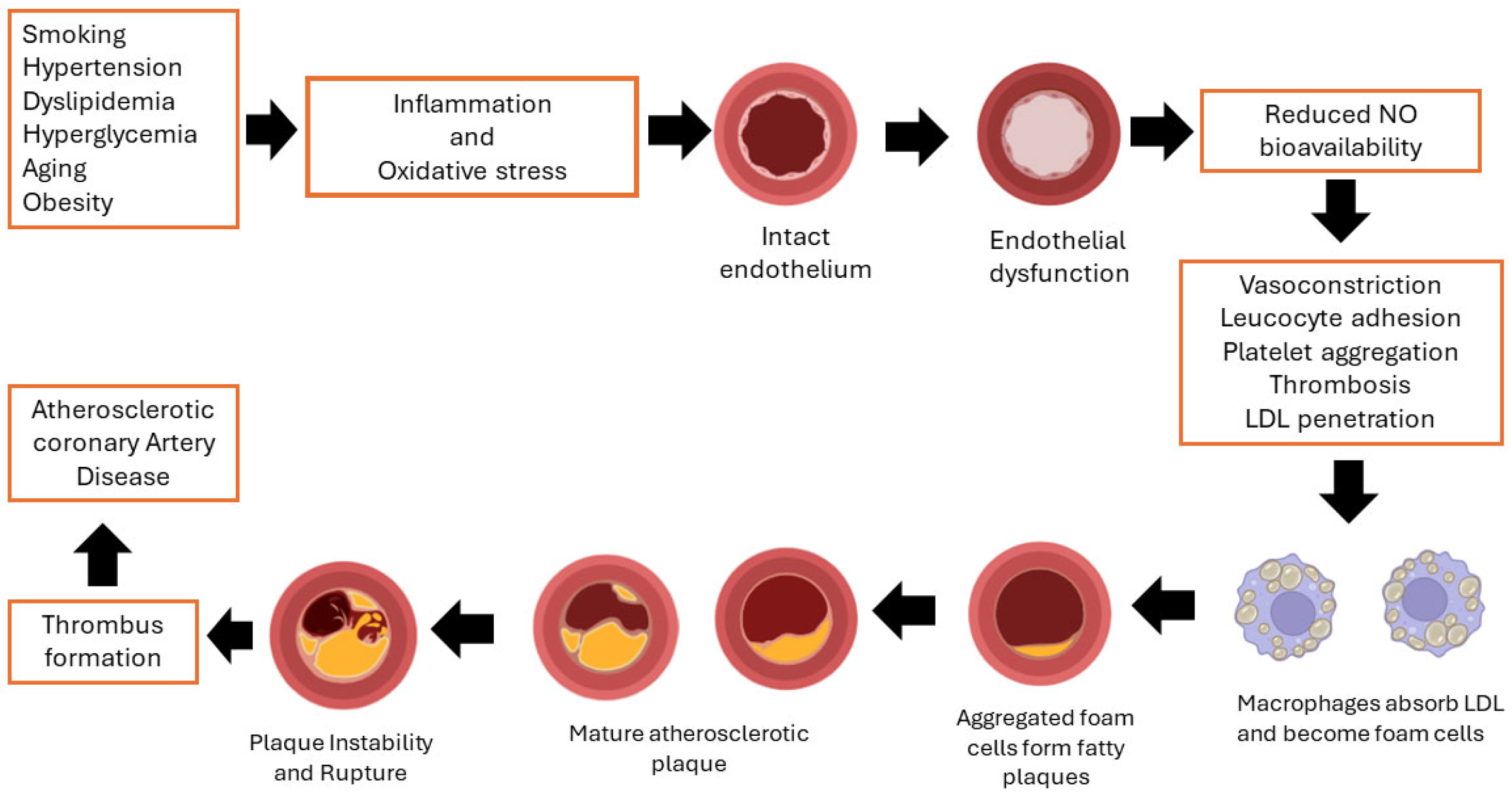
Figure 1. Development of atherosclerotic coronary artery disease. Endothelial dysfunction, caused by factors like hypertension, hyperlipidemia, smoking, and diabetes, involves changes in the production and availability of endothelial-derived nitric oxide (NO), prostacyclin, and endothelin, impacting vascular reactivity. This leads to reactive oxygen species reaching regulatory molecules, causing blood vessel constrictions and promoting blood clotting. The endothelium becomes compromised, increasing activity and buildup within the artery wall. Low-density lipoprotein (LDL) cholesterol accumulates in the artery wall, triggering inflammation and lipid peroxidation. Macrophages absorb cholesterol-rich lipoproteins and secrete pro-oxidant substances, leading to inflammation and fatty streaks. Advanced plaques can become unstable, leading to platelet aggregation and thrombus formation, reducing blood flow to the myocardium and potentially causing myocardial infarction [23,24].
Jvd 03 00020 g001
Figure 2. Different modifications of Human Serum Albumin (HSA) [48]. Pre-pro albumin, the first translation product, has a molecular weight of 69.3 kDa and 609 amino acids (AA). It includes an N-terminal endoplasmic reticulum (ER) import signal sequence. Next, Pre-pro albumin is made in the liver and modified post-translationally in the Golgi apparatus. Before the mature HSA protein is released into the circulation, the first 24 amino acids from the N-terminus (1–18 signal peptide and 19–24 pro-peptide) are cleaved. The mature HSA protein consists of 585 amino acids and has a molecular weight of 66.5 kDa. In ischemic conditions, IMA undergoes several amino acid modifications due to increased reactive oxygen species (ROS) and hypoxia, resulting in 581 amino acids and ~65 kDa molecular weight.
Jvd 03 00020 g002
Figure 3. (a) Human Serum Albumin secondary structure motifs (sequence colored by residue conservation). Images created using PDB data (PDB ID: 1bm0) and PDB sum-EMBL EBI. The protein sequence is colored by residue conservation, with highly conserved residues highlighted in red and less conserved residues in lighter shades. The secondary structure motifs are annotated, showing alpha-helices, beta and gamma turns, and loop regions. Disulfide bridges are numbered from 1–17. Variant residues are also indicated, showing differences that may affect protein function. (b) Crystal structure of Human Serum Albumin (HSA). HSA is made up of three topologically identical and structurally similar domains: domain IA (AA 5–107), IB (AA 108–196), IIA domain (AA 197–297), IIB (AA 298–383), and domain IIIA (AA 384–497), IIIB (AA 498–585) [51].
Jvd 03 00020 g003
Figure 4. Auto degradation reaction: Amine nitrogen of Asp1 exerts a nucleophilic attack on the carbonyl of the peptide bond between Ala2 and His3, forming a cyclic dipeptide [53].
Jvd 03 00020 g004
Figure 5. Proposed mechanism of N-terminus of HSA modification by free radicals [27,54,55]. Localized ischemia, hypoxia, and activation of anaerobic glycolysis result in acidosis. Cu2+ ions are released from their weak binding sites in a localized acidic environment. In the presence of a reducing agent such as ascorbic acid, free Cu2+ is converted to Cu+ ions. These Cu+ ions react with oxygen to form Cu2+, generating superoxide free radicals. This reaction between Cu2+ or Fe3+ with ascorbate generates free radicals, which can modify proteins in various ways. Cu2+ ions (formed during Cu+ oxidation) are immediately scavenged by HSA and tightly bind with the N-terminal. In the presence of Cu2+ or Fe3+ ions, they undergo Fenton reaction and generate hydroxyl free radicals. These hydroxyl radicals damage the copper-bound N-terminus of HSA, causing the amino acids to cleave and release Cu2+ ions. The process will repeat in a chain reaction.
Jvd 03 00020 g005
Table 1. Summary of detection methods for measuring IMA.
MethodPrincipleAdvantagesLimitations
Albumin Cobalt Binding (ACB) Test [29,30,42,43,58,60,69]This is the most widely used traditional method. It relies on the decreased binding capacity of cobalt to the N-terminus of albumin due to ischemic modifications.Simple, cost-effective, and can be performed with standard laboratory equipment.Results can vary due to factors like albumin concentration and the presence of other interfering substances. Sensitivity and specificity can be lower compared to newer methods.
Cobalt Albumin Binding (CAB) assay [42]An improved version of the ACB assay.This assay reduces the impact of pH fluctuations on the binding of cobalt ions to albumin by optimizing pH conditions and using NaH2PO4 buffer at pH 7.4.
The assay requires smaller volumes of specimens and reagents, making it more efficient and cost-effective.
Lack of standard Human Serum Albumin (HSA) as a control.
Effect of pH on cobalt-DTT Binding: Although pH optimization has been addressed, cobalt binding to DTT decreases significantly with pH reduction.
Albumin Copper Binding (ACuB) assay [63]It relies on the decreased binding capacity of copper to the N-terminus of albumin due to ischemic modifications.Cu2+ ions, which have a higher binding affinity for the N-terminus of HSA, enhance the assay’s sensitivity.
Lucifer yellow is a copper-selective agent that doesn’t denature protein structures and releases metal ions from binding sites like DTT.
Direct fluorescence measurements can be obtained.
Environmental factors such as pH changes and the presence of competing ions could still impact the binding efficiency.
Fluorescence measurement requires specific equipment and dyes.
Nickel-Albumin Binding assay [64,65]It relies on the decreased binding capacity of nickel to the N-terminus of albumin due to ischemic modifications.Using nickel ions, which have a higher affinity for binding to the N-terminus of HSA than cobalt, enhances detection sensitivity.Using DTT as a colorizing agent can denature protein structures and release metal ions from binding sites, potentially affecting the accuracy and reliability of the assay results.
Requires precise control of reaction conditions
While Ni2+ ions have a relatively high binding affinity, they are not as strong as Cu2+, which may limit the assay’s sensitivity compared to the Albumin Copper Binding (ACuB) assay.
Quantum Dot coupled X-ray Fluorescence Spectroscopy (Q-XFR) [66,67,68]Primary X-ray irradiates the target element, and that interaction emits a characteristic secondary X-ray, which an X-ray fluorescence detector will detect. This characteristic secondary X-ray is unique to a particular aspect of interest. The XRF peak’s intensity is related to its number of elements.Higher specificity (95.9%) compared to the ACB assay (82.9%)
This method effectively removes interferences from excessively high or low albumin levels by calculating the difference between total HSA and intact HSA levels.
Rapid and accurate detection
Specialized X-ray fluorescence spectroscopy equipment is required, and specialized training and expertise are needed to operate it.
Complexity of sample preparation
Enzyme-Linked Immunosorbent Assay (ELISA) [69,70,72,73,74,75,76]Specific antibodies bind the target antigen and detect the presence and quantity of antigens binding.Precise detection of IMA using antibodies, targeting the specific protein modification associated with ischemia.
Provides quantitative measurement of IMA levels
Adjusting IMA values based on serum albumin levels can minimize false positives and negatives, ensuring more accurate results.
ELISA results may vary based on the timing of sample collection
The procedure involves multiple steps.
Despite high specificity, there is always a risk of non-specific binding, which can affect the accuracy of the assay results.
Liquid crystal biosensor [77,78,79]Liquid crystals are phase transition metals that exist between liquid and crystal states. They have both liquid and crystal properties. They respond readily to external stimuli such as changes in surface properties (binding specific biomolecules onto the surface), temperature, pressure, and electromagnetic field. These stimuli will alter the orientation of the LC and change the color/brightness of the sensor.High sensitivity and specificity
The method is simple and provides quick results.
Changes in the LC orientation result in optical signal changes observable through a polarizing microscope, allowing for immediate visual detection of IMA levels.
LC biosensors can detect various biomolecules, including proteins, antibodies, and cells, making them versatile tools for different diagnostic applications.
Need for specialized equipment
External stimuli sensitivity must be controlled
Surface Plasmon Resonance (SPR) immunosensor for IMA [81]SPR is an optical-electrical phenomenon that occurs when a photon of incoming light strikes a metal surface. A portion of the light energy interacts with the metal surface, and the electrons in the metal surface move due to excitation. Electron movements are known as plasmons, traveling parallel to the metal surface. The plasmon oscillation, in turn, generates an electric field from the metal surface. This allows for more precise measurements of molecule adsorption on the metal surface and subsequent interactions with specific ligands.High sensitivity in detecting IMA, particularly when enhanced with colloidal gold nanoparticles
Real-time, in situ monitoring of biomolecular interactions
Label-free detection
Low interference
Versatility and biocompatibility
Rapid and simple
SPR systems require sophisticated and expensive equipment, which may limit their availability in resource-constrained settings.
Specialized training is required to operate the instruments.
Over time, the metal surface can become fouled with non-specific proteins or other contaminants.
Disclaimer/Publisher’s Note: The statements, opinions and data contained in all publications are solely those of the individual author(s) and contributor(s) and not of MDPI and/or the editor(s). MDPI and/or the editor(s) disclaim responsibility for any injury to people or property resulting from any ideas, methods, instructions or products referred to in the content.