
Effect of Microencapsulated Cocoa Polyphenols on Macro- and Microvascular Function after Eccentric Exercise

 ,
, 
Abstract
1. Introduction
2. Methods
2.1. Participants
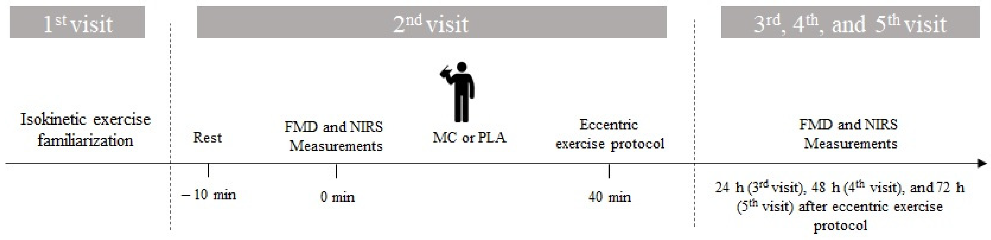
2.2. Study Design
2.3. Microencapsulated Cocoa/Placebo Preparation
2.4. Eccentric Exercise Protocol
2.5. Flow-Mediated Dilatation
2.6. Muscle Oxygen Saturation Measurement
2.7. Statistical Analysis
3. Results
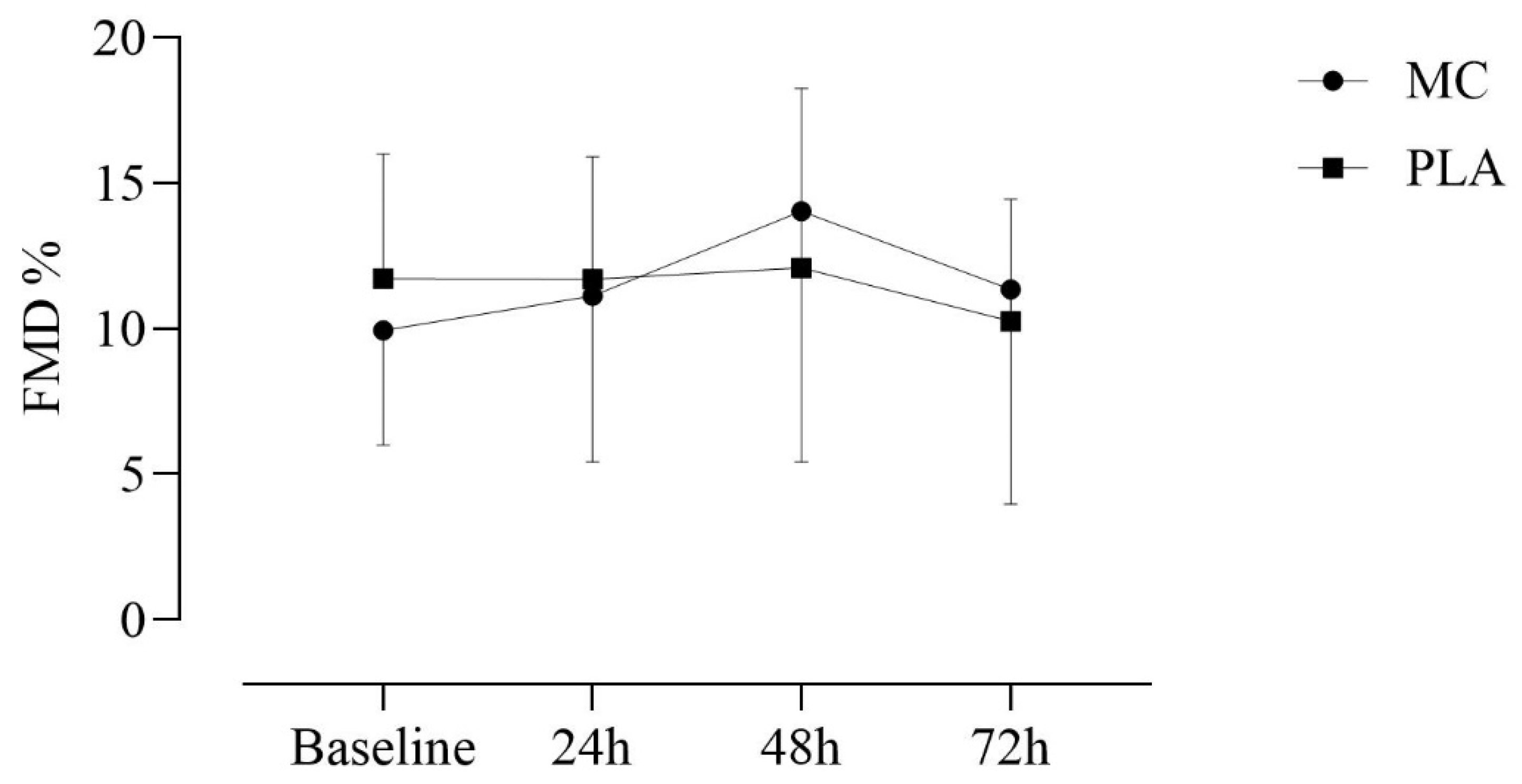
3.1. Flow-Mediated Dilation
3.2. Muscle Oxygen Saturation Parameters
4. Discussion
Experimental Consideration
5. Conclusions
Supplementary Materials
Author Contributions
Funding
Institutional Review Board Statement
Informed Consent Statement
Data Availability Statement
Acknowledgments
Conflicts of Interest
References
- Gimbrone, M.A.; García-Cardeña, G. Endothelial Cell Dysfunction and the Pathobiology of Atherosclerosis. Circ. Res. 2016, 118, 620–636. [Google Scholar] [CrossRef] [PubMed]
- Mensah, G.A.; Roth, G.A.; Fuster, V. The Global Burden of Cardiovascular Diseases and Risk Factors. J. Am. Coll. Cardiol. 2019, 74, 2529–2532. [Google Scholar] [CrossRef] [PubMed]
- Xu, S.; Ilyas, I.; Little, P.J.; Li, H.; Kamato, D.; Zheng, X.; Luo, S.; Li, Z.; Liu, P.; Han, J.; et al. Endothelial Dysfunction in Atherosclerotic Cardiovascular Diseases and Beyond: From Mechanism to Pharmacotherapies. Pharmacol. Rev. 2021, 73, 924–967. [Google Scholar] [CrossRef] [PubMed]
- Förstermann, U.; Xia, N.; Li, H. Roles of Vascular Oxidative Stress and Nitric Oxide in the Pathogenesis of Atherosclerosis. Circ. Res. 2017, 120, 713–735. [Google Scholar] [CrossRef] [PubMed]
- Cyr, A.R.; Huckaby, L.V.; Shiva, S.S.; Zuckerbraun, B.S. Nitric Oxide and Endothelial Dysfunction. Crit. Care Clin. 2020, 36, 307–321. [Google Scholar] [CrossRef] [PubMed]
- Barnes, M.J.; Mündel, T.; Stannard, S.R. Post-exercise alcohol ingestion exacerbates eccentric-exercise induced losses in performance. Eur. J. Appl. Physiol. 2010, 108, 1009–1014. [Google Scholar] [CrossRef] [PubMed]
- Stacy, M.R.; Bladon, K.J.; Lawrence, J.L.; McGlinchy, S.A.; Scheuermann, B.W. Serial assessment of local peripheral vascular function after eccentric exercise. Appl. Physiol. Nutr. Metab. 2013, 38, 1181–1186. [Google Scholar] [CrossRef]
- Oliveira, G.V.; Cordeiro, E.M.; Volino-Souza, M.; Rezende, C.; Conte-Junior, C.A.; Alvares, T.S. Flow-Mediated Dilation in Healthy Young Individuals Is Impaired after a Single Resistance Exercise Session. Int. J. Environ. Res. Public Health 2020, 17, 5194. [Google Scholar] [CrossRef] [PubMed]
- Green, D.J.; Jones, H.; Thijssen, D.; Cable, N.T.; Atkinson, G. Flow-mediated dilation and cardiovascular event prediction: Does nitric oxide matter? Hypertension 2011, 57, 363–369. [Google Scholar] [CrossRef] [PubMed]
- Choi, Y.; Akazawa, N.; Zempo-Miyaki, A.; Ra, S.-G.; Shiraki, H.; Ajisaka, R.; Maeda, S. Acute Effect of High-Intensity Eccentric Exercise on Vascular Endothelial Function in Young Men. J. Strength Cond. Res. 2016, 30, 2279–2285. [Google Scholar] [CrossRef] [PubMed]
- Santos, K.S.; Junior, O.J.F.R.; Tavares, I.R.G.; Volino-Souza, M.; Oliveira, G.V.; Alvares, T.S. A single dose of microencapsulated cocoa supplementation attenuated eccentric exercise-induced endothelial dysfunction. Int. J. Food Sci. Nutr. 2023, 74, 373–381. [Google Scholar] [CrossRef] [PubMed]
- Cuevas, A.M.; Germain, A.M. Diet and endothelial function. Biol. Res. 2004, 37, 225–230. [Google Scholar] [CrossRef] [PubMed]
- Yamagata, K.; Tagami, M.; Yamori, Y. Dietary polyphenols regulate endothelial function and prevent cardiovascular disease. Nutrition 2015, 31, 28–37. [Google Scholar] [CrossRef] [PubMed]
- Parsamanesh, N.; Asghari, A.; Sardari, S.; Tasbandi, A.; Jamialahmadi, T.; Xu, S.; Sahebkar, A. Resveratrol and endothelial function: A literature review. Pharmacol. Res. 2021, 170, 105725. [Google Scholar] [CrossRef] [PubMed]
- Cheng, Y.C.; Sheen, J.M.; Hu, W.L.; Hung, Y.C. Polyphenols and Oxidative Stress in Atherosclerosis-Related Ischemic Heart Disease and Stroke. Oxid. Med. Cell Longev. 2017, 2017, 8526438. [Google Scholar] [CrossRef]
- Katz, D.L.; Doughty, K.; Ali, A. Cocoa and Chocolate in Human Health and Disease. Antioxid. Redox Signal. 2011, 15, 2779–2811. [Google Scholar] [CrossRef] [PubMed]
- Tan, T.Y.C.; Lim, X.Y.; Yeo, J.H.H.; Lee, S.W.H.; Lai, N.M. The Health Effects of Chocolate and Cocoa: A Systematic Review. Nutrients 2021, 13, 2909. [Google Scholar] [CrossRef] [PubMed]
- Scalbert, A.; Williamson, G. Dietary Intake and Bioavailability of Polyphenols. J. Nutr. 2000, 130, 2073S–2085S. [Google Scholar] [CrossRef] [PubMed]
- Manach, C.; Williamson, G.; Morand, C.; Scalbert, A.; Rémésy, C. Bioavailability and bioefficacy of polyphenols in humans. I. Review of 97 bioavailability studies. Am. J. Clin. Nutr. 2005, 81, 230S–242S. [Google Scholar] [CrossRef] [PubMed]
- Oliveira, G.; Volino-Souza, M.; Conte-Junior, C.A.; Alvares, T.S. Food-derived polyphenol compounds and cardiovascular health: A nano-technological perspective. Food Biosci. 2021, 41, 101033. [Google Scholar] [CrossRef]
- Sasaki, H.; Sunagawa, Y.; Takahashi, K.; Imaizumi, A.; Fukuda, H.; Hashimoto, T.; Wada, H.; Katanasaka, Y.; Kake-ya, H.; Fujita, M.; et al. Innovative preparation of curcumin for improved oral bioavailability. Biol. Pharm. Bull. 2011, 34, 660–665. [Google Scholar] [CrossRef] [PubMed]
- Aird, W.C. Endothelial cell heterogeneity. Cold Spring Harb. Perspect. Med. 2012, 2, a006429. [Google Scholar] [CrossRef] [PubMed]
- Thijssen, D.H.J.; Bruno, R.M.; van Mil, A.C.C.M.; Holder, S.M.; Faita, F.; Greyling, A.; Zock, P.L.; Taddei, S.; Deanfield, J.E.; Luscher, T.; et al. Expert consensus and evidence-based recommendations for the assessment of flow-mediated dilation in humans. Eur. Heart J. 2019, 40, 2534–2547. [Google Scholar] [CrossRef] [PubMed]
- Ludovici, V.; Barthelmes, J.; Nägele, M.P.; Enseleit, F.; Ferri, C.; Flammer, A.J.; Ruschitzka, F.; Sudano, I. Cocoa, Blood Pressure, and Vascular Function. Front. Nutr. 2017, 4, 36. [Google Scholar] [CrossRef] [PubMed]
- McHugh, M.P. Recent advances in the understanding of the repeated bout effect: The protective effect against muscle damage from a single bout of eccentric exercise. Scand. J. Med. Sci. Sports 2003, 13, 88–97. [Google Scholar] [CrossRef] [PubMed]
- Grassi, D.; Desideri, G.; Necozione, S.; di Giosia, P.; Barnabei, R.; Allegaert, L.; Bernaert, H.; Ferri, C. Cocoa consumption dose-dependently improves flow-mediated dilation and arterial stiffness decreasing blood pressure in healthy individuals. J. Hypertens. 2015, 33, 294–303. [Google Scholar] [CrossRef]
- Junior, O.J.F.R.; dos Santos, K.S.; Tavares, I.R.G.; de Oliveira, G.V.; Alvares, T.S. A Single Dose of Microencapsulated Cocoa Supplementation Does Not Speed up Muscle Force Recovery after Eccentric Exercise-Induced Muscle Damage: A Placebo-Controlled, Double-Blind, Crossover Study. Appl. Biosci. 2023, 3, 1–13. [Google Scholar] [CrossRef]
- Hou, L.; Liu, Y.; Qian, L.; Zheng, Y.; Gao, J.; Cao, W.; Shang, Y. Portable Near-Infrared Technologies and Devices for Noninvasive Assessment of Tissue Hemodynamics. J. Healthc. Eng. 2019, 2019, 3750495. [Google Scholar] [CrossRef] [PubMed]
- McLay, K.M.; Fontana, F.Y.; Nederveen, J.P.; Guida, F.F.; Paterson, D.H.; Pogliaghi, S.; Murias, J.M. Vascular responsiveness determined by near-infrared spectroscopy measures of oxygen saturation. Exp. Physiol. 2016, 101, 34–40. [Google Scholar] [CrossRef] [PubMed]
- Cunningham, J.B.; McCrum-Gardner, E. Power, effect and sample size using GPower: Practical issues for researchers and members of research ethics committees. Evid. Based Midwifery 2007, 5, 132–136. [Google Scholar]
- Sullivan, G.M.; Feinn, R. Using Effect Size—Or Why the P Value Is Not Enough. J. Grad. Med. Educ. 2012, 4, 279–282. [Google Scholar] [CrossRef] [PubMed]
- Choi, Y.; Tanabe, Y.; Akazawa, N.; Zempo-Miyaki, A.; Maeda, S. Curcumin supplementation attenuates the decrease in endothelial function following eccentric exercise. J. Exerc. Nutr. Biochem. 2019, 23, 7–12. [Google Scholar] [CrossRef] [PubMed]
- Gayda, M.; Juneau, M.; Tardif, J.C.; Harel, F.; Levesque, S.; Nigam, A. Cardiometabolic and traditional cardiovascular risk factors and their potential impact on macrovascular and microvascular function: Preliminary data. Clin. Hemorheol. Microcirc. 2015, 59, 53–65. [Google Scholar] [CrossRef] [PubMed]
- Caldwell, J.T.; Wardlow, G.C.; Branch, P.A.; Ramos, M.; Black, C.D.; Ade, C.J. Effect of exercise-induced muscle damage on vascular function and skeletal muscle microvascular deoxygenation. Physiol. Rep. 2016, 4, e13032. [Google Scholar] [CrossRef] [PubMed]
- Goldfarb, A.H.; Bloomer, R.J.; McKenzie, M.J. Combined Antioxidant Treatment Effects on Blood Oxidative Stress after Eccentric Exercise. Med. Sci. Sport. Exerc. 2005, 37, 234–239. [Google Scholar] [CrossRef] [PubMed]
- Proske, U.; Morgan, D.L. Muscle damage from eccentric exercise: Mechanism, mechanical signs, adaptation and clinical applications. J. Physiol. 2001, 537, 333–345. [Google Scholar] [CrossRef] [PubMed]
- Lin, M.J.; Nosaka, K.; Ho, C.C.; Chen, H.L.; Tseng, K.W.; Ratel, S.; Chen, T.C. Influence of Maturation Status on Eccentric Exercise-Induced Muscle Damage and the Repeated Bout Effect in Females. Front. Physiol. 2018, 5, 1118. [Google Scholar] [CrossRef] [PubMed]
- Gröne, M.; Sansone, R.; Höffken, P.; Horn, P.; Rodriguez-Mateos, A.; Schroeter, H.; Kelm, M.; Heiss, C. Cocoa Flavanols Improve Endothelial Functional Integrity in Healthy Young and Elderly Subjects. J. Agric. Food Chem. 2020, 68, 1871–1876. [Google Scholar] [CrossRef] [PubMed]
- Buchanan, C.E.; Kadlec, A.O.; Hoch, A.Z.; Gutterman, D.D.; Durand, M.J. Hypertension during Weight Lifting Reduces Flow-Mediated Dilation in Nonathletes. Med. Sci. Sports Exerc. 2017, 49, 669–675. [Google Scholar] [CrossRef] [PubMed]

| Demographics | |
|---|---|
| N | 13 |
| Age (years) | 25 ± 4 |
| Body mass (kg) | 66.8 ± 12.0 |
| Body mass index (kg/m2) | 23.5 ± 3.3 |
| Clinical | |
| SBP (mm Hg) | 118 ± 8 |
| DBP (mm Hg) | 73 ± 7 |
| HR (beats/min) | 64 ± 3 |
| Variables | T0 | T24 | T48 | T72 | |
|---|---|---|---|---|---|
| FMD (%) | MC | 9.9 ± 3.9 | 11.1 ± 5.6 | 14.02 ± 8.6 | 11.3 ± 7.3 |
| PLA | 11.7 ± 4.2 | 11.6 ± 4.1 | 12.1 ± 6.1 | 10.2 ± 4.1 | |
| BAD (mm) | MC | 0.32 ± 0.09 | 0.32 ± 0.08 | 0.33 ± 0.06 | 0.32 ± 0.07 |
| PLA | 0.30 ± 0.09 | 0.33 ± 0.08 | 0.32 ± 0.07 | 0.31 ± 0.06 | |
| PAD (mm) | MC | 0.35 ± 0.1 | 0.35 ± 0.1 | 0.38 ± 0.07 | 0.36 ± 0.08 |
| PLA | 0.35 ± 0.1 | 0.37 ± 0.08 | 0.36 ± 0.09 | 0.35 ± 0.06 | |
| StO2upslope (%.s−1) | MC | 2.1 ± 0.5 | 2.01 ± 0.6 | 1.9 ± 0.6 | 2.0 ± 0.6 |
| PLA | 2.04 ± 0.3 | 1.8 ± 0.6 | 1.9 ± 0.7 | 2.1 ± 0.4 | |
| StO2downslope (%.s−1) | MC | −0.08 ± 0.02 | −0.09 ± 0.02 | −0.08 ± 0.03 | −0.08 ± 0.02 |
| PLA | −0.08 ± 0.01 | −0.08 ± 0.02 | −0.07 ± 0.02 | −0.09 ± 0.01 | |
| △StO2 (%) | MC | 32.5 ± 7.7 | 32.4 ± 6.9 | 36.01 ± 7.5 | 33.7 ± 8.3 |
| PLA | 32.3 ± 6.7 | 31.8 ± 6.1 | 35.8 ± 6.2 | 31.2 ± 6.1 |
Disclaimer/Publisher’s Note: The statements, opinions and data contained in all publications are solely those of the individual author(s) and contributor(s) and not of MDPI and/or the editor(s). MDPI and/or the editor(s) disclaim responsibility for any injury to people or property resulting from any ideas, methods, instructions or products referred to in the content. |
© 2024 by the authors. Licensee MDPI, Basel, Switzerland. This article is an open access article distributed under the terms and conditions of the Creative Commons Attribution (CC BY) license (https://creativecommons.org/licenses/by/4.0/).
Share and Cite
de Oliveira, G.V.; Miranda de Souza, L.V.; Junior, O.J.F.R.; Volino-Souza, M.; Alvares, T.S. Effect of Microencapsulated Cocoa Polyphenols on Macro- and Microvascular Function after Eccentric Exercise. J. Vasc. Dis. 2024, 3, 235-244. https://doi.org/10.3390/jvd3030019
de Oliveira GV, Miranda de Souza LV, Junior OJFR, Volino-Souza M, Alvares TS. Effect of Microencapsulated Cocoa Polyphenols on Macro- and Microvascular Function after Eccentric Exercise. Journal of Vascular Diseases. 2024; 3(3):235-244. https://doi.org/10.3390/jvd3030019
Chicago/Turabian Stylede Oliveira, Gustavo Vieira, Leonardo Victor Miranda de Souza, Olavo João Frederico Ramos Junior, Mônica Volino-Souza, and Thiago Silveira Alvares. 2024. "Effect of Microencapsulated Cocoa Polyphenols on Macro- and Microvascular Function after Eccentric Exercise" Journal of Vascular Diseases 3, no. 3: 235-244. https://doi.org/10.3390/jvd3030019
APA Stylede Oliveira, G. V., Miranda de Souza, L. V., Junior, O. J. F. R., Volino-Souza, M., & Alvares, T. S. (2024). Effect of Microencapsulated Cocoa Polyphenols on Macro- and Microvascular Function after Eccentric Exercise. Journal of Vascular Diseases, 3(3), 235-244. https://doi.org/10.3390/jvd3030019