Article

Intraoperative Flow Measurement as a Quality Control during Carotid Endarterectomy in a Teaching Hospital Setting

1
Division of Vascular and Endovascular Surgery, Department of General, Visceral and Transplant Surgery, University Hospital Essen, University Duisburg-Essen, 45147 Essen, Germany
2
Contilia Heart- and Vascular Center, Clinic of Vascular Surgery and Phlebology, 45138 Essen, Germany
3
Department of General, Visceral and Transplant Surgery, University Hospital Essen, University of Duisburg-Essen, 45147 Essen, Germany
4
Institute of Pharmacology and Toxicology, Ruhr-University Bochum, 44801 Bochum, Germany
5
Institute for Medical Informatics, Biometry and Epidemiology, University Hospital, University Duisburg-Essen, 45147 Essen, Germany
6
BDH-Clinic Elzach, 79215 Elzach, Germany
*
Author to whom correspondence should be addressed.
J. Vasc. Dis. 2022, 1(1), 71-79; https://doi.org/10.3390/jvd1010008
Submission received: 19 July 2022 / Revised: 8 August 2022 / Accepted: 26 August 2022 / Published: 5 September 2022
(This article belongs to the Section Neurovascular Diseases)

Abstract

:
Background: To evaluate the technical results of an arterial repair, a variety of intraoperative imaging and assessment techniques can be used during carotid endarterectomy (CEA). The aim of the study was to evaluate the usefulness of intraoperative ultrasound flow measurement as a quality control after primary CEA in a teaching hospital setting. Methods: Over 36 months, 107 consecutive CEAs were performed at our institution. Retrospectively acquired demographics, intraoperative flow measurements, duplex results, revisions, and surgical outcomes were reviewed. Postoperative 30-day transient ischemic attack (TIA), stroke, and death rates were analyzed. Results were compared with ultrasound flow measurement and duplex ultrasonography. Results: From March 2012 to March 2015, 107 primary consecutive CEAs were performed in 107 patients (71% male, 29% female), whose age ranged from 51 to 81 years with a mean age of 68 ± 4 years. Associated risk factors included diabetes for 89 (83%), smoking for 92 (86%), hypertension for 94 (87.8%), chronic renal insufficiency for 71 (66%), and coronary artery disease for 57 (53%) of the patients. Early postoperative duplex scans in all 107 patients showed no significant changes from intraoperative findings. The ipsilateral stroke and death rate in this study was 0 (0/107) and the 30-day death and stroke rate was also 0 (0/107), with no significant difference between trainees and senior surgeons. Three patients (2.8%) had flow < 100 mL/Min and two of them were revised after completion of contrast angiography. Conclusions: The findings of this study indicate that the intraoperative flow measurement is an alternative method for detecting technical errors and a tool for quality-control imaging. Especially for trainees, it makes sense to ensure effectiveness of the procedure upon its completion and to assess the technical adequacy of CEA.

1. Introduction

A hemodynamically impaired cerebral perfusion represents an increased risk for cerebral ischemia [1]. Of all ischemic strokes, 7.5–15% are caused by internal carotid artery stenosis [2,3]. Carotid endarterectomy (CEA) has been considered the standard treatment for high-grade asymptomatic and symptomatic internal carotid stenosis. The benefit of CEA as a preventative treatment depends upon achieving an extremely low rate of perioperative complications. The main suggested causes of intraoperative stroke (IOS) related to carotid endarterectomy are embolization, intraoperative hypoperfusion, thrombotic occlusion of the ipsilateral or contralateral carotid artery, or hyperperfusion syndrome. The most common underlying cause in surgical therapy is inadvertent technical error due to retained intraluminal thrombus, residual stenosis, and intimal flaps [4,5,6].
For vascular surgeons, it is of utmost importance to confirm the adequacy of their arterial reconstructions to ensure the best possible outcome for their patients and to minimize perioperative and late complications. To judge this adequacy, simple clinical assessments such as pulse palpation, Doppler examination, transit time ultrasound flowmeter measurements, duplex ultrasonography, and contrast arteriography have been described for intraoperative quality-control examinations during carotid operations [7]. However, the availability of more expensive and technically demanding techniques such as duplex ultrasonography or contrast arteriography is limited in many institutions. Alternatively, transit time ultrasound flowmeter measurement is an easy procedure that does not require trained technicians. In addition, it has been successfully used in other anatomic areas before [8]. Performance of transit time flow measurement is easily applicable under surgical conditions and artefacts affecting results are thought to be less severe than in other intraoperative control techniques [9,10,11]. Clinical studies showed a positive effect of intraoperative duplex sonography after carotid endarterectomy detected lesions that might cause postoperative strokes [12,13,14,15,16,17].
In an attempt to identify these lesions, we have routinely used intraoperative transit time flow measurement. We report our recent experience with intraoperative time flow measurement in 107 consecutive primary CEA procedures performed at our institution over 36 months. The purpose of this retrospective study was to evaluate whether intraoperative flow measurement is an optimal method for detecting technical errors after CEA. The best way to determine the hemodynamic status of the repair is intraoperative evaluation, because immediate operative revision is possible in case of a technical defect. To our knowledge, there have been no reports evaluating the role of intraoperative transit time flow measurement in the internal carotid artery and its potential prediction of postoperative strokes.

2. Materials and Methods

2.1. Study Population

This study was a single-center retrospective observational study of 107 consecutive patients undergoing 107 primary CEAs between March 2012 and March 2015 at a university hospital. The primary endpoint of our analysis was to assess the value of intra-operative blood flow measurements and the correlation with postoperative strokes.
Data were analyzed retrospectively. The criterion for study inclusion was an indication for CEA based upon established criteria as outlined in the Asymptomatic Carotid Atherosclerosis Study (ACAS) and North American Symptomatic Carotid Endarterectomy Trial (NASCET) [18,19].
Patients who underwent revision CEAs and surgery for carotid body tumors, aneurysms, trauma, fibromuscular dysplasia, or Takayasu disease were not included in this study. All of the study procedures adhered to the Declaration of Helsinki. Ethical approval was waived by the local Ethics Committee of Duisburg-Essen University in view of the retrospective nature of the study, and all the procedures being performed were part of routine care. All patients were provided written informed consent for CEA. Finally, data of a total of 107 CEAs were available for complete analysis.
Preoperative physical examination was performed in order to detect any signs of neurological deficit. All patients underwent a preoperative vascular duplex ultrasound (Siemens ACUSON S2000, Siemens AG, Erlangen, Germany) and a second predominantly within 96 h after CEA based on a standardized protocol. Our protocol included insonation of the common carotid artery (CCA), internal carotid artery (ICA), and external carotid artery (ECA) with acquisition of real time B-mode imaging in sagittal and transverse planes. In addition, all three arterial segments were subjected to color-flow imaging, power Doppler scanning, and spectral waveform analyses. In addition, angle-corrected systolic and diastolic flow velocity in cm/sec was measured and analyzed. In many instances power Doppler scanning was used to improve definition and to obtain a more accurate measurement of the residual lumen. Color-flow disturbances were used to assess the hemodynamic significance of the technical defects. Lastly, the luminal diameter of the ICA was measured and recorded in all cases. All duplex examinations were performed by one of two vascular technologists in a neurological vascular laboratory.
Patients were followed for every 3 months or until patient death. Each follow-up visit consisted of a duplex scan examination and clinical evaluation.
Stroke was clinically defined as any persistent focal or global neurological deficit lasting longer than 24 h and presumed to be of no other than vascular origin. All perioperative stroke events were assessed by a study neurologist. Stroke was defined as ipsilateral when occurring in the territory of the operated carotid artery.

2.2. Surgical Procedures

All procedures were performed with general endotracheal anesthesia with continuous radial artery pressure monitoring. The patient’s head was turned to the contralateral side and extended. Attempts were made to keep mean arterial blood pressure at a level of 80 mmHg or more during cross-clamping of the carotid artery. CEA was performed via longitudinal arteriotomy in a standardized manner by one consultant in vascular surgery or by a advanced surgical fellow under consultant supervision. Intravenous heparin (5000 IE) was routinely given before carotid artery clamping in all cases and all patients underwent routine shunting (Flexcel Carotid Shunt, LeMaitre®, North Brunswick, NJ, USA). Additionally, somatosensory evoked potentials (SEPs) were used to monitor cerebral status during surgical intervention. All carotid arteriotomies were reconstructed using bovine pericardium patches (Bovine pericardium, Vascu-Guard®, Saint Paul, MN, USA) and the proximal and distal intimal steps were tacked down with 7:0 prolene (Ethicon, Germany).
Intraoperative transit time flow measurements were performed in all patients and all data were entered into a computerized database. After completion of the operation and recovery from anesthesia, the patient was examined neurologically and transferred to the intensive care unit for postoperative monitoring. Any new neurological deficit apparent upon recovery from anesthesia was recorded and the patient assessed by a neurologist. Postoperative neurological complications were also documented. The decision to re-operate was left to the discretion of the surgeon. All neurological deficits occurring in the postoperative period were investigated by Duplex ultrasound, transcranial Doppler, and computed tomography (CT) scan.

2.3. Intraoperative Assessment

The adequacy of arterial repair was assessed intraoperatively by transit time flow measurement (T206, Transonic Inc, Ithaca, NY, USA) and predominantly within 96 h by duplex ultrasonography (Siemens ACUSON S2000, Siemens AG, Erlangen, Germany). Every patient in our cohort underwent complete flow measurement of the exposed artery upon completion of the endarterectomy and reconstruction of the bifurcation. Absolute flow data of the ICA and the CCA were measured intraoperatively. ICA blood flow was obtained without changing the position of the probe by clamping the ECA and the superior thyroid artery. During flow measurements the systolic blood pressure, pulse rate, and pCO2 were protocolled by the anesthesiologist to ensure that local blood flow was not influenced by general hemodynamic changes or metabolically caused hyperemia. The intraoperative measurement was performed by vascular fellows or a vascular consultant.
The transit time method uses two piezoelectric crystals transmitting ultrasound through the blood vessel toward a reflector on the other side of the vessel (Figure 1). The probe transmits an ultrasonic beam of a known volume through the vessel upstream. After passage, the beam is reflected and passes through the vessel again. The flow advances the arrival time of the sound beam, and a phase detector senses this as a phase difference compared to a master oscillator signal. The flowmeter stores the phase difference, and a second beam is sent downstream. When it reflects, the flow slows the ultrasonic beam, and this second phase difference is also stored. Because the volume of the ultrasonic beam is known, the flow can be calculated through the difference between the two signals. Only the moving parts within the blood vessel give a phase difference, and therefore nonmoving cells are not analyzed. Low flow rates using flow measurement were expected to be associated with technical problems [18,19,20]. Multiple probe sizes were available to accommodate blood vessels with outside diameters ranging from 4 to 8 mm. The circuit included a thermoregulated reservoir and a water-filled tank, kept at room temperature. For optimal ultrasound signal coupling, the arterial pressure was increased to >80 mmHg and the neck of the probe was placed in a flat position. In case of persistently abnormal flow measurements, an intraoperative arteriogram was obtained to identify potential technical defects or occlusive disease. A 21-gauge needle, connected to a syringe containing 10 mL of contrast medium, was introduced in the common carotid artery and retrograde injections were used. It was possible to obtain good visualization of intra- and extracranial vessels in all the cases. After angiography, the carotid vessel was closed only by manual compression. Technical defects were defined as major flaws in the presence of residual stenosis > 50% or mobile flap longer than 2 mm and minor flaws in the presence of residual stenosis < 50% or mobile flap less than 2 mm. Otherwise, complete duplex ultrasonography was performed predominantly within 96 h after carotid revascularization.

2.4. Statistical Analysis

Numerical variables were presented as mean +/− SD, and discrete variables by percentages. Two-sided-Student’s t-test was used to determine the differences in continuous variables. Categorical variables were presented as percentages. Statistical analysis was performed using IBM SPSS Statistics version 24 (SPSS, Chicago, IL, USA). Graphs were created using SigmaPlot for Windows version 10.0 (Systat Software GmbH, Erkrath, Germany).
The cut-off point of 100 mL/Min was established after preliminary review of the data, with the aim of achieving a sufficient number of patients for statistical analysis.

3. Results

3.1. Participants

Of the 107 consecutive patients reviewed, 76 were men (71%) and 31 women (29%). Their age ranged from 51 to 81 years with a mean age of 68 ± 4 years. Associated risk factors were common and included diabetes for 89 (83%), smoking for 92 (86%), hypertension for 94 (87.8%), chronic renal insufficiency for 71 (66%), and coronary artery disease for 57 (53%) of the patients. Of the 107 patients, 61.7% (66 patients) were operated on with symptomatic and 38.3% (41 patients) with asymptomatic disease. Prior to operation, 58% (38 patients) with symptomatic disease had a complete stroke and 42% (28 patients) had TIA symptoms (amaurosis fugax, reversible cerebral ischemia, or minor stroke). All patients with symptomatic disease had an ipsilateral ICA stenosis of 75% (NASCET) or more detected by duplex ultrasound scanning. Asymptomatic patients presented bilateral stenosis in 63% (26 patients), and in 37% (15 patients) a contralateral closure of the ICA. Baseline demographic characteristics and indications for CEA are shown in Table 1.

3.2. Intraoperative Assessment

Intraoperative flow measurement was performed in all 107 CEA patients. The mean ICA flow was 216 mL/min (range, 90–550 mL/min), as seen in Figure 2. Only three patients (2.8%) had an arterial flow of <100 mL/min. In these cases the angiography was examined immediately by the operating surgeon. An intimal flap was seen at the distal end of the endarterectomy on the ICA in two patients (1.9%) and was corrected by re-opening the ICA with the use of tacking stitches. These two patients were successfully revised and had a normal repeated angiogram with no technical defects or residual disease. Intraoperative angiography in the third patient was normal and the ICA was not re-explored. No complication in relation to carotid catheterization or injection of contrast media occurred. Postoperative duplex control (in 85% of cases within 96 h) performed by one of two neurologists was clearly visualized in its entirety and no stenosis or wall abnormalities were detected. Early postoperative duplex scans in all 107 patients showed no significant changes from intraoperative findings.

3.3. Postoperative TIA, Stroke, and Death

The perioperative stroke and death rate in this study was 0 (0/107). However, in one initially asymptomatic patient TIA symptoms occurred temporarily after surgery, and completely resolved within 24 h. Blood flow measurement in this patient was >150 mL/min. Extracranial and transcranial duplex studies were normal, and a CT scan revealed no cerebral infarction. Follow-up carotid duplex scans at 1 and 3 months and a neurological examination showed no significant changes from examination and scans obtained immediately after the repair.
The 30-day death and stroke rate was also 0 (0/107), with no significant difference between trainees and senior surgeons.

3.4. Local Complications

A total of 8 patients (8.4%) had local complications. Four patients (3.7%) were subject to postoperative local bleeding with revision of hematomas and 3.7% (4 patients) experienced peripheral nerve lesions. Wound infection or early postoperative ICA occlusion did not occur.

3.5. Postoperative Follow-Up

All 107 patients were followed for up to 24 months with no postoperative stroke. Follow-up carotid duplex scans at 1, 3, 6, and 12 months showed no significant changes when compared with scans obtained immediately after CEA. Five patients (4.7%) showed restenosis of less than 50% (NASCET). All restenotic patients were asymptomatic and no reoperations were performed.

4. Discussion

Based on our initial experience, flow measurement is a safe and effective method of ensuring the technical success of CEA. Presently, the role of flow measurement remains controversial, mainly because of numerous reports on other diagnostic methods such as duplex scans or even angiography. Applying these methods, excellent neurologic outcomes and survival rates after CEA were demonstrated [21,22,23]. However, there is no evidence of an intraoperative quality control during extracranial carotid surgery to support its routine application. Because most complications of CEA are related to flow disturbances [4,5,6], it makes sense to ensure the effectiveness of the procedure upon its completion.
In our opinion, the main advantage of flow measurement during CEA is the elimination of possible vessel injury by puncture of the carotid artery and subintimal injection of contrast agent during angiography. In addition, blood flow can be directly measured and the flow probe can be relatively simply applied. The intraoperative blood flow application of this technique has been advocated for different anatomic regions such as the femoral region or during creation of dialysis fistulas [8,24,25]. Flow measurements can be used to determine patency of the reconstructed carotid artery. While transit time technology is inexpensive and abnormal signal recognition requires minimal training, the assessment is subjective and audible interpretation is not quantitative.
Considering the carotid artery, the natural history of flow changes with carotid stenosis progression and significant changes that occur immediately following endarterectomy have been previously demonstrated using duplex ultrasound flow rates [7,26,27]. Duplex ultrasound, which we utilized predominantly within 96 h after carotid revascularization, identified no technical errors, although none of the patients in our series had any neurological complications that could be attributed to mechanical defects.
Zannetti et al. [28] have suggested that repair of intraoperative major defects is associated with increased postoperative neurologic sequelae. However, this experience could not be reproduced by others [12,13,14,15,16]. One other advantage of using flow measurement routinely is to ensure that the surgeon will pay even more attention to technical details.
Other investigators have also stressed the importance of flow rates for intraoperative quality-control studies following CEA. Asher et al. demonstrated that mean ICA volume flow rates of <100 mL/min were related to technical problems that required re-exploration or treatment of post endarterectomy spasm [4]. It was clearly shown that a spasm could result in thrombus formation when performing CEA with patch. Three patients in our cohort showed flow rates of less than 100 mL/min. An intimal flap was seen at the distal end of the endarterectomy on the ICA in two patients (1.9%) and was corrected by re-opening the ICA with the use of tacking stitches.
The relatively wide range of measurements for ICA volume flow in our study (90–550 mL/min) did not correlate with adverse outcome at the ends of the volume flow measurement spectrum. The use of intraluminal shunts was not a significant factor associated with low flow in the ICA. Patients with a relatively low-flow state in the ICA may be more susceptible to thrombosis, particularly if synthetic patches are used. However, we recognize that flow of 100 mL/Min was an arbitrary number picked at random after preliminary review of the data, with the aim of achieving a sufficient number of patients for statistical analysis.
Our overall perioperative neurologic complication rate was only 0.93% (1 of 107), which included a TIA in an initially asymptomatic patient. These symptoms completely disappeared after a few minutes and there were no further problems until discharge. There was no correlation with blood flow.
They were no deaths from any cause in the series and none of these patients had a major neurologic event. Our restenosis rate was 4.7%. It is likewise competitive with accepted rates in the literature. All restenosis patients in our study were <50% and remained stable or regressed during further course of time. Furthermore, all patients were asymptomatic and none required intervention. These results suggest that the routine inclusion of completion flow measurement as a quality-control measure may represent one of the many technological advances that make CEA safer.
To achieve a better outcome after CEA than is currently reported in the literature, the surgeon must identify and immediately correct possible technical problems causing perioperative strokes. Despite optimal surgical techniques, clinically unsuspected intraluminal defects may occur, which if left untreated may cause major neurological deficits. Such defects (flaps, residual plaque, thrombus, dissection, or persistent stenosis) occur in 6% to 30% of patients undergoing CEA. The rate of repeat interventions to correct these lesions varies in the literature from 3.5% to 10% [13,16]. Our data show that in 107 consecutive patients only two surgical revisions (1.9%) were needed after CEA. We have shown in this study that completion of flow measurement was an accurate method of assessing the technical adequacy of CEA. We became more aggressive in stitching of the sectioned intima of the CCA and extending the endarterectomy proximal on the CCA to avoid any significant flap. In this aspect as well, completion of flow measurement was an accurate method to assess and to improve our technique. One other advantage of using flow measurements routinely is to ensure that the surgeon pays more attention to technical details. Moreover, intraoperative flow measurement has also been shown to provide feedback and quality control of CEA performed by trainees, especially at a teaching hospital. It may therefore provide a strong incentive for the surgeon to perform the operation as precisely as possible.
Considering the intraoperative revision rate being a measure of ability to detect defects, angioscopy might have the highest sensitivity of all intraoperative quality-control techniques. The fact that angioscopy is performed before restoration of the blood flow holds advantages and disadvantages compared to measures performed thereafter. The major strength of the technique is that residual thrombi persisting after irrigation can be prevented from embolizing into the brain [29].
Lingenfelter et al. tried to assess sensitivities and specificities of different control techniques after carotid revascularization. Intraoperative duplex ultrasonography was found to possess higher sensitivities and specificities than angiography, followed by flow measurement [30]. However, the major limitation of the study was an erroneous calculation of sensitivities, as the possibility of false negative findings was disregarded. The unknown amount of false negative findings prohibits an accurate calculation of sensitivities for all intraoperative control techniques applied. This finding underlines the need for further large-scale studies to assess the effect of intraoperative control in CEA [29].

5. Limitations

Transit time flow measurement is not without limitations. First, when compared with a duplex scan, flow measurement does not allow for quantitative measurement of hemodynamic parameters and B-mode imaging. These parameters have become increasingly utilized and provide valuable information regarding the mechanical improvement in flow, especially when used in conjunction with preoperative duplex ultrasounds. Secondly, this retrospective study was not designed to compare the sensitivity of flow measurement and duplex ultrasound. Third, this study included retrospectively collected data of a small sample from one institution. A larger population is needed to confirm the results of this study. Therefore, this study is useful for hypothesis generation and for planning of future studies but is not ideal for definitive conclusions.
Nevertheless, this study shows that the intraoperative measurement of blood flow is an optimal method for detecting technical errors and a tool for quality-control imaging, and may also be useful for predicting postoperative stroke.
In summary, intraoperative blood flow rates can be determined with relative ease and accuracy in vascular surgery using intraoperative flow measurement. The information gathered is more objective and reliable than findings obtained by physical examination in the operating room.

6. Conclusions

Routine intraoperative flow measurement in the setting of carotid revascularization is a safe and reliable diagnostic method for intraoperative quality control. Technical errors and hemodynamic flow irregularities can be detected and remedied immediately. This method can also be helpful in a teaching setting to examine surgical technique and results performed by trainees.

Author Contributions

Data curation, A.E.C.; formal analysis, A.E.C., P.H., S.R. and A.P.; methodology, A.E.C., C.W. and J.N.H.; project administration, A.E.C. and J.N.H.; software, A.P.; supervision, J.N.H.; visualization, A.P.; writing—original draft, A.E.C.; writing—review and editing, A.E.C., J.B., B.J., W.B., P.H., S.R., A.P., C.W., J.T. and J.N.H. All authors have read and agreed to the published version of the manuscript.

Funding

This research received no external funding.

Institutional Review Board Statement

The study was conducted in accordance with the Declaration of Helsinki. Ethical approval was waived by the local Ethics Committee of University Duisburg-Essen (2015-BO) in view of the retrospective nature of the study and all the procedures being performed as part of routine care.

Informed Consent Statement

Informed consent was obtained from all subjects involved in the study.

Data Availability Statement

Not applicable.

Conflicts of Interest

The authors declare no conflict of interest.

References

  1. Silvestrini, M.; Vernieri, F.; Troisi, E.; Passarelli, F.; Matteis, M.; Pasqualetti, P.; Rossini, P.M.; Caltagirone, C. Cerebrovascular reactivity in carotid artery occlusion: Possible implications for surgical management of selected groups of patients. Acta Neurol. Scand. 1999, 99, 187–191. [Google Scholar] [CrossRef] [PubMed]
  2. Hass, W.K.; Fields, W.S.; North, R.R.; Kricheff, I.I.; Chase, N.E.; Bauer, R.B. Joint study of extracranial arterial occlusion. II. Arteriography, techniques, sites, and complications. JAMA 1968, 203, 961–968. [Google Scholar] [CrossRef] [PubMed]
  3. Weimar, C.; Goertler, M.; Harms, L.; Diener, H.-C. Distribution and outcome of symptomatic stenoses and occlusions in patients with acute cerebral ischemia. Arch. Neurol. 2006, 63, 1287–1291. [Google Scholar] [CrossRef]
  4. Riles, T.S.; Imparato, A.M.; Jacobowitz, G.R.; Lamparello, P.J.; Giangola, G.; Adelman, M.A.; Landis, R. The causes of perioperative stroke after carotid endarterectomy. J. Vasc. Surg. 1994, 19, 206–216. [Google Scholar] [CrossRef]
  5. Pappada, G.; Vergani, F.; Parolin, M.; Cesana, C.; Pirillo, D.; Pirovano, M.; Santoro, P.; Landi, A.; Ferrarese, C. Early acute hemispheric stroke after carotid endarterectomy. Pathogenesis and management. Acta Neurochir. 2010, 152, 579–587. [Google Scholar] [CrossRef]
  6. Halliday, A.; Harrison, M.; Hayter, E.; Kong, X.; Mansfield, A.; Marro, J.; Pan, H.; Peto, R.; Potter, J.; Rahimi, K.; et al. 10-year stroke prevention after successful carot-id endarterectomy for asymptomatic stenosis (ACST-1): A multicentre ran-domised trial. Lancet 2010, 376, 1074–1084. [Google Scholar] [CrossRef]
  7. Ascher, E.; Markevich, N.; Hingorani, A.P.; Kallakuri, S.; Gunduz, Y. Internal carotid artery flow volume measurement and other intraoperative duplex scanning parameters as predictors of stroke after carotid endarterectomy. J. Vasc. Surg. 2002, 35, 439–444. [Google Scholar] [CrossRef]
  8. Lundell, A.; Bergqvist, D. Prediction of early graft occlusion in femoropopliteal and femorodistal reconstruction by measurement of volume flow with a transit time flowmeter and calculation of peripheral resistance. Eur. J. Vasc. Surg. 1993, 7, 704–708. [Google Scholar] [CrossRef]
  9. Hartman, J.C.; Olszanski, D.A.; Hullinger, T.G.; Brunden, M.N. In vivo validation of a transit time ultrasonic volume flowmeter. J. Pharmacol. Toxicol. Methods 1994, 31, 153–160. [Google Scholar] [CrossRef]
  10. Laustsen, J.; Pedersen, E.M.; Terp, K.; Steinbrüchel, D.; Kure, H.; Paulsen, P.; Jørgensen, H.; Paaske, W. Validation of a new transit time ultrasound flowmeter in man. Eur. J. Endovasc. Surg. 1996, 12, 91–96. [Google Scholar] [CrossRef] [Green Version]
  11. Walpoth, B.H.; Bosshard, A.; Genyk, I.; Kipfer, B.; Berdat, P.A.; Hess, O.M.; Althaus, U.; Carrel, T.P. Transit time flow measurement for Detection of early graft failure during myocardial revascularisation. Ann. Thorac. Surg. 1998, 66, 1097–1100. [Google Scholar] [CrossRef]
  12. Panetton, J.M.; Berger, M.W.; Lewis, B.D.; Hallett, J.W.; Bower, T.C.; Gloviczki, P.; Cherry, K.J. Intraoperative duplex ultrasound during carotid endarterectomy. Vasc. Surg. 2001, 35, 1–9. [Google Scholar] [CrossRef] [PubMed]
  13. Mays, B.W.; Towne, J.B.; Seabrook, G.R.; Cambria, R.A.; Jean-Claude, J. Intraoperative carotid evaluation. Arch. Surg. 2000, 135, 525–528. [Google Scholar] [CrossRef] [PubMed]
  14. Steinmetz, O.; MacKenzie, K.; Nault, P.; Singher, F.; Dumaine, J. Intraoperative duplex scanning for carotid endarterectomy. Eur. J. Vasc. Endovasc. Surg. 1998, 16, 153–158. [Google Scholar] [CrossRef]
  15. Dykes, J.R., II; Bergamini, T.M.; Lipski, D.A.; Fulton, R.L.; Garrison, R.N. Intraoperative duplex scanning reduces both residual stenosis and postoperative morbidity of carotid endarterectomy. Am. Surg. 1997, 63, 51–54. [Google Scholar]
  16. Walker, R.A.; Fox, A.D.; Magee, T.R.; Horrocks, M. Intraoperative duplex scanning as a means of quality control during carotid endarterectomy. Eur. J. Vasc. Endovasc. Surg. 1996, 11, 364–367. [Google Scholar] [CrossRef]
  17. Kinney, E.V.; Seabrook, G.R.; Kinney, L.Y.; Bandyk, D.F.; Towne, J.B. The importance of intraoperative detection of residual flow abnormalities after carotid artery endarterectomy. J. Vasc. Surg. 1993, 17, 912–922. [Google Scholar] [CrossRef]
  18. Executive Committee for the Asymptomatic Carotid Atherosclerosis Study. Endarterectomy for asymptomatic carotid artery stenosis. JAMA 1995, 273, 1421–1428. [Google Scholar] [CrossRef]
  19. North American Symptomatic Carotid Endarterectomy Trial Collaborators. Beneficial effect of carotid endarterectomy in symptomatic patients with high-grade carotid stenosis. N. Engl. J. Med. 1991, 325, 445–453. [Google Scholar] [CrossRef]
  20. Plass, K.G. A New Ultrasonic Flowmeter for Intravascular Application. IEEE Trans. Biomed. Eng. 1964, 11, 154–156. [Google Scholar] [CrossRef]
  21. Burton, R.G.; Gorewit, R. Ultrasonic flowmeter uses widebeam transit-time technique. Med. Electr. 1984, 15, 68–73. [Google Scholar]
  22. Bamford, J.; Sandercock, P.; Dennis, M.; Warlow, C.; Jones, L.; McPherson, K.; Vessey, M.; Fowler, G.; Molyneux, A.; Hughes, T. A prospective study of acute cerebrovascular disease in the community: The Oxfordshire Community Stroke Project 1981–1986. Part 2. Incidence, case fatality rates and overall outcome at one year of cerebral infarction, primary intracerebral and subarachnoid hemorrhage. J. Neurol. Neurosurg. Psychiatry 1990, 53, 16–22. [Google Scholar] [PubMed] [Green Version]
  23. Rockman, C.B.; Jacobowitz, G.R.; Lamparello, P.J.; Rockman, C.B.; Jacobowitz, G.R.; Lamparello, P.J.; Adelman, M.A.; Woo, D.; Schanzer, A.; Riles, T.S. Immediate reexploration for the perioperative neurologic event after carotid endarterectomy: Is it worthwhile? J. Vasc. Surg. 2000, 32, 1062–1070. [Google Scholar] [CrossRef] [PubMed]
  24. Johnson, C.P.; Zhu, Y.; Matt, C.; Pelz, C.; Roza, A.M.; Adams, M.B. Prognostic value of intraoperative blood flow measurements in vascular acess surgery. Surgery 1998, 124, 729–737. [Google Scholar] [CrossRef]
  25. Cyrek, A.E.; Bernheim, J.; Juntermanns, B.; Husen, P.; Pacha, A.; Hoffmann, J.N. Intraoperative use of transit time flow measurement improves patency of newly created radiocephalic arteriovenous fistulas in patients requiring hemodialysis. J. Vasc. Access 2020, 21, 990–996. [Google Scholar] [CrossRef]
  26. Gordon, I.L.; Stemmer, E.A.; Williams, R.A.; Arafi, M.; Wilson, S.E. Changes in internal carotid blood flow after carotid endarterectomy correlate with preoperative stenosis. Am. J. Surg. 1994, 168, 127–130. [Google Scholar] [CrossRef]
  27. Gordon, I.L.; Stemmer, E.A.; Wilson, S.E. Redistribution of blood flow after carotid endarterectomy. J. Vasc. Surg. 1995, 22, 349–360. [Google Scholar] [CrossRef]
  28. Zannetti, S.; Cao, P.; De Rango, P.; Giordano, G.; Parlani, G.; Lenti, M.; Nora, A. Intraoperative assessment of technical perfection in carotid endarterectomy: A prospective analysis of 1305 completion procedures—Collaborators of the EVEREST study group. Eversion versus standard carotid endarterectomy. Eur. J. Vasc. Endovasc. Surg. 1999, 18, 52–58. [Google Scholar] [CrossRef]
  29. Knappich, C.; Lang, T.; Tsantilas, P.; Schmid, S.; Kallmayer, M.; Haller, B.; Eckstein, H.-H. Intraoperative completion studies in carotid endarterectomy: Systematic review and meta-analysis of techniques and outcomes. Ann. Transl. Med. 2021, 9, 1201. [Google Scholar] [CrossRef]
  30. Lingenfelter, K.A.; Fuller, B.C.; Sullivan, T.M. Intraoperative Assessment of Carotid Endarterectomy: A Comparison of Techniques. Ann. Vasc. Surg. 1995, 9, 235–240. [Google Scholar] [CrossRef]
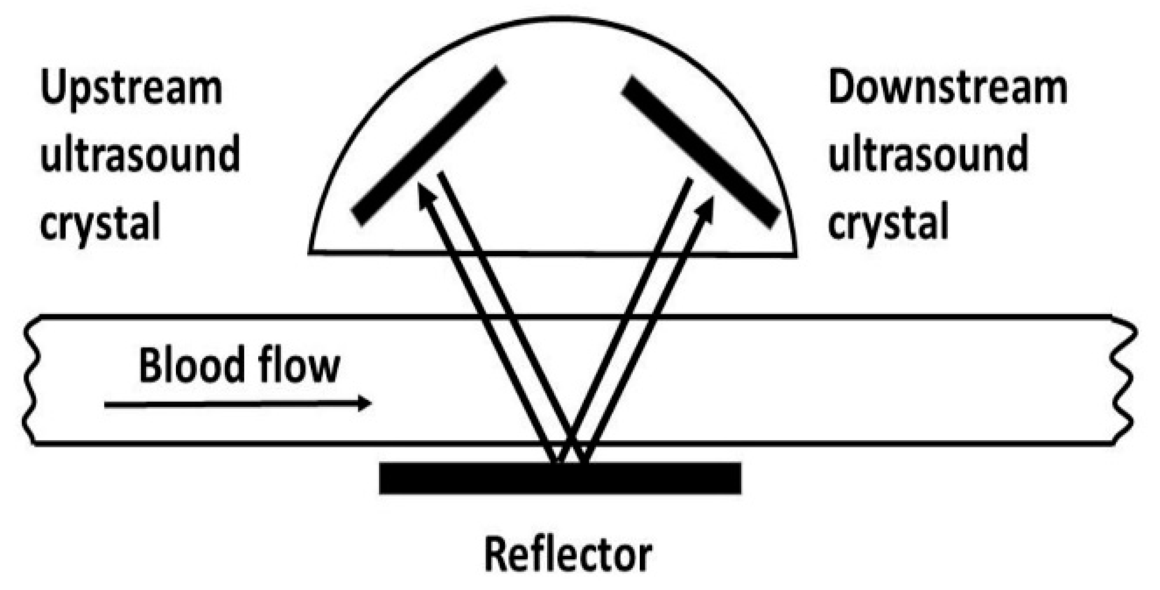
Figure 1. Principle of transit time flow measurement.
Jvd 01 00008 g001
Figure 2. Intraoperative flow measurement in mL/Min.
Jvd 01 00008 g002
Table 1. Patient demographic data and operative indications.
Baseline Characteristicsn = 107 (%)
    Men76 (71%)
    Women31 (29%)
    Age68 ± 4
    Hypertension94 (87.8%)
    Diabetes89 (83%)
    Smoking92 (86%)
    Chronic renal insufficiency71 (66%)
    Coronary artery disease (CAD)57 (53%)
Asymptomatic patients41 (38.3%)
    Bilateral carotid artery stenosis > 70% (NASCET)26 (63%)
    Contralateral closure of internal carotid artery (ICA)15 (37%)
Symptomatic patients66 (61.7%)
    Stroke38 (57.6%)
    TIA: amaurosis fugax
       reversible cerebral ischemia
       minor stroke
28 (42.4%)
Publisher’s Note: MDPI stays neutral with regard to jurisdictional claims in published maps and institutional affiliations.