Article

Probiotics Alter the Microbial and Behavioral Consequences of Methamphetamine Exposure in a Sex-Selective Manner

by
Shadab Forouzan
1,
Kristi L. Hoffman
2 and
Therese A. Kosten
1,*
1
Department of Psychology, University of Houston, Houston, TX 77204, USA
2
Molecular Virology and Microbiolgy, Baylor College of Medicine, Houston, TX 77030, USA
*
Author to whom correspondence should be addressed.
Psychoactives 2024, 3(3), 318-336; https://doi.org/10.3390/psychoactives3030020
Submission received: 25 April 2024 / Revised: 23 June 2024 / Accepted: 25 June 2024 / Published: 1 July 2024

Abstract

:
Methamphetamine use disorder (MuD) is a global health problem, with no FDA-approved medications. Our prior work demonstrated that repeated methamphetamine exposure alters the gut microbiota in male rats and results in depressive-like behaviors. In this study, we extend our findings to females and determine whether probiotics block these effects. Male and female rats were administered methamphetamine (2 mg/kg; SC) or saline twice daily with either a combination of two probiotics (Lactobacillus helveticus R0052 and Bifidobacterium longum R0175) or placebo solution for 14 days. Fecal samples were collected at baseline and other days after treatment cessation. Tests of anxiety- and depressive-like behaviors were conducted using open-field and forced-swim assays. Methamphetamine induced anxiety-like behavior in females and anxiety-like and depressive-like behaviors in males. Probiotics blocked the depressive-like effect in males but did not alter anxiety-like effects in either sex. Methamphetamine exposure decreased levels of alpha diversity in both sexes, but sex differences were seen in the ability of probiotics or methamphetamine to alter levels of various bacteria. These findings support the role of the gut–brain microbiome in the depressive effects of repeated methamphetamine exposure in males, suggesting that probiotics may be a viable treatment option for MuD.

1. Introduction

Methamphetamine (METH) is a powerful psychostimulant with a high potential for abuse. In the United States, 0.9 percent of the population over the age of 12 (or about 2.6 million people) report using METH in the past 12 months, and the number of individuals diagnosed with methamphetamine use disorder (MuD) is nearly 1.5 million [1]. METH is self-administered through different routes, including injection, oral ingestion, smoking, and nasal inhalation. The injection route is associated with a longer, more intense withdrawal [2,3]. The core symptoms of METH withdrawal during the first several weeks of abstinence are depression and anxiety [4,5,6]. Experiencing withdrawal symptoms often triggers relapse to compulsive use [7], suggesting that reversal or prevention of METH withdrawal symptoms may be a viable treatment approach [5,8,9]. Pharmacotherapies have been used in combination with behavioral–cognitive therapies in an attempt to improve treatment engagement and retention and to manage withdrawal symptoms, but results are inconsistent [10,11]. Indeed, because there are currently no FDA-approved medications to treat MuD, novel therapeutic targets for MuD, explicitly targeting the management of withdrawal symptoms, are needed.
One novel treatment approach is to target the gut microbiota due to its profound impact on the brain, behavior, and health via the microbiota–gut–brain axis [12,13]. Studies show altered gut microbiota in persons with various psychiatric disorders [12,14,15], including substance use disorders [16], although findings are mixed. For example, bacterial richness and alpha diversity, two measures of the health of the gut microbiota, negatively correlated with severity measures of anxiety and depressive symptoms in an inpatient population [17]. However, currently depressed inpatients showed greater fecal bacterial alpha diversity compared to both healthy controls and to a group of recovering depressed inpatients [18]. Indeed, the relations between measures of alpha and beta diversity and anxiety and depression signs were found to be inconsistent in a meta-analysis study [19]. Other research found various taxa differences at both the family and genus levels between those with depression versus healthy controls, but no consistent pattern was seen [20,21,22]. However, studies in animals showed that fecal transfer from depressed patients to rodents induced depressive-like behaviors [23,24] and that stress-induced behavioral impairments can be alleviated with the administration of probiotics [25,26,27,28]. Probiotics also reduced depressive- and anxiety-like behaviors in rodents with normal gut microbiota [29,30].
Our prior study demonstrated that cessation from chronic METH exposure led to depressive-like behavior and alterations in the gut microbiota in male rats. Specifically, we observed increased immobility in the forced-swim test, but no effect on anxiety-like behaviors (elevated plus maze and open-field tests) and decreased alpha diversity, as well as altered levels in several taxa immediately after cessation of chronic METH exposure [31]. At that time, we also observed increased immobility in the forced-swim test (FST) and decreased activity in the open-field test (OFT) that reflected depressive-like effects. However, there were no effects on anxiety-like behaviors, as assessed with elevated plus maze (EPM) or OFT. Most measures of the gut microbiota were normalized by seven days. The purpose of the present study is to test if concurrent administration of probiotics can ameliorate both the behavioral and microbiome changes due to cessation from chronic METH exposure. Specifically, we chose to administer a probiotic combination that showed efficacy for the treatment of depression [32,33]. We also wanted to extend the research to female rats because some research suggests that associations between the gut–brain axis with psychiatric symptomology are not seen in females [34]. We assessed depressive-like behavior with the forced-swim test and anxiety-like behavior with center time in the open-field test. Although we failed to find an effect on anxiety-like behaviors in our previous study, cessation from chronic METH exposure may affect this behavior in females. Findings are expected to advance our understanding of the role of gut microbiota and guide the choice of target therapeutics for MuD and, perhaps, for other substance use disorders.

2. Materials and Methods

2.1. Animals and Housing

Thirty-two male and thirty-two female Sprague-Dawley rats (60–90 days old) were purchased from Charles River Laboratory (Wilmington, MA, USA) and group-housed (two to a cage) with cage-mates of the same sex and treatment condition in amber polysulfone cages in a temperature- and humidity-controlled colony room with a 12:12 light/dark cycle (lights on at 7:00 a.m.). Animals were provided ad libitum access to food (Purina chow) and tap water. The Institutional Animal Care and Use Committee at the University of Houston approved the experimental procedures, in accordance with the guidelines set forth in “Guide for the Care and Use of Laboratory Animals” (8th Edition) [35].

2.2. Groups and Experimental Timeline

The estimated total sample size was 80 rats (10/group) based on G*Power (version 3.1) calculation at 80% power for moderate effects (f = 0.25) of interest for 3-way interaction effects (of Group × Treatment × Time), treating sex as a measure of a separate independent factor. Due to logistics, a smaller sample size (8/group, instead of 10/group) was sufficiently powered to detect a difference between the groups. The previous study used 8 animals/groups to reduce animal use. There were two groups of rats per sex (n = 16 each), in which rats in one group per sex were randomly assigned to be administered twice daily (8 a.m. and 3 p.m.) injections (SC) of methamphetamine at a dose of 2 mg/kg for 14 consecutive days. Rats in the other group per sex were randomly assigned to receive vehicle (isotonic saline) injections at the same times and served as controls. Prior to the 14 days, all rats were injected with isotonic saline once daily for five consecutive days (Days −4 to 0) to habituate them to the procedure. Half of each of the groups (n = 8 each) was also randomly to be administered a probiotic solution, whereas the other half was assigned to receive a placebo probiotic solution, as described below. Thus, the four groups per sex were designated Control-Placebo, Control-Probiotic, METH-Placebo, and METH-Probiotic (n = 8 each).
All treatments began on Day 0 and terminated on Day 14. Behavioral tests were conducted over the next 3 days (Days 15–17), as described below. Microbiome data obtained on Day 0 after the five habituation sessions was used as the baseline measure. The effects of 14 days of METH and probiotics (or vehicle and placebo) treatments were assessed on Day 14. The effects of acute and chronic cessation of METH administration on the microbiome were assessed on Days 17 and 40, respectively. See Figure 1 for the experimental timeline.

2.3. Methamphetamine Administration

Methamphetamine HCl (Sigma-Aldrich, St. Louis, MO, USA) was dissolved in isotonic saline and administered subcutaneously (SC) in a volume of 2 mg/mL based on our previous study [31].

2.4. Probiotics

The probiotic groups received a combination of commercially available probiotics (L. helveticus R0052 and B. longum R0175, generously supplied by Lallemand Health Solutions Inc., Mirabel, QC, Canada) at a concentration of 1 billion colony-forming units (CFUs) per mL per day (i.e., 0.001 g of probiotic powder per 0.5 mL of phosphate-buffered saline based on a high-concentration stock at 500 × 109 CFU/g) over the same timeframe as the methamphetamine (or vehicle) injections. This treatment procedure was based on findings that administration of concentrations of 109 and 1010 CFU for 2 weeks shows beneficial effects in animals [36]. Sucrose powder (0.1 g) was added to the probiotic solution. An equivalent amount of placebo solution was prepared (i.e., 0.1 g of sucrose powder per 0.5 mL of phosphate-buffered saline) and administered to the placebo groups. The probiotic or placebo was delivered via syringe feeding between 8 and 9 a.m., as per the protocol developed in [37] to reduce handling stress and for more precise individual dosing of probiotics. The probiotic solution inside the syringe was monitored to ensure that enough bacteria were ingested. Solutions were made fresh each morning and maintained at 4 (±4) °C until administration [38].

2.5. Syringe Feeding

All animals were trained to drink probiotic or placebo solutions via syringe feeding for 3–4 days prior to the start of the experiment (adapted from [37]). A rat was placed in a towel and held gently against the experimenter’s chest and slowly fed 0.5 mL of solution using a 1 mL oral syringe so that the syringe did not enter the mouth cavity beyond its tip. Once hand-training was complete, the rat was held inside the cage to learn the association of syringe feeding with the home cage. The rat was ready for the next phase if it approached the syringe in the cage and drank from it on its own. The solutions were slowly and forcefully administered to rats by the side of the mouth behind the teeth to make sure the solutions were swallowed. Some METH-exposed animals stopped drinking the solutions voluntarily and had to be force-fed. Notably, the data derived from these animals did not differ from the free-feeding rats. Feeding was conducted at the same time daily (8 a.m. ± 1 h) and a standard single-use syringe was used for individual rats.

2.6. Behavioral Testing

Behavioral tests were conducted on Days 15 and 17 following cessation of repeated METH exposure (see Figure 1). The forced-swim test (FST) requires a swim pre-exposure on one day followed by the test day. The pre-exposure occurred on Day 16, two days following cessation of repeated METH exposure, with the test session conducted on Day 17. The open-field test (OFT) was performed on Day 15. This order was chosen so that the more stressful test (FST) was run last [39].
Open-field test (OFT). We used time in the center of the open field as a measure of anxiety-like behavior, with lower levels indicative of anxiety and higher levels indicative of novelty-seeking or risk-taking behavior [40,41]. Rats were tested on Day 15. Animals were individually placed in the center of the apparatus (43.0 cm × 43.2 cm × 30.5 cm; MED Associates, Fairfax, VT, USA) that used an infrared emitter and detectors located around the perimeter to tabulate measures of activity (total distance traveled in cm) and time spent (s) in the center of the arena. The apparatuses were cleaned with 70% ethanol and water after each rat was tested.
Forced-swim test (FST). The forced-swim test (FST) is a widely used model to assess depressive-like behavior, specifically behavioral despair, as indicated by greater time spent immobile during the test session [42,43,44]. One day before the test day (Day 16), rats were placed in a cylindrical tank (40 cm H × 20 cm D) filled to about 10 cm from the top with water (24–25 °C) for 15 min and then removed. The water depth was set to prevent animals from touching the bottom of the tank with their hind limbs. On the following day (Day 17), rats were again placed in the tank for 5 min and videotaped for later behavioral analysis, conducted using a computerized program (Ethovision; Noldus; Wageningen, The Netherlands) to avoid any experiment bias. The length of time spent immobile, defined as floating or the least amount of movement needed to maintain the head above the water, was recorded. The apparatuses were cleaned with 70% ethanol and water after each rat was tested.

2.7. Data Analysis for Behavioral Assessments

Data were analyzed using 2 (Group: METH vs. Control) × 2 (Treatment: Probiotic vs. Placebo) ANOVA. The measures analyzed included total time spent in the center (s) and total distance traveled (cm) in the OFT, and time (s) spent immobile in the FST. Separate analyses were conducted by sex. Post hoc Tukey tests were used to follow-up on significant main effects and interactions. Significance was set at p < 0.05 for all tests. Statistical analyses were performed using Statistica 13.0 software.

2.8. Fecal Sample Collection

Fecal samples were collected from all groups 1 h after the morning injection at the following time points: baseline (Day 0), Day 14 (repeated treatment assessment), and on Day 17 of acute METH withdrawal and Day 40 of chronic METH withdrawal (see Figure 1). On the fecal collection days, each rat was taken out of the cage, placed on the sterilized countertop, and allowed to defecate normally. The fecal pellets per animal were collected into an empty sterilized tube and labeled such that no identifying information was provided. The tubes containing fecal pellets were placed on ice as soon as possible and stored at −80 °C until further analyzed using 16s rRNA sequencing, performed by the Center for Metagenomics and Microbiome Research (CMMR) at Baylor College of Medicine.

2.9. 16S rRNA Gene Sequencing and Compositional Analysis

Total genomic DNA was extracted using the MOBIO PowerMag Soil DNA Isolation Kit (MO BIO Laboratories, Carlsbad, CA, USA), according to the manufacturer’s protocol. The 16S rDNA V4 region was amplified by PCR (GGACTACHVGGGTWTCTAAT and GTGCCAGCMGCCGCGGTAA) [45] and sequenced on the MiSeq platform (Illumina, San Diego, CA, USA) using the 2 × 250 bp paired-end protocol, yielding paired-end reads that overlapped almost completely. The primers used for amplification contained adapters for MiSeq sequencing and single-end barcodes, allowing pooling and direct sequencing of PCR products [45].
The 16S rRNA gene data pipeline incorporates phylogenetic and alignment-based approaches to maximize data resolution. The read pairs were demultiplexed based on the unique molecular barcodes, and reads were merged using USEARCH v7.0.1090 (Edgar 2010). Sequences were clustered into operational taxonomic units (OTUs) at a similarity cutoff value of 97% using the UPARSE algorithm [46]. OTUs were mapped to an optimized version of the SILVA Database (version 128, https://www.arb-silva.de/documentation/release-128/ 1 June 2023) [47] containing only the 16S v4 region to determine taxonomies. UPARSE centroid OTU sequences were queried via the Basic Local Alignment Search Tool (BLAST) [48] to identify likely representative bacterial species. ATIMA (Agile Toolkit for Incisive Microbial Analyses), a visualization toolkit developed at the CMMR, was used to evaluate alpha diversity, beta diversity, and phylogenetic trends using a rarefied OTU table (2964 reads/sample). The significance of categorical variables was determined using the non-parametric Mann–Whitney test for two-category comparisons or the Kruskal–Wallis test when comparing three or more categories. Correlations between two continuous variables were determined with R (version 4.3.0) base “lm” function for linear regression models), where p-values indicated the probability that the slope of the regression line is zero. PCoA plots employed the Monte Carlo permutation test [49] to estimate p-values. All p-values were adjusted for multiple comparisons with the FDR algorithm [50].

2.10. Gut Microbiome Analysis

Taxonomy assignment and diversity analyses were computed through ATIMA with default settings to compare bacterial species richness between the control and METH groups. Bacterial alpha diversity (within-sample diversity) was measured using observed OTU (total number of unique OTUs per sample). Statistical significance in alpha diversity was calculated by Kruskal–Wallis and Mann–Whitney U tests. Beta diversity (between-sample diversity) was measured using the weighted UniFrac distance metric [51]. UniFrac incorporates phylogenetic information for the comparison of bacterial communities. Principal coordinate analysis (PCoA) was used to visualize clustering patterns between samples based on beta diversity distances generated from the normalized OTU table. The relative abundance of various bacterial taxa at phylum and genus taxonomic levels was compared between groups at different time points using Kruskal–Wallis and Mann–Whitney U tests. Results are expressed as means ± SEM. A value of p < 0.05 was used for all biomarkers. All procedures were chosen based on our prior work [31].

3. Results

3.1. Open-Field Test (OFT)
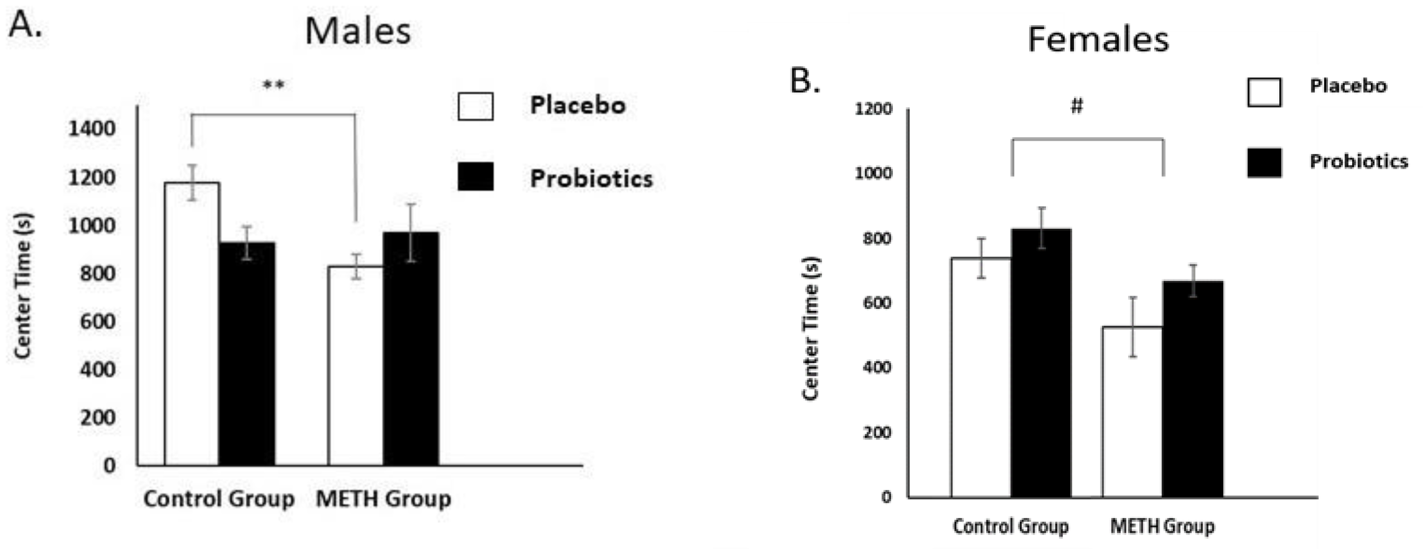
The behavioral measures obtained from the open field test (OFT) included time spent (s) in the center area of the open-field arena and total distance traveled (cm) across the 30 min test session. Decreased time in the center area indicated anxiety-like behavior. Total distance traveled reflected exploratory behavior. Data were analyzed separately by sex.

3.1.1. Male Rats

Time spent in the center of the arena for male rats showed a significant interaction of Group × Treatment (F (1, 28) = 5.83, p < 0.0225), although neither main effect was significant (p > 0.05). As seen in Figure 2A, the METH-Placebo group spent less time in the center of the open-field arena than the Control-Placebo group (Tukey HSD: p < 0.05), suggesting that acute cessation from METH resulted in anxiety-like effects. The Group × Treatment interaction may also reflect that the probiotic treatment increased anxiety-like behavior (e.g., lower center time) in the control group, and that both METH groups showed anxiety-like effects regardless of probiotic treatment. There were no significant effects on measures of distance traveled at either time (p > 0.10), as seen in Table 1.

3.1.2. Female Rats

Time spent in the center of the arena for female rats showed a significant main effect of Group (F (1, 28) = 7.7747, p < 0.01), as seen in Figure 2B. Neither the main effect of Treatment nor its interaction with Group was significant (p > 0.10). The METH groups spent significantly less time in the center compared to control groups, demonstrating greater anxiety-like effects. The probiotic treatment did not affect this behavior. There were no significant Group or Treatment effects for total distance traveled (p > 0.10), as seen in Table 1.

3.2. Forced-Swim Test (FST)

The behavioral measure obtained from the forced-swim test (FST) was time spent immobile during the 5 min test session. Increased immobility time reflects behavioral despair and is a measure of depressive-like behavior. Immobility time for male rats showed a significant interaction of Group × Treatment (F (1, 28) = 10.508, p < 0.01), although neither main effect was significant (p > 0.10). The METH-Placebo group spent significantly more time immobile compared to both control groups (p > 0.05) and the METH-Probiotics group (p < 0.01), as seen in Figure 3A. This suggests that cessation from METH resulted in depressive-like effects and that probiotics alleviated this effect. Immobility time for females showed no significant main effects of Group or Treatment or an interaction effect (p > 0.10), as seen in Figure 3B.

3.3. Microbial Diversity

We assessed alpha and beta diversity to explore alterations in microbiota diversity between groups and treatments. Separate analyses for each measure were conducted by sex. We analyzed observed OTUs to measure alpha diversity or within-sample diversity. Beta diversity, which examines differences in the overarching microbial community, was analyzed using the weighted UniFrac distance metric using principal coordinate analysis. However, overall, microbial communities did not cluster into separate populations after METH exposure and probiotic treatments and no significant differences in beta diversity were found for either sex.

3.3.1. Male Rats

Alpha diversity did not differ between groups of male rats at baseline (Day 0) prior to probiotic and METH (or Placebo and Vehicle) administrations, as seen in Figure 4A (p > 0.10). Bacterial richness, as assessed by the number of observed OTUs, showed a significant Group × Time interaction (Kruskal–Wallis test; p < 0.05) that reflected the lower alpha diversity of the METH groups, relative to control groups, on Day 14, the last day of the drug and probiotic administrations (Wilcoxon matched-pairs test; p < 0.05). These changes were normalized by acute and chronic METH withdrawal days (Days 17 and 40). There was no effect of probiotic treatment (p > 0.01).

3.3.2. Female Rats

Alpha diversity did not differ between groups of female rats at baseline (Day 0), as seen in Figure 4B. Bacterial richness, or observed OTUs, showed a significant interaction of Treatment × Time (Kruskal–Wallis test; p < 0.05). Probiotic treatment in female rats was associated with significantly higher alpha diversity compared to placebo treatments at Day 14 and on Days 17 and 40 (Mann–Whitney U test; p < 0.05). Also, on Day 14, the METH-Placebo group had significantly lower alpha diversity than the Control-Placebo group (Mann–Whitney U test; p < 0.05). The within-group comparison showed that both METH-Placebo and METH-Probiotics groups had significantly lower alpha diversity at Day 14 compared to baseline (Wilcoxon matched-pairs test; p < 0.05). These changes were normalized by Day 40 following chronic METH withdrawal.

3.4. Microbial Composition
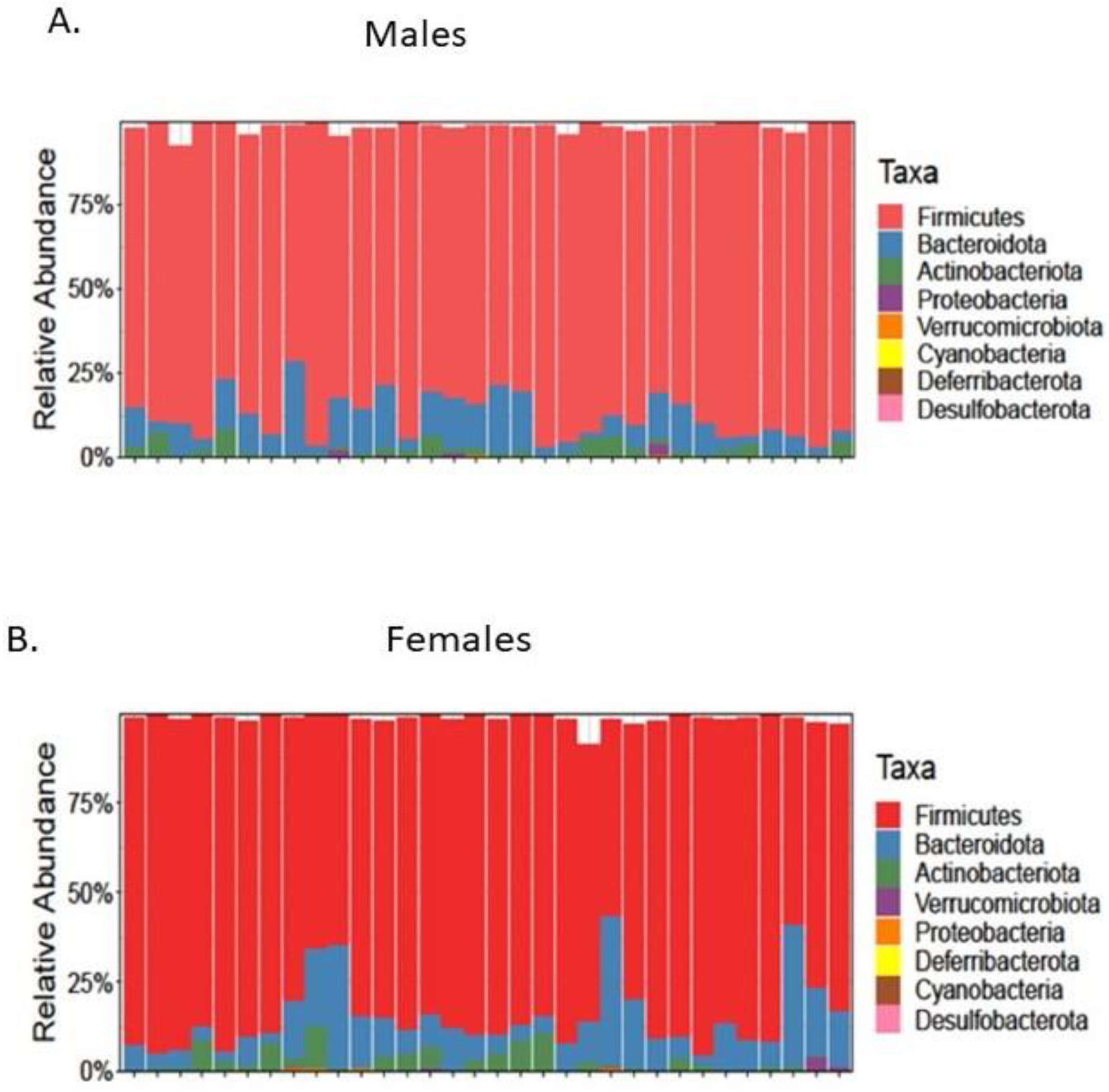
We assessed differences in microbial composition between groups by comparing the relative abundances of taxa at the phylum and genus levels. Separate analyses were conducted by sex. The effects of METH exposure and probiotics on gut microbial composition on Day 14 and on Days 17 and 40 following acute and chronic METH withdrawal, respectively, are shown in Table 2.

3.4.1. Male Rats

The intestinal microbiota of male rats at baseline (Day 0) was typical for healthy individuals—it was largely dominated by the phyla Firmicutes (86%), Bacteroidota (9%), and Actinobacteria (2%), followed by Proteobacteria (0.4%), Verrucomicrobia (0.2%), and Cyanobacteria (0.02%), as seen in Figure 5A. At the phylum level, the relative abundance of Firmicutes, Bacteroidota, Actinobacteria, and Proteobacteria of male rats were as shown in Table 2. The within-group comparison showed that the relative abundance of Bacteroidota in the METH-Placebo group decreased significantly from baseline to Day 14 and from baseline to Day 40 of chronic METH withdrawal (Wilcoxon matched-pairs test; p < 0.05). In addition, the within-group comparison revealed a significant decrease in the relative abundance of Actinobacteria from baseline to Day 40 of chronic METH withdrawal (Wilcoxon matched-pairs test; p < 0.05). There were no Group or Treatment effects for Firmicutes or Proteobacteria at any test time (Kruskal–Wallis test; p > 0.05).
At the genus level, the relative abundance of Bifidobacterium and Lactobacillus in male rats were as shown in Table 2. The within-group comparison showed that the relative abundance of Lactobacillus increased significantly from baseline to Day 14 in the METH-Placebo group (Wilcoxon matched-pairs test; p < 0.05). Also, the relative abundance of Lactobacillus decreased significantly from baseline to Day 14 in the METH-Probiotics group (Wilcoxon matched-pairs test; p < 0.05). These changes were normalized by Days 17 and 40 (acute and chronic METH withdrawal, respectively). There were no significant main effects or interactions in the relative abundance of Bifidobacterium (Kruskal–Wallis test; p > 0.05).

3.4.2. Female Rats

The intestinal microbiota of female rats at baseline (Day 0) was typical for healthy individuals—it was dominated largely by the phyla Firmicutes (83%), Bacteroidota (11%), and Actinobacteria (3%), followed by Verrucomicrobia (0.3%), Proteobacteria (0.2%), and Deferribacterota (0.03%), as seen in Figure 5B. The effects of METH exposure and probiotics on gut microbial composition at Day 14 and Days 17 and 40 following acute and chronic METH withdrawal are shown in Table 3.
At the phylum level, for the relative abundance of Firmicutes, Bacteroidota, Actinobacteria, and Proteobacteria in female rats, there was an interaction of Treatment × Time in the relative abundance of Firmicutes at Days 17 and 40 (acute and chronic METH withdrawal, respectively; Kruskal–Wallis test; p < 0.05). The placebo groups showed a significantly higher level of Firmicutes than probiotic groups (Mann–Whitney U test; p < 0.05). The relative abundance of Firmicutes in the METH-Placebo group increased significantly from baseline to Day 17 of acute METH withdrawal and remained high even at chronic withdrawal on Day 40 (Wilcoxon matched-pairs test; p < 0.05). The within-group comparison also showed that relative abundance of Firmicutes in the METH-Probiotics group decreased significantly on Day 17 following acute METH withdrawal, compared to Day 14 (Wilcoxon matched-pairs test; p < 0.05). These changes were normalized after three weeks of chronic METH withdrawal (Day 40).
There was a main effect of Treatment on Day 40 following chronic METH withdrawal and an interaction effect of Group × Treatment × Time in the relative abundance of the phylum Bacteroidota in female rats (Kruskal–Wallis test; p < 0.05). On Day 14, the Control-Probiotics group had significantly higher levels of Bacteroidota than the Control-Placebo group. Also, on Day 40 following chronic METH withdrawal, probiotic groups had significantly higher levels of Bacteroidota than placebo groups (Mann–Whitney U test; p < 0.05). The within-group comparison showed that relative abundance of Bacteroidota in the METH-Probiotics group increased significantly on Day 17 following acute METH withdrawal, compared to Day 14 (Wilcoxon matched-pairs test; p < 0.05).
There was a main effect of Treatment and an interaction effect of Group × Treatment × Time in the relative abundance of the phylum Actinobacteria in female rats (Kruskal–Wallis test; p < 0.05). The Control-Probiotic group had lower levels of Actinobacteria than placebo groups on Day 40 following chronic METH withdrawal. On Day 17 following acute METH withdrawal, the METH-Probiotics group had a significantly higher relative abundance of Actinobacteria than the METH-Placebo group (Mann–Whitney U test; p < 0.05). Furthermore, the Control-Placebo group had a significantly higher relative abundance of Actinobacteria than the METH-Placebo group (Mann–Whitney U test; p < 0.05). The within-group comparison showed that relative abundance of Actinobacteria in the METH-Placebo group decreased significantly on Day 17 following acute METH withdrawal, compared to Day 14 (Wilcoxon matched-pairs test; p < 0.05). These changes were normalized after three weeks of chronic METH withdrawal (Day 40).
There was a significant interaction of Group × Treatment × Time in the relative abundance of the phylum Proteobacteria in female rats (Kruskal–Wallis test; p < 0.05). Immediately after cessation of treatments on Day 14, the Control-Probiotic group had significantly higher levels of Proteobacteria than the Control-Placebo group, but this difference reversed on Day 40, such that the Control-Probiotic group had lower levels of Proteobacteria (Mann–Whitney U test; p < 0.05). The within-group comparison showed that the relative abundance of Proteobacteria significantly increased in the Control-Probiotics group from baseline to Day 40 (Wilcoxon matched-pairs test; p < 0.05). Levels of Proteobacteria in the METH-Placebo group decreased significantly on Days 17 and 40 following acute and chronic METH withdrawal, compared to baseline (Wilcoxon matched-pairs test; p < 0.05). The relative abundance of Proteobacteria in the METH-Probiotics group also decreased significantly from baseline to Day 14, and these changes normalized by Day 17 following acute METH withdrawal (Wilcoxon matched-pairs test; p < 0.05).
At the genus level, the relative abundance of Bifidobacterium and Lactobacillus in female rats were as shown in Table 3. There was a significant main effect of Treatment in the relative abundance of Bifidobacterium on Day 40 (Kruskal–Wallis test; p > 0.05), in which the placebo groups showed a higher level of Bifidobacterium than probiotic groups. The within-group comparison showed that the relative abundance of Bifidobacterium in the METH-Probiotics group increased significantly on Day 14 compared to baseline, and these changes were normalized after three weeks of METH withdrawal (Day 40, Wilcoxon matched-pairs test; p < 0.05). Also, the relative abundance of Bifidobacterium decreased significantly in the METH-Placebo group from Day 14 to Day 17 (acute METH withdrawal), and it normalized after three weeks of METH withdrawal (Day 40, Wilcoxon matched-pairs test; p < 0.05). Furthermore, there was a significant Group × Time interaction effect in the relative abundance of Lactobacillus (Kruskal–Wallis test; p < 0.05). The METH groups had significantly higher levels of Lactobacillus than control groups at Day 14. The within-group comparison showed that the relative abundance of Lactobacillus in the METH-Placebo group decreased significantly from baseline and Day 14 to Day 40 (chronic METH withdrawal, Wilcoxon matched-pairs test; p < 0.05). Finally, the relative abundance of Lactobacillus for the METH-Probiotics group increased significantly from baseline to Day 14, and these changes were normalized by Days 17 and 40 of acute and chronic withdrawal, respectively (Wilcoxon matched-pairs test; p < 0.05).

4. Discussion

The results of the current study demonstrated that acute cessation from 14 days of repeated, twice-daily METH injections induced anxiety- and depressive-like behaviors in male rats and anxiety-like behavior in females. Administration of the combination of two probiotic strains (L. helveticus R0052 and B. longum R0175) during the METH exposure blocked the depressive-like effect in males but did not alter anxiety-like behavior in either sex. We also observed decreases in alpha diversity and alterations in the relative abundance of specific bacterial taxa due to cessation from repeated METH exposure. Administration of the probiotic combination produced a number of microbial changes in females and restored some aspects of the METH-induced alterations in the composition of the gut microbiome in a sex-dependent manner.
Behavioral effects of METH. The findings that cessation from repeated METH exposure induced anxiety- and depressive-like behavioral effects in male rats and anxiety-like effects in females confirmed and extended our prior work, in which we found depressive-like effects in male rats [31]. This supports the notion that cessation from repeated METH exposure leads to altered behaviors that may be indicative of withdrawal distress. The depressive-like effects in male rats were seen in the forced-swim test (FST). The FST is a widely used model to evaluate depressive-like behavior [42,43,44], in which increased time spent immobile during the test session indicates behavioral despair. However, the female rats tested in the current study did not show this effect of cessation from METH exposure. That females showed higher immobility times compared to males may suggest that the stress of the FST had a greater impact on them regardless of METH or probiotic administration, which could mask any effect of the treatments.
Cessation from METH exposure did lead to anxiety-like effects in the female rats in the current study, as shown by lower times spent in the center of the open field. The open-field test (OFT) is widely used to measure anxiety-like behavior [52]. Male rats also exhibited greater anxiety-like behavior in the OFT in the present study. However, this finding was not seen in our prior work, in which there was no effect of METH exposure in either the OFT or in the elevated plus maze in male rats [31]. The discrepancy in the center time data between these two studies may reflect that in our prior work, we conducted the OFT at baseline, prior to METH administration, and re-tested after cessation of these treatments. In the current study, rats were tested only once in the OFT after treatments. The initial exposure to the novel apparatus (e.g., the open field) is thought to be a mild stressor and induced heightened activity levels and greater fear compared to what was seen in subsequent sessions. This procedural difference may also explain why we failed to find an effect of cessation from METH administration on distance traveled in the OFT in the current study in either sex but did see a decrease in METH-exposed male rats in our prior work, an effect we suggested may reflect depressive-like effects [31]. The lack of group differences in distance traveled in the current study suggests that we can rule out impaired motoric effects of cessation from METH exposure as an explanation for the greater immobility times seen in the FST in male rats. These preclinical data are consistent with the fact that both anxiety and depression are reported symptoms of METH withdrawal in humans [4,5,6] and that anxiety symptoms are more common during METH withdrawal in women compared to men [6,53].
Microbial effects of METH. Alterations in the gut microbiome were seen with cessation from repeated METH exposure. Alpha diversity, assayed by observed OTUs, was lower in the METH groups immediately after cessation of METH administration in rats of both sexes (Day 14). These changes normalized after three days of METH withdrawal (Day 17) and remained that way three weeks after METH withdrawal (Day 40). This effect is consistent with our prior findings in male rats [31] and is now extended to female rats. However, there was no effect of METH exposure on beta diversity in the present study in either sex, whereas we did see changes in our prior study [31]. Beta diversity reflects the overarching microbial composition that reflects the change in diversity of species across ecosystems. In contrast, alpha diversity reflects the diversity of species or species richness within an ecosystem. It is unclear why we failed to find an effect of METH exposure on beta diversity in the present study. Nonetheless, these findings suggest that METH exposure negatively impacted bacterial richness but had inconsistent effects on evenness in both males and females.
The relative abundance of four major phyla, Firmicutes, Bacteroidota, Actinobacteria, and Proteobacteria, was assessed in the present study. As seen in previous studies [54,55], we found that phyla Firmicutes and Bacteroidota were the most predominant and represented 90% of the gut microbiota of all groups of both sexes. METH exposure increased levels of the genus Lactobacillus on Day 14 compared to baseline in rats of both sexes but did not alter levels of Bifidobacterium. At the phylum level, METH had sex-specific effects. Male rats showed lower levels of Bacteroidota immediately after cessation of METH exposure (Day 14) compared to baseline. These levels were also lower on Day 40, when we also found a decrease in levels of Actinobacteria. Female rats showed an elevated abundance of Firmicutes and lower levels of Proteobacteria on Day 17, with no differences seen immediately after cessation of METH administration. Increased levels of Firmicutes were seen in studies in which alcohol, nicotine, or opioids were administered [19], but this effect was not seen in male rats in the present study or in our previous one with male rats [31]. The results of the current study replicated our prior findings of the effects of Lactobacillus and Bacteroidota but we did not observe a METH-induced enhancement of Bifidobacterium levels, as we did previously [31].
The present results are consistent with prior reports that METH exposure is associated with lower levels of Bacteroidota, suggesting a potential role for gut dysbiosis in the pathogenesis of MuD [31,56,57]. These alterations in the gut microbiome due to METH exposure showed some similarities to those seen in the depression and anxiety literature [18,23,58], but a direct causal relationship between changes in bacteria and METH-induced anxiety and depressive behaviors could not be concluded from this study. Contrary to previous studies that reported a lower relative abundance of beneficial gut bacteria, Bifidobacterium and Lactobacillus, in depressed patients [23,59], we found higher relative abundance of these taxa in the METH groups. Perhaps METH withdrawal resulted in different mechanisms for bacterial changes than depression, even though the observed behaviors were similar. Alternatively, increased abundance, similar to decreased abundance, may disrupt brain circuitry to result in similar alterations in behavior.
Probiotic effects. Administration of the combination of two probiotic strains (L. helveticus R0052 and B. longum R0175) was associated with alterations in the gut microbiome in females but not in males. Probiotic treatment in female rats led to significantly higher alpha diversity compared to placebo treatment immediately after cessation of treatment on Day 14 and was also seen on later days (Days 17 and 40). This suggests that probiotics had a lasting effect even after termination of the treatment, at least in females. There were also some effects of the probiotic treatments in female rats at the phyla and genus levels. We found increased levels of Bacteroidota and Proteobacteria immediately after cessation of treatments on Day 14. This effect was reversed on Day 40, when levels were lower than the control group. On Day 40, we also observed decreased levels of Bifidobacterium and Actinobacteria due to probiotics treatment.
We closely monitored phyla Firmicutes and Actinobacteria compositions and abundances because the probiotic supplement used in this study contained Lactobacillus spp., belonging to Firmicutes, and Bifidobacterium spp., belonging to Actinobacteria. Our expectation was that administration of the probiotic combination of L. helveticus R0052 and B. longum R0175 would increase levels of its component parts. That is, we anticipated it would increase levels of Lactobacillus and Bifidobacterium at the genus level, as well as Firmicutes and Actinobacteria at the phylum level. However, the only effect of probiotic treatment on these bacterium types was a decrease in Actinobacteria in females, and only on Day 40.
Did probiotics alter METH effects on behavior or the gut microbiome? A major aim of the present study was to determine if administration of probiotics would counter the behavioral and microbial consequences of repeated METH exposure. The choice of testing the probiotic combination of L. helveticus R0052 and B. longum R0175 was based on the literature on the gut microbiome and depression and our prior findings that METH induced depressive-like effects and increased relative abundance of Actinobacteria, Lactobacillus, and Bifidobacterium [31].
The depressive-like consequence of cessation from repeated METH exposure was prevented by co-administration of the probiotic combination in male rats. However, we found no effect of the probiotics on alpha diversity in male rats, and in fact, the METH group treated with probiotics had the lowest alpha diversity of the four groups. Although the probiotic administration did not alter the relative abundance of the various phyla taxa assessed in males, it did normalize levels of Lactobacillus and Bacteroidota. Probiotic administration failed to alter the anxiety-like behavior induced by METH in either sex. However, some alterations in the gut microbiome were seen in female rats. For example, the relative abundance of Bacteroidota in the METH-Probiotics group increased significantly on Day 17, as did the relative abundance of Proteobacteria on Day 14, rescuing the apparent METH-induced decreases in these levels. The beneficial effects of probiotics on anxiety and depression may be explained by competitive exclusion of harmful gut pathogens, decreases in pro-inflammatory cytokines, and communication with the central nervous system via vagal sensory fibers, leading to changes in neurotransmitter levels or function [60]. Moreover, exogenously administered probiotics may help maintain the elevation in the relative abundance of the Gram-positive Bifidobacterium during acute METH withdrawal, which might reflect a recovery of the intestinal microbial homeostasis and modulation of inflammatory immune responses in the gut.
Sex differences. Several sex differences were seen in the present study at the microbial level. While METH exposure lowered alpha diversity and increased levels of Lactobacillus in both sexes, it affected the relative abundances of various other taxa differently by sex. Only females showed microbial changes due to the probiotic treatment. This may be driven, in part, by circulating sex hormone concentrations or by sex differences in dose efficacy. While rats of both sexes showed anxiety-like effects due to METH exposure, only male rats expressed a depressive-like effect, perhaps due to sex differences in the effectiveness of the FST in females. Although the literature is mixed on sex differences in the FST in rats [61], our findings of greater immobility times in female rats in the FST compared to male rats are consistent with previous studies [62,63]. Further, immobility times do not differ across the estrous stage [62].
We analyzed the behavioral and microbial data separately by sex due, in part, to complications of including a fourth variable (sex) in the analyses that would have made interpretation of the effects of probiotic treatment and METH exposure groups over time difficult. Further, there were baseline sex differences at both the behavioral and microbial levels. We tested for sex differences in behavior by comparing the two Placebo-Control groups (male vs. female) and for microbial differences by comparing the data obtained on Day 0, prior to the initiation of drug and probiotic administrations. Examination of the OFT data showed that females tended to spend less time in the center than males, t(14) = 1.89; p < 0.08. Female rats also spent more time immobile in the FST than male rats, t(14) = 6.81; p < 0.0001. Further, the baseline levels of alpha diversity were lower in females compared to males, t(61) = 3.87; p < 0.001. The sex differences in the behavioral tests were not unexpected and, as we have argued previously [64], likely reflect that procedures were developed with male subjects using apparatuses more suited to their body size and weight, which are much greater than female rats.
Limitations. Although we obtained significant and meaningful data that built upon our previous research, there are limitations to the current study design. We did not monitor the estrous stage in the female rats. It would have been difficult to assess if there were any such effects considering that the treatments occurred over 14 consecutive days and it was necessary to conduct the behavioral tests of withdrawal immediately after the cessation of treatments.
We used the novel delivery method of syringe feeding as an alternative to oral gavage or water bottles to administer the probiotics. This delivery method controls for bacterial stability, animal welfare, dosing accuracy, and ease of chronic administration and is more translational (closer to human probiotic intake) than other techniques [37]. Not all animals acclimated well to this method of delivery, and some had to be force-fed, which may have been stressful.
The experimental design of the current study examined whether co-administration of probiotics altered the development of behavioral and microbial consequences of repeated METH exposure. While it would have been interesting to determine if probiotics could reverse the expression of METH-induced effects, it was not possible to use this design. Thus, by necessity, we tested whether the probiotics altered the development of the effects of METH administration. Further, we do not know if the behavioral indices of METH withdrawal would be seen weeks after cessation of METH administration. In fact, we attempted to assess withdrawal at 40 days but this within-subject assessment was compromised by clear repetition effects. To address this question would require separate groups that were not tested immediately post-cessation from METH administration. Future studies are needed to delineate some behavioral changes that are seen long after cessation of METH exposure to assess whether probiotics can reverse the withdrawal effects. Finally, it is unclear how these preclinical findings would be transferable to humans. The existing literature in humans is derived from correlational research and shows inconsistent results. Future studies in clinical populations could include more careful analysis of specific withdrawal symptomology expressed (e.g., depression- and anxiety-like effects) and gender in conjunction with alterations in the microbiota. This may provide insight into the treatment efficacy of probiotics, including the length of treatment needed to restore the microbiome.

5. Conclusions

The results of the present study demonstrated that cessation from repeated METH exposure altered the gut microbiota and induced anxiety-like behavior in both males and females and depressive-like behavior in male rats. The findings confirmed and extended our prior work and provided evidence that a probiotics combination (L. helveticus R0052 and B. longum R0175), shown to have efficacy for depression [32,33], mitigated the depressive-like effects of METH withdrawal and enhanced the functionality of existing microbial communities that may prevent METH-induced dysbiosis and intestinal permeability. While the focus of the current study was on withdrawal distress, other research supports the role of the gut–brain axis in the rewarding effects of stimulants [65,66] that may be sex-selective [67]. Indeed, investigations into the role of gut bacteria in substance use disorders, including alcohol, opioids, and other stimulants, show strong support for the contribution of the gut–brain axis to responses to various abused substances [63,68,69,70,71,72]. This literature, along with results from the present study, supports the potential utility for therapies that target the gut microbiota in the treatment of substance use disorders.

Author Contributions

Conceptualization, S.F. and T.A.K.; methodology, S.F., K.L.H. and T.A.K.; software, K.L.H.; validation, S.F., K.L.H. and T.A.K.; formal analysis, S.F.; investigation, S.F.; resources, T.A.K.; data curation, S.F.; writing—original draft preparation, S.F.; writing—review and editing, T.A.K.; visualization, S.F. and T.A.K.; supervision, T.A.K. and K.L.H.; project administration, S.F.; funding acquisition, T.A.K. All authors have read and agreed to the published version of the manuscript.

Funding

This research was funded by The Michael C. Gibson Addiction Research Fund, University of Houston.

Institutional Review Board Statement

The study was conducted in accordance with the Declaration of Helsinki and approved by the Institutional Animal Care and Use Committee of the University of Houston (protocol code 202000053; approval date 2 September 2021).

Informed Consent Statement

Not applicable.

Data Availability Statement

Data are available upon request.

Acknowledgments

Lallemand Health Solutions Inc., Mirabel, QC, Canada, generously supplied the probiotic powder. We thank Richard Meisch for insightful comments on the manuscript.

Conflicts of Interest

The authors declare no conflicts of interest.

References

  1. NIDA. What Is Methamphetamine? Available online: https://nida.nih.gov/publications/research-reports/methamphetamine/what-methamphetamine (accessed on 1 June 2023).
  2. McLellan, A.T.; Lewis, D.C.; O’Brien, C.P.; Kleber, H.D. Drug dependence, a chronic medical illness: Implications for treatment, insurance, and outcomes evaluation. JAMA 2000, 284, 1689–1695. [Google Scholar] [CrossRef]
  3. Novak, S.P.; Kral, A.H. Comparing injection and non-injection routes of administration for heroin, methamphetamine, and cocaine users in the United States. J. Addict. Dis. 2011, 30, 248–257. [Google Scholar] [CrossRef] [PubMed]
  4. London, E.D.; Simon, S.L.; Berman, S.M.; Mandelkern, M.A.; Lichtman, A.M.; Bramen, J.; Shinn, A.K.; Miotto, K.; Learn, J.; Dong, Y.; et al. Mood disturbances and regional cerebral metabolic abnormalities in recently abstinent methamphetamine abusers. Arch. Gen. Psychiatry 2004, 61, 73–84. [Google Scholar] [CrossRef] [PubMed]
  5. Zorick, T.; Nestor, L.; Miotto, K.; Sugar, C.; Hellemann, G.; Scanlon, G.; Rawson, R.; London, E.D. Withdrawal symptoms in abstinent methamphetamine-dependent subjects. Addiction 2010, 105, 1809–1818. [Google Scholar] [CrossRef] [PubMed]
  6. Su, H.; Zhang, J.; Ren, W.; Xie, Y.; Tao, J.; Zhang, X.; He, J. Anxiety level and correlates in methamphetamine-dependent patients during acute withdrawal. Medicine 2017, 96, e6434. [Google Scholar] [CrossRef]
  7. Brecht, M.L.; O’Brien, A.; von Mayrhauser, C.; Anglin, M.D. Methamphetamine use behaviors and gender differences. Addict. Behav. 2004, 29, 89–106. [Google Scholar] [CrossRef]
  8. Newton, T.; Kalechstein, A.; Duran, S.; Vansluis, N.; Ling, W. Methamphetamine abstinence syndrome: Preliminary findings. Am. J. Addict. 2004, 13, 248–255. [Google Scholar] [CrossRef]
  9. McGregor, C.; Srisurapanont, M.; Jittiwutikarn, J.; Laobhripatr, S.; Wongtan, T.; White, J.M. The nature, time course and severity of methamphetamine withdrawal. Addiction 2005, 100, 1320–1329. [Google Scholar] [CrossRef]
  10. Khoramizadeh, M.; Effatpanah, M.; Mostaghimi, A.; Rezaei, M.; Mahjoub, A.; Shishehgar, S. Treatment of amphetamine abuse/use disorder: A systematic review of a recent health concern. Daru J. Fac. Pharm. Tehran Univ. Med. Sci. 2019, 27, 743–753. [Google Scholar] [CrossRef]
  11. Ray, L.A.; Meredith, L.R.; Kiluk, B.D.; Walthers, J.; Carroll, K.M.; Magill, M. Combined Pharmacotherapy and Cognitive Behavioral Therapy for Adults With Alcohol or Substance Use Disorders: A Systematic Review and Meta-analysis. JAMA Netw. Open 2020, 3, e208279. [Google Scholar] [CrossRef]
  12. Mayer, E.A.; Tillisch, K.; Gupta, A. Gut/brain axis and the microbiota. J. Clin. Investig. 2015, 125, 926–938. [Google Scholar] [CrossRef] [PubMed]
  13. Cryan, J.F.; Dinan, T.G. Mind-altering microorganisms: The impact of the gut microbiota on brain and behaviour. Nat. Rev. Neurosci. 2012, 13, 701–712. [Google Scholar] [CrossRef] [PubMed]
  14. Bastiaanssen, T.F.; Cowan, C.S.; Claesson, M.J.; Dinan, T.G.; Cryan, J.F. Making sense of… the microbiome in psychiatry. Int. J. Neuropsychopharmacol. 2018, 22, 37–52. [Google Scholar] [CrossRef] [PubMed]
  15. Yarandi, S.S.; Peterson, D.A.; Treisman, G.J.; Moran, T.H.; Pasricha, P.J. Modulatory effects of gut microbiota on the central nervous system: How gut could play a role in neuropsychiatric health and diseases. J. Neurogastroenterol. Motil. 2016, 22, 201–212. [Google Scholar] [CrossRef] [PubMed]
  16. Skosnik, P.D.; Cortes-Briones, J.A. Targeting the ecology within: The role of the gut–brain axis and human microbiota in drug addiction. Med. Hypotheses 2016, 93, 77–80. [Google Scholar] [CrossRef] [PubMed]
  17. Madan, A.; Thompson, D.; Fowler, J.C.; Ajami, N.J.; Salas, R.; Frueh, B.C.; Bradshaw, M.R.; Weinstein, B.L.; Oldham, J.M.; Petrosino, J.F. The gut microbiota is associated with psychiatric symptom severity and treatment outcome among individuals with serious mental illness. J. Affect. Disord. 2020, 264, 98–106. [Google Scholar] [CrossRef] [PubMed]
  18. Jiang, H.; Ling, Z.; Zhang, Y.; Mao, H.; Ma, Z.; Yin, Y.; Wang, W.; Tang, W.; Tan, Z.; Shi, J.; et al. Altered fecal microbiota composition in patients with major depressive disorder. Brain Behav. Immun. 2015, 48, 186–194. [Google Scholar] [CrossRef] [PubMed]
  19. Simpson, C.A.; Diaz-Arteche, C.; Eliby, D.; Schwartz, O.S.; Simmons, J.G.; Cowan, C.S.M. The gut microbiota in anxiety and depression—A systematic review. Clin. Psychol. Rev. 2021, 83, 101943. [Google Scholar] [CrossRef]
  20. Cheung, S.G.; Goldenthal, A.R.; Uhlemann, A.C.; Mann, J.J.; Miller, J.M.; Sublette, M.E. Systematic Review of Gut Microbiota and Major Depression. Front. Psychiatry 2019, 10, 34. [Google Scholar] [CrossRef]
  21. Mason, B.L.; Li, Q.; Minhajuddin, A.; Czysz, A.H.; Coughlin, L.A.; Hussain, S.K.; Koh, A.Y.; Trivedi, M.H. Reduced anti-inflammatory gut microbiota are associated with depression and anhedonia. J. Affect. Disord. 2020, 266, 394–401. [Google Scholar] [CrossRef]
  22. Sanada, K.; Nakajima, S.; Kurokawa, S.; Barceló-Soler, A.; Ikuse, D.; Hirata, A.; Yoshizawa, A.; Tomizawa, Y.; Salas-Valero, M.; Noda, Y.; et al. Gut microbiota and major depressive disorder: A systematic review and meta-analysis. J. Affect. Disord. 2020, 266, 1–13. [Google Scholar] [CrossRef] [PubMed]
  23. Zheng, P.; Zeng, B.; Zhou, C.; Liu, M.; Fan, Z.; Xu, X.; Zeng, L.; Chen, J.; Fan, S.; Du, X.; et al. Gut microbiome remodeling induces depressive-like behaviors through a pathway mediated by the host’s metabolism. Mol. Psychiatry 2016, 21, 786–796. [Google Scholar] [CrossRef]
  24. Kelly, J.R.; Borre, Y.; O’Brien, C.; Patterson, E.; El Aidy, S.; Deane, J.; Kennedy, P.J.; Beers, S.; Scott, K.; Moloney, G.; et al. Transferring the blues: Depression-associated gut microbiota induces neurobehavioural changes in the rat. J. Psychiatr. Res. 2016, 82, 109–118. [Google Scholar] [CrossRef] [PubMed]
  25. Li, N.; Wang, Q.; Wang, Y.; Sun, A.; Lin, Y.; Jin, Y.; Li, X. Oral probiotics ameliorate the behavioral deficits induced by chronic mild stress in mice via the gut microbiota-inflammation axis. Front. Behav. Neurosci. 2018, 12, 266. [Google Scholar] [CrossRef] [PubMed]
  26. Marin, I.A.; Goertz, J.E.; Ren, T.; Rich, S.S.; Onengut-Gumuscu, S.; Farber, E.; Wu, M.; Overall, C.C.; Kipnis, J.; Gaultier, A. Microbiota alteration is associated with the development of stress-induced despair behavior. Sci. Rep. 2017, 7, 43859. [Google Scholar] [CrossRef] [PubMed]
  27. Desbonnet, L.; Garrett, L.; Clarke, G.; Kiely, B.; Cryan, J.F.; Dinan, T.G. Effects of the probiotic Bifidobacterium infantis in the maternal separation model of depression. Neuroscience 2010, 170, 1179–1188. [Google Scholar] [CrossRef] [PubMed]
  28. Savignac, H.M.; Kiely, B.; Dinan, T.G.; Cryan, J.F. Bifidobacteria exert strain-specific effects on stress-related behavior and physiology in BALB/c mice. Neurogastroenterol. Motil. 2014, 26, 1615–1627. [Google Scholar] [CrossRef] [PubMed]
  29. Bravo, J.A.; Forsythe, P.; Chew, M.V.; Escaravage, E.; Savignac, H.M.; Dinan, T.G.; Bienenstock, J.; Cryan, J.F. Ingestion of Lactobacillus strain regulates emotional behavior and central GABA receptor expression in a mouse via the vagus nerve. Proc. Natl. Acad. Sci. USA 2011, 108, 16050–16055. [Google Scholar] [CrossRef] [PubMed]
  30. Arseneault-Breard, J.; Rondeau, I.; Gilbert, K.; Girard, S.A.; Tompkins, T.A.; Godbout, R.; Rousseau, G. Combination of Lactobacillus helveticus R0052 and Bifidobacterium longum R0175 reduces post-myocardial infarction depression symptoms and restores intestinal permeability in a rat model. Br. J. Nutr. 2012, 107, 1793–1799. [Google Scholar] [CrossRef]
  31. Forouzan, S.; Hoffman, K.L.; Kosten, T.A. Methamphetamine exposure and its cessation alter gut microbiota and induce depressive-like behavioral effects on rats. Psychopharmacology 2021, 238, 281–292. [Google Scholar] [CrossRef]
  32. Wallace, C.J.K.; Milev, R.V. The Efficacy, Safety, and Tolerability of Probiotics on Depression: Clinical Results From an Open-Label Pilot Study. Front. Psychiatry 2021, 12, 618279. [Google Scholar] [CrossRef] [PubMed]
  33. Karakula-Juchnowicz, H.; Rog, J.; Juchnowicz, D.; Łoniewski, I.; Skonieczna-Żydecka, K.; Krukow, P.; Futyma-Jedrzejewska, M.; Kaczmarczyk, M. The study evaluating the effect of probiotic supplementation on the mental status, inflammation, and intestinal barrier in major depressive disorder patients using gluten-free or gluten-containing diet (SANGUT study): A 12-week, randomized, double-blind, and placebo-controlled clinical study protocol. Nutr. J. 2019, 18, 50. [Google Scholar] [CrossRef] [PubMed]
  34. Kleiman, S.C.; Bulik-Sullivan, E.C.; Glenny, E.M.; Zerwas, S.C.; Huh, E.Y.; Tsilimigras, M.C.; Fodor, A.A.; Bulik, C.M.; Carroll, I.M. The Gut-Brain Axis in Healthy Females: Lack of Significant Association between Microbial Composition and Diversity with Psychiatric Measures. PLoS ONE 2017, 12, e0170208. [Google Scholar] [CrossRef] [PubMed]
  35. National_Research_Council. Guide for the Care and Use of Laboratory Animals, 8th ed.; National Academies Press: Washington, DC, USA, 2011. [Google Scholar]
  36. Wang, H.; Lee, I.S.; Braun, C.; Enck, P. Effect of Probiotics on Central Nervous System Functions in Animals and Humans: A Systematic Review. J. Neurogastroenterol. Motil. 2016, 22, 589–605. [Google Scholar] [CrossRef] [PubMed]
  37. Tillmann, S.; Wegener, G. Syringe-feeding as a novel delivery method for accurate individual dosing of probiotics in rats. Benef. Microbes 2018, 9, 311–315. [Google Scholar] [CrossRef] [PubMed]
  38. Myles, E.M.; O’Leary, M.E.; Romkey, I.D.; Piano, A.; de Carvalho, V.; Tompkins, T.A.; Perrot, T.S. Guidelines for best practice in placebo-controlled experimental studies on probiotics in rodent animal models. Benef. Microbes 2020, 11, 245–254. [Google Scholar] [CrossRef] [PubMed]
  39. McIlwain, K.L.; Merriweather, M.Y.; Yuva-Paylor, L.A.; Paylor, R. The use of behavioral test batteries: Effects of training history. Physiol. Behav. 2001, 73, 705–717. [Google Scholar] [CrossRef] [PubMed]
  40. Hall, C.S. Emotional behavior in the rat. III. The relationship between emotionality and ambulatory activity. J. Comp. Physiol. Psychol. 1936, 22, 345–352. [Google Scholar] [CrossRef]
  41. Weiss, J.M.; Bailey, W.H.; Pohrecky, L.A.; Korzeniowski, D.; Grillione, G. Stress-induced depression of motor activity correlates with regional changes in brain norepinephrine but not in dopamine. Neurochem. Res. 1980, 5, 9–22. [Google Scholar] [CrossRef]
  42. Caspi, A.; Sugden, K.; Moffitt, T.E.; Taylor, A.; Craig, I.W.; Harrington, H.; McCaly, J.; Mill, J.; Martin, J.; Braithwaite, A.; et al. Influence of life stress on depression: Moderation by a polymorphism in the 5-HTT gene. Science 2003, 301, 386–389. [Google Scholar] [CrossRef]
  43. Detke, M.J.; Rickels, M.; Lucki, I. Active behaviors in the rat forced swimming test differentially produced by serotonergic and noradrenergic antidepressants. Psychopharmacology 1995, 121, 66–72. [Google Scholar] [CrossRef] [PubMed]
  44. Porsolt, R.D.; Bertin, A.; Jalfre, M. Behavioral despair in mice: A primary screening test for antidepressants. Arch. Int. De Pharmacodyn. Et. De Ther. 1977, 229, 327–336. [Google Scholar]
  45. Caporaso, J.G.; Lauber, C.L.; Walters, W.A.; Berg-Lyons, D.; Huntley, J.; Fierer, N.; Owens, S.M.; Betley, J.; Fraser, L.; Bauer, M.; et al. Ultra-high-throughput microbial community analysis on the Illumina HiSeq and MiSeq platforms. ISME J. 2012, 6, 1621–1624. [Google Scholar] [CrossRef] [PubMed]
  46. Edgar, R.C. Search and clustering orders of magnitude faster than BLAST. Bioinformatics 2010, 26, 2460–2461. [Google Scholar] [CrossRef] [PubMed]
  47. Quast, C.; Pruesse, E.; Yilmaz, P.; Gerken, J.; Schweer, T.; Yarza, P.; Peplies, J.; Glöckner, F.O. The SILVA ribosomal RNA gene database project: Improved data processing and web-based tools. Nucleic Acids Res. 2013, 41, D590–D596. [Google Scholar] [CrossRef] [PubMed]
  48. Zhang, Z.; Schwartz, S.; Wagner, L.; Miller, W. A greedy algorithm for aligning DNA sequences. J. Comput. Biol. A J. Comput. Mol. Cell Biol. 2000, 7, 203–214. [Google Scholar] [CrossRef] [PubMed]
  49. Dwass, M. Modified tests for nonparametric hypotheses. Ann. Math. Stat. 1957, 28, 181–187. [Google Scholar] [CrossRef]
  50. Benjamin, Y.; Hochberg, Y. Controlling the false discovery rate: A practical and powerful approach to multiple testing. J. R. Stat. Soc. Ser. B 1995, 57, 289–300. [Google Scholar] [CrossRef]
  51. Lozupone, C.; Knight, R. UniFracL: A new phylogenetic method for comparing microbial communities. Appl. Environ. Microbiol. 2005, 71, 828–8235. [Google Scholar] [CrossRef]
  52. Uys, J.D.; Stein, D.J.; Daniels, W.M.; Harvey, B.H. Animal models of anxiety disorders. Curr. Psychiatry Rep. 2003, 5, 274–281. [Google Scholar] [CrossRef]
  53. Vesga-López, O.; Schneier, F.R.; Wang, S.; Heimberg, R.G.; Liu, S.M.; Hasin, D.S.; Blanco, C. Gender differences in generalized anxiety disorder: Results from the National Epidemiologic Survey on Alcohol and Related Conditions (NESARC). J. Clin. Psychiatry 2008, 69, 1606–1616. [Google Scholar] [CrossRef] [PubMed]
  54. Eckburg, P.B.; Bik, E.M.; Bernstein, C.N.; Purdom, E.; Dethlefsen, L.; Sargent, M.; Gill, S.R.; Nelson, K.E.; Relman, D.A. Diversity of the human intestinal microbial flora. Science 2005, 308, 1635–1638. [Google Scholar] [CrossRef] [PubMed]
  55. Nguyen, T.L.; Vieira-Silva, S.; Liston, A.; Raes, J. How informative is the mouse for human gut microbiota research? Dis. Models Mech. 2015, 8, 1–16. [Google Scholar] [CrossRef] [PubMed]
  56. Deng, D.; Su, H.; Song, Y.; Chen, T.; Sun, Q.; Jiang, H.; Zhao, M. Altered Fecal Microbiota Correlated With Systemic Inflammation in Male Subjects With Methamphetamine Use Disorder. Front. Cell. Infect. Microbiol. 2021, 11, 783917. [Google Scholar] [CrossRef]
  57. Yang, C.; Fu, X.; Hao, W.; Xiang, X.; Liu, T.; Yang, B.Z.; Zhang, X. Gut dysbiosis associated with the rats’ responses in methamphetamine-induced conditioned place preference. Addict. Biol. 2021, 26, e12975. [Google Scholar] [CrossRef] [PubMed]
  58. Chen, J.J.; Zheng, P.; Liu, Y.Y.; Zhong, X.G.; Wang, H.Y.; Guo, Y.J.; Xie, P. Sex differences in gut microbiota in patients with major depressive disorder. Neuropsychiatr. Dis. Treat. 2018, 14, 647–655. [Google Scholar] [CrossRef] [PubMed]
  59. Aizawa, E.; Tsuji, H.; Asahar, T.; Takahashi, T.; Teraishi, T.; Yoshida, S.; Ota, M.; Koga, N.; Hattori, K.; Kunugi, H. Possible association of Bifidobacterium and Lactobacillus in the gut microbiota of patients with major depressive disorder. J. Affect. Disord. 2016, 202, 254–257. [Google Scholar] [CrossRef]
  60. Messaoudi, M.; Violle, N.; Bisson, J.F.; Desor, D.; Javelot, H.; Rougeot, C. Beneficial psychological effects of a probiotic formulation (Lactobacillus helveticus R0052 and Bifidobacterium longum R0175) in healthy human volunteers. Gut Microbes 2011, 2, 256–261. [Google Scholar] [CrossRef] [PubMed]
  61. Kokras, N.; Dalla, C. Sex differences in animal models of psychiatric disorders. Br. J. Pharmacol. 2014, 171, 4595–4619. [Google Scholar] [CrossRef]
  62. Alonso, S.J.; Castellano, M.A.; Afonso, D.; Rodriguez, M. Sex differences in behavioral despair: Relationships between behavioral despair and open field activity. Physiol. Behav. 1991, 49, 69–72. [Google Scholar] [CrossRef]
  63. Abderrahim, L.; Hicham, E.M.; Aboubaker, E.; Fatima, A.; Tarik, T.; Soufiane, B.; Abdelhalim, M. Sex differences in behavioral, cognitive and voluntary ethanol-intake effects in Dexamethasone-induced depression-like state in Wistar rat. AIMS Neurosci. 2022, 9, 228–249. [Google Scholar] [CrossRef] [PubMed]
  64. Cortez, I.; Rodgers, S.P.; Kosten, T.A.; Leasure, J.L. Sex and Age Effects on Neurobehavioral Toxicity Induced by Binge Alcohol. Brain Plast. 2020, 6, 5–25. [Google Scholar] [CrossRef] [PubMed]
  65. Meckel, K.R.; Simpson, S.S.; Godino, A.; Peck, E.G.; Sens, J.P.; Leonard, M.Z.; George, O.; Calipari, E.S.; Hofford, R.S.; Kiraly, D.D. Microbial short-chain fatty acids regulate drug seeking and transcriptional control in a model of cocaine seeking. Neuropsychopharmacology 2024, 49, 386–395. [Google Scholar] [CrossRef] [PubMed]
  66. Acuña, A.M.; Olive, M.F. Influence of gut microbiome metabolites on cocaine demand and cocaine-seeking behavior. Neuropsychopharmacology 2024, 49, 357–358. [Google Scholar] [CrossRef] [PubMed]
  67. Peterson, V.L.; Richards, J.B.; Meyer, P.J.; Cabrera-Rubio, R.; Tripi, J.A.; King, C.P.; Polesskaya, O.; Baud, A.; Chitre, A.S.; Bastiaanssen, T.F.S.; et al. Sex-dependent associations between addiction-related behaviors and the microbiome in outbred rats. EBioMedicine 2020, 55, 102769. [Google Scholar] [CrossRef] [PubMed]
  68. Forouzan, S.; McGrew, K.; Kosten, T.A. Drugs and bugs: Negative affect, psychostimulant use and withdrawal, and the microbiome. Am. J. Addict. 2021, 30, 525–538. [Google Scholar] [CrossRef] [PubMed]
  69. Hofford, R.S.; Kiraly, D.D. Clinical and preclinical evidence for gut microbiome mechanisms in substance use disorder. Biol. Psychiatry 2024, 95, 329–338. [Google Scholar] [CrossRef] [PubMed]
  70. Chivero, E.T.; Sil, S.; Kumar, M.; Buch, S. Substance use, microbiome and psychiatric disorders. Pharmacol. Biochem. Behav. 2022, 219, 173432. [Google Scholar] [CrossRef] [PubMed]
  71. Fu, X.; Chen, T.; Cai, J.; Liu, B.; Zeng, Y.; Zhang, X. The Microbiome-Gut-Brain Axis, a Potential Therapeutic Target for Substance-Related Disorders. Front. Microbiol. 2021, 12, 738401. [Google Scholar] [CrossRef]
  72. Russell, J.T.; Zhou, Y.; Weinstock, G.M.; Bubier, J.A. The Gut Microbiome and Substance Use Disorder. Front. Neurosci. 2021, 15, 725500. [Google Scholar] [CrossRef]
Figure 1. Experimental timeline.
Psychoactives 03 00020 g001
Figure 2. Center time in the open-field test. Time spent (s) in the center of the open-field arena during the 30 min test is shown as mean ± SEM for groups injected with methamphetamine (METH) or vehicle (Control) that were administered probiotics (shaded bars) or placebo probiotic (open bars). (A) Male rats in the METH-Placebo group spent significantly less time in the center of the open-field arena compared to male rats in the Control-Placebo group: Group × Time, ** p < 0.01. (B) Female rats in both METH groups spent significantly less time in the center compared to control groups: Group, # p < 0.01. There was no effect of probiotics in female rats.
Psychoactives 03 00020 g002
Figure 3. Immobility times in the forced-swim test. Times spent (s) immobile during the 5 min tests are shown as mean ± SEM for groups injected with methamphetamine (METH) or vehicle (Control) that were administered probiotics (shaded bars) or placebo probiotic (open bars). (A) Male rats in the METH-Placebo group spent significantly more time immobile than the control groups and compared to the METH-Probiotic group. This indicates that cessation from repeated METH exposure induced depressive-like effects that were attenuated by co-administration of probiotics. (B) There were no significant effects of METH or probiotics treatment in female rats. ** p < 0.01.
Psychoactives 03 00020 g003
Figure 4. Alpha diversity. Alpha diversity richness (observed OTUs) across days is shown as mean ± SEM for groups injected with methamphetamine (squares) or vehicle (triangles) that were administered probiotics (closed symbols) or placebo probiotic (open symbols). Assessments were performed prior to all treatments (baseline), on the last day of treatments (Day 14), and 3 days (Day 17) and 26 days (Day 40) after cessation of treatments. (A) Male rats showed no significant differences in microbial diversity between METH and control groups at baseline. Observed OTUs were significantly lower in METH groups on Day 14, especially in the METH-Probiotics group. These changes normalized after 3 days of METH withdrawal (day 17) in METH groups. (B) Female rats showed no significant differences in microbial diversity between METH and control groups at baseline. Observed OTUS were significantly lower in METH groups on Day 14 compared to baseline. Observed OTUS were significantly higher in probiotic groups at Days 14, 17, and 40 compared to placebo groups. + Significant differences between METH and control groups (p < 0.05). ^^ Significant differences between placebo and probiotics groups (p < 0.01). All data are shown as mean ± SEM.
Psychoactives 03 00020 g004
Figure 5. Phyla taxa abundance at baseline. Prior to treatments (baseline), the relative abundance of partial sequences of bacterial 16S rRNA genes from fecal samples of all rats were classified at the phylum level, with different colors representing each phylum. (A) In male rats, the relative abundance was predominated by Firmicutes, Bacteroidetes, Actinobacteria, and Proteobacteria, followed by Verrucomicrobia, Cyanobacteria, Deferribacterota, and Desulfobacterota. Colors represent each phylum. (B) In female rats, the relative abundance was predominated by Firmicutes, Bacteroidetes, Actinobacteria, Verrucomicrobia, and Proteobacteria, followed by Deferribacterota, Cyanobacteria, and Desulfobacterota.
Psychoactives 03 00020 g005
Table 1. Distance traveled (cm) in the open-field test (OFT) by METH (vs. Control) and probiotic (vs. Placebo) treatment groups and sex *.
METH and Probiotic GroupsMales (n = 8/Group)Females (n = 8/Group)
Control-Placebo11,260 ± 58910,482 ± 995
Control-Probiotic8087 ± 84111,091 ± 626
METH-Placebo9915 ± 127711,387 ± 1022
METH-Probiotic10,027 ± 35310,419 ± 632
* Sessions were 30 min in length. Separate analyses by sex revealed no significant differences between groups.
Table 2. Relative abundance of bacteria at phylum and genus levels by group at different time points in male rats *.
PhylumGroupBaselineDay 14Day 17Day 40
FirmacutesControl-Placebo0.830.870.90.89
METH-Placebo0.830.90.920.86
Control-Probiotics0.850.850.880.83
METH-Probiotics0.90.860.850.84
BacteroidotaControl-Placebo0.1110.0740.0730.052
METH-Placebo0.1180.0510.0810.052
Control-Probiotics0.0910.0960.1040.07
METH-Probiotics0.0590.0750.0950.089
ActinobacteriaControl-Placebo0.0290.0260.0170.015
METH-Placebo0.0190.0180.0140.006
Control-Probiotics0.0240.0270.0210.024
METH-Probiotics0.0180.0370.0260.019
ProteobacteriaControl-Placebo0.0020.0030.0020.001
METH-Placebo0.0060.0040.0040.002
Control-Probiotics0.0060.0030.0040.002
METH-Probiotics0.0010.0010.0010.001
Genus
BifidobacteriumControl-Placebo0.0250.0220.0130.011
METH-Placebo0.0110.0140.0080.002
Control-Probiotics0.0160.0220.0190.016
METH-Probiotics0.0130.0360.0220.017
LactobacillusControl-Placebo0.1140.1470.1420.118
METH-Placebo0.1290.2270.1570.123
Control-Probiotics0.1780.1410.1320.087
METH-Probiotics0.1610.1710.1430.082
* Samples taken on Day 14 occurred on the last day of the 14 days of treatments, whereas Days 17 and 40 reflect 3 and 26 days after cessation of treatments, respectively. The bold font shows significant within-group (Wilcoxon matched-pairs test) comparisons.
Table 3. Relative abundance of bacteria at phylum and genus levels by group at different time points in female rats *.
PhylumGroupBaselineDay 14Day 17Day 40
FirmacutesControl-Placebo0.8920.9070.860.918
METH-Placebo0.7980.850.8830.882
Control-Probiotics0.8050.80.820.8
METH-Probiotics0.8230.870.7690.835
BacteroidotaControl-Placebo0.0610.0470.0860.029
METH-Placebo0.1450.0920.0840.062
Control-Probiotics0.1280.1370.1130.147
METH-Probiotics0.1390.0840.1780.116
ActinobacteriaControl-Placebo0.0320.030.0350.039
METH-Placebo0.0390.0450.010.038
Control-Probiotics0.0350.0280.0080.018
METH-Probiotics0.0110.0320.0280.017
ProteobacteriaControl-Placebo0.00160.000720.00160.0011
METH-Placebo0.00390.00210.00060.0009
Control-Probiotics0.00280.00390.00180.0053
METH-Probiotics0.00140.000770.00290.0021
Genus
BifidobacteriumControl-Placebo0.02760.0340.0320.037
METH-Placebo0.0350.0420.0080.034
Control-Probiotics0.0320.0240.0050.015
METH-Probiotics0.0080.0290.0250.011
LactobacillusControl-Placebo0.1010.07160.1110.072
METH-Placebo0.1510.22770.1510.092
Control-Probiotics0.1010.12510.1080.066
METH-Probiotics0.1140.20030.09690.102
* Samples taken on Day 14 occurred on the last day of the 14 days of treatments, whereas Days 17 and 40 reflect 3 and 26 days after cessation of treatments, respectively. The bold font shows significant within-group (Wilcoxon matched-pairs test) comparisons. Italic font shows significant between-group comparisons (Kruskal–Wallis test).
Disclaimer/Publisher’s Note: The statements, opinions and data contained in all publications are solely those of the individual author(s) and contributor(s) and not of MDPI and/or the editor(s). MDPI and/or the editor(s) disclaim responsibility for any injury to people or property resulting from any ideas, methods, instructions or products referred to in the content.

