Review

Multimodal Imaging of Choroidal Tumors: What Ultrasonography Still Uniquely Provides

by
Thomas Desmettre
1,2,* and
Livia Lumbroso-Le Rouic
3
1
Department of Ophthalmology, University of Kansas School of Medicine, Kansas City, KS 66208, USA
2
Centre de Rétine Médicale, 187 Rue de Menin, 59520 Marquette-Lez-Lille, France
3
Service d’Oncologie Oculaire, Institut Curie, 26 Rue d’Ulm, 75005 Paris, France
*
Author to whom correspondence should be addressed.
J. Clin. Transl. Ophthalmol. 2026, 4(3), 23; https://doi.org/10.3390/jcto4030023
Submission received: 2 July 2026 / Revised: 4 September 2026 / Accepted: 10 September 2026 / Published: 17 September 2026

Abstract

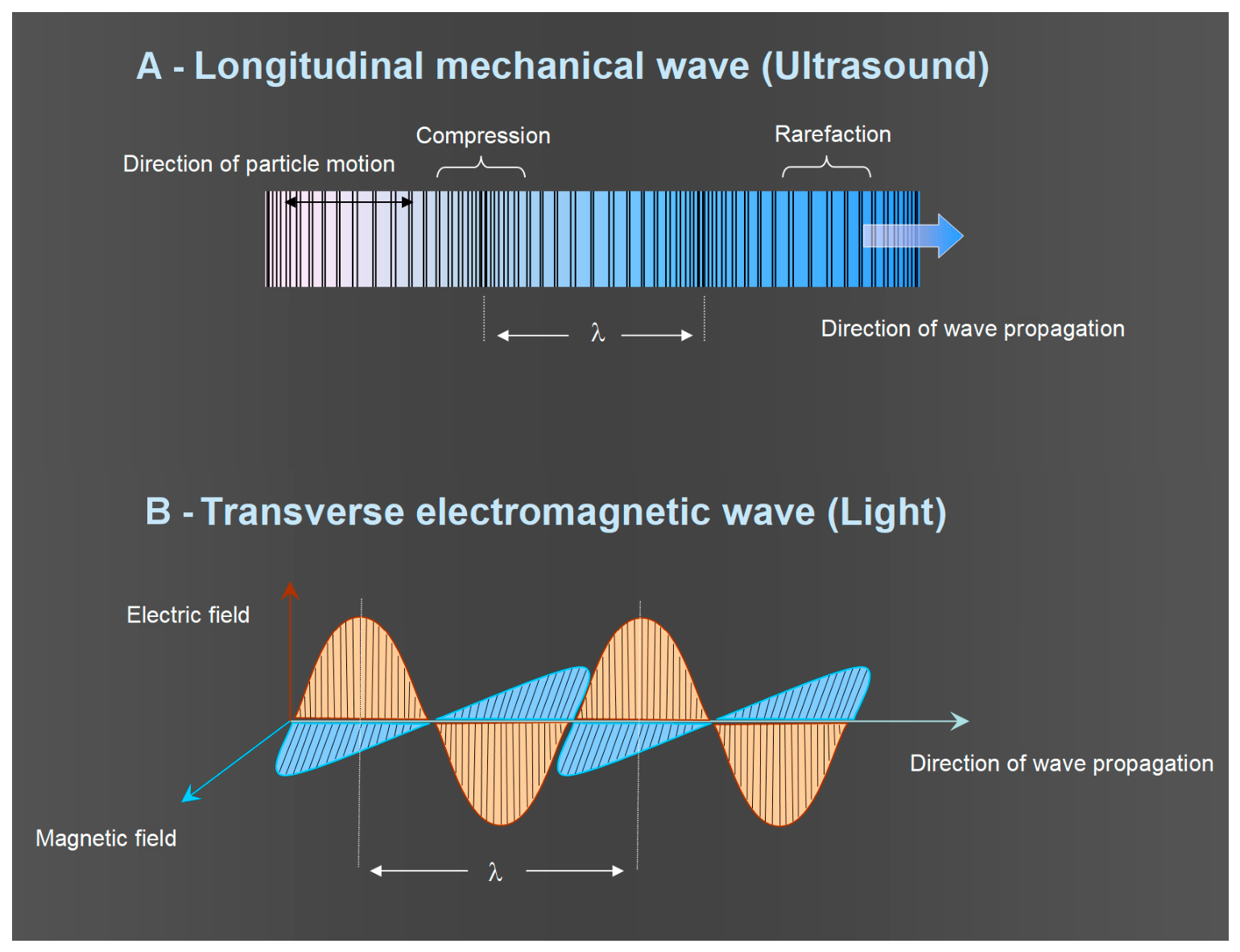
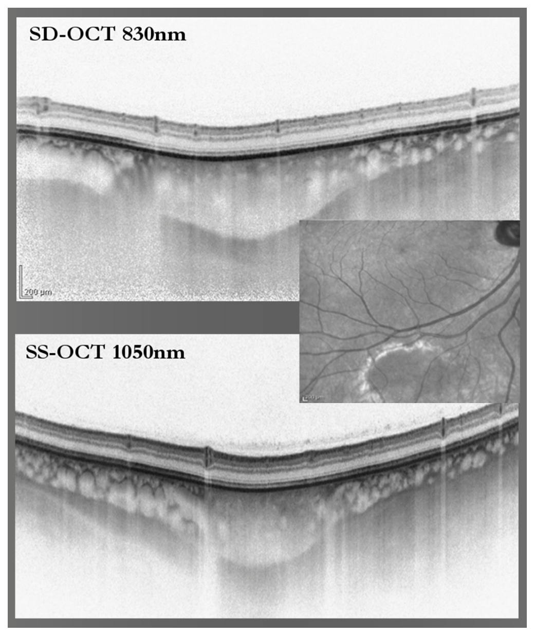
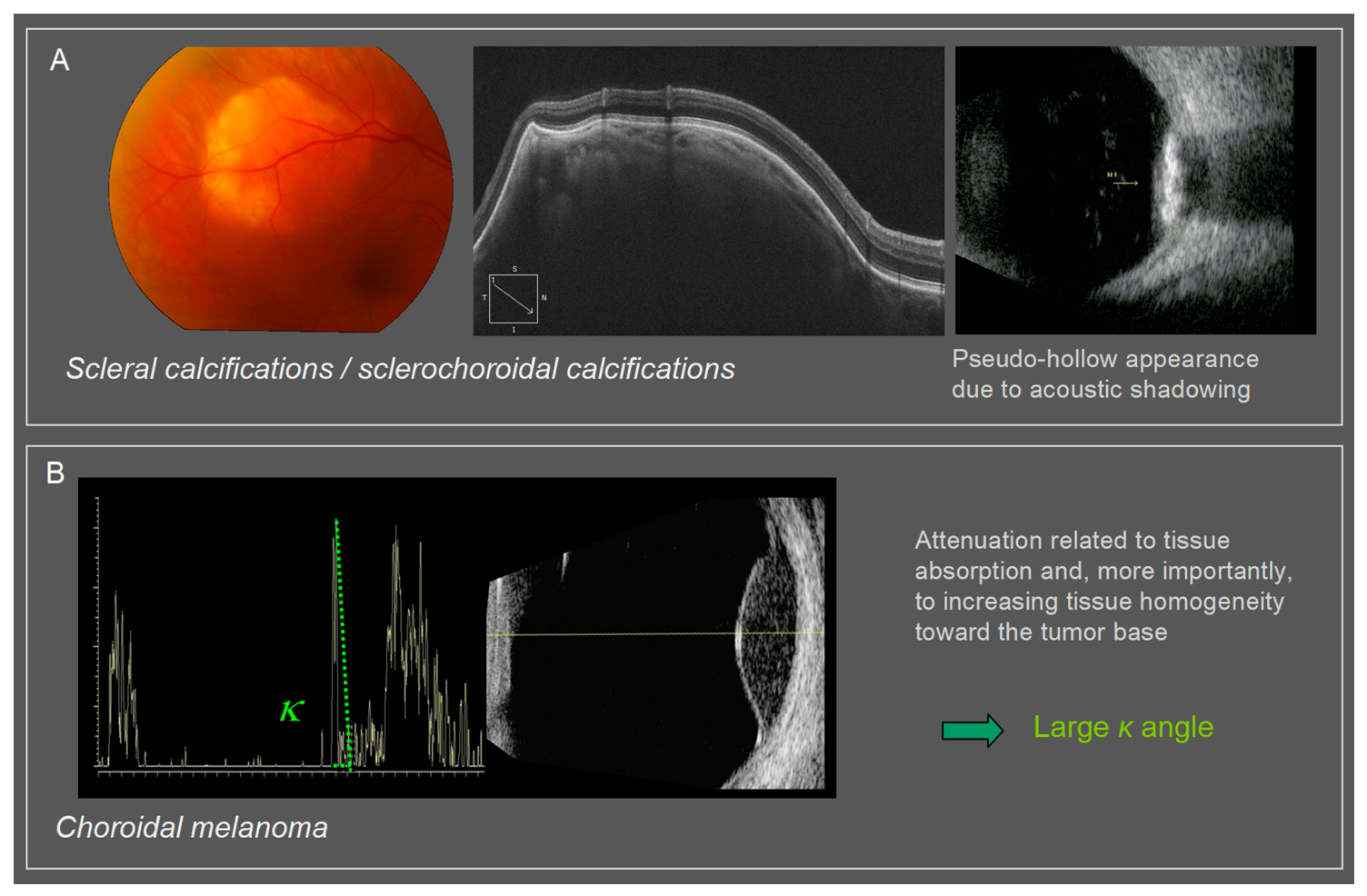
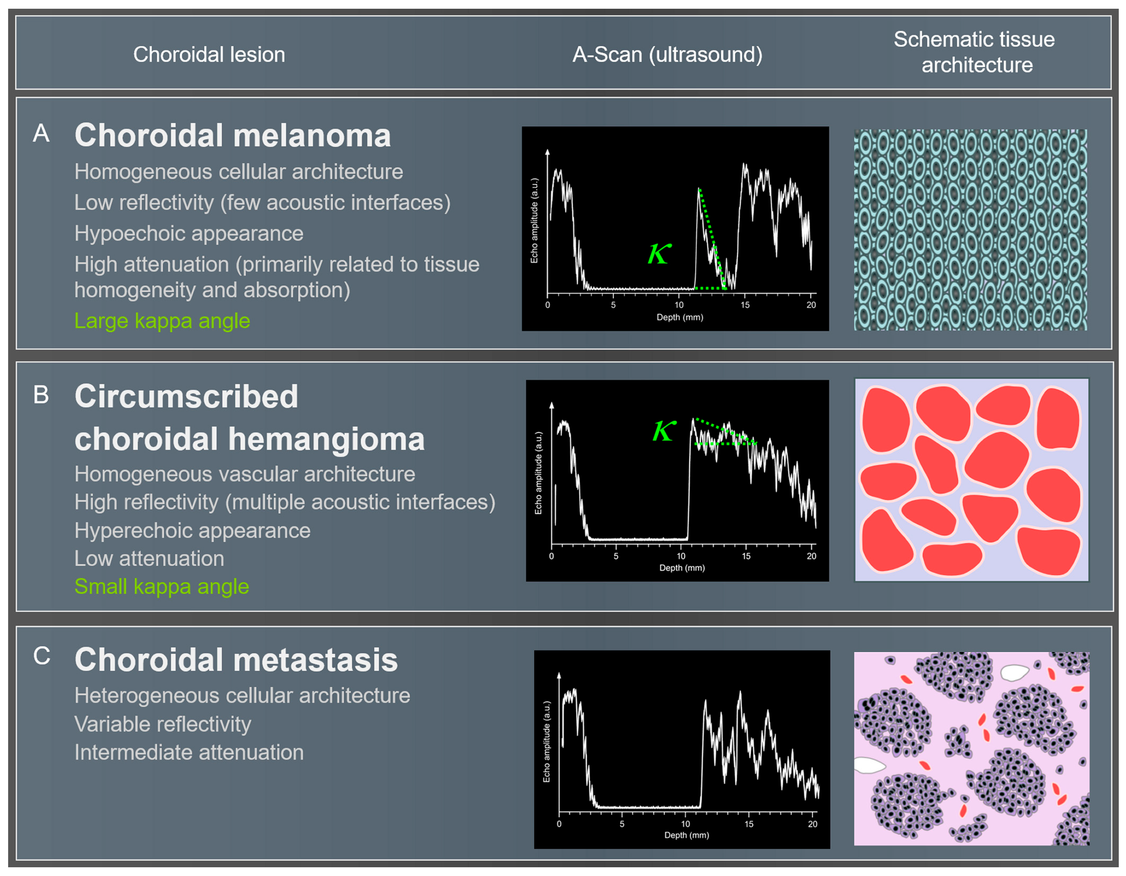
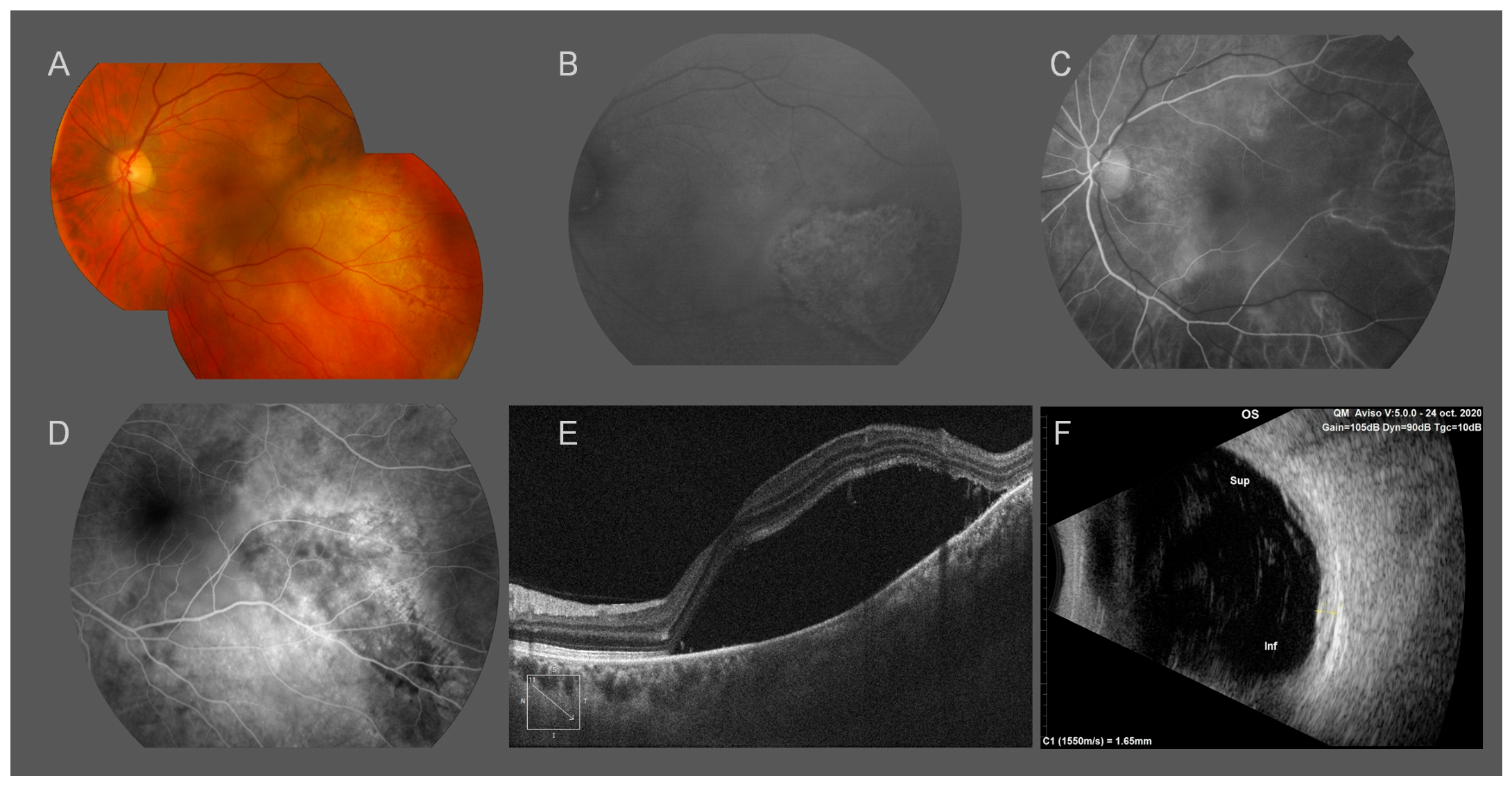
Choroidal tumors encompass a heterogeneous group of lesions ranging from benign melanocytic nevi and vascular tumors to malignant melanomas and metastatic lesions. Their evaluation increasingly relies on multimodal imaging combining techniques that interrogate different physical and biological properties of the lesion and surrounding tissues. This narrative review examines the principles, clinical contributions, and limitations of optical and acoustic imaging in the assessment of choroidal tumors. Modern optical modalities—including spectral-domain and swept-source optical coherence tomography (OCT), OCT angiography (OCT-A), fundus autofluorescence, ultra-widefield imaging, and scanning laser ophthalmoscope (SLO)-based techniques—provide high-resolution information on lesion morphology where optically accessible, as well as on associated retinal pigment epithelium alterations, subretinal fluid, photoreceptor changes, fluorophore-related signals, and vascular flow or permeability. However, many of these findings reflect responses of the retina, RPE, or choroidal vasculature adjacent to the tumor rather than intrinsic tumor characteristics. Ultrasonography provides different information based on acoustic backscatter and attenuation, including lesion dimensions, internal reflectivity and echo patterns, and remains particularly important for larger or peripheral lesions and when optical visualization is limited by media opacity. Its interpretation and measurement are nevertheless operator- and scan-dependent, with limitations related to spatial resolution, insonation geometry, boundary definition, and reproducibility, particularly for small lesions. Representative examples of choroidal nevi, melanomas, circumscribed choroidal hemangiomas, metastases, and selected pseudotumoral lesions illustrate how multimodal findings contribute to differential diagnosis, longitudinal assessment, and clinical decision-making. Emerging techniques such as OCT-A and Retro mode imaging provide additional structural or flow-related information, although their tumor-specific diagnostic performance remains incompletely validated. Multimodal imaging can substantially narrow the differential diagnosis but cannot always establish tissue diagnosis, and biopsy or systemic investigation may remain necessary in selected cases. Accurate evaluation of choroidal tumors therefore depends on integrating the distinct information and limitations of each modality with clinical findings and longitudinal evolution.
Keywords: choroidal tumors; choroidal melanoma; choroidal nevus; circumscribed choroidal hemangioma; choroidal metastasis; ocular oncology; multimodal imaging; optical coherence tomography; OCT angiography; ultrasonography; retro mode imaging; fundus autofluorescence; acoustic reflectivity; choriocapillaris remodeling choroidal tumors; choroidal melanoma; choroidal nevus; circumscribed choroidal hemangioma; choroidal metastasis; ocular oncology; multimodal imaging; optical coherence tomography; OCT angiography; ultrasonography; retro mode imaging; fundus autofluorescence; acoustic reflectivity; choriocapillaris remodeling