Article

The Source of Melanocytes in Ortho- and Heterotopic Tail Regenerates of Axolotls and the Dependence of the Regenerative Response on the Presence of Neural Tissue

1
Institute for Evolution and Biodiversity, University of Münster, 48149 Münster, Germany
2
Department of Biology, University of Hildesheim, 31141 Hildesheim, Germany
3
Independent Researcher, 55232 Alzey, Germany
*
Author to whom correspondence should be addressed.
Anatomia 2024, 3(1), 29-49; https://doi.org/10.3390/anatomia3010004
Submission received: 31 January 2024 / Revised: 26 February 2024 / Accepted: 27 February 2024 / Published: 29 February 2024

Abstract

:
We studied the regeneration of orthotopic and heterotopic tails in larval axolotls. First, we analyzed tail regeneration following reciprocal exchange of cuffs of tail integument between dark-colored (wild-type) and yellow-colored (hybrid) larval animals. Second, we studied tail regeneration in larval axolotls following transplantation of cuffs of tail integument from metamorphosed dark-colored conspecifics and from an adult fire salamander. In all cases, the amputation planes involved the transplanted integumental cuffs. In the first experiment, the regenerated tails showed the color of the host animals, not that of the transplanted cuffs. This suggests that the melanocytes of the regenerated tails were derived from the host hypodermis. Following transplantation of metamorphosed skin from axolotls and a fire salamander onto larval axolotls, the metamorphosed epidermis reverted to a larval condition. This indicates that the state of differentiation of the metamorphosed epidermis was not permanent. Rather, in order to maintain the metamorphosed epidermal structure, a continuous exposure of the animals to sufficient levels of thyroid hormones was required. Transplantation of tail buds from yellow-colored onto dark-colored axolotl embryos caused the formation of yellow-colored tails both in the head and the anterior limb region of the hosts. Incomplete resection of these heterotopic tails was followed by tail regeneration, while no tail regeneration occurred following complete resection of the heterotopic tails. Successful tail regeneration depended on the presence of neural tissue along the resection plane.

1. Introduction

While all tetrapods are capable of wound healing, the ability to regenerate lost body parts is unevenly distributed in this taxon. The capacity for epimorphic regeneration is most pronounced in amphibians, particularly urodeles (order Caudata), which can regenerate lost body parts (limbs, tails and jaws) throughout life [1,2]. In contrast, in anuran amphibians (e.g., frogs and toads), the ability for epimorphic regeneration is largely restricted to larval stages and lost at metamorphosis [1,2]. However, recent studies have demonstrated that it is possible to experimentally overcome this metamorphosis-related loss of regenerative ability in an anuran species (Xenopus laevis) by pro-regenerative multidrug treatment, thereby facilitating limb regeneration and functional recovery also in adult individuals [3].
Examples of extensive epimorphic regeneration in amniotes are tail regeneration in lizards [4] and the annual regrowth of antlers in deer [5]. A case of more limited appendage regeneration in mammals is the regrowth of the distal digit tip, which depends on the presence of the nail organ in the stump [6,7]. Elucidating and comparing the mechanisms underlying epimorphic regeneration processes in different vertebrate taxa is considered crucial for achieving the ultimate goal of regenerative medicine, viz., the regrowth of amputated or otherwise lost limbs in humans [8,9].
Among urodele amphibians, the axolotl (Ambystoma mexicanum) is a preferred species in regeneration studies [10,11]. The species is paedomorphic, attaining sexual maturity as an aquatic larva. The present study addressed the roles of the dermis and hypodermis (subcutis) as potential local sources of melanocytes during tail regeneration in wild-type (dark-colored) A. mexicanum and in artificial hybrids between A. mexicanum and the closely related A. tigrinum [12]. These hybrids are of a yellow color, and, contrary to the wild-type, their integument is free of melanocytes but contains xanthophores [13]. Adult individuals from both groups are partially metamorphosed and composed of a mosaic of larval and metamorphosed tissues and organs [14,15].
The amphibian skin is composed of an epidermis, dermis and hypodermis. The dermis consists of a stratum spongiosum underlain by a stratum compactum, which borders on the loose connective tissue of the hypodermis [16]. The pigment cells of the vertebrate skin are derived from the neural crest, a transient structure during embryonic development that gives rise to a multitude of migratory cells [17]. The presence of Leydig cells is a diagnostic feature of the epidermis of larval and paedomorphic urodeles [18,19,20,21]. Triggered by a short-term increase in thyroid hormones (triiodothyronine (T3) and thyroxine (T4)), rare instances of spontaneous metamorphosis in axolotls were associated with a loss of the Leydig cells [22].
Metamorphosis in A. mexicanum can be experimentally induced by exogenous thyroid hormone application [23,24,25,26,27]. Once metamorphosed, a complete reversal to the larval condition is not possible [28,29]. As was first demonstrated in the fire salamander (Salamandra salamandra), localized metamorphosis, i.e., metamorphosis affecting only certain body parts as opposed to the complete organism, can be achieved by local application of thyroxine [30]. Few studies addressed the question whether metamorphosed epidermis present in a ‘larval environment’ can revert to a larval condition and whether the retention of the metamorphosed state of the epidermis depends on the continuous presence of thyroid hormones [31,32]. Table 1 lists diagnostic criteria for distinguishing pre- and post-metamorphic epidermis in Ambystoma and Salamandra.
Using a classical transplantation approach, the present study addressed (1) the source of melanocytes in the integument of regenerated tails in Ambystoma, (2) the possibility of a reversal of transplanted epidermis derived from metamorphosed axolotls and a metamorphosed fire salamander to a larval condition as well as the formation of larval epidermis in tail regenerates, and (3) the regeneration of heterotopic tails derived from the tail buds of yellow axolotl embryos transplanted onto dark host embryos, and the size of these tail regenerates depending on the amputation level.

2. Materials and Methods

2.1. Animals

The dark-colored A. mexicanum and yellow-colored hybrid Ambystoma individuals [12] as well as the male fire salamander used in the study originated from the respective breeding populations held at the Institute of Special and Comparative Embryology of the University of Münster, Germany. All experiments were performed on animals fully anesthetized with MS-222 (tricaine) [33]. Excision and transplantation of cuffs of integument, tail amputation and sampling of pieces of integument from transplanted and regenerated tails were performed while the animals were exposed to concentrations of 300 mg MS-222/L of Ringer’s solution for amphibians [34]. For prolonged anesthesia, a concentration of 100 mg MS-222/L water was used. Prior to sacrifice (decapitation), the metamorphosed axolotls were kept at 600 mg MS-222/L water and the fire salamander at 800 mg/L.
The animal experiments reported here conformed to the animal care guidelines and animal ethics regulations effective at the time of performance and had been approved by the responsible animal care authorities of the provincial government of Münster, Germany (Az 26.II-0834 (36/88)).

2.2. Experiments

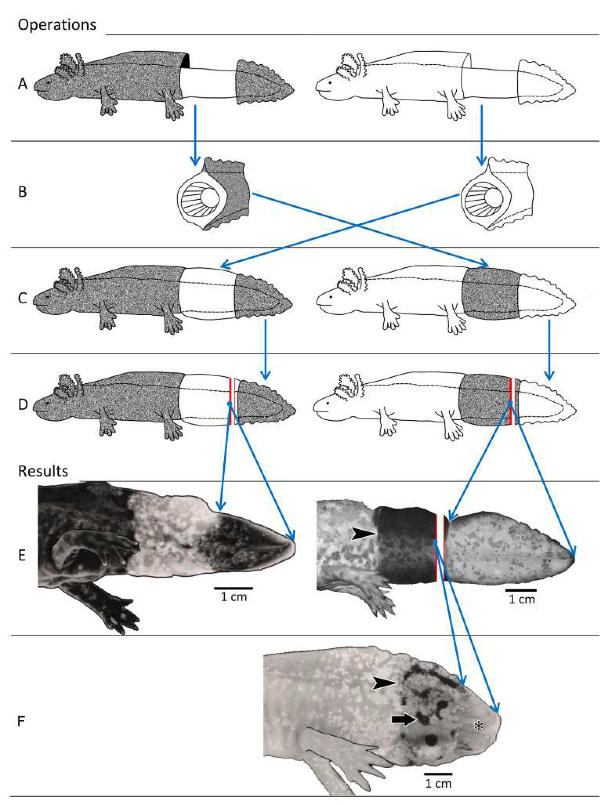
2.2.1. Experiment I: Reciprocal Exchange of Cuffs of Tail Integument between Axolotl Larvae

In this experiment, a cuff of (dark-colored) tail integument from a larval A. mexicanum (donor) was transplanted onto the tail of a larval yellow A. mexicanum/A.tigrinum hybrid (host) whose tail had previously been denuded of its integument. This was followed by amputation of the latter’s tail tip 17 days after cuff transplantation. The same procedure was also executed in reciprocal order, i.e., an integumental cuff from a yellow donor was transplanted onto a dark-colored host (Figure 1). The experiment was performed on late larvae (total n = 6, 3 per group) with a body length between 10.3 and 12.0 cm. These animals had not yet developed bicuspid teeth in the upper jaw, which start to appear at the onset of the semi-adult phase of life [14].
For harvesting of integumental cuffs, the integument (including the fin edges) was incised with fine scissors and a scalpel down to the mid-hypodermis directly posterior to the cloaca and about 2.5 cm further posterior. Using fine forceps and a small glass rod with rounded tips, the circumcised portion of the integument, consisting of epidermis, dermis and the outer hypodermis, was carefully separated from the underlying basal portion of the hypodermis (bordering on the tail musculature) and pulled over the tail tip. The cuff of donor integument was then placed in anatomical orientation onto the host tail region from which a corresponding portion of integument had previously been removed. The donor cuffs were locally fixed by thermocauterization. Following surgery, the animals remained anesthetized for 24 h.
At postoperative day 17, the tip region of the tail was resected at a level about 3 to 5 mm cranial to its end, i.e., in the region of the transplanted cuff, and processed for light and electron microscopy. Tail regeneration was assessed 24 months after cuff transplantation. In a single yellow individual, the meanwhile regenerated tail was amputated about 4 months prior to the termination of the experiment. This was followed by the formation of a second, smaller tail regenerate (Figure 1F).

2.2.2. Experiment II: Transplantation of Cuffs of Tail Integument from Metamorphosed, Dark-Colored Axolotls onto Larvae of Dark and Yellow-Colored Axolotls

Five individually held late axolotl larvae (dark color, body length between 10.5 and 11.7 cm) were each treated with 70 mg L-thyroxine (Merck, Rahway, NJ, USA) dissolved in the water, which induced metamorphosis (moult 6 to 8 days after treatment, transition to land first observed on day 9). At either day 28 or day 45 after thyroxine application, the metamorphosed animals were sacrificed and the cuffs of tail integument harvested as described above. Three of these cuffs (from day 28 after induction of metamorphosis) were transplanted on the tails (denuded of integument) of three yellow Ambystoma larvae (body length between 10.5 and 11.7 cm) (Figure 2A), and two (from day 45 after induction of metamorphosis) on the denuded tails of two dark-colored axolotl larvae (body length of, respectively, 10.7 and 11.4 cm) (Figure 2B), applying the previously described technique.
Two weeks after the transplantation, the tails were amputated approximately 3 mm cranial to the posterior end of the transplanted cuffs. Twelve weeks after transplantation, the animals were sacrificed and portions of epidermis and subepidermal tissues of the regenerated tail were excised and processed for light and electron microscopy.
Analysis of the regenerated tails provided information on the source of the melanocytes in the regenerated tail (same question as in experiment 1) and on the stability of the metamorphosed condition of the transplanted cuff epidermis, and the nature of the regenerated epidermis.

2.2.3. Experiment III: Xeno-Transplantation of a Cuff of Tail Integument from an Adult Fire Salamander on a Dark-Colored Axolotl Larva

Following sacrifice, a cuff of tail integument (length 2.5 cm) was removed from an adult (5 yr-old) male fire salamander (body length 17.4 cm) as described above for Ambystoma. The cuff was thoroughly rinsed in Ringer’s solution for amphibians to remove the secretions of the damaged poison glands of the salamander skin. The cuff was then transplanted onto the tail of a dark-colored Ambystoma larva, from which a corresponding portion of integument had previously been removed (Figure 2C). The experiment then followed the course outlined for experiment 1.
Again, analysis of the regenerated tail provided information on the source of the melanocytes in the regenerated tails and on the stability of the determination of the cells derived from the donor epidermis, i.e., their possibility to revert to a larval condition.

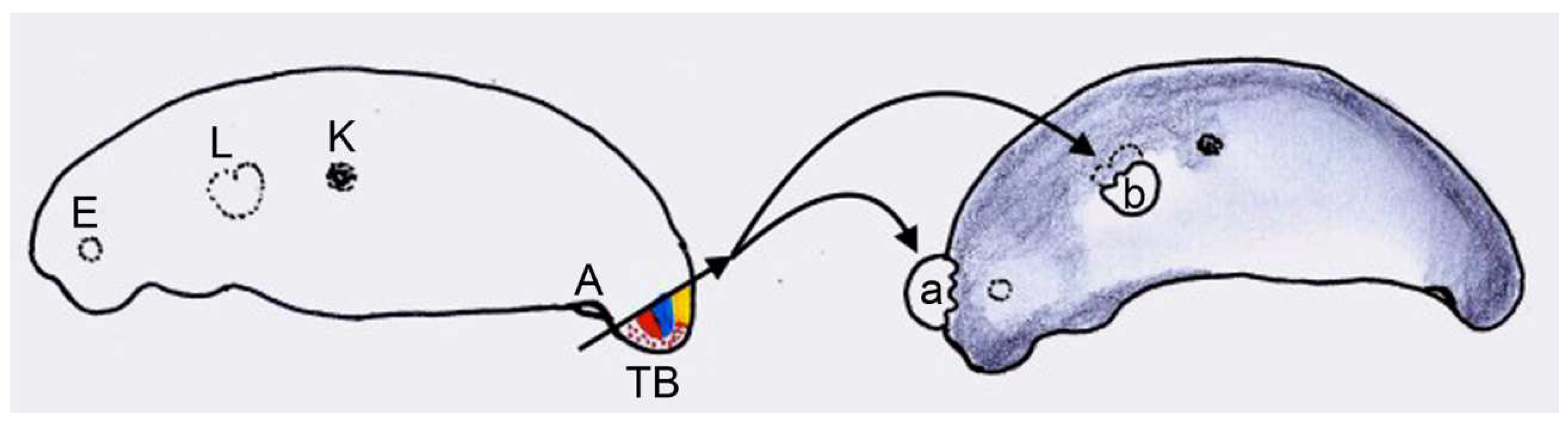
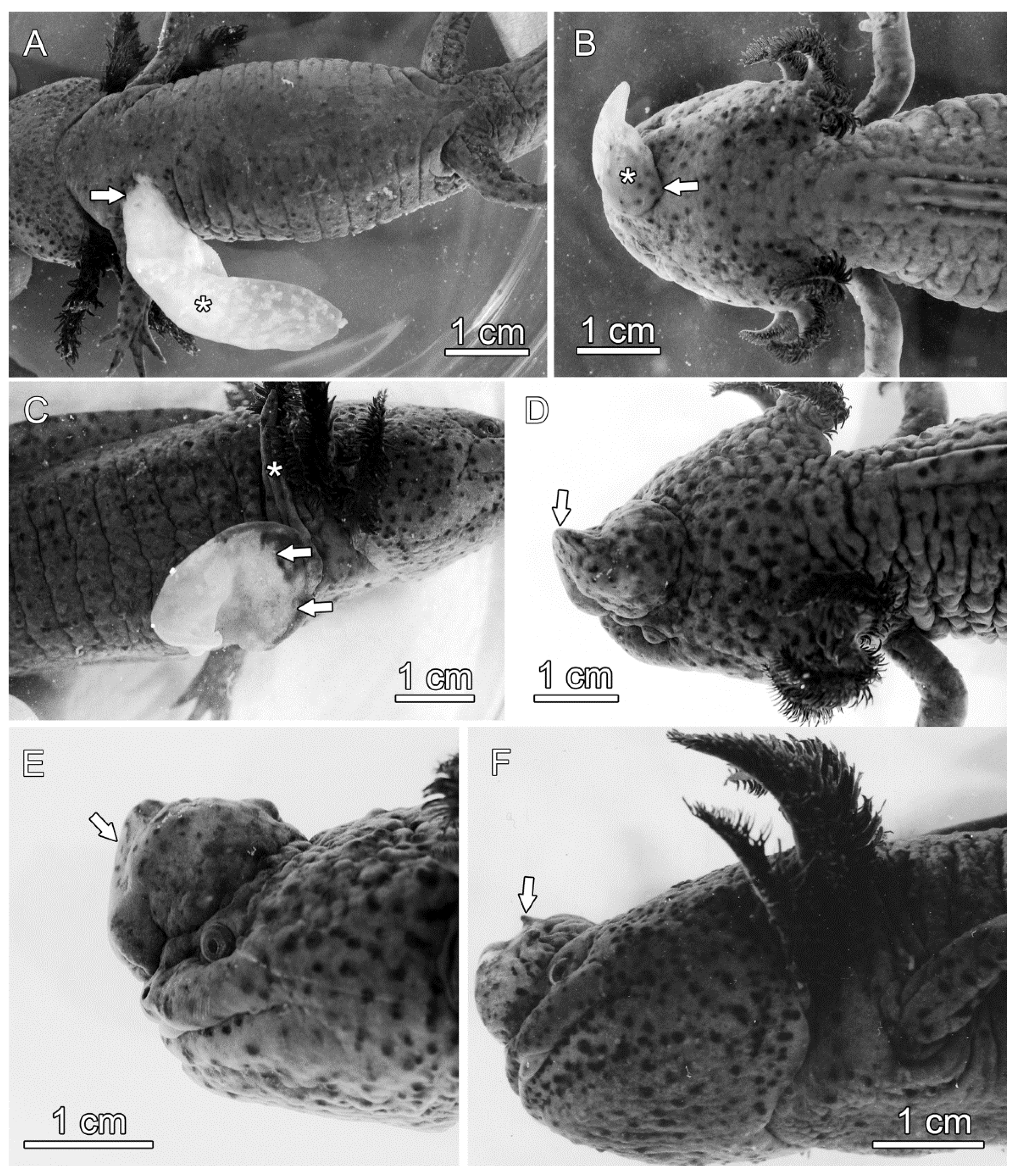
2.2.4. Experiment IV: Heterotopic Transplantation of Tail Buds from Yellow-Colored on Dark-Colored Axolotl Embryos, and Analysis of Tail Regeneration following Resection of the Heterotopic Tails

Following removal of the egg membranes, the tail buds of embryos of the yellow-colored Ambystoma hybrids (donors) were transplanted onto the head or the anterior limb bud region of dark-colored Ambystoma embryos (hosts) of similar developmental stage (Figure 3). Removal of the tail buds from the donors and transfer to the hosts were performed with instruments made from fine platinum wire.
Prior to transplantation of the tail buds, the host ectoderm was removed at the transplantation site. Following transplantation, the host embryos were transferred to petri dishes with Ringer’s solution for amphibians and kept at 14 °C. The solution was changed daily until the embryos had reached stage 40 [35], after which they were transferred to water. At the earliest 22 months after the transplantation, a variable portion (25 to 75%) of the yellow tail (derived from the transplant) was amputated in six animals (Nos. 1–6 in Table 2). After a further 12 to 18 months, the regenerated tails were resected in these animals (body length of 24 to 26 cm) and analyzed by light and electron microscopy. In a second group of four animals (Nos. 7–10 in Table 2), the heterotopic tails were almost completely (95%) resected between 15 and 24 months after transplantation. In a third group of three animals (Nos. 11–13 in Table 2), the heterotopic tails were completely resected 19 to 20 months after transplantation, the resection plane being entirely located in host tissue.
The experiment analyzed the dependence of tail regeneration from the level of amputation and the dependence of the pigmentation of the regenerated tails from the location of the amputation plane in donor or host tissues. Furthermore, the presence of components of the neural tube in the regenerated tails and their possible influence on the extent of regeneration were addressed.

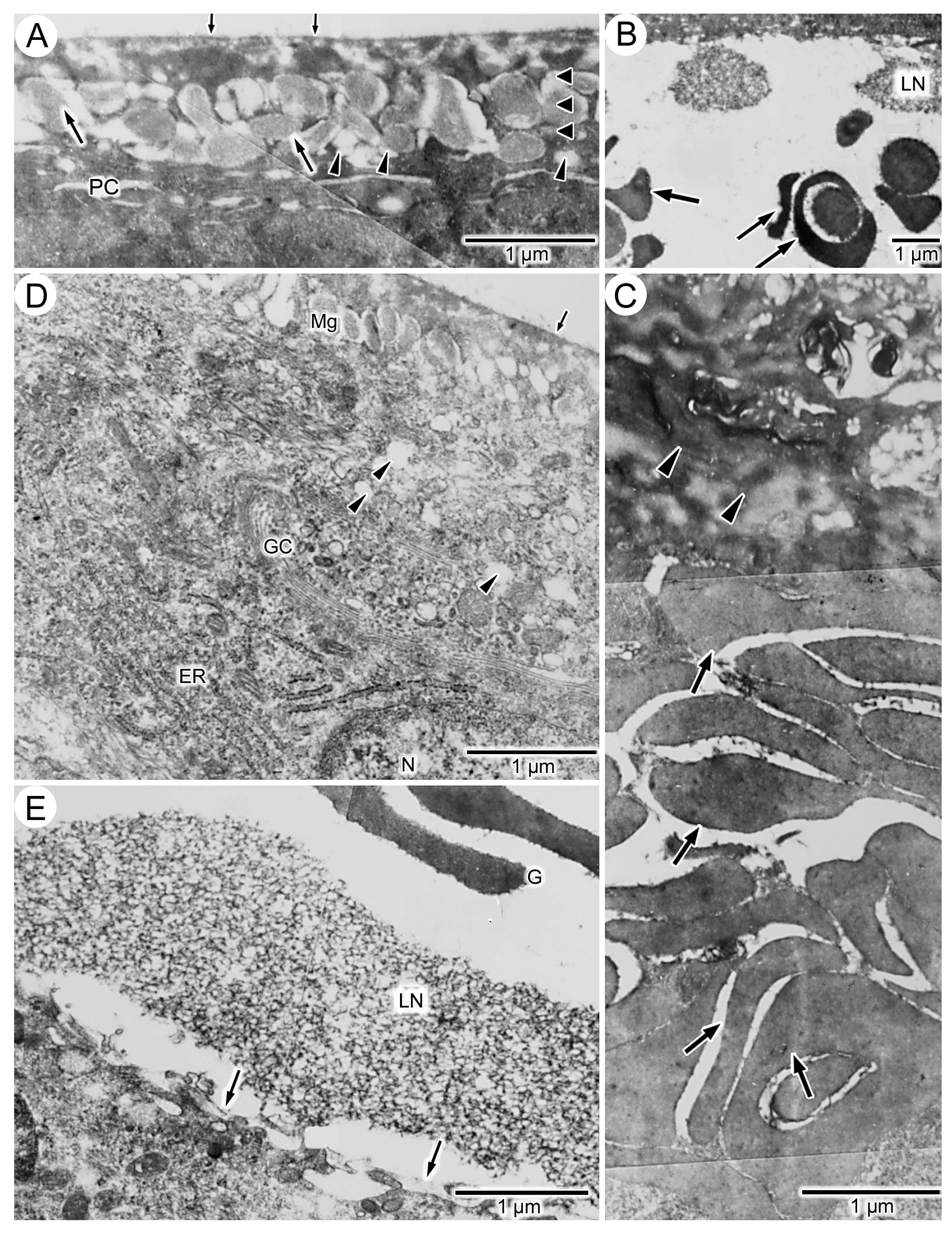
2.3. Light and Electron Microscopic Methods

For light microscopy (LM), pieces of integument were fixed (48 h) in Bouin’s solution, dehydrated in a graded series of ethanol, transferred to butanol and embedded in paraplast. Deparaffinized sections of about 7 µm thickness were stained with Heidenhain’s Azan, hemalum/eosin (HE), or alcian blue and periodic-acid Schiff’s reagent (AB-PAS) according to [36]. The stained sections were viewed and photographed in a Zeiss Axio Imager 2 microscope (Zeiss, Oberkochen, Germany), equipped with a digital camera (Zeiss Axiocam 503 color) or a Keyence VHX 7000 digital microscope (Keyence, Osaka, Japan).
For transmission electron microscopy (TEM), samples of the integument were fixed for 1 to 3 h in an ice-cold solution of 1% formaldehyde, 1.5% glutaraldehyde, 1% osmium and 3% saccharose in 0.07 mol/L phosphate buffer (pH 7.4), followed by rinsing (3 × 15 min) in distilled water, dehydration in ethanol and embedding in styrene–methacrylate resin [37]. Ultrathin sections were cut from the blocks, stained with lead acetate for 10 to 20 min, and studied in a transmission electron microscope (Elmiskop 101; Siemens, Munich, Germany).

2.4. Diagnostic Criteria Used in the Analysis of the Integumental Samples

As controls and for establishing possible differences between dark-colored and yellow-colored Ambystoma individuals, the integumental samples obtained at the days of operation were histologically analyzed. The analyses were based on studies on the histology of the integument in urodeles [18,38], and the epidermis of dark-colored A. mexicanum [19,39,40] and that of S. salamandra [41,42] (Table 1).

3. Results

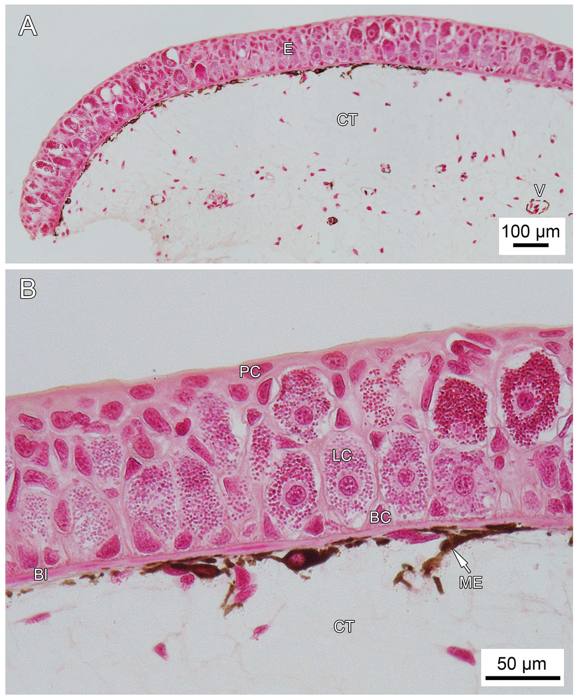
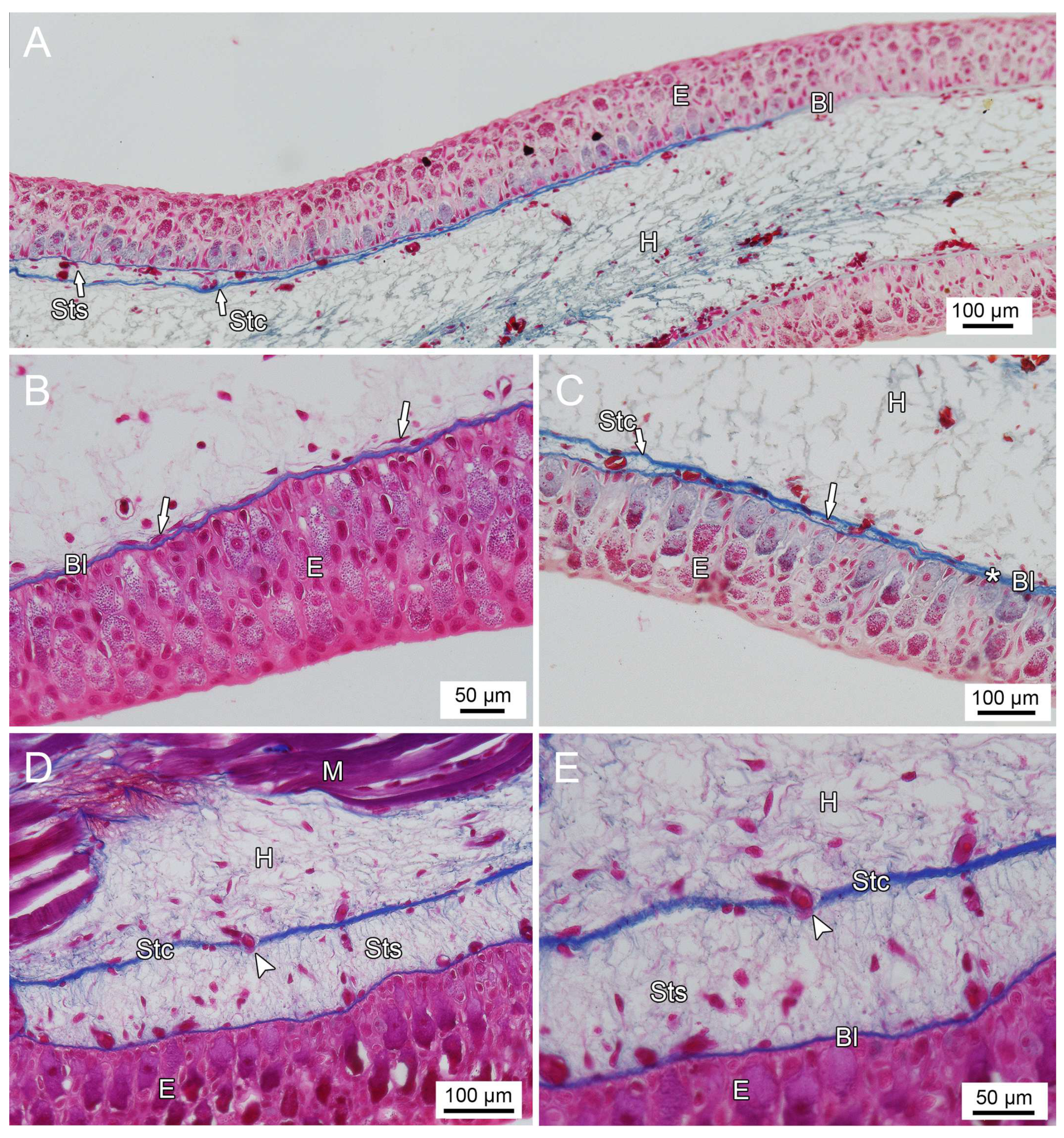
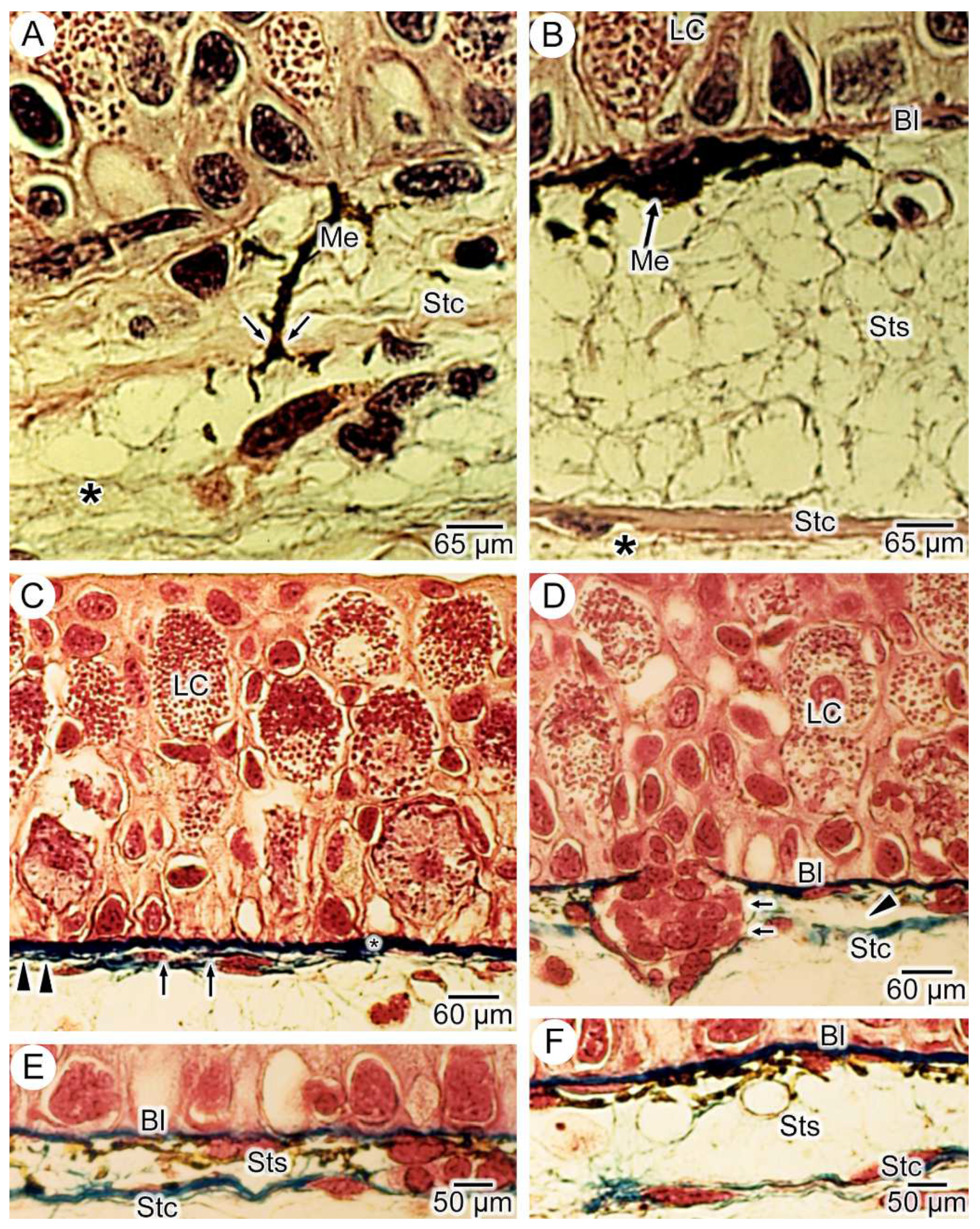
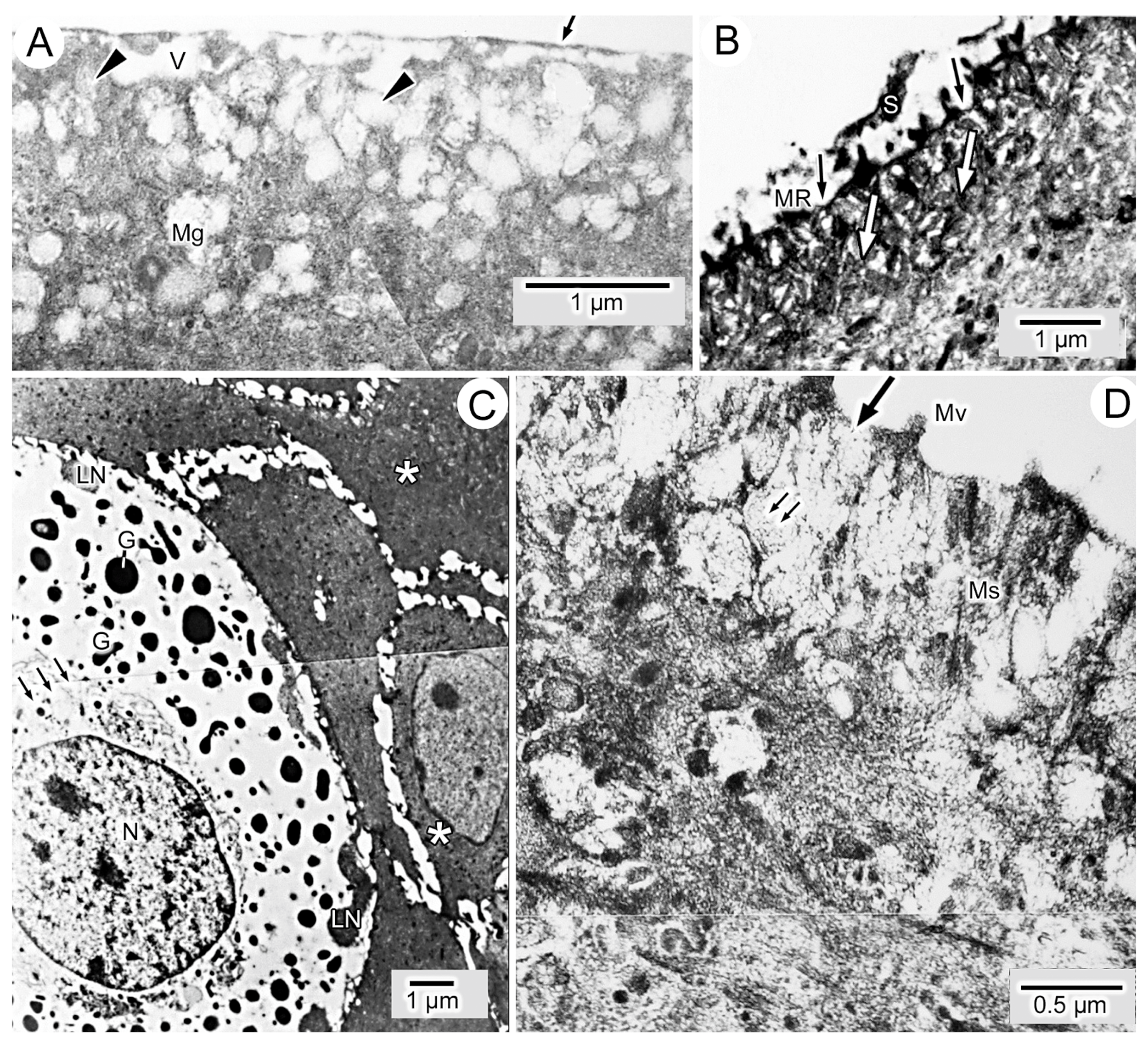
3.1. General Findings on Integumental Structure

Except for the lack of melanin granules in the latter, the integument of dark-colored and yellow-colored Ambystoma larvae showed no structural differences. The larval epidermis consisted of a single-layered stratum basale, a multi-layered stratum intermedium and a single-layered stratum superficiale. The latter consisted of pavement cells with membrane-bound mucin granules that contained a flocculent material. The plasmalemma of the pavement cells exhibited microvilli and/or microridges, and in places keratinization of single or multiple pavement cells was observed. The nucleus of the pavement cell was surrounded by organelles (rough endoplasmic reticulum, Golgi complex and mitochondria), while sporadic bundles of tonofilaments were present apicolaterally. The stratum intermedium was dominated by Leydig cells, the larger of which were anchored to extensions of the cells from the basal layer (stratum basale) and extended up to the pavement cells, as was previously reported [43]. The basal lamella (basement membrane) was underlain by the stratum spongiosum and the stratum compactum of the dermis. Between the stratum compactum and the tail musculature the hypodermis was situated.
No differences were observed in the structure of the multi-layered epidermis of dark-colored A. mexicanum at days 28 and 45 after induced metamorphosis. Leydig cells were missing and the outer layer of the epidermis was a stratum corneum of anucleated keratinized cell rests. The keratinocytes as well as the basal cells possessed bundles of tonofilaments. The cytoplasm of the keratinocytes in the metamorphosed S. salamandra was characterized by multiple bundles of tonofilaments and lysosomes, which were both also present in replacement cells.

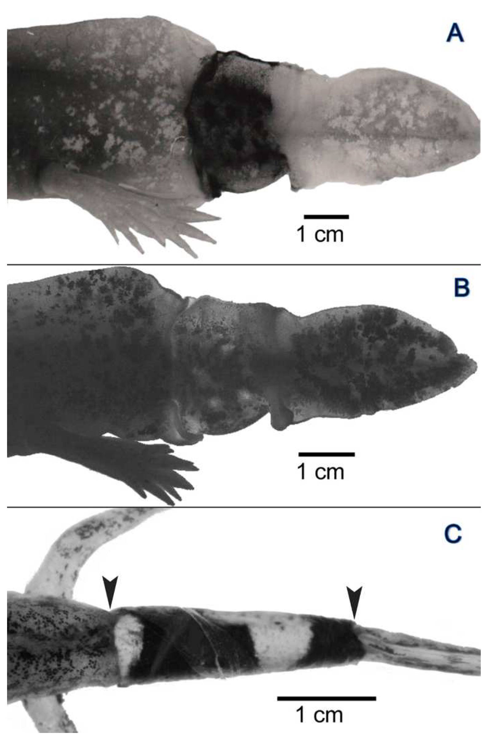
3.2. Experiment I: Reciprocal Exchange of Cuffs of Tail Integument between Axolotl Larvae

Two years after transplantation (the hosts were now adult), the transplanted integumental cuffs were still clearly demarcated from the host integument (Figure 1E). Apical to the hypodermis of the transplanted yellow cuffs, some melanocytes, originating from the host hypodermis, could be found at locations where nerves and blood vessels had penetrated the stratum compactum (consisting of densely packed collagen fibers) of the cuff’s dermis (Figure 1E and Figure 4A,B), thereby causing the formation of small pigmented spots. Some loss of pigmentation was observed in the dark-colored cuffs at 24 months after their transplantation (Figure 1E,F). The dermis of the transplanted cuffs showed a clear separation into a stratum spongiosum and stratum compactum (Figure 4A,B).
In all experimental animals, the regenerated tail tips showed the color of the host integument, not that of the transplanted cuffs (Figure 1E,F). Light microscopic observation revealed that in the older portions of the regenerate, i.e., those closer to the amputation level, a stratum spongiosum and a stratum compactum could be distinguished in the dermis, while in the younger portions of the regenerate such a differentiation of the dermis had not yet developed (Figure 4C–F). In the case of dark-colored axolotl hosts, melanocytes were present in the regenerate (Figure 1E). In contrast to the dermis, the light microscopic appearance of the epidermis did not differ between transplanted cuffs (Figure 4A,B) and regenerated tails (Figure 4C,D).

3.3. Experiment II: Transplantation of Cuffs of Tail Integument from Metamorphosed, Dark-Colored Axolotls onto Larvae of Dark-Colored and Yellow-Colored Axolotls

Twelve weeks after transplantation, the epidermis of the transplanted cuffs had reverted to a larval condition, as was indicated by the presence of Leydig cells and the lack of a stratum corneum (Figure 5A–C). Two types of pavement cells were recognized in the stratum superficiale of the cuff. The first type lacked microvilli, whereas the second type, which was considered to be of a larval nature, exhibited numerous microvilli and microridges (Figure 5B). In addition, larval replacement pavement cells were present (Figure 5C).
The epidermis of the regenerated tails also possessed Leydig cells, indicating its larval condition. The structure of the pavement cells and basal cells of the regenerated tails also corresponded to that of the cuffs (Figure 5D).

3.4. Experiment III: Xeno-Transplantation of a Cuff of Tail Integument from an Adult Fire Salamander on a Dark-Colored Axolotl Larva

Six months after transplantation, the epidermis of the salamander integument lacked a stratum corneum, microvilli and microridges (Figure 6A), and contained Leydig cells with a relatively sparsely developed Langerhans’ net, which is a characteristic of larval salamander epidermis (Figure 6B). At that time, also the other cell types of the salamander epidermis had largely reverted to a larval condition. Two types of pavement cells, exhibiting either high or low electron-density, were observed (Figure 6A,D). Both types contained mucin and promucin granules that differed in electron density (Figure 6A,D).
The color of the regenerated tail corresponded to that of the host. Differences in epidermal structure and epidermal organelles between transplanted cuff and regenerated tail were not observed, except for the presence of condensed Leydig cells with irregularly-shaped, elongated granules in the stratum intermedium of the latter (Figure 6C,E).

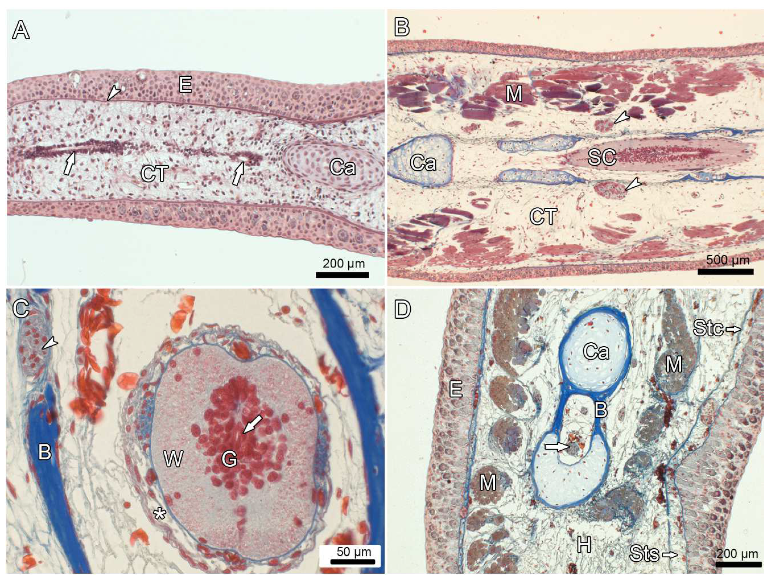
3.5. Experiment IV: Heterotopic Transplantation of Tail Buds from Yellow-Colored on Dark-Colored Axolotl Embryos, and Tail Regeneration following Resection of the Heterotopic Tails

At both transplantation sites (head, anterior limb bud region), the transplanted tail buds of the yellow-colored donor embryos developed into immotile yellow tails.
Following partial (25–75%) tail resection along a plane located entirely in the yellow tail, the regenerated tail was free of melanin (Figure 7A). However, when the resection plane included portions of the (dark) host integument, the basal portion of the regenerated tails contained pigmented areas adjacent to the regions where the host integument had been severed (Figure 7B,C). The amount of tail regeneration decreased sharply when about 95% of the heterotopic tail were resected (Table 2). When the heterotopic tail was completely resected, only a bulgy outgrowth developed from the severed host tissue. These outgrowths lacked elements of the spinal cord and morphologically did not resemble tails (Figure 7D–F). At the top of the bulgy outgrowths, a small crista or spike-like protuberance was present.
The spinal cord of the tails that developed from the transplanted tail buds as well as that of the tail regenerates formed after partial tail amputation consisted of grey and white matter and an ependyma-lined central canal (Figure 8B,C). However, during tail regeneration a clear proximal to distal sequence was evident with respect to spinal cord regeneration and the formation of other axial tail structures (muscles and skeletal elements). Thus, in the tip region of the regenerating tail only an ependyma-lined central canal was present (Figure 8A). A fully regenerated spinal cord, as well as spinal ganglia, axial muscles and skeletal elements (cartilage and bone) were only present in the more proximal i.e., in the older portions of the regenerated tails (Figure 8B–D). The heterotopic tails were immotile and did not respond to tactile stimulation.
In regenerated tails formed in the limb region following extensive resection (95%) of the heterotopic tails, which partly included severing of the host integument, melanocytes were present in the proximal portions of the regenerates adjacent to the severed host integument. The melanocytes were located directly underneath the basal lamella (Figure 9A,B). As a dermis (stratum compactum and stratum spongiosum) had not formed in the regenerated tails, a layer of un-stratified connective tissue directly bordered on the basal lamella (Figure 9B). The epidermis of the regenerated tails was of a larval nature, with numerous Leydig cells and flattened pavement cells (Figure 9B).
Table 2. Extent of tail regeneration following resection of heterotopic tails derived from transplanted tail buds of yellow-colored axolotl embryos on dark-colored axolotl host embryos. Embryonic stages according to [35].
Animal No.Sex (m/f)Donor StageHost StageTime between Transplantation and Amputation of Tail (Months)Body Length at Amputation (cm)Location of Amputated Tail Transplant (Head, Limb)Length of Amputated Tail Transplant (cm, % of Total Transplant)Age at End of Experiment (months)Body Length at End of Experiment (cm)Tail Regenerate (yes/no), Length of Tail Regenerate (cm)Presence of Spinal Cord (sc) or Only Ependymal Tissue (e) in Tail RegenerateFigures
1m32322419.5head3.9 (50)4025.0yes, 7.0scFigure 8D
2f32322419.0limb4.0 (50)4024.0yes, 8.1sc-
3m32292418.0head3.6 (50)3624.0yes, 7.1sc-
4f32292419.0limb4.1 (50)4226.0yes, 8.6scFigure 7A
5m29292221.0head2.1 (25)3624.0yes, 8.2scFigure 8B
6f29332218.5head5.5 (75)4024.5yes, 6.9scFigure 8C
7f31331818.0head7.4 (95)3026.0yes, 2.8eFigure 7B and Figure 10A,C
8f30322419.0limb7.6 (95)3625.5yes, 4.1eFigure 7C and Figure 9A,B
9m32301818.5head7.5 (95)2822.5yes, 1.7eFigure 8A
10m29291518.5head5.7 (95)2820.0yes, 2.2eFigure 10B,D,E
11f28301918.0head7.6 (100)3022.5no *neitherFigure 7D,E
12m29321918.5head7.4 (100)2823.0no *neitherFigure 7F
13f29322018.0limb7.8 (100)4026.0no *neither-
* only formation of a bulgy outgrowth that is not considered to represent a tail regenerate.
Figure 7. Regenerated tails formed after partial resection of heterotopic tails in the anterior limb region (A,C) and the head region (B) of dark-colored Ambystoma hosts, and bulgy outgrowths from the head following complete resection of the heterotopic tails (DF) (cf. Table 2). (A): Regenerated tail (asterisk) following resection of the distal half of the transplanted tail. Note the distinct border (arrow) between the yellow-colored transplant/regenerate and the dark-colored host. (B): Regenerated tail following nearly complete resection (about 95%) of the transplant. The resection plane was partly located in the integument of the host. In these areas, the proximal portion of the regenerate is covered by dark-colored integument (asterisk). Note border (arrow) between the host’s head and the base of the regenerated tail. (C): Regenerated tail in the limb region following nearly complete resection (about 95%) of the transplant. Note border (arrows) between a basal dark-colored and a distal yellow-colored portion of the regenerate. The right forelimb (asterisk) is directed upwards. (D): Dark-colored bulgy outgrowth with median crista (arrow) that had formed following complete resection (100%) of the transplanted tail. (E): Lateral view of the outgrowth with crista (arrow) shown in D. (F): Bulgy outgrowth with small pointed protuberance (arrow) that had formed following complete resection (100%) of the transplanted tail.
Anatomia 03 00004 g007
Figure 8. Light micrographs of sections through regenerated tails (cf. Table 2). (A): HE staining, (BD): Azan staining. (A): Oblique section through the distal portion of a regenerated tail on the head. Cartilage (Ca) and tangentially cut ependyma-lined central canal (arrows). Epidermis (E) with underlying basal lamella (arrowhead). The subepithelial connective tissue (CT) shows no stratification. (B): Oblique section through the proximal portion of a regenerated tail on the head. The spinal cord (SC) is surrounded by developing cartilaginous elements (Ca). Spinal ganglia (arrowheads) are visible. CT: connective tissue. M: musculature. (C): Transverse section through the proximal portion of a regenerated tail on the head. The spinal cord contains grey (G) and white (W) matter and ependymal tissue (arrow). The spinal cord is covered by developing meninges with interspersed capillaries (asterisk) and surrounded by perichondral bone (B). Arrowhead: spinal ganglion. (D): Transverse section through the distal portion of a regenerated tail on the head. Terminal portion of spinal cord (arrow) surrounded by cartilage (Ca) and perichondral bone (B). Note presence of dermis with stratum compactum (Stc) and stratum spongiosum (Sts). E: epidermis, H: hypodermis, M: musculature.
Anatomia 03 00004 g008
Figure 9. Light micrographs showing proximal portion of regenerated heterotopic tail from yellow-colored donor in anterior limb region (cf. Figure 7C); HE-stained. (A): Overview image showing epidermis (E) and underlying connective tissue (CT) with blood vessels (V). Melanocytes are attached to the basal lamella and present in the vessel walls. (B): Higher magnification of epidermis with Leydig cells (LC) and apically located, flattened pavement cells (PC). Note cuboidal basal cells (BC) near basal lamella (Bl). CT: connective tissue, ME: melanocyte.
Anatomia 03 00004 g009
Contrary to the regenerates in the limb region, the regenerated tails in the head region contained a dermis. There was a successive differentiation of the subepithelial connective tissue in proximal to distal direction that led to the formation of the dermal strata and the underlying hypodermis (Figure 8D). The first step in dermal development was the separation of the stratum compactum from the basal lamella, followed by the invasion of fibrocytes into the cleft developing between the two structures (Figure 10A–C). In more proximal portions of the regenerated tails, the initial cleft widened and became populated by fibrocytes forming a stratum spongiosum (Figure 10D,E). In places, the stratum compactum was breached by blood vessels that had formed in the underlying hypodermis (Figure 10D,E).
Figure 10. Light micrographs showing different regions of regenerated heterotopic tails from yellow -colored donors in the head region. Longitudinal sections; Azan stained. (A): Overview image near distal tip (to the right of the image) showing beginning differentiation of the dermis into stratum compactum (Stc) and stratum spongiosum (Sts). Bl: basal lamella, E: epidermis, H: hypodermis. (B): Detail of distalmost (tip) region, showing epidermis (E) underlain by basal lamella. Note spindle-shaped fibrocytes (arrows) attached to the basal lamella. (C): Regenerated tail portion located further proximally than B, showing beginning differentiation of the dermis by separation (asterisk) of the stratum compactum (Stc) from the basal lamella (Bl) and invasion of the resulting cleft space by fibrocytes (arrow). E: epidermis, H: hypodermis. (D): Further proximally, the stratum spongiosum (Sts) is present between basal lamella and stratum compactum (Stc). The latter is perforated by blood vessels (arrowhead). E: epidermis, H: hypodermis, M: musculature. (E): Higher magnification of region depicted in (D), showing blood vessels (arrowhead) penetrating the stratum compactum (Stc). Note that stratum spongiosum (Sts) shows a denser structure than the hypodermis (H). Bl: basal lamella, E: epidermis.
Anatomia 03 00004 g010

4. Discussion

In experiment I, the color of the regenerated tails corresponded to that of the host, not that of the transplanted cuff. The formation of yellow-colored tail regenerates in yellow-colored host animals with dark-colored integumental cuffs suggested that the dermis of the transplanted cuff had not contributed melanocytes to the regenerate. In the case of dark-colored hosts, the black pigmentation of the regenerated tails was derived from melanocytes of the host. The dark patches that developed in the lateral portions of the yellow cuffs are likewise attributed to host melanocytes. The loss of pigmentation in long-standing dark-colored cuffs on yellow-colored hosts is attributed to a lack of supply of new melanocytes from the host.
Our findings suggest that the (deeper layer of the) hypodermis rather than the dermis is the likely source of the melanocytes causing the dark pigmentation of the regenerated tails. This interpretation is in line with findings of a previous study on cysts of normal skin that developed after autologous transplantation of skin into the hypodermis of fire salamanders (Clemen, unpubl. observ.). Also here, the melanocytes present in the newly formed connective tissue of the cyst wall were derived exclusively from the surrounding hypodermis.
Using lineage tracing of GFP-labeled neural precursor cells (radial glial cells) of the spinal cord, it has been demonstrated that these cells can differentiate into melanocytes in the regenerated axolotl tail [44]. In the study by Echeverri and Tanaka [44], GFP-labeled melanocytes were observed in 8% (2 of 25) of the experimental animals. In our experiment, a contribution by spinal cord-derived precursor cells to the melanocytes of the tail regenerates in dark-colored hosts is therefore considered possible. However, given the low frequency of animals with melanocytes of neural origin in the study by [44], we suggest that in our case the hypodermis of the dark-colored hosts served as the main source of the melanocytes present in the regenerated tails and the pigment patches of the yellow cuffs.
Resection of heterotopic yellow-colored tails along a plane partly located in the dark-colored host integument caused a dark pigmentation of basal portions of the regenerated tails. In these cases, the host integument is considered the only potential source of melanocytes, as no spinal cord of the host was present along the resection plane.
Amputation of the tail tip is followed by re-epithelization of the wound from the surrounding epidermis, dedifferentiation in the stump, and formation of a regeneration blastema, i.e., an accumulation of undifferentiated/dedifferentiated cells [2]. The regenerated subepidermal structures are derived from this blastema. Using cell lineage tracing (GFP-labeling), it has been demonstrated that the cells of the blastema formed during axolotl limb regeneration do not represent a homogeneous population of pluripotent progenitor cells, but constitute a heterogeneous assemblage, with individual cells retaining a memory of their tissue of origin and re-differentiating according to this origin [11]. That is, blastemal cells derived from the dermis make new dermis, former muscle cells remain largely or exclusively restricted to the myogenic lineage, and cells derived from cartilaginous elements form new cartilage. It has, however, been demonstrated that blastemal cells derived from the hypodermis are capable to also differentiate into skeletogenic cells [11,45,46,47,48]. As we did not perform lineage tracing in our experiments, the relative contribution of cells derived from the dermis and hypodermis to the mesodermal component of the regenerated tails, remains unknown.
Differentiation of the dermis during tail regeneration started with the separation of the stratum compactum from the basal lamella, followed by invasion of fibrocytes and blood vessels into the resulting cleft. During further development, this cleft became enlarged and populated by fibrocytes that formed a stratum spongiosum. It is presently unclear why no dermis had formed in the regenerated heterotopic tails in the anterior limb region, while it was present in the regenerates formed in the head region.
Experiment II demonstrated that the epidermis of the regenerated tail, which was derived from the metamorphosed epidermis of the transplanted cuff, exhibited a larval condition, as was demonstrated by the presence of Leydig cells. Also the epidermis of the remaining transplanted cuff reverted to a larval condition with presence of Leydig cells. Twelve weeks after transplantation, this process was completed. This shows that the metamorphosed state of differentiation of the epidermis cannot be maintained in a larval environment.
In experiment III (xenoplastic transplantation), the larval condition of the epidermis of the regenerated tail was demonstrated by the occurrence of salamander-typical pavement cells and of atypical Leydig cells, as well as the lack of a stratum corneum. Thus, even several years after metamorphosis there existed no stable state of differentiation of the adult Salamandra epidermis. Our observations are in line with previous findings demonstrating that both the initiation of metamorphosis and the maintenance of the metamorphosed state depends on the presence of sufficient amounts of thyroid hormones (T3/T4) [49,50].
Experiment IV demonstrated heterotopic tail development following transplantation of tail buds to the head or anterior limb bud region. In all cases of heterotopic tail formation, the spinal cord comprised grey and white matter surrounding an ependyma-lined central canal. The lack of motility of the heterotopic tails and the failure of the animals to react to tactile stimulation of the heterotopic tails suggests that a functional innervation of these structures had not been established.
In the large tail regenerates that had formed after partial (25–75%) resection of the heterotopic tails, a spinal cord and other axial structures (muscles and skeletal elements) were eventually regenerated in a clear disto-proximal sequence. First evidence for the onset of spinal cord regeneration was a tubular arrangement of ependymal cells around a central canal. This stage was followed by the re-establishment of grey and white matter and of spinal ganglia. However, in the small tail regenerates that had formed after almost complete resection (95%) of the heterotopic tails, the regeneration of the spinal cord did until the end of the experiment not proceed further than the formation of an ependyma-lined canal, i.e., the regeneration process was truncated at an early stage of spinal cord restitution. The bulgy outgrowths that had formed after complete (100%) resection of the heterotopic tails contained neither a spinal cord nor an ependyma-lined canal. The formation of these outgrowths is, thus, not considered a case of tail regeneration but may instead represent an abortive regenerative attempt or an exaggerated unspecific wound healing response.
The dependence of vertebrate appendage regeneration on molecular signals produced by neural tissue cells has repeatedly been demonstrated [51,52,53]. There is convincing evidence that in regeneration-competent taxa, ependymal layer cells (ELCs) play a crucial role in the process of spinal cord regeneration. The ELC population contains resident neural stem/progenitor cells (radial glia cells) that following injury are activated to contribute new neurons and new glia cells to the regenerating spinal cord [54,55].
The findings of the present study indicate that also in the case of the heterotopic tails in axolotls, the presence of neural tissue along the resection plane is a prerequisite for their regeneration. Thus, after complete resection of the heterotopic tails, no neural tissue was present along the resection plane and, in consequence, tail regeneration did not occur. These findings are in line with results from studies on neural-tissue dependence in orthotopic appendage regeneration [51,52,53]. Our results further indicate that the amount of neural tissue present in the stump area may be a crucial determinant for the extent of the regenerative response. It has been shown that a first event following injury/amputation is a retraction of the ruptured spinal cord into the vertebral canal. This is followed by cell proliferation and blastema formation, with a first sign of spinal cord regeneration being the tubular arrangement of an ependymal cell layer [55,56]. Later differentiation of cells of grey and white matter occurs by differentiation of stem/progenitor cells from the ependyma.
It may be speculated that after nearly complete resection (95%) of the heterotopic tails, the amount of remaining neural stem/progenitor cells in the stump area was too low to enable full regeneration of the spinal cord so that the regeneration process was truncated at an early stage (formation of an ependyma-lined canal). Only when sufficient neural tissue was present in the stump area, i.e., after only partial (25–75%) resection of the heterotopic tails, could a complete spinal cord eventually be regenerated. In the latter case, also the size of the tail regenerate markedly exceeded that formed after nearly complete tail resection.
Interestingly, the originally formed as well as the regenerated heterotopic tails of the axolotls from our experiments were immotile. This constitutes a difference to the situation in spinal cord regeneration following orthotopic spinal cord ablation in axolotl larvae [56]. Here, neural function for a coordinated mobility of hind limbs and tails was re-established in a high percentage of the experimental animals. This indicates that a functional innervation had not been established in the heterotopic tails.
In conclusion, the present study provided evidence for (1) the role of the hypodermis as a local source of melanocytes in regenerated tails of axolotls, (2) the labile state of differentiation of the metamorphosed epidermis and its reversal to a larval condition in the absence of stimulation by thyroid hormones, and (3) the dependence of heterotopic tail regeneration on the presence and the amount of neural tissue in the amputation plane. Studies using cell lineage tracing to further clarify the source of melanocytes in regenerating appendages of amphibians are encouraged.

Author Contributions

Conceptualization, G.C.; methodology, G.C., M.H., H.K. and U.K.; software, H.K. and U.K.; validation, G.C., H.K. and U.K.; formal analysis, G.C., H.K. and U.K.; investigation, G.C., H.K. and U.K.; resources, G.C. and H.K.; data curation, G.C.; writing-original draft preparation G.C., H.K. and U.K.; writing-review and editing, G.C., H.K. and U.K.; visualization, G.C., M.H. and H.K.; supervision, G.C.; project administration, G.C. All authors have read and agreed to the published version of the manuscript.

Funding

This research received no external funding.

Institutional Review Board Statement

The experiments reported were approved by the responsible animal care authorities of the provincial government Münster [Permission: Az 26.II-0834 (36/88)] and complied with all relevant animal care legislation effective at the time of performance.

Informed Consent Statement

Not applicable.

Data Availability Statement

The data that support the findings of this study are available from the first author upon reasonable request.

Acknowledgments

We thank Karin Wacker for preparing Figure 1 and Figure 3. The helpful comments of the two reviewers are gratefully acknowledged.

Conflicts of Interest

The authors declare no conflicts of interest.

References

  1. Carlson, B.M. Principles of Regenerative Biology; Elsevier Academic Press: Amsterdam, The Netherlands, 2007. [Google Scholar]
  2. Stocum, D.L. Regenerative Biology and Medicine, 2nd ed.; Academic Press: Burlington, NJ, USA, 2012. [Google Scholar]
  3. Murugan, N.J.; Vigran, H.J.; Miller, K.A.; Golding, A.; Pham, Q.L.; Sperry, K.A.; Rasmussen-Ivey, C.; Kane, A.W.; Kaplan, D.L.; Levin, A. Acute multidrug delivery via a wearable bioreactor facilitates long-term limb regeneration and functional recovery in adult Xenopus laevis. Sci. Adv. 2022, 8, eabj2164. [Google Scholar] [CrossRef]
  4. Alibardi, L. Morphological and cellular aspects of tail and limb regeneration in lizards. In Advances in Anatomy, Embryology and Cell Biology; Springer: Berlin, Germany, 2010; Volume 207. [Google Scholar]
  5. Kierdorf, U.; Kierdorf, H. Antler regrowth as a form of epimorphic regeneration in vertebrates—A comparative view. Front. Bioscience 2012, E4, 1606–1624. [Google Scholar] [CrossRef]
  6. Neufeld, D.A.; Zhao, W. Bone regrowth after digit tip amputation in mice is equivalent in adults and neonates. Wound Repair Regen. 1995, 3, 461–466. [Google Scholar] [CrossRef]
  7. Seifert, A.W.; Muneoka, K. The blastema and epimorphic regeneration in mammals. Dev. Biol. 2018, 433, 190–199. [Google Scholar] [CrossRef]
  8. Alibardi, L. Review: Limb regeneration in humans: Dream or reality? Ann. Anat. 2018, 217, 1–6. [Google Scholar] [CrossRef]
  9. Daponte, V.; Tylzanowski, P.; Forlino, A. Appendage regeneration in vertebrates: What makes this possible? Cells 2021, 10, 242. [Google Scholar] [CrossRef]
  10. Hutchinson, C.; Pilote, M.; Roy, S. The axolotl limb: A model for bone development, regeneration and fracture healing. Bone 2007, 40, 45–56. [Google Scholar] [CrossRef]
  11. Kragl, M.; Knapp, D.; Nacu, E.; Khattak, S.; Maden, M.; Epperlein, H.H.; Tanaka, E.M. Cells keep a memory of their tissue origin during axolotl limb regeneration. Nature 2009, 460, 60–67. [Google Scholar] [CrossRef] [PubMed]
  12. Humphrey, R.R. Albino axolotl from an albino tiger salamander through hybridization. J. Heredity 1967, 58, 95–101. [Google Scholar] [CrossRef] [PubMed]
  13. Epperlein, H.H.; Löfberg, J. Xanthophores in chromatophore groups of the premigratory neural crest initiate the pigment pattern of the axolotl larva. Roux’s Arch. Dev. Biol. 1984, 193, 357–369. [Google Scholar] [CrossRef] [PubMed]
  14. Clemen, G.; Greven, H. Morphologische Untersuchungen an der Mundhöhle von Urodelen. III. Die Munddachbezahnung von Ambystoma mexicanum Cope (Ambystomatidae, Amphibia). Zool. Jb. Anat. 1977, 98, 95–136. [Google Scholar]
  15. Clemen, G. Competence and reactions of early- and late-larval dental laminae in original and not-original dental systems of Ambystoma mexicanum Shaw. Arch. Biol. 1988, 99, 307–324. [Google Scholar]
  16. Akat, E.; Yenmis, M.; Pombal, M.A.; Molist, P.; Arman, S.; Vesely, M.; Anderson, A.; Ayaz, D. Comparison of vertebrate skin structure at class level: A review. Anat. Rec. 2022, 305, 3543–3608. [Google Scholar] [CrossRef]
  17. Dupin, E.; Le Douarin, N.M. Development of melanocyte precursors from the vertebrate neural crest. Oncogene 2003, 22, 3016–3023. [Google Scholar] [CrossRef] [PubMed]
  18. Theis, A. Untersuchungen über die Epidermis im Individualcyclus von Salamandra maculosa. Z. Wiss. Zool. 1932, 14, 356–420. [Google Scholar]
  19. Fährmann, W. Die Morphodynamik der Epidermis des Axolotls (Siredon mexicanum SHAW) unter dem Einfluß von exogen appliziertem Thyroxin. I. Die Epidermis des neotenen Axolotls. Z. Mikros. Anat. Forsch. Leipzig 1971, 83, 472–506. [Google Scholar]
  20. Jarial, M.S. Fine structure of the epidermal Leydig cells in the axolotl Ambystoma mexicanum in relation to their function. J. Anat. 1989, 67, 95–102. [Google Scholar]
  21. Gerling, S.; D’Haese, J.; Greven, H. Number and distribution of Leydig cells (LC) in the epidermis of the growing axolotl, Ambystoma mexicanum (Amphibia: Urodela). Vert. Zool. 2012, 62, 97–111. [Google Scholar] [CrossRef]
  22. Clemen, G.; Greven, H. Organization of the palate in a spontaneously transforming Mexican axolotl (Ambystoma mexicanum): A case report. Vert Zool. 2015, 65, 351–356. [Google Scholar] [CrossRef]
  23. Taurog, A.; Oliver, C.; Eskay, R.L.; Porter, J.C.; McKenzie, J.M. The role of TRH in the neoteny of the Mexican axolotl (Ambystoma mexicanum). Gen. Comp. Endocrinol. 1974, 24, 267–279. [Google Scholar] [CrossRef]
  24. Larras-Regard, E. Hormonal determination of neoteny in facultative neotenic Urodeles. In Metamorphosis; Balls, M., Bownes, M., Eds.; Clarendon Press: Oxford, UK, 1985; pp. 294–311. [Google Scholar]
  25. Page, R.B.; Monaghan, J.; Walker, J.; Voss, R. A model of transcriptional and morphological changes during thyroid hormone-induced metamorphosis of the axolotl. Gen. Comp. Endocrinol. 2009, 162, 219–232. [Google Scholar] [CrossRef]
  26. Laudet, V. The origin and evolution of vertebrate metamorphosis. Curr. Biol. 2011, 21, R726–R737. [Google Scholar] [CrossRef]
  27. Denver, R.J. Neuroendocrinology of amphibian metamorphosis. In Current Topics in Developmental Biology: Animal Metamorphosis; Shi, Y.-B., Ed.; Elsevier: Amsterdam, The Netherlands, 2013; Volume 103, pp. 195–228. [Google Scholar]
  28. Geigy, R. Die Metamorphose als Folge gewebsspezifischer Determination. Rev. Suisse Zool. 1941, 48, 483–494. [Google Scholar]
  29. Johnson, C.K.; Voss, S.R. Salamander paedomorphosis: Linking thyroid hormone to life history and life cycle evolution. In Current Topics in Developmental Biology; Shi, Y.-B., Ed.; Academic Press: Burlington, NJ, USA, 2013; Volume 103, pp. 229–258. [Google Scholar]
  30. Hartwig, H. Metamorphose-Reaktionen auf einen lokalisierten Hormonreiz. Biol. Zentralbl. 1940, 60, 473–478. [Google Scholar]
  31. Hartwig, H. Gestaltungstendenzen und Metamorphosereaktion bei subcutan transplantierten Hautfragmenten. Naturwissenschaften 1962, 49, 165. [Google Scholar] [CrossRef]
  32. Clemen, G. Untersuchungen zur Bildung der Vomerspange bei Salamandra salamandra (L.). Roux’s Arch. Dev. Biol. 1979, 185, 305–321. [Google Scholar] [CrossRef] [PubMed]
  33. Cakir, Y.; Strauch, S.M. Tricaine (MS-222) is a safe anesthetic compound compared to benzocaine and pentobarbital to induce anesthesia in leopard frogs (Rana pipiens). Pharmacol. Rep. 2005, 57, 467–474. [Google Scholar]
  34. Böck, P. (Ed.) Romeis Histologische Technik, 17th ed.; Urban & Schwarzenberg: München, Germany, 1989. [Google Scholar]
  35. Schreckenberg, G.M.; Jacobsen, A.G. Normal stages of development of the axolotl Ambystoma mexicanum. Dev. Biol. 1975, 42, 391–400. [Google Scholar] [CrossRef] [PubMed]
  36. Johannes, M.L.; Klessen, C. Alcianblue/PAS or PAS/Alcianblue? Remarks on a classical technique used in carbohydrate histochemistry. Histochemie 1984, 80, 129–132. [Google Scholar] [CrossRef] [PubMed]
  37. Kushida, H. A styrene-methacrylate resin embedding method for ultrathin sectioning. J. Electron Microsc. 1961, 10, 16–19. [Google Scholar]
  38. Gersch, H. Aufbau und Differenzierung des Integuments vom Axolotl auf Grund einer vergleichenden Untersuchung mit Hilfe vitaler und histologischer Färbemethoden. Z. Mikrosk. Anat. Forsch. 1942, 51, 513–554. [Google Scholar]
  39. Fährmann, W. Die Morphodynamik der Epidermis des Axolotls (Siredon mexicanum SHAW) unter dem Einfluß von exogen appliziertem Thyroxin. II. Die Epidermis während der Metamorphose. Z. Mikrosk. Anat. Forsch. 1971, 83, 535–568. [Google Scholar] [PubMed]
  40. Fährmann, W. Die Morphodynamik der Epidermis des Axolotls (Siredon mexicanum SHAW) unter dem Einfluß von exogen appliziertem Thyroxin. III. Die Epidermis des metamorphosierten Axolotls. Z. Mikrosk. Anat. Forsch. 1971, 84, 1–25. [Google Scholar]
  41. Warburg, M.R.; Lewinson, D. Ultrastructure of epidermis of Salamandra salamandra followed throughout ontogenesis. Cell Tissue Res. 1977, 181, 369–393. [Google Scholar] [CrossRef] [PubMed]
  42. Rosenberg, M.; Lewinson, D.; Warburg, M.R. Ultrastructural studies of the epidermal Leydig Cell in larvae of Salamandra salamandra (Caudata, Salamandrida). J. Morphol. 1982, 174, 275–281. [Google Scholar] [CrossRef] [PubMed]
  43. Kantorek, S.; Clemen, G. The origin and development of Leydig cells in the epithelium of the oropharynx of Ambystoma mexicanum SHAW (Urodela: Ambystomatidae)—An ultrastructural analysis. Zool. Anz. 1991, 227, 13–24. [Google Scholar]
  44. Echeverri, K.; Tanaka, E.M. Ectoderm to mesoderm lineage switching during axolotl tail regeneration. Science 2002, 298, 1993–1996. [Google Scholar] [CrossRef]
  45. Wallace, R.B. Regeneration; Academic Press: New York, NY, USA, 1981. [Google Scholar]
  46. Kragl, M.; Roensch, K.; Nüsslein, I.; Tazaki, A.; Taniguchi, Y.; Tarui, H.; Hayashi, T.; Agata, K.; Tanaka, E.M. Muscle and connective tissue progenitor populations show distinct Twist1 and Twist3 expression profiles during axolotl limb regeneration. Dev. Biol. 2013, 373, 196–204. [Google Scholar] [CrossRef]
  47. Ferretti, P. Is there a relationship between adult neurogenesis and neuron generation following injury across evolution? Eur. J. Neurosci. 2011, 34, 951–962. [Google Scholar] [CrossRef]
  48. Seifert, A.W.; Monaghan, J.R.; Smith, M.D.; Pasch, B.; Stier, A.C.; Michonneau, F.; Maden, M. The influence of fundamental traits on mechanisms controlling appendage regeneration. Biol. Rev. 2012, 87, 330–345. [Google Scholar] [CrossRef]
  49. Norris, D.O.; Platt, J.E. T3- and T4 induced rates of metamorphosis in immature and sexually mature larvae of Ambystoma tigrinum (Amphibia: Caudata). J. Exp. Zool. 1974, 189, 303–310. [Google Scholar] [CrossRef]
  50. Norris, D.O.; Gern, W.A. Thyroxine-induced activation of hypothalamo-hypophysial axis in neotenic salamander larvae. Science 1976, 194, 525–527. [Google Scholar] [CrossRef]
  51. Kumar, A.; Godwin, J.W.; Gates, P.B.; Garza-Garcia, A.A.; Brockes, J.P. Molecular basis for the nerve dependence of limb regeneration in an adult vertebrate. Science 2007, 318, 772–777. [Google Scholar] [CrossRef] [PubMed]
  52. Yin, V.P.; Poss, K.D. New regulators of vertebrate appendage regeneration. Curr. Opin. Genet. Dev. 2008, 18, 381–386. [Google Scholar] [CrossRef]
  53. Makanae, A.; Mitogawa, K.; Satoh, A. Cooperative inputs of Bmp and Fgf signaling induce tail regeneration in urodele amphibians. Dev. Biol. 2016, 410, 45–55. [Google Scholar] [CrossRef] [PubMed]
  54. Tanaka, E.M.; Ferretti, P. Considering the evolution of regeneration in the central nervous system. Nat. Rev. Neurosci. 2009, 10, 713–723. [Google Scholar] [CrossRef] [PubMed]
  55. Gilbert, E.A.B.; Vickaryous, M.K. Neural stem/progenitor cells are activated during tail regeneration in the leopard gecko (Eublepharis macularius). J. Comp. Neurol. 2018, 526, 285–309. [Google Scholar] [CrossRef]
  56. Butler, E.G.; Ward, M.B. Reconstitution of the spinal cord following ablation in urodele larvae. J. Exp. Zool. 1965, 160, 47–66. [Google Scholar] [CrossRef]
Figure 1. Schematic illustration (AD) of the sequential transplantation and amputation experiments in Ambystoma larvae and the results of the experiments (E,F). (AC). Resection of integumental cuffs and reciprocal transplantation of cuffs between dark-colored (grey) and yellow-colored (white) Ambystoma larvae. (D) Tail amputation in the region of the transplanted integumental cuff. Amputation plane marked by red line. (E) Tail regeneration following amputation 24 months after transplantation. The integument of the regenerated tail is dark in the wild-type host (left) and yellow in the yellow-colored host (right). Arrowhead: anterior border of transplanted cuff in the yellow-colored host. (F) Stage of tail regeneration at four months after second tail amputation in the yellow-colored host, with the amputation plane located in the dark-colored transplanted cuff. The dark pigmentation of the transplanted cuff is markedly reduced (arrow) and the regenerate (asterisk) is yellow-colored. Arrowhead: anterior border of transplanted cuff.
Anatomia 03 00004 g001
Figure 2. Results of transplantation of integumental cuffs from dark-colored metamorphosed axolotls on yellow-colored and dark-colored Ambystoma larvae (A,B), and transplantation of an integumental cuff from an adult fire salamander on a dark-colored Ambystoma larva (C). (A,B) The regenerated tails (yellow-colored in (A), dark-colored in (B)) show larval morphology (tail fin edges). In (C), the situation prior to tail amputation is shown. Arrowheads: edges of transplanted integumental cuff.
Anatomia 03 00004 g002
Figure 3. Operation scheme showing transplantation of tail buds (TB) from yellow-colored donors (stage 32, left) to dark-colored hosts (stage 29, right). The tail bud of the donor was either transplanted to the head (a) or to the anterior limb bud region (b) of the host. Tissue distribution in the tail bud of the donor is schematically indicated by different colors: yellow = neural crest; blue = neural tube; red: notochordal mesoderm; stippled red: other mesodermal components. A = anal region; E = eye region; K = kidney anlage; L = limb bud. Note irregular border between transplant and host. Embryonic stages according to [35].
Anatomia 03 00004 g003
Figure 4. (AF) Light micrographs showing details of integumental cuff (A,B) of a yellow-colored Ambystoma donor larva transplanted onto a dark-colored Ambystoma host larva, and of the regenerated tail (CF) following tail amputation in the posterior cuff region. For macroscopic appearance, see Figure 1E. (A,B) AB/PAS staining, (CF) Azan staining. (A): Melanocyte (Me) in the stratum spongiosum with multiple dendritic cytoplasmic extensions (arrows) that penetrate the underlying stratum compactum (Stc) of the dermis. Asterisk: hypodermis. (B): Melanocyte (Me) in the stratum spongiosum (Sts) opposed to the basal lamella (Bl). Stc: stratum compactum, LC: Leydig cell, asterisk: hypodermis. The patches of dark pigmentation within the yellow cuff are also visible on macroscopic inspection (Figure 1E). (CF): distoproximal sequence of regenerated tail showing successive differentiation stages of the dermis. (C). In the distalmost (tip) region, the stratum compactum borders directly on the basal lamella (asterisk). Slightly more proximally, a small cleft (arrowheads) separates the two structures. Arrows: local fenestration of the stratum compactum. LC: Leydig cells in epidermis. (D): Widened cleft (arrowhead) between basal lamella and stratum compactum (Stc). Protrusion of a poison gland (arrows) that has penetrated the basal lamella LC: Leydig cell. (E): Early stage of forming stratum spongiosum (Sts). Stc: stratum compactum, Bl: basal lamella. (F): Close to the amputation level (proximal portion of regenerate) the stratum spongiosum (Sts) is more expanded. Stc: stratum compactum, Bl: basal lamella.
Anatomia 03 00004 g004
Figure 5. Transmission electron microscopic images showing details of the epidermis of the transplanted cuff originating from a dark-colored larval Ambystoma donor (AC), and the epidermis of the regenerated tail (D) at the end of the experiment. (A): Apical portion of larval pavement cell with membrane-bound mucin granules (Mg) containing flocculent secretions (arrowheads). Near the apical cell border, the granules become confluent and the resulting vesicles (V) extrude their content by exocytosis. Arrow: plasmalemma. (B): Dark-stained mucin granules (white arrows) near the apical plasmalemma that exhibits invaginations (black arrows). MR: microridge of plasmalemma, S: extruded material. (C): Detail of Leydig cell and adjacent replacement pavement cells (asterisks). N: nucleus of Leydig cell surrounded by perinuclear cytoplasm (arrows). Note granules (G) of different size and sparsely developed Langerhans’ net (LN). (D): Typical larval pavement cell. Mucin granules with flocculent content (double arrow) form a seam (Ms) near the apical plasmalemma (large arrow). The plasmalemma exhibits microvilli (Mv) and microridges.
Anatomia 03 00004 g005
Figure 6. Transmission electron microscopic images showing details of the epidermis of the transplanted cuff (A,B,E); originating from an adult Salamandra donor transplanted to a dark-colored Ambystoma host), and epidermis of the regenerated tail (C,D) at the end of the experiment. (A): Smooth apical plasmalemma (small arrows) of pavement cell (PC) lacking microvilli and microridges. Seam of mucin granules (extension indicated by horizontal arrowheads) of higher (large arrows) or lower (vertical arrowheads) electron density. (B): Detail of Leydig cell with Langerhans’ net (LN) attached to the plasmalemma and irregularly shaped granules (arrows). (C): Portion of Leydig cell from regenerated tail epidermis with clustered, partly confluent, atypically shaped granules (arrows) overlain by an apoptotic pavement cell with condensed chromatin (arrowheads). (D): Portion of newly established pavement cell from regenerated tail epidermis with smooth apical plasmalemma (arrow). Note mucin granules (Mg) and promucin granules (arrowheads), Golgi complex (GC), rough endoplasmic reticulum (ER), and nucleus (N). (E): Detail of Leydig cell from transplanted cuff showing extended Langerhans’ net (LN) attached to the plasmalemma (arrow). G: Elongated, irregularly shaped granules.
Anatomia 03 00004 g006
Table 1. Basic diagnostic criteria for distinguishing between pre- and post-metamorphic epidermis in Ambystoma and Salamandra.
FeatureAmbystomaSalamandra
Pre-MetamorphicPost-MetamorphicPre-MetamorphicPost-Metamorphic
Basal layer of epidermisYes, few tonofilamentsYes, many tonofilamentsYes, few tonofilamentsYes, many tonofilaments
Intermediate layers of epidermisYes, replacement pavement cellsYes, replacement keratinocytesYes, replacement pavement cellsYes, replacement keratinocytes
Superficial layer of epidermisApical cell seam (pavement cells) with layers of roundish mucin granules. Apical plasmalemma with microvilli/-ridges. Single or groups of keratinized cellsLayer of living keratinocytes overlain by a stratum corneum Apical cell seam (pavement cells) with layers of roundish mucin granules. Apical plasmalemma with microvilli/-ridges. Single or groups of keratinized cellsLayer of living keratinocytes overlain by a thick stratum corneum
Leydig cells in epidermisYes, larger granules than in SalamandraNoYes, smaller, more irregularly shaped granules than in AmbystomaNo
Disclaimer/Publisher’s Note: The statements, opinions and data contained in all publications are solely those of the individual author(s) and contributor(s) and not of MDPI and/or the editor(s). MDPI and/or the editor(s) disclaim responsibility for any injury to people or property resulting from any ideas, methods, instructions or products referred to in the content.

