Article

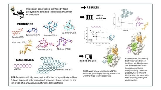
The Inhibition of Pancreatic α-Amylase by Monomeric, Dimeric and Trimeric Procyanidins Is Dependent upon the Structural Characteristics of Inhibitors and Substrates

by
Jocelin Violeta Aguilar-López
,
Ana V. Arras-Gardea
,
Alejandra I. Martinez-Gonzalez
,
Emilio Alvarez-Parrilla
and
Laura A. de la Rosa
*
Department of Chemical Biological Sciences, Institute of Biomedical Sciences, Universidad Autónoma de Ciudad Juárez, Anillo Envolvente del Pronaf y Estocolmo s/n, Ciudad Juárez 32400, Chihuahua, Mexico
*
Author to whom correspondence should be addressed.
Appl. Biosci. 2025, 4(4), 49; https://doi.org/10.3390/applbiosci4040049 (registering DOI)
Submission received: 19 September 2025 / Revised: 15 October 2025 / Accepted: 24 October 2025 / Published: 31 October 2025

Abstract

Procyanidins are oligomeric flavonoids with several bioactive properties. Their antidiabetic potential is related to their capacity to inhibit enzymes responsible for the absorption of dietary carbohydrates, such as pancreatic α-amylase. Procyanidins possess great structural diversity, including types of monomers and interflavanic bonds (A- or B-), and the degree of polymerization. However, there is a lack of evidence that systematically analyzes the effect of these structural features on their α-amylase inhibitory activity. In this paper, the activity of a mammalian pancreatic α-amylase was assessed using two different substrates, and the inhibitory activity of five commercially available procyanidins and three monomeric flavonoids was compared. The enzyme-binding sites of the eight compounds were predicted by in silico analysis to help explain the different enzyme-inhibitory activities. The inhibitory activity of procyanidins and monomeric flavonoids depended on the substrate used. A-type dimers presented the best activity against a polymeric substrate, while a B-type dimer was the best inhibitor for an oligomeric substrate. The predicted binding site for dimers and monomers was close to the active site. For the B-type trimer, the binding site was on the back side (approximately 180°) of the catalytic triad. In silico predictions suggested that dimeric procyanidins, especially A-type, could better enter the active site cavity, which could explain their superior inhibitory activity. We may conclude that inhibition of pancreatic α-amylase by procyanidins is mainly related to the type of interflavanic bond and the degree of polymerization. Dimers could be the most effective procyanidins to mildly inhibit this enzyme and present antidiabetic potential.

Graphical Abstract

1. Introduction

Diabetes mellitus is currently a major health concern in many countries. According to the WHO, more than 420 million people worldwide have diabetes. The majority live in low- and middle-income countries, where prevalence and mortality rates are rising. Diabetes is characterized by constantly elevated blood sugar levels. These high blood sugar levels contribute to diabetes-related vascular complications, leading to serious conditions such as blindness, renal failure, and myocardial infarction [1]. Several approaches are needed to prevent and treat diabetes. These include a healthy diet, regular exercise, and pharmacological treatment, which may include drugs that inhibit glucose absorption in the small intestine.
Acarbose and miglitol are commercial antidiabetic drugs that inhibit a-glucosidases and pancreatic a-amylase. These enzymes break down dietary starch to glucose in the small intestine. Their inhibition slows glucose absorption [2]. Many natural products also inhibit these enzymes. Among them, flavonoids are of special interest [3]. Flavonoids are abundant in many plant foods. They are known for antioxidant activity and many health-related actions, including antidiabetic and heart-protective properties. There are different flavonoid families, including flavones, flavonols, flavanones, and flavan-3-ols, among others [4]. Since flavonoids are abundant in foods but generally poorly bioavailable [5], it is reasonable to assume they exert many health-protective actions in the gastrointestinal tract; this is especially true for procyanidins [6].
Procyanidins are oligomeric and polymeric flavan-3-ols found in many foodstuffs. These include tea, chocolate, red wine, nuts, and some fruits. Procyanidins are composed of 2 or more catechin/epicatechin units. They can be classified by the number of interflavanic bonds. B-type procyanidins have one C-C bond, while A-type have one C-C and one C-O (ether) bond (Figure 1) [4]. B-type procyanidins with degrees of polymerization between 2 and 11 are most widely distributed in foods. A-type oligomers are found mainly in cranberries, cinnamon, apricots, and avocado [6].
There has been recent interest in understanding the structure-activity relationship for the inhibition of α-glucosidase and α-amylase by flavonoids. For monomeric flavonoids, some structural characteristics that enhance the inhibitory activity are the presence of a double bond between C2 and C3, a keto group in C4, and a catechol moiety in ring B [3,7]. Monomeric flavan-3-ols lack the first two features and, therefore, are not considered good inhibitors of these enzymes. However, less information exists for the oligomeric and polymeric flavan-3-ols. In a recent study, Visvanathan et al. [8] tested more than 50 polyphenols as inhibitors of human salivary and pancreatic α-amylases, using an oligosaccharide substrate. However, no procyanidins were used in the study. Other authors have shown that procyanidins are good inhibitors of α-amylase. It has been hypothesized that small oligomeric procyanidins with degrees of polymerization of 3 or 4 would be the best inhibitors of α-amylase and other digestive enzymes [9]. A-type procyanidins have been considered better inhibitors than B-type procyanidins [10]. Most studies, however, have been carried out with extracts of medicinal or edible plants that contain mixtures of procyanidins of different sizes and types. This complicates the comparison of results and inhibitory potency of various compounds. Moreover, different substrates and assay methods have been used to assess the activity and inhibition of α-amylase. The substrate plays an important role in the inhibitory mechanism of flavonoids on α-amylase. If oligosaccharides are used as substrates, the inhibition is due to enzyme-flavonoid interactions. If a polysaccharide (starch) is used, the inhibitory mechanism may also involve substrate-flavonoid interactions [8,11].
There is a lack of evidence that systematically analyzes the effect of the type of procyanidin interflavanic bond (A- or B-), procyanidin degree of polymerization, or substrate characteristics on the inhibition of α-amylase. Therefore, in the present paper, five commercially available procyanidins (two type-B dimers, two type-A dimers, and one type-B trimer) and three monomeric flavonoids (catechin, epicatechin, and quercetin) have been studied as inhibitors of pancreatic α-amylase. Their enzyme-binding sites were predicted by in silico analysis. These compounds were selected because they are the most abundant procyanidins in foods. Their different structures allowed us to systematically test the effect of the interflavanic bond type (by using A- and B-type dimers, which only differ in their interflavanic bond type) and degree of polymerization (by using the monomers catechin and epicatechin, the B-type dimers, and the B-type trimer). Moreover, two substrates (one oligosaccharide and one polysaccharide) were used to explore substrate-dependent effects on the procyanidins’ inhibitory activity.

2. Materials and Methods

2.1. Materials

Porcine pancreatic α-amylase (Type I-A), p-nitrophenyl-α-D-maltohexaoside (p-NPG6), HEPES (4-(2-hydroxyethyl)-1-piperazineethanesulfonic acid) were purchased from Sigma Aldrich® (Toluca, Mexico). p-Nitrophenol, starch azure, Tris(tris(hydroxymethyl)aminomethane), catechin, epicatechin, quercetin, and starch azure were purchased from J.T. Baker® (Phillipsburg, NJ, USA). Calcium chloride, acetic acid, and methanol from TEDIA® (Fairfield, OH, USA). Procyanidins A1, A2, B1, B2 and C1 were purchased from Phytolab phyproof® (Vestenbergsgreuth, Germany).

2.2. Enzyme Activity of Pancreatic α-Amylase

2.2.1. p-NPG6 as Substrate

The steady-state enzymatic activity of pancreatic α-amylase was determined according to Martinez-Gonzalez et al. [3] with some modifications. A calibration curve of p-nitrophenol (product of the hydrolysis of p-NPG6) was performed with p-nitrophenol concentrations of 0.05–0.8 mM. The absorbance was recorded on a Bio-Rad® (Hercules, CA, USA) XMark model UV-Vis microplate spectrophotometer at a wavelength of 400 nm. For the activity assay, enzyme (10 μM) and substrate (30 mM) solutions were prepared in 50 mM HEPES buffer at pH 7.0. In a microplate well, buffer solution, substrate solution (final concentrations 0.25–10 mM), and pancreatic α-amylase solution (final concentration 1 μM) were mixed to initiate the reaction. The absorbance was recorded (λ = 400 nm; 120 min at 37 °C). Each measurement was carried out in triplicate. The initial velocity (V0) was expressed as mmolL−1 p-nitrophenol min−1. The results were used to determine the apparent values of the Michaelis–Menten constant (KM) and maximum velocity (Vmax) from a nonlinear fit to the Hill Equation (1), using Sigma Plot software v.10 (USA).
V 0 = V m a x S h K M h + S h
where V0 is the initial rate of the enzymatic reaction at a given substrate concentration [S]. Vmax and KM are the apparent kinetic constants, and h is the Hill number. When h = 1, Equation (1) is the Michaelis–Menten Equation, which indicates zero cooperativity in substrate binding.

2.2.2. Starch Azure as Substrate

The activity of pancreatic α-amylase was also determined using starch azure as a substrate, according to the method proposed by Liu et al. [12] with some modifications. A Tris buffer solution (0.5 M, pH 6.9, CaCl2∙2H2O 0.01 M) was prepared in which the enzyme (10 mM) and substrate (different concentrations) were dissolved. Starch azure was diluted with the Tris buffer solution in five capped tubes to obtain final concentrations of 1, 2.5, 5, 7.5, and 10 mg mL−1 at a final volume of 3 mL. The solutions were incubated at 100 °C for 5 min, after which they were allowed to cool to room temperature. Subsequently, they were incubated at 37 °C for 10 min and shaken at 80 rpm. The reaction was started by adding porcine pancreatic α-amylase (600 μL) to a final concentration of 2 mM. After homogenizing the starch-enzyme mixture, 500 μL was removed at 0, 2, 5, 10, 60, and 120 min, respectively, and mixed with 500 μL of acetic acid (50% v/v). The new mixtures were centrifuged (Thermo Scientific® Sorvall® ST 16R model centrifuge; Waltham, MA, USA) at 5000 rpm for 10 min at 4 °C. The absorbance of the supernatant (λ = 595 nm) associated with the release of the hydrolysis product Remazol brilliant blue R was recorded in an Agilent Technologies® (Santa Clara, CA, USA) model 8453 UV-Vis spectrophotometer. Each measurement was performed in triplicate. The initial velocity (V0) was expressed as AU 595 min−1, and the results were used to determine the apparent values of the constants KM and Vmax by nonlinear analysis of Equation (1), using Sigma Plot v.10 software.

2.3. Inhibition of Pancreatic α-Amylase Enzyme Activity

2.3.1. Determination of the Percent Inhibition with p-NPG6 as Substrate

The percent inhibition of each polyphenolic compound was determined according to the methodology of Martinez-Gonzalez et al. [3] with some modifications. The flavonoids catechin (CAT), epicatechin (EPI), quercetin (QUE), and procyanidins A1 (PCA1), A2 (PCA2), B1 (PCB1), B2 (PCB2), and C1 (PCC1) were dissolved separately in methanol at a concentration of 400 µM. The assay was carried out in a microplate with final concentrations of pancreatic α-amylase and p-NPG6 of 1 µM and 3 mM, respectively, while those of the polyphenolic compounds were 0–250 µM. The order of addition of the solutions in the microplate was substrate, inhibitor (polyphenolic compound), and finally, enzyme. The absorbance (λ = 400 nm) was recorded for 120 min at 37 °C in the UV-Vis microplate spectrophotometer. Each assay was performed in triplicate, and the percent inhibition (%I) values were determined according to Equation (2):
% I = A c     A c b       ( A s   A s b ) A c       A c b     · 100
where Acb is the absorbance of the control blank (HEPES buffer only), Ac is the absorbance of the control (substrate and enzyme solutions), Asb is the absorbance of the sample blank (buffer and polyphenolic compound), and As is the absorbance of the sample (substrate, enzyme, and polyphenolic compound).

2.3.2. Determination of the Percent Inhibition with Starch Azure as Substrate

The percent inhibition of polyphenolic compounds was determined as previously described [12,13,14], with some modifications. The polyphenolic compounds were dissolved in methanol at 2 mM each. Porcine pancreatic α-amylase was suspended in the Tris buffer solution at a concentration of 10 mM. Starch azure was also suspended in the Tris buffer solution at a concentration of 7 mg mL−1. It was kept at 100 °C for 5 min, then allowed to cool to room temperature before being incubated at 37 °C for 10 min in a shaking water bath. An aliquot of the treated starch was taken, and the polyphenolic compound and enzyme solutions were added. The final concentrations were 5 mg mL−1 of starch azure, 2 µM of α-amylase, and 0–500 µM of each polyphenolic compound. The reaction was carried out at 37 °C for 10 min with shaking at 80 rpm and stopped by adding 50% (v/v) acetic acid. The mixture was centrifuged at 5000 rpm for 10 min at 4 °C, and the absorbance of the supernatant was recorded in the UV-Vis spectrophotometer (λ = 595 nm).
Enzyme-inhibitor preincubation assays were also performed. The enzyme and polyphenolic compound solutions were mixed at the same concentrations mentioned above for 10 min in a water bath at 37 °C. The reaction was then started by adding the starch azure solution (5 mg mL−1 final concentration). The mixture was incubated and centrifuged, and absorbance was measured as described above. In both cases (with and without preincubation), measurements were performed in triplicate, and the percent inhibition of each polyphenolic compound was determined according to Equation (2).

2.4. Molecular Docking Analysis

In silico analysis was performed to predict the interactions between the enzyme porcine pancreatic α-amylase and various polyphenolic compounds. The structure of porcine pancreatic α-amylase was obtained from the Protein Data Bank (USA); PDB code 1pif. Porcine pancreatic α-amylase structure was chosen since it presents the highest homology (87.1%) to the human enzyme among mammalian α-amylases [15]. The 3D structure of the enzyme was prepared by removing the surface water molecules. The 3D structures of the polyphenolic compounds (the ligands) were obtained from the 2D structures in PubChem® (NIH, USA) and prepared for analysis using the Avogadro V 1.95 program by using its auto-optimization tool (Molecular Dynamics 300K Algorithm) until the change in energy value was equal to 0 (dE = 0). The codes of the ligands in this database were as follows: 9064 (CAT), 72,276 (EPI), 5,280,343 (QUE), 9,872,976 (PCA1), 124,025 (PCA2), 11,250,133 (PCB1), 122,738 (PCB2), 169,853 (PCC1), and 9,811,704 (acarbose). Docking studies were performed by the SwissDock® server (Swiss Institute of Bioinformatics, Lausanne, Switzerland), using a flexible ligand and rigid protein system. The grid box that includes the catalytic residues (Asp300, Glu233, and Asp197) had the dimensions of 45.02 × 66.25 × 58.63 ((x)(y)(z)). The possible interactions of the best-scored results (energy values, ΔG) of the models for each polyphenolic compound were analyzed. Interactions and distances were evaluated with the UCSF Chimera® V 1.18 program (University of California, San Francisco, CA, USA). The molecular docking results were also analyzed in 2D using the BIOVIA Discovery Studio Visualizer® program (Dassault Systemes, Vélizy-Villacoublay, France). RMSD (Root Mean Square Deviation) values were used to evaluate model accuracy; values lower than 2.0 Å were considered acceptable.

2.5. Data Analysis

All experimental trials were performed in triplicate. Results were expressed as means ± standard deviation (SD). A nonparametric analysis of variance (Kruskal–Wallis) and a pairwise multiple comparison analysis (Dunn) were performed using XLSTAT v. 1.3 software (USA) to determine significant differences (p < 0.05) between treatments.

3. Results and Discussion

3.1. Enzyme Activity of α-Amylase Towards Two Model Substrates

The results of α-amylase activity at different substrate concentrations are shown in Figure 2, and their respective kinetic parameters are summarized in Table 1. The first substrate was the chromogen p-NPG6, a maltooligosaccharide of 6 glucose units linked to a nitrophenyl group (p-nitrophenol-α-D-maltohexaoside). Due to the excess enzyme present in the mixture, the substrate is rapidly hydrolyzed to glucose, oligosaccharides, and free p-nitrophenol. Thus, the amount of p-nitrophenol released is proportional to the activity of α-amylase [16]. Starch azure is also a chromogenic substrate for α-amylase; it is a corn starch covalently bound to Remazol brilliant blue R, whose release into the reaction medium is proportional to the degree of starch hydrolysis [17,18]. For both substrates, the experimental results show a better fit to the Hill equation with Hill coefficient (h) values close to 2 (Table 1). This indicates that both substrates, p-NPG6 and starch azure, exhibit apparent cooperativity in their binding to the enzyme. Previous studies [3] showed that this same enzyme (porcine pancreatic α-amylase) exhibited sigmoidal kinetics and a degree of cooperativity similar to that calculated here, with a substrate similar to p-NPG6 (p-NPG5). The Hill coefficient is commonly used to estimate the number of ligand molecules that must bind to a receptor to produce a functional effect [19]. However, it is more appropriate to consider it as a measure of interaction, which reflects the degree of cooperativity between multiple binding sites in a protein [20]. In monomeric enzymes, such as α-amylase, false cooperativity can be observed due to the presence of two isoenzymes or isoforms in the assay preparation that act on the same substrate with different kinetic properties [21]. It is also possible that α-amylase has more than one binding site. In a bacterial enzyme, the existence of two binding sites for a small substrate has been reported, and it is considered that, in addition to the active site, an activator site is necessary for optimal substrate binding [22,23]. This phenomenon has also been studied in other amylases against various substrates, ranging from starches to different oligomers, where it is considered that there are not just one but multiple secondary carbohydrate binding sites [24,25]. The cooperativity exhibited by α-amylase could also be explained by a kinetic cooperativity mechanism, where at least two forms of the enzyme are present in equilibrium [21], as proposed in previous studies [3].
Regarding starch azure, saturation kinetics showed that the reaction rate increased to a maximum with a substrate concentration of 5 mg mL−1 and then decreased, suggesting substrate inhibition or inhibition by the products released from its hydrolysis. It has been shown that glucose and maltose, products of the starch hydrolysis reaction, inhibit α-amylase, reducing its reaction rate [26]. Thus, the best fit to a sigmoidal behavior for the saturation kinetics of starch azure (Figure 2 and Table 1) could be due to the substrate-inhibition phenomenon that gives the appearance of a false cooperative behavior. Furthermore, a possible mechanism has been suggested that involves the formation of non-productive enzyme-substrate (E-S) complexes when excess substrate competitively binds to the active site, leading to a lower turnover rate. Another mechanism suggests that substrate molecules at high concentrations may bind non-specifically to sites other than the catalytic one, thus slowing down catalysis through allosteric coupling [27,28]. Recently, substrate inhibition has also been attributed to a slowdown in the product release step rather than the formation of the catalytically competent E-S complex [29]. Furthermore, since blue starch is an insoluble and structurally modified substrate, other factors could contribute to the observed sigmoidal behavior, such as enzyme adsorption onto the insoluble substrate surface, diffusion limitations, and heterogeneous accessibility to hydrolysis sites within the blue starch granules [30]. These effects could produce an apparent cooperative behavior that deviates from classical Michaelis–Menten kinetics, as previously observed for insoluble starches [31,32].

3.2. Inhibition of α-Amylase Using p-NPG6 as Substrate

The inhibitory effects of two dimeric A-type procyanidins (PCA1 and PCA2, Figure 3A), two B-type dimers and one trimer (PCB1, PCB2, and PCC1, Figure 3B), and their monomeric units (CAT and EPI, Figure 3C), as well as a flavonoid that has been described as a good inhibitor of pancreatic α-amylase (QUE, Figure 3C), were compared. PCB1, PCA2, and PCA1 inhibited α-amylase hydrolysis of p-NPG6, with maximum inhibition rates of 49.68%, 32.54%, and 21.16%, respectively. PCB1 exhibited a much stronger inhibition effect than A-type procyanidins at the highest concentration tested (Table S1 in Supplementary Materials). CAT, EPI, QUE, and PCC1 did not show inhibitory activity against α-amylase at concentrations of 250 µM but did so at 50 µM (inhibition values close to those of PCB1); however, at lower concentrations, no inhibition was observed with these compounds either. PCB2 did not show significant inhibition of α-amylase activity with p-NPG6 substrate at any inhibitor concentration.
That is to say, the capacity to inhibit the enzymatic activity of α-amylase on an oligomeric substrate did not show a clear dependence on any of the structural characteristics (degree of polymerization, type of bond, or stereochemistry) of the procyanidins studied here, nor could a significant difference be observed between procyanidins (belonging to the group of flavan-3-ols) and other flavonoids such as quercetin (a flavonol).
Previous studies have shown that CAT has a lower inhibitory effect on α-amylase than QUE in the presence of an oligomeric substrate (p-NPG5) and that CAT and EPI generally have little inhibitory capacity on digestive enzymes [3,33]. In the present work, an inconsistent inhibitory effect of the three monomeric flavonoids (CAT, EPI, and QUE) was observed, suggesting little effectiveness of these compounds. In the case of QUE, its low inhibitory activity, compared to previous works, could be related to the concentration or size of the substrate used or even the degree of purity of the enzyme [34,35]. The existence of solubility and stability limitations may also play a crucial role; at increasing concentrations, these monomeric inhibitors, especially QUE, can self-associate through π–π stacking and hydrogen bonding, leading to colloidal aggregation that decreases their effective free concentration and accessibility to the enzyme’s active site [36,37]. Additionally, inhibitor–substrate complex formation could also explain the observed decline in inhibition at high concentrations [8]. On the other hand, the fact that the inhibitory effect of the monomers and the trimer decreased with increasing concentration of these compounds could be consistent with the sigmoid model of substrate saturation kinetics, indicating that these compounds could have interactions with a possible activating site or differential interactions with the different isoforms of the enzyme [8,23,35].
The degree of polymerization (DP) of procyanidins is one of the structural characteristics critical to determining their inhibitory capacity. Procyanidins with a higher DP (higher molecular weight and complexity) are considered to have better inhibitory activity due to their polyhydroxylation character [10,38]. However, procyanidin oligomers are likely to be better inhibitors than polymers due to their ability to enter and bind to specific cavities of enzymes [9]. Another determining structural characteristic is the type of interflavanic bond, with type A procyanidins being considered the best inhibitors because their two interflavanic bonds confer greater rigidity and stability to the enzyme-inhibitor complex [39,40].
In the present work, dimeric procyanidins showed the highest inhibition of oligomeric substrate hydrolysis, except for PCB2, which had no effect. However, PCB1, with only one interflavanic bond (C4-C8), was a better inhibitor than both PCAs. This could indicate that the presence of an extra hydroxyl group, possibly involved in hydrogen bond formation, would be relevant for the inhibitory activity of the dimers [41]. However, it is important to mention that PCB2 did not show significant inhibitory activity. The main structural difference between PCB1 and PCB2 lies in the stereochemistry of their monomeric units: PCB1 is composed of EPI-CAT and PCB2 of EPI-EPI. There is evidence indicating that the affinity of CAT for proteins, particularly for the amino acid proline, is stronger than that of EPI, while the degrees of specific binding of PCB2 and EPI are similar [42,43]. This suggests that PCB1 could have a stronger binding with α-amylase, which could explain its higher inhibitory effect compared to PCB2. In vitro analysis with a different substrate and docking analysis were then carried out to further understand the differences in inhibitory activity.

3.3. Inhibition of α-Amylase Using Starch Azure as Substrate

Inhibition of α-amylase activity using starch azure as substrate showed a dependence on the concentration and structural characteristics of procyanidins (Figure 4). The variability in response (high standard deviations) may be related to the heterogeneous structure and solubility of starch azure (corn starch covalently bound to Remazol brilliant blue R); however, statistical differences were obtained between some concentrations of the inhibitors. PCA1, PCA2, and PCC1 were the best inhibitors under two experimental conditions: with and without enzyme-inhibitor (E-I) preincubation. The maximum inhibition rates were 37.56%, 34.27%, and 25.39% without preincubation and 45.83%, 31.24%, and 29.01% with preincubation, respectively (Table S2). B-type dimers (PCB1 and PCB2) had similar inhibition rates to the trimer (PCC1).
However, unlike the latter, their effectiveness did not increase with preincubation, suggesting a faster binding of the dimers than that of the trimer. On the other hand, CAT, EPI, and QUE showed little inhibitory activity without preincubation. However, preincubation with the enzyme increased the effectiveness of the flavan-3-ols (CAT and EPI) but not the flavonol (QUE), suggesting differences in their binding mechanisms. Actually, molecular dynamics simulations have shown some differences in the binding dynamics of EPI and QUE to α-amylase, although no detailed analysis of the differences was performed [44].
These results indicate that both the type of interflavanic bond and the degree of polymerization have an important effect on the inhibitory activity of procyanidins on α-amylase. This observation is relevant since starch azure is a substrate more similar to the enzyme’s natural substrate than p-NPG6. Under these experimental conditions, A-type procyanidins were better inhibitors than B-type procyanidins, which was the expected outcome. This phenomenon is attributed to the presence of the interflavanic ether bond, which enhances the molecule’s hydrophobicity, thereby increasing the potential for hydrophobic interactions [10]. In fact, molecular dynamic simulations have predicted that hydrophobic interactions are the major drivers of procyanidin-a-amylase complex formation [11]. The greater rigidity of A-type procyanidins has also been described as a favorable characteristic for their inhibitory effectiveness [39,40].
On the other hand, the degree of polymerization also showed an effect, although it was not as consistent. In the absence of preincubation, the B-type trimer was slightly more effective than the dimers of the same type, and all oligomers were better than the monomers (including the flavonol). However, preincubation increased the inhibitory effect of monomers and trimer, and decreased that of dimers. In this sense, we could conclude that the most determining structural characteristic for a better inhibitory effect of small oligomeric proanthocyanidins would be the type of interflavanic bond, and the degree of polymerization would be secondary in this size range. Enzyme-inhibitor preincubation generally produced a slight increase in inhibitory activity (except in B-type dimers), so it would be advisable to study the mechanisms of enzyme-procyanidin interaction further. It is also noteworthy that, according to the evidence shown here, oligomeric procyanidins showed a better inhibitory effect than the flavonol QUE. However, it would be necessary to compare them with representatives of other flavonoid families to confirm their degree of effectiveness.
However, the inhibitory effectiveness also depended on the substrate used; in the presence of the oligomeric substrate (p-NPG6), a B-type dimer was the best inhibitor, followed by the A-type dimers. This could be explained by a different form of interaction between a small substrate and the enzyme, although it could also be due to the interaction between procyanidins and substrates. Previous studies have shown that α-amylase inhibition by flavonoids is partly due to the formation of starch-flavonoid complexes through hydrophobic interactions [45,46]. These complexes prevent the interaction of α-amylase with starch granules, thus reducing starch hydrolysis. Therefore, the observed inhibition of α-amylase by procyanidins in the presence of starch azure could be due to specific interactions between amylose and linear amylopectin fragments with these inhibitors, suggesting that hydrophobic substrate-inhibitor interactions are involved [45], a case that does not occur with p-NPG6. Therefore, the mode of inhibition of flavonoids, more specifically of procyanidins, may depend on the substrates used for the experiments [47], which may bind only to the catalytic site or bind to both the catalytic site and the activating (or secondary) site. This suggests that the inhibitions also depend on the size of the substrate and not only on the hydrophobic or hydrophilic character of the procyanidin [26,48].
From an applied point of view, mild inhibition of α-amylase is beneficial to mitigate postprandial glycemic spikes. Excessive inhibition can induce abdominal pain, bloating, or cramps due to the blockage of starch digestion and abnormal bacterial fermentation —side effects related to currently used drugs, such as acarbose [49]. Procyanidins are one of the most widely distributed compounds in foods, so they are ingested daily [39], and their natural presence in the diet could have beneficial effects to mitigate postprandial glycemic spikes and help prevent or control diabetes.

3.4. Molecular Docking

Molecular docking analyses allow us to simulate optimal interactions and predict the affinity between proteins and ligands [50]. In the present work, the validity of the docking protocol was assessed by analyzing the binding of acarbose, a known ligand (inhibitor) of pancreatic α-amylase. Binding energy (ΔG) value for acarbose was −7.21 (Kcal mol−1) and its RMSD was 0.71 Å, similar to previously reported values that labeled models as correct poses [51]. Next, the most probable binding sites, were obtained for the ligands (oligomeric and monomeric procyanidins and a flavonol) in the structure of the pancreatic α-amylase protein (Figure 5, Table 2). All ΔG values were lower than that of acarbose (<−7.21 Kcal mol−1), which was considered as a cutoff value for an effective binding. The dimeric and trimeric procyanidins presented lower ΔG values than the monomeric flavonoids, CAT, EPI, and QUE (Table 2), suggesting a higher affinity or spontaneity in their binding with α-amylase. The ΔG values were lowest for the trimer (PCC1) with −9.4 Kcal mol−1, followed by the dimers PCA1 > PCB1 > PCB2 > PCA2, and the monomers, QUE > CAT > EPI, the latter with a value of −7.6 Kcal mol−1. This could be related to a greater capacity of larger compounds, which have more hydroxyl groups and aromatic rings, to interact with the enzyme.
The most probable binding site was similar for all compounds except PCC1. Monomeric flavonoids and dimeric procyanidins would bind very close to the active site, while the PCC1 trimer would bind behind this site (Figure 5). This difference could be due to the larger size of the trimer and a steric hindrance to access the active site cavity [9]. On the other hand, both binding sites would be mainly made up of amino acid residues with charged R groups, such as Asp, Glu, and Arg, and aromatic ones, such as Phe and Tyr, where the interactions would be carried out mainly through hydrogen bonds and π-stacking, respectively (Table 2).
The results observed in the present in silico analysis predict differences in the interactions between pancreatic α-amylase and monomeric and oligomeric procyanidins, consistent with their differences as inhibitors. For example, a major difference between the binding of dimers PCA1 and PCB1 (Figure 5) and monomers CAT, EPI, and QUE (Figure S1) is that dimers were oriented such that the conjugated rings of one subunit were located between catalytic residues Asp300 and Glu233, which was not the case for monomers. The type of interflavanic bond (A- or B-), which appears to be the main structural feature responsible for the differences between the inhibitory activity of oligomeric procyanidins, also showed an effect on the manner of interaction between procyanidins and pancreatic α-amylase. Figure 5 shows that for PCA1 (5B), the B ring of one subunit can enter the pocket between Asp197 and Trp59, forming interactions with the catalytic residue Asp197 and allowing the C ring (dihydropyran heterocycle) of the EPI unit to orient itself parallel to the aromatic ring of Trp59. In this way, π-stacking interactions would occur between the C ring and Trp59, as well as between the B ring of the same unit and Tyr62 (see the two-dimensional model), and this could be related to its high ΔG value (only below that of PCC1). Something similar occurs with PCA2, which also enters the cavity and has π-stacking interactions with Trp59 and Tyr62 (Table 2 and Figure S1). This does not happen with PCB1 (Figure 5C), which has a flatter structure, so it does not fit as far into the cavity, does not interact with Tyr62, and although the B ring of a subunit does interact with Trp59, the interaction could be weaker. These observations suggest that the second interflavanic bond of A-type procyanidins may enable an EPI unit to enter the active site cavity more effectively, and its interactions with the Trp59 and Tyr62 residues could be crucial to its inhibitory capacity against large substrates, such as starch or starch azure.
In contrast, PCB1 seems to be the only one that interacts, through hydrogen bonds, with the three catalytic residues (Asp197, Glu233, and Asp300, Table 2), which could be important for its inhibitory activity against the small substrate (p-NPG6). To our knowledge, there are currently no published molecular docking studies describing the interaction between PCB1 and α-amylase. This highlights the novelty and relevance of our findings. The most closely related report is that of Dai et al. [52], who studied the interaction between porcine pancreatic α-amylase and a B-type procyanidin dimer (PB2). Their molecular docking indicated that hydrophobic interactions mediate binding of PB2 at or near the active site, but it did not interact directly with all the catalytic residues, which agrees with our predictions for PCB2. Actually, PCB2 (formed by two EPI units) shows interactions more like those of A-type dimers (Table 2 and Figure S1) but with a different alignment of rings B and C entering the cavity, which could explain its lower inhibitory capacity. It should be noted that these predictions do not rule out the possibility that other binding sites contribute to the inhibitory activity of procyanidins. Finally, the EPI subunits in all dimers seem to have a greater tendency than the CAT units to enter the enzyme cavities.
Previous molecular docking studies have suggested that residues such as Trp59, Tyr62, and Asp197 could be key for the binding of flavonoids to the active site of pancreatic α-amylase and, consequently, could be related to their inhibitory activity [3,53]. Very few studies have investigated the molecular interactions between procyanidins and pancreatic α-amylase, and to the best of our knowledge, none of the interactions have been analyzed by crystallography. Molecular dynamics simulation of the binding of α-amylase with procyanidin (a commercial B-type dimer) also indicated that Trp59, Tyr62, and Asp197 in addition to Gln63 and Asp300, were among the amino acid residues that contributed significantly to the total binding energy [11]. Crystallographic studies have been carried out for complexes formed between human pancreatic α-amylase and flavonoids such as myricetin, mombretin, and derivatives (reviewed in [7]). These studies have shown that flavonoids indeed bind to the active site of the enzyme and interact with the catalytic residues through the B ring, as demonstrated by the models of the present work, especially for A-type dimers. Flavonoids also showed π-stacking interactions with Tyr62 and hydrogen bonds with Gln63, and it was suggested that these interactions could improve the inhibitory activity and the stability of the ligand-enzyme complex [7]. This also agrees with the models obtained in the present work, where A-type dimers, which presented the best inhibitory activity against starch azure, also showed interactions with Gln63 (Table 2).
The trimeric procyanidin (PCC1), which showed good inhibitory effectiveness on starch azure but not on p-NPG6, presented, according to the in silico model, a binding site different from that of the rest of the evaluated compounds (Figure 5). PCC1, which is made up of three monomeric units of EPI, showed interactions on the back side (approximately 180°) of the catalytic triad. They were mainly hydrophobic interactions between the B rings of each unit with the residues Phe406 and Tyr2 and hydrogen bonds with Glu282, Gly403, and Arg421 (Table 2 and Figure 5). That is, the trimer could exert its inhibitory action without entering the catalytic cavity, but by blocking this other site, which would be essential for the binding or catalysis of starch azure, but not of the smaller substrate (p-NPG6). The presence of more than one ligand binding site in pancreatic α-amylase has been suggested by previous studies [24] and by our results of steady-state enzymatic activity. However, there is no evidence that the predicted binding site for PCC1 could be involved with substrate binding. On the other hand, one of the few studies where the structure of complexes between human pancreatic α-amylase and phenolic compounds has been described showed that ethyl caffeate (a non-flavonoid phenolic compound) could bind to this α-amylase at three different sites, none of them corresponding to the active site, but the binding of the compound produced a disordering of the active site regions [54]. It would be interesting to explore the possibility that PCC1 possesses a similar binding and inhibition mechanism.

4. Conclusions

The present work has systematically explored the pancreatic α-amylase inhibitory activity of dimeric and trimeric procyanidins compared to monomeric flavonoids. It shows that inhibition is mostly related to the type of interflavanic bond, the degree of polymerization of procyanidins and substrates, and to a lesser extent to the stereochemistry of the monomeric units of the procyanidins. A-type dimers presented the best inhibitory activity against a polymeric substrate (starch azure), while a B-type dimer was the best inhibitor of the hydrolysis of an oligomeric substrate. So, it could be suggested that dimers are the most effective procyanidins for mildly inhibiting α-amylase, reducing postprandial glycemia peaks, and presenting few undesirable side effects. In silico studies predict that procyanidins could interact differently with the catalytic and secondary sites of the enzyme, which would explain the differences in their inhibitory effectiveness. Dimeric procyanidins, especially A-type, showed a tendency to enter the active site cavity better and interact with catalytic residues (Asp197, Glu233, and Asp300) and others that could be key for the binding and stability of the E-I complex, such as Trp59, Tyr62, and Gln63. Predictions indicated that the trimeric procyanidin would present a binding site far from the active site, which could be involved in the binding or catalysis of the polymeric substrate (starch azure). This study highlights the relevance of exploring in more detail the precise molecular mechanisms of natural compounds as inhibitors of pancreatic α-amylase. A deeper understanding of these aspects will allow for design optimization and the use of strategies based on natural inhibitors to prevent or control diabetes mellitus.

Supplementary Materials

The following supporting information can be downloaded at: https://www.mdpi.com/article/10.3390/applbiosci4040049/s1, Table S1: Percent inhibition of α-amylase with the substrate p-NPG6 in the presence of different concentrations of phenolic compounds; Table S2: Percent inhibition of α-amylase with the substrate starch azure in the presence of different concentrations of phenolic compounds. Figure S1: Molecular docking simulations of the interaction between flavonoids and procyanidins, and pancreatic α-amylase.

Author Contributions

J.V.A.-L.: Investigation, Methodology, Formal analysis, Visualization, Writing—original draft. A.V.A.-G.: Investigation, Methodology, Formal analysis, Visualization, Writing—original draft. A.I.M.-G.: Methodology, Supervision, Visualization, Writing—original draft. E.A.-P.: Conceptualization, Supervision, Resources, Writing—review and editing. L.A.d.l.R.: Conceptualization, Supervision, Resources, Project administration, Writing—review and editing. All authors have read and agreed to the published version of the manuscript.

Funding

E.A.-P. and L.A.d.l.R. were partially funded by UACJ through the internal project RIPI2023ICB1. A.I.M-G. received a postdoctoral fellowship from CONAHCYT.

Institutional Review Board Statement

Not applicable.

Informed Consent Statement

Not applicable.

Data Availability Statement

The original contributions presented in this study are included in the article and Supplementary Materials. Further inquiries can be directed to the corresponding author.

Conflicts of Interest

The authors declare no conflicts of interest.

Abbreviations

The following abbreviations are used in this manuscript:
p-NPG6p-nitrophenyl-α-D-maltohexaoside
CATCatechin
EPIEpicatechin
QUEQuercetin
PCA1Procyanidin A1
PCA2Procyanidin A2
PCB1Procyanidin B1
PCB2Procyanidin B2
PCC1Procyanidin C1

References

  1. Kanter, J.E.; Bornfeldt, K.E. Impact of Diabetes Mellitus. Arterioscler. Thromb. Vasc. Biol. 2016, 36, 1049–1053. [Google Scholar] [CrossRef]
  2. Kaur, N.; Kumar, V.; Nayak, S.K.; Wadhwa, P.; Kaur, P.; Sahu, S.K. Alpha-amylase as Molecular Target for Treatment of Diabetes Mellitus: A Comprehensive Review. Chem. Biol. Drug Des. 2021, 98, 539–560. [Google Scholar] [CrossRef]
  3. Martinez-Gonzalez, A.I.; Díaz-Sánchez, Á.G.; de la Rosa, L.A.; Bustos-Jaimes, I.; Alvarez-Parrilla, E. Inhibition of α-Amylase by Flavonoids: Structure Activity Relationship (SAR). Spectrochim. Acta A Mol. Biomol. Spectrosc. 2019, 206, 437–447. [Google Scholar] [CrossRef]
  4. de la Rosa, L.A.; Moreno-Escamilla, J.O.; Rodrigo-García, J.; Alvarez-Parrilla, E. Phenolic Compounds. In Postharvest Physiology and Biochemistry of Fruits and Vegetables; Yahia, E., Ed.; Elsevier: Amsterdam, The Netherlands, 2019; pp. 253–271. [Google Scholar]
  5. Yang, H.; Tuo, X.; Wang, L.; Tundis, R.; Portillo, M.P.; Simal-Gandara, J.; Yu, Y.; Zou, L.; Xiao, J.; Deng, J. Bioactive Procyanidins from Dietary Sources: The Relationship between Bioactivity and Polymerization Degree. Trends Food Sci. Technol. 2021, 111, 114–127. [Google Scholar] [CrossRef]
  6. Cires, M.J.; Wong, X.; Carrasco-Pozo, C.; Gotteland, M. The Gastrointestinal Tract as a Key Target Organ for the Health-Promoting Effects of Dietary Proanthocyanidins. Front. Nutr. 2017, 3, 57. [Google Scholar] [CrossRef]
  7. Lam, T.-P.; Tran, N.-V.N.; Pham, L.-H.D.; Lai, N.V.-T.; Dang, B.-T.N.; Truong, N.-L.N.; Nguyen-Vo, S.-K.; Hoang, T.-L.; Mai, T.T.; Tran, T.-D. Flavonoids as Dual-Target Inhibitors against α-Glucosidase and α-Amylase: A Systematic Review of in Vitro Studies. Nat. Prod. Bioprospect 2024, 14, 4. [Google Scholar] [CrossRef] [PubMed]
  8. Visvanathan, R.; Houghton, M.J.; Barber, E.; Williamson, G. Structure-Function Relationships in (Poly)Phenol-Enzyme Binding: Direct Inhibition of Human Salivary and Pancreatic α-Amylases. Food Res. Int. 2024, 188, 114504. [Google Scholar] [CrossRef] [PubMed]
  9. Vazquez-Flores, A.A.; Martinez-Gonzalez, A.I.; Alvarez-Parrilla, E.; Díaz-Sánchez, Á.G.; de la Rosa, L.A.; González-Aguilar, G.A.; Aguilar, C.N. Proanthocyanidins with a Low Degree of Polymerization Are Good Inhibitors of Digestive Enzymes Because of Their Ability to Form Specific Interactions: A Hypothesis. J. Food Sci. 2018, 83, 2895–2902. [Google Scholar] [CrossRef]
  10. Le Bourvellec, C.; Renard, C.M.G.C. Interactions between Polyphenols and Macromolecules: Effect of Tannin Structure. In Encyclopedia of Food Chemistry; Elsevier: Amsterdam, The Netherlands, 2019; pp. 515–521. [Google Scholar]
  11. Jiang, C.; Chen, Y.; Ye, X.; Wang, L.; Shao, J.; Jing, H.; Jiang, C.; Wang, H.; Ma, C. Three Flavanols Delay Starch Digestion by Inhibiting α-Amylase and Binding with Starch. Int. J. Biol. Macromol. 2021, 172, 503–514. [Google Scholar] [CrossRef] [PubMed]
  12. Liu, J.; Lu, J.; Kan, J.; Wen, X.; Jin, C. Synthesis, Characterization and in Vitro Anti-Diabetic Activity of Catechin Grafted Inulin. Int. J. Biol. Macromol. 2014, 64, 76–83. [Google Scholar] [CrossRef]
  13. Wang, H.; Du, Y.-J.; Song, H.-C. α-Glucosidase and α-Amylase Inhibitory Activities of Guava Leaves. Food Chem. 2010, 123, 6–13. [Google Scholar] [CrossRef]
  14. Tao, W.; Pan, H.; Jiang, H.; Wang, M.; Ye, X.; Chen, S. Extraction and Identification of Proanthocyanidins from the Leaves of Persimmon and Loquat. Food Chem. 2022, 372, 130780. [Google Scholar] [CrossRef]
  15. Darnis, S.; Juge, N.; Guo, X.-J.; Marchis-Mouren, G.; Puigserver, A.; Chaix, J.-C. Molecular Cloning and Primary Structure Analysis of Porcine Pancreatic α-Amylase. BBA-Protein Struct. Mol. Enzymol. 1999, 1430, 281–289. [Google Scholar] [CrossRef]
  16. Baks, T.; Janssen, A.E.M.; Boom, R.M. A Kinetic Model to Explain the Maximum in A-amylase Activity Measurements in the Presence of Small Carbohydrates. Biotechnol. Bioeng. 2006, 94, 431–440. [Google Scholar] [CrossRef]
  17. Gaquere-Parker, A.; Taylor, T.; Hutson, R.; Rizzo, A.; Folds, A.; Crittenden, S.; Zahoor, N.; Hussein, B.; Arruda, A. Low Frequency Ultrasonic-Assisted Hydrolysis of Starch in the Presence of α-Amylase. Ultrason. Sonochem 2018, 41, 404–409. [Google Scholar] [CrossRef]
  18. Rinderknecht, H.; Wilding, P.; Haverback, B.J. A New Method for the Determination of α-Amylase. Experientia 1967, 23, 805. [Google Scholar] [CrossRef] [PubMed]
  19. Abeliovich, H. On Hill Coefficients and Subunit Interaction Energies. J. Math. Biol. 2016, 73, 1399–1411. [Google Scholar] [CrossRef] [PubMed]
  20. Weiss, J.N. The Hill Equation Revisited: Uses and Misuses. FASEB J. 1997, 11, 835–841. [Google Scholar] [CrossRef] [PubMed]
  21. Cappiello, M.; Balestri, F.; Moschini, R.; Del-Corso, A.; Mura, U. Apparent Cooperativity and Apparent Hyperbolic Behavior of Enzyme Mixtures Acting on the Same Substrate. J. Enzyme Inhib. Med. Chem. 2016, 31, 1556–1559. [Google Scholar] [CrossRef]
  22. Ishikura, K.; Nitta, Y.; Watanabe, T. Hydrolysis of Phenyl β-Maltoside Catalyzed by Saccharifying α-Amylase from Bacillus subtilis. J. Biochem. 1977, 81, 1187–1192. [Google Scholar] [PubMed]
  23. Yoshida, H.; Hiromi, K.; Ono, S. Kinetics and Mechanism of Hydrolysis of Phenyl α-Maltoside by Saccharifying α-Amylase of Bacillus subtilis. J. Biochem. 1969, 65, 741–750. [Google Scholar] [CrossRef]
  24. Nielsen, J.W.; Kramhøft, B.; Bozonnet, S.; Abou Hachem, M.; Stipp, S.L.S.; Svensson, B.; Willemoës, M. Degradation of the Starch Components Amylopectin and Amylose by Barley α-Amylase 1: Role of Surface Binding Site 2. Arch. Biochem. Biophys. 2012, 528, 1–6. [Google Scholar] [CrossRef]
  25. Nielsen, M.M.; Seo, E.-S.; Bozonnet, S.; Aghajari, N.; Robert, X.; Haser, R.; Svensson, B. Multi-site Substrate Binding and Interplay in Barley A-amylase 1. FEBS Lett. 2008, 582, 2567–2571. [Google Scholar] [CrossRef]
  26. Apar, D.K.; Özbek, B. Estamation of Kinetic Parameters for Rice Starch Hydrolysis Inhibited by Added Materials. Chem. Eng. Commun. 2007, 194, 334–344. [Google Scholar] [CrossRef]
  27. Yoshino, M.; Murakami, K. Analysis of the Substrate Inhibition of Complete and Partial Types. Springerplus 2015, 4, 292. [Google Scholar] [CrossRef] [PubMed]
  28. Zhang, J.; Li, C.; Wang, G.; Cao, J.; Yang, X.; Liu, X.; Sun, L. α-Amylase Inhibition of a Certain Dietary Polyphenol Is Predominantly Affected by the Concentration of α-1, 4-Glucosidic Bonds in Starchy and Artificial Substrates. Food Res. Int. 2022, 157, 111210. [Google Scholar] [CrossRef]
  29. Zhang, Y.; Chen, M.; Lu, J.; Li, W.; Wolynes, P.G.; Wang, W. Frustration and the Kinetic Repartitioning Mechanism of Substrate Inhibition in Enzyme Catalysis. J. Phys. Chem. B 2022, 126, 6792–6801. [Google Scholar] [CrossRef] [PubMed]
  30. Kong, B.-W.; Kim, J.-I.; Kim, M.-J.; Kim, J.C. Porcine Pancreatic α-Amylase Hydrolysis of Native Starch Granules as a Function of Granule Surface Area. Biotechnol. Prog. 2008, 19, 1162–1166. [Google Scholar] [CrossRef]
  31. Lehoczki, G.; Kandra, L.; Gyémánt, G. The Use of Starch Azure for Measurement of Alpha-Amylase Activity. Carbohydr. Polym. 2018, 183, 263–266. [Google Scholar] [CrossRef]
  32. Guerra, N.P.; Pastrana Castro, L. Modelling the Effects of Ageing Time of Starch on the Enzymatic Activity of Three Amylolytic Enzymes. Sci. World J. 2012, 2012, 1–13. [Google Scholar] [CrossRef]
  33. Zhao, L.; Wen, L.; Lu, Q.; Liu, R. Interaction Mechanism between α-Glucosidase and A-Type Trimer Procyanidin Revealed by Integrated Spectroscopic Analysis Techniques. Int. J. Biol. Macromol. 2020, 143, 173–180. [Google Scholar] [CrossRef] [PubMed]
  34. Barber, E.; Houghton, M.J.; Williamson, G. Flavonoids as Human Intestinal α-Glucosidase Inhibitors. Foods 2021, 10, 1939. [Google Scholar] [CrossRef]
  35. Shen, H.; Wang, J.; Ao, J.; Hou, Y.; Xi, M.; Cai, Y.; Li, M.; Luo, A. Structure-Activity Relationships and the Underlying Mechanism of α-Amylase Inhibition by Hyperoside and Quercetin: Multi-Spectroscopy and Molecular Docking Analyses. Spectrochim. Acta A Mol. Biomol. Spectrosc. 2023, 285, 121797. [Google Scholar] [CrossRef]
  36. Peng, Q.; Ma, Y.; Wang, Z.; Wang, J. Inhibition Mechanism of Different Structural Polyphenols against α-Amylase Studied by Solid-State NMR and Molecular Docking. Int. J. Biol. Macromol. 2024, 275, 133757. [Google Scholar] [CrossRef]
  37. Deogratias, G.; Shadrack, D.M.; Munissi, J.J.E.; Kinunda, G.A.; Jacob, F.R.; Mtei, R.P.; Masalu, R.J.; Mwakyula, I.; Kiruri, L.W.; Nyandoro, S.S. Hydrophobic π-π Stacking Interactions and Hydrogen Bonds Drive Self-Aggregation of Luteolin in Water. J. Mol. Graph. Model. 2022, 116, 108243. [Google Scholar] [CrossRef]
  38. Barrett, A.H.; Farhadi, N.F.; Smith, T.J. Slowing Starch Digestion and Inhibiting Digestive Enzyme Activity Using Plant Flavanols/Tannins—A Review of Efficacy and Mechanisms. LWT Food Sci. Technol. 2018, 87, 394–399. [Google Scholar] [CrossRef]
  39. Rue, E.A.; Rush, M.D.; van Breemen, R.B. Procyanidins: A Comprehensive Review Encompassing Structure Elucidation via Mass Spectrometry. Phytochem. Rev. 2018, 17, 1–16. [Google Scholar] [CrossRef]
  40. Spiegler, V. Anthelmintic A-Type Procyanidins and Further Characterization of the Phenolic Composition of a Root Extract from Paullinia pinnata. Molecules 2020, 25, 2287. [Google Scholar] [CrossRef] [PubMed]
  41. Prigent, S.V.E.; Voragen, A.G.J.; van Koningsveld, G.A.; Baron, A.; Renard, C.M.G.C.; Gruppen, H. Interactions between Globular Proteins and Procyanidins of Different Degrees of Polymerization. J. Dairy. Sci. 2009, 92, 5843–5853. [Google Scholar] [CrossRef]
  42. Li, B.; Fu, R.; Tan, H.; Zhang, Y.; Teng, W.; Li, Z.; Tian, J. Characteristics of the Interaction Mechanisms of Procyanidin B1 and Procyanidin B2 with Protein Tyrosine Phosphatase-1B: Analysis by Kinetics, Spectroscopy Methods and Molecular Docking. Spectrochim. Acta A Mol. Biomol. Spectrosc. 2021, 259, 119910. [Google Scholar] [CrossRef]
  43. de Freitas, V.; Mateus, N. Structural Features of Procyanidin Interactions with Salivary Proteins. J. Agric. Food Chem. 2001, 49, 940–945. [Google Scholar] [CrossRef]
  44. Sharma, P.; Joshi, T.; Joshi, T.; Chandra, S.; Tamta, S. Molecular Dynamics Simulation for Screening Phytochemicals as α-Amylase Inhibitors from Medicinal Plants. J. Biomol. Struct. Dyn. 2020, 39, 6524–6538. [Google Scholar] [CrossRef]
  45. Takahama, U.; Hirota, S. Interactions of Flavonoids with α-Amylase and Starch Slowing down Its Digestion. Food Funct. 2018, 9, 677–687. [Google Scholar] [CrossRef]
  46. Barros, F.; Awika, J.M.; Rooney, L.W. Interaction of Tannins and Other Sorghum Phenolic Compounds with Starch and Effects on in Vitro Starch Digestibility. J. Agric. Food Chem. 2012, 60, 11609–11617. [Google Scholar] [CrossRef]
  47. Tadera, K.; Minami, Y.; Takamatsu, K.; Matsuoka, T. Inhibition of α-Glucosidase and α-Amylase by Flavonoids. J. Nutr. Sci. Vitaminol. 2006, 52, 149–153. [Google Scholar] [CrossRef]
  48. Bozonnet, S.; Jensen, M.T.; Nielsen, M.M.; Aghajari, N.; Jensen, M.H.; Kramhøft, B.; Willemoës, M.; Tranier, S.; Haser, R.; Svensson, B. The ‘Pair of Sugar Tongs’ Site on the Non-catalytic Domain C of Barley A-amylase Participates in Substrate Binding and Activity. FEBS J. 2007, 274, 5055–5067. [Google Scholar] [CrossRef]
  49. Agu, K.C.; Eluehike, N.; Ofeimun, R.O.; Abile, D.; Ideho, G.; Ogedengbe, M.O.; Onose, P.O.; Elekofehinti, O.O. Possible Anti-Diabetic Potentials of Annona muricata (Soursop): Inhibition of α-Amylase and α-Glucosidase Activities. Clin. Phytosci. 2019, 5, 21. [Google Scholar] [CrossRef]
  50. Fan, J.; Fu, A.; Zhang, L. Progress in Molecular Docking. Quant. Biol. 2019, 7, 83–89. [Google Scholar] [CrossRef]
  51. Nada, A.A.; Metwally, A.M.; Asaad, A.M.; Celik, I.; Ibrahim, R.S.; Eldin, S.M.S. Synergistic Effect of Potential Alpha-Amylase Inhibitors from Egyptian Propolis with Acarbose Using in Silico and in Vitro Combination Analysis. BMC Complement. Med. Ther. 2024, 24, 65. [Google Scholar] [CrossRef] [PubMed]
  52. Dai, T.; Chen, J.; McClements, D.J.; Li, T.; Liu, C. Investigation the Interaction between Procyanidin Dimer and α-Glucosidase: Spectroscopic Analyses and Molecular Docking Simulation. Int. J. Biol. Macromol. 2019, 130, 315–322. [Google Scholar] [CrossRef]
  53. Proença, C.; Freitas, M.; Ribeiro, D.; Tomé, S.M.; Oliveira, E.F.T.; Viegas, M.F.; Araújo, A.N.; Ramos, M.J.; Silva, A.M.S.; Fernandes, P.A.; et al. Evaluation of a Flavonoids Library for Inhibition of Pancreatic α-Amylase towards a Structure–Activity Relationship. J. Enzyme Inhib. Med. Chem. 2019, 34, 577–588. [Google Scholar] [CrossRef] [PubMed]
  54. Williamson, G. Possible Effects of Dietary Polyphenols on Sugar Absorption and Digestion. Mol. Nutr. Food Res. 2013, 57, 48–57. [Google Scholar] [CrossRef] [PubMed]
Figure 1. General schematic representation of the chemical structures of monomeric flavan-3-ols, catechin, and epicatechin, and B-type and A-type procyanidin dimers. B1 and A1 dimers contain one catechin and one epicatechin unit; B2 and A2 contain two epicatechin units. Carbon atom numbers are indicated for reference, and red lines denote the interflavanic bonds characteristic of each dimer type (C4→C8 for B-type and C4→C8/C2→O→C7 for A-type linkages).
Applbiosci 04 00049 g001
Figure 2. α-Amylase activity with two substrates. (A) p-NPG6 (B) starch azure. The symbols (●) represent the average of the experimental data ± the standard deviation of at least three experiments, and the lines are the curves obtained from the Hill (red lines), and Michaelis–Menten (M-M, dashed lines) fits, respectively.
Applbiosci 04 00049 g002
Figure 3. Inhibition of α-amylase by procyanidins and monomeric flavonoids, with p-NPG6 as substrate. (A) A-type dimers: PCA1 and PCA2. (B) B-type dimers and trimers: PCB1, PCB2 and PCC1. (C) Monomers: CAT, EPI, and QUE. Symbols represent experimental data ± standard deviation of at least three experiments. * indicates significant differences (p < 0.05) between treatments.
Applbiosci 04 00049 g003
Figure 4. Inhibition of α-amylase by procyanidins and monomeric flavonoids, with starch azure as substrate. (A) A-type dimers without preincubation. (B) B-type dimers and trimer without preincubation. (C) Monomers without preincubation. (D) A-type dimers with 10 min preincubation with the enzyme. (E) B-type dimers and trimer with 10 min of preincubation with the enzyme. (F) Monomers with 10 min of preincubation with the enzyme. Symbols represent experimental data ± standard deviation of at least three experiments. * indicates significant differences (p < 0.05) between treatments.
Applbiosci 04 00049 g004
Figure 5. Predictions of procyanidin binding sites in porcine pancreatic α-amylase. (A) Space-filling representations of α-amylase, showing the active site in green and the binding site for the procyanidin trimer (PCC1) in orange. (BD) Molecular docking simulations in 3D and 2D of the interaction between procyanidins PCA1 (B), PCB1 (C), and PCC1 (D) with porcine pancreatic α-amylase. For 3D simulations, H-bonds and π-π stacking interactions are shown as dashed lines with the distances between the ligand and the residues. Catalytic site residues are shown in green.
Applbiosci 04 00049 g005
Table 1. Apparent kinetic constants of α-amylase using two different substrates.
SubstrateFitVmaxKMR2h
p-NPG6Hill0.0383 ± 0.0038 mM min−13.1447 ± 0.4526 mM0.97451.871 ± 0.2965
Michaelis–Menten0.0698 ± 0.0144 mM min−19.1077 ± 2.9978 mM0.9444NA
Starch azureHill0.0167 ± 0.0018 AU min−11.3601 ± 0.345 mg mL−10.89832.3454 ± 1.3201
Michaelis–Menten0.0201 ± 0.0036 AU min−11.6591 ± 1.0779 mg mL−10.7901NA
Values expressed as means ± SD of at least three experiments. KM—Michaelis–Menten constant, Vmax—maximum velocity; h—Hill coefficient. NA: not applicable.
Table 2. H-bonds and π-π stacking interactions of flavonoids and procyanidins with specific residues of pancreatic α-amylase. Catalytic triad residues are shown in bold characters.
LigandΔG (Kcal mol−1)Molecular StructureH-Bondsπ-π Stacking
Residuesd (Å)Residuesd (Å)
PCA1−8.88Applbiosci 04 00049 i001Glu3521.88–2.94Trp594.47–4.63
Gln633.27–3.29
Asp1971.81–2.54
Asp3003.81
PCA2−8.42Applbiosci 04 00049 i002Arg1952.71Trp593.91–4.41
Asp1972.64Tyr623.66–4.67
Asp3001.70
Hse3052.77
Gln632.98
PCB1−8.85Applbiosci 04 00049 i003Asp3001.77Trp593.85–4.95
Glu2332.06
Asp1971.94
Tyr1513.51
Lys2002.38
PCB2−8.68Applbiosci 04 00049 i004Hse3052.34–2.69Tyr623.67–4.50
Asp3562.36Trp593.94–4.26
Asp3001.94
Asp1972.69
PCC1−9.41Applbiosci 04 00049 i005Asp2903.65Phe4064.59–4.59
Glu2822.12–2.53Tyr24.60–4.84
Asp4022.78
Gly4032.14
Arg4211.97
CAT−7.58Applbiosci 04 00049 i006Asp1971.93–2.11Tyr623.79
Hse1012.37Trp593.97–4.20
Asp3001.84
EPI−7.56Applbiosci 04 00049 i007Glu2331.98Tyr624.35
Tyr1512.53
Asp1972.77
Gln632.41
QUE−7.74Applbiosci 04 00049 i008Hse3052.59Trp583.50–4.29
Asp3563.61Tyr623.72–4.21
Gln632.78
Asp1972.24–2.37
Disclaimer/Publisher’s Note: The statements, opinions and data contained in all publications are solely those of the individual author(s) and contributor(s) and not of MDPI and/or the editor(s). MDPI and/or the editor(s) disclaim responsibility for any injury to people or property resulting from any ideas, methods, instructions or products referred to in the content.

