Systematic Review

Effects of Detraining on Muscle Strength and Hypertrophy Induced by Resistance Training: A Systematic Review

by
Irismar G. A. Encarnação
1,2,
Ricardo B. Viana
3,4,
Saulo R. S. Soares
5,
Eduardo D. S. Freitas
6,
Claudio A. B. de Lira
7 and
João B. Ferreira-Junior
1,*
1
Department of Education, Federal Institute of Sudeste of Minas Gerais, Rio Pomba 36180-000, Brazil
2
School of Education, Polytechnic Institute of Bragança, 5300-252 Bragança, Portugal
3
Superior School of Physical Education and Physiotherapy, State University of Goiás, Goiânia 74643-010, Brazil
4
Faculdade Estácio de Sá de Goiás (FESGO), Goiânia 74063-010, Brazil
5
College of Physical Education, University of Brasilia, Brasilia 70910-900, Brazil
6
School of Life Sciences, Arizona State University, Phoenix, AZ 85004, USA
7
Faculty of Physical Education and Dance, Federal University of Goiás, Goiânia 74690-900, Brazil
*
Author to whom correspondence should be addressed.
Muscles 2022, 1(1), 1-15; https://doi.org/10.3390/muscles1010001
Submission received: 3 November 2021 / Revised: 14 December 2021 / Accepted: 23 December 2021 / Published: 7 January 2022

Abstract

:
A detraining period after resistance training causes a significant decrease in trained-induced muscular adaptations. However, it is unclear how long muscle strength and hypertrophy gains last after different detraining periods. Thus, the present systematic review with meta-analysis aimed to evaluate the chronic effects of detraining on muscle strength and hypertrophy induced by resistance training. Searches were conducted on PubMed, Scopus, EBSCO, CINAHL, CENTRAL, and Web of Science. The difference in means and pooled standard deviations of outcomes were converted into Hedges’ g effect sizes (g). Twenty randomized and non-randomized trials (high and moderate risks of bias, respectively, and fair quality) were included for qualitative analysis of muscle strength and hypertrophy, while only two studies were included in the meta-analysis for maximum muscle strength. The resistance training group presented a significant increase in one-repetition maximum (1RM) chest press (g: 4.43 [3.65; 5.22], p < 0.001) and 1RM leg press strength (g: 4.47 [2.12; 6.82], p < 0.001) after training. The strength gains observed in the resistance training group were also maintained after 16–24 weeks of detraining (g: 1.99 [0.62; 3.36], p = 0.004; and g: 3.16 [0.82; 5.50], p = 0.008; respectively), when compared to the non-exercise control group. However, 1RM chest press and leg press strength level was similar between groups after 32 (g: 1.81 [−0.59; 4.21], p = 0.139; and g: 2.34 [−0.48; 5.16], p = 0.104; respectively) and 48 weeks of detraining (g: 1.01 [−0.76; 2.79], p = 0.263; and g: 1.16 [−1.09; 3.42], p = 0.311; respectively). There was not enough data to conduct a meta-analysis on muscular hypertrophy. In conclusion, the present systematic review and meta-analysis demonstrated that, when taking random error into account, there is no sufficient high-quality evidence to make any unbiased claim about how long changes in muscle strength induced by RT last after a DT period. Moreover, the effect of different DT periods on muscle hypertrophy induced by RT remains unknown since there was not enough data to conduct a meta-analysis with this variable.

1. Introduction

It is currently well understood that muscle strength and hypertrophy gains result from continuously performing resistance training (RT) [1]. The appropriate manipulation of acute training variables must be considered to ensure that RT will elicit positive chronic adaptations, such as volume, intensity, frequency, contraction velocity, exercise order, and the length of the rest intervals between sets [1,2,3,4]. Current standard recommendations to improve muscle function and size state that RT sessions should be performed continuously and in a non-interrupted fashion for at least 12 weeks [1].
Training interruption or a complete loss of trained-induced adaptations have been defined as “strength detraining” [5,6,7]. Previous studies have shown various physiological effects of detraining (DT) periods after RT, such as reduced maximal function, decreased muscle size [5,8], and decreased neural drive to the muscle [9,10,11]. Conversely, some studies have reported the possibility of partially maintaining muscle strength after short-term or even prolonged DT periods. For example, Häakkinen et al. [12] reported that a short-term DT period of 3 weeks led to only minimal changes in training-induced neuromuscular adaptations, while a more prolonged DT period of 24 weeks was associated with significant muscle atrophy and a 4–12% reduction on muscle strength. According to [13], muscle strength decreased 17% after a prolonged DT period of 24 weeks. Despite similar findings, these studies had methodological differences regarding the subjects (i.e., middle-aged versus elderly people), RT conditioning level (i.e., untrained versus trained subjects), and the type of exercise performed (i.e., explosive versus traditional resistance exercises). Other studies have shown that neuromuscular adaptations to RT may be partially maintained even after DT periods as long as 32 [14] and 48 weeks [15,16]. Additionally, DT seems to have a higher impact on muscle size than on muscle strength. Taaffe and Marcus [17] observed that cross-sectional muscle area returned to baseline values after 12 weeks of DT, while muscle strength was only partially lost. Similar findings were obtained by Correa et al. [8], who reported that muscle strength remained 12% superior to baseline values after 12 weeks of DT, while the RT-induced muscle mass gains were almost completely lost. The mechanisms explaining the maintenance of strength gains while muscle mass is completely lost are not yet fully understood [8].
It seems that DT periods cause significant decreases in trained-induced neural and muscular adaptations. However, it is plausible for the muscle to have the capacity to maintain some of the strength gains and muscle mass. Since muscle strength and hypertrophy are variables related to health status and sports performance, it is necessary to understand the magnitude and the time course of these neuromuscular adaptations. During the training routines, there are interruptions of training due to holiday/vacation or any other unexpected reason such as those related to diseases/injuries, intense day-to-day routine (e.g., studying and/or working), and most recently due to the closing of physical exercise facilities due to COVID-19 outbreak. Thus, evaluating the effects of DT periods on the RT-induced adaptations may assist health professionals and trainers to better understand the effect of scheduled interruptions of training on these variables. Therefore, it is critically important to investigate the underlying mechanisms and time course of changes in muscle strength and size after different DT periods. Thus, the present study aimed to conduct a systematic review and meta-analysis to evaluate the chronic effects of RT and DT on muscle strength and hypertrophy in humans.

2. Methods

The current study was carried out according to the Preferred Reporting Items for Systematic Reviews and Meta-Analyses (PRISMA) methodology [18]. This review was registered on PROSPERO before the beginning of the search process (7 July 2020, No CRD42020187470) and was last updated on 11 March 2021.

2.1. Search Strategy

Two researchers (I.G.A.E. and J.B.F.-J.) independently searched for articles from 28 May to 1 June 2021 on six databases: PubMed, Scopus, EBSCO, CINAHL, Cochrane Central Register of Controlled Trials (CENTRAL), and Web of Science. The search terms were all the possible combinations of the following terms: “resistance”, “strength”, “weight”, “resistance exercise”, “weightlifting”, “muscle power training”, “periodic training”, “interrupted training”, “detraining”, and “retraining”. Both researchers initially selected articles with potential relevance by reading their titles and abstracts. Then, the selected articles were completely read to check if they met all the inclusion criteria. Thereafter, the researchers compared their findings, and duplicated articles were eliminated. Both researchers had to unanimously on the inclusion or exclusion of a study, otherwise, the final decision would be made based on the opinion of a third reviewer (R.B.V.).

2.2. Eligibility Criteria

All studies involving both healthy adults and older adults who performed RT followed by a DT period were included in the initial analysis. These studies should have included RT protocols using machines, free weights, or barbells for at least three consecutive weeks, followed by at least two weeks of DT. Only studies published in English and those that included an experimental and a non-exercise control group were selected. The assessed variables should involve at least one of the following parameters: objective measurement of muscle strength (e.g., isokinetic strength or maximal strength (one-repetition maximum, 1RM)), or muscle hypertrophy (ultrasound, magnetic resonance imaging, or computed tomography). Muscle strength and hypertrophy parameters were measured pre- and post-RT and post-DT for both experimental and control groups. Publications consisting of systematic reviews or meta-analyses, letters to the editor, conference papers, guidelines, studies in the pediatric and clinical population (e.g., unhealthy individuals), animal studies, studies using RT combined with other interventions, studies that showed no outcomes on strength and/or muscle size, and studies that did not provide consistent data concerning the training protocols were not considered.

2.3. Data Extraction

In the data extraction step, the following data were extracted: authors’ names, year of publication, sample size, participants’ characteristics (age, sex, and training status), RT characteristics, maximum muscle strength and muscle size outcomes, methods used to assess maximum muscle strength and muscle size, and length of the DT periods. These data were extracted independently by two researchers (I.G.A.E. and J.B.F.-J.), with disagreements being resolved by a third researcher (R.B.V.).

2.4. Study Quality and Risk of Bias Assessment

The quality of the studies selected was independently assessed by two authors (I.G.A.E. and J.B.F.-J.) using the Physiotherapy Evidence Database (PEDro) scale [19,20]. Two authors (I.G.A.E. and J.B.F.-J.) independently assessed the risk of bias using the second version of the Cochrane risk-of-bias tool for randomized trials (RoB 2) [21] and the risk-of-bias tool for non-randomized studies (ROBINS-I) [22]. Traffic light and weighted summary risk-of-bias plots for randomized and non-randomized included studies were produced by the Risk-of-bias visualization (robvis) online tool [23]. There was an agreement of approximately 70 and 98% on study quality and risk of bias assessment, respectively. Any discrepancies were resolved through discussion.

2.5. Statistical Analysis

The available data from the included studies only allowed the conduction of a between-group meta-analysis using random-effects models [24] to compare the effects of RT and DT periods versus a non-exercise control intervention (control group) on 1RM muscle strength. There was not enough data to conduct a meta-analysis on muscular hypertrophy. Therefore, we compared three different mean changes between the RT and control groups: (i) post-RT—baseline, (ii) post-DT—post-RT, and (iii) post-DT—baseline, using 1RM muscle strength data. All between-group meta-analyses were conducted by plotting standardized mean differences (Hedges’ g effect size) [25] and their respective standard errors (SEg) [25] into Jeffreys’s Amazing Statistics Program (JASP) open-source software (version 0.12.2.0, University of Amsterdam, Amsterdam, The Netherlands).
As most of the included studies did not report the mean differences for 1RM muscle strength, mean differences were calculated for the following three-time points: (i) post-RT mean value—baseline mean value, (ii) post-DT mean value—post-RT mean value, and (iii) post-DT mean value—baseline mean value. The standard deviations of mean difference values were used as reported in the original studies or otherwise calculated assuming a within-participant pre-post correlation coefficient, as none of the original studies reported this statistic [25].
Sensitivity analysis was used to examine the effects of a range of assumed reliability coefficients (r = 0.5, 0.7, and 0.9). The statistical heterogeneity of the treatment effect among studies was assessed using the Q statistic and the inconsistency I2 test. The I2 statistic provides an estimate of the degree of heterogeneity in effects within a set of studies between 0 and 100%, in which values above 30 and 50% were considered indicative of moderate and high heterogeneity, respectively [26]. Publication bias was visually assessed using only a visual analysis of funnel plot by plotting the Hedges’ g effect size of each trial against its standard error. To improve our results, we conducted several sensitivity analyses (the one study removed method) to consider the individual influence of each study on the overall results.

3. Results

3.1. Included Studies

The search strategy retrieved a total of 8414 records. Two records were identified through individual hand sources. After screening for duplicates and language examination, 5084 studies were excluded from the review process. A total of 3275 studies were also excluded after analysis of title and/or abstract, resulting in 55 full-text copies of the remaining studies to be submitted for further evaluation. After reading full-text copies, 35 studies were excluded from this review due to the following reasons: (i) 20 studies used a within-group design and/or did not include a control group; (ii) 11 studies did not apply traditional RT and/or DT; (iii) two studies investigated clinical populations; (iv) one study performed an indirect assessment of 1RM strength; and (v) one study measured 10RM. At the end of the process, 20 publications meeting the eligibility criteria were included for qualitative analysis: 20 studies related to muscle strength [10,16,27,28,29,30,31,32,33,34,35,36,37,38,39,40,41,42,43,44] and seven studies [10,27,28,32,37,43,44] related to muscle hypertrophy. Out of these 20 studies, 18 studies were excluded from the quantitative analysis due to the following reasons: (i) 11 studies applied different maximum strength tests, avoiding a pooled analysis [29,30,31,33,35,36,38,39,40,41,42]; (ii) five studies reported information about different hypertrophy outcomes, avoiding a pooled analysis [10,28,32,37,44]; and (iii) two studies did not report muscle hypertrophy data for the control group [27,43]. Therefore, two studies [16,34] were included in the meta-analysis for maximum muscle strength (1RM). In addition, it was not possible to perform a meta-analysis for the variable muscle hypertrophy. Figure 1 shows the diagram flow of outcomes of the review.

3.2. Studies’ Characteristics

In total, 616 and 82 participants were included in the qualitative and quantitative analyses, respectively. The number of participants in each study varied from 12 to 100. Thirteen studies examined exclusively men, three exclusively women, whilst four studies assessed men and women. In total, 477 men and 139 women participated in the included studies. Seven studies included older adults (≥60 years), 12 included young and adult participants (≥18 to <60 years), and only one study included both young and older adults (58 ± 4 years). In two studies, the sample was composed of individuals with experience in RT, five studies included moderately active individuals, and four studies included sedentary or inactive individuals. In the remaining studies, the level of training experience with physical exercise or level of physical activity was not described.
The RT interventions’ characteristics are summarized in Table 1. RT interventions lasted from 6 weeks to two years, with most interventions lasting 8, 10, 12, and 24 weeks. The weekly frequency of RT ranged from two to four times per week, with thrice per week as the most common weekly frequency. The number of exercises per session ranged from one to 10. Ten studies had RT interventions composed of only lower body exercises, two studies included only upper body exercises, three studies contained both upper and lower body exercises, and five studies included whole-body exercises. The number of sets per exercise ranged from one to 4–5 sets, with three sets as the most common number of sets per exercise. The number of repetitions per set ranged from six to 16 repetitions; however, as many of the included studies applied a progressive decrease or increase in the number of repetitions per set over intervention, it was not possible to identify the most common number of repetitions per set. Only five studies reported adjusted workload based on a specific range of repetitions per set, such as 6–8RM, 7–10RM, 8–10RM, 8–12RM, and 10–12RM. Workload ranged from 50 to 100% of 1RM in the studies that reported intensity as a percentage of 1RM, with 80% of 1RM as the most common intensity. Only seven studies reported the rest intervals between sets, ranging from one to six minutes. The most common rest interval between sets adopted was 2 min. Ten studies provided supervision to RT sessions.
The DT periods ranged from three weeks to 4 years. There was also data after 6 weeks, 8 weeks, 12 weeks, 24 weeks, one year, and three years of DT. One study presented data after 16, 32, and 48 weeks of DT, and another presented data after 1, 4, 6, and 8 weeks of DT.
Different methods were used to assess muscle strength levels, such as lower and upper limbs 1RM strength, only lower limbs 1RM strength, handgrip strength, concentric isokinetic knee extension strength (at 30°/s, 45°/s, 60°/s, 90°/s, 120°/s, 180°/s, 240°/s, 270°/s), concentric and eccentric internal and external shoulder rotation, bilateral knee extension maximum voluntary isometric contraction (MVIC), unilateral knee extension MVIC, bilateral squat MVIC; unilateral plantar flexion MVIC, and unilateral elbow flexion MVIC.
Two studies measured quadriceps cross-sectional area through magnetic resonance images at baseline, after RT, and after 12 weeks of DT. One study measured muscle fiber cross-sectional area using slow and fast-twitch biopsies from the vastus lateralis at baseline, after RT intervention, and after 8 weeks of DT, while two studies measured these same variables at baseline, after RT, and after 12 weeks of DT. Only Houston et al. [44] measured changes in muscle slow- and fast-twitch muscle fiber cross-sectional area using muscle biopsies obtained from the vastus lateralis at baseline, after RT, and after 12 weeks of DT. Only McMahon et al. [37] measured vastus lateralis normalized physiological cross-sectional area (physiological cross-sectional area divided by body mass) through ultrasonography at baseline, after RT, and after four weeks of DT.

3.3. Quality and Risk of Bias Assessment

A fair quality was observed for both randomized (5 ± 1, ranging from 4 to 6) [16,29,30,31,33,34,35,36,37,38,39,40] and non-randomized trials (4 ± 1, ranging from 2 to 5) [10,27,28,32,41,42,43,44] (Table 2).
Overall, the randomized and non-randomized trials presented a high and moderate risk of bias, respectively (Figure 2). Figure S1 shows the traffic light risk-of-bias plots for randomized and non-randomized included studies.

3.4. Meta-Analyses (1RM Strength)

As the studies included in the between-group meta-analysis used different DT periods and 1RM strength tests [16,34], this analysis was separated by 1RM chest and leg press strength tests according to the following DT periods: 16–24 weeks [16,34], 32 weeks [16], and 48 weeks [16]. One of the selected studies included two different RT groups (low-intensity RT and high-intensity RT) [16]. Therefore, this study was computed twice in the analyses. Moreover, the three coefficients of correlations used (r = 0.5, 0.7, and 0.9) did not influence the results of the meta-analyses (Table 3). Thus, the results below are presented by adopting a more conservative approach (r = 0.7).
In the analysis of training effect and DT period of 16–24 weeks, the RT group presented a significant increase in 1RM chest press and 1RM leg press strength after the RT period compared to the control group. It also remained elevated after 16–24 weeks of DT when compared to the control group (Table 3). Additionally, the control group presented a significant decrease in 1RM chest press and 1RM leg press strength from after RT to post 16–24 weeks of DT when compared to the RT group (Table 3). There was significant heterogeneity for the analysis on changes in 1RM chest press strength from baseline to post DT period (p = 0.001) and for changes in 1RM leg press strength from baseline to the post-RT period (p = 0.011) and from baseline to post-DT (p = 0.002) (Figures S2 and S3).
In the analysis of training effect and DT period of 32 weeks, the RT group presented a significant increase in 1RM chest press and 1RM leg press strength post-RT compared to the control group (Table 3). However, after 32 weeks of DT, the RT group did not differ from the control group (Table 3). Moreover, the control group presented a significant decrease in 1RM chest press and 1RM leg press strength from post-RT to post 32 weeks of DT in comparison to the RT group (Table 3). There was significant heterogeneity for the changes in 1RM chest press strength from baseline to the post-DT period (p < 0.001), and for changes in 1RM leg press strength from baseline to the post-RT period (p = 0.011) and from baseline to the post-DT period (p < 0.001) (Figures S4 and S5).
In the analysis of training effect and DT period of 48 weeks, the RT group presented a significant increase in 1RM chest press and 1RM leg press strength post-RT compared to the control group (Table 3). Nevertheless, it did not differ from the control group after 48 weeks of DT (Table 3). Additionally, the control group presented a decrease in 1RM chest press and 1RM leg press strength from the post-RT period to post 48 weeks of DT in comparison to the RT group (Table 3). There was significant heterogeneity for the changes in 1RM chest press strength from baseline to the post-DT period (p < 0.001), and for changes in 1RM leg press strength from baseline to the post-RT period (p = 0.011) and from baseline to the post-DT period (p < 0.001) (Figures S6 and S7). Figure 3 shows the summary of the effects of RT and DT periods on 1RM chest press and leg press.

3.5. Sensitivity Analyses and Publication Bias

As mentioned above, there were only three studies that reported data on changes in 1RM chest press strength over the DT period of 16–24 weeks [16,34], in which one of them [16] included two different RT groups. Thus, it was possible to conduct a sensitivity analysis only for this DT period. It was observed that a significant increase (p < 0.01) in 1RM chest press strength in the RT group post-RT compared to the control group remained after the removal of each one of the included studies. However, the significant increase in 1RM chest press strength in the RT group compared to the control group after 16–24 weeks of DT did not remain after removal of the low-intensity RT group [16] (g = 2.18 [−0.33; 4.69], p = 0.088; heterogeneity: p < 0.001). Visual analysis of the funnel plot demonstrated that publication bias was present in the studies included in the between-group meta-analysis for RT versus control group for changes in 1RM chest press strength over 16–24 weeks of DT (Figure S8).

4. Discussion

Overall, individual analysis of the included studies showed that muscle strength and muscle hypertrophy gains obtained after RT were not totally lost after different DT periods. They also remained at least slightly above pre-training values. In addition, a meta-analysis was performed only for the muscle strength variable due to the lack of studies and available data about muscular hypertrophy and DT.
Our qualitative analysis demonstrated that most of the included studies presented a fair quality and moderate-high risk of bias (Table 2 and Figure 2, respectively). Furthermore, the included studies applied DT periods that were similar in length to the RT period, which was not able to reverse the improvements obtained through RT. This aspect indicates that the rate of gains in muscle strength and muscle hypertrophy seems to be higher than the rate of loss. However, although there were decreases in both muscle strength and muscle hypertrophy after DT in the included studies [10,16,27,28,29,30,31,32,33,34,35,36,37,38,39,40,41,42,43,44], it is still unknown if the loss rate of muscle strength and muscle hypertrophy after DT is different. However, the initial losses of muscular hypertrophy may not be considered as being due to the loss of muscle fiber size, but possibly due to decreases in muscle glycogen and water content, which are quickly replaced after training is resumed [45].
The classical proposed mechanism for muscle strength adaptations consists of neural adaptations followed by contributions from muscle hypertrophy [46,47]. However, it is important to note that recent experimental studies [48,49,50] showed evidence against changes in muscle size as a major mechanism contributing to increases in muscular strength, which more recently increased the discussion about this topic [51,52,53]. Neural adaptations such as changes in the primary motor cortex [54], spinal cord [55], motor neuron alterations [56], and/or fiber level alterations (e.g., myosin motors, myofibrillar ATPase adaptations, the pattern of calcium release, and/or changes in the major components involved in the excitation-contraction coupling process) [57,58] are proposed to explain the gains in muscle strength following RT.
Specific to the DT period, it can be considered as a short-term (≤4 weeks) or long-term period (>4 weeks) [6], which might differently influence the changes in neuromuscular parameters following RT. It was observed in the qualitative analysis that short-term periods of DT did not induce significant muscle strength loss [39], but the same could not be said regarding muscle hypertrophy, since no studies with short-term periods of DT were found for this variable. Besides, it was observed through meta-analysis (only two studies) that, when counting for random error, both upper (1RM chest press) and lower limb (1RM leg press) muscle strength gains remained after 16–24 weeks of DT (large effect size) [16,34]. However, muscle strength for both upper and lower limbs returned to pre-RT levels after 32 and 48 weeks of DT (Figure 3) [16]. Fatouros et al. [16] distributed 52 healthy and physically inactive older individuals to either a control group (n = 14) or to two experimental groups that underwent full-body RT, with low-intensity training (n = 18; 55% 1RM) or high-intensity training (n = 20; 82% 1RM), followed by 48 weeks of DT. Although low-intensity training improved strength (42–66%), high-intensity training elicited greater gains (63–91%). The strength gains induced by training in the low-intensity group were lost after 16 to 32 weeks of DT, while the strength gains were maintained following DT in the high-intensity group. In another study, healthy university men were assigned to a RT group (n = 10) or a control group (n = 10). The RT group was submitted to a resistance circuit training three times a week for 24 weeks. In the first 8 weeks, RT was performed at an intensity that allowed participants to complete 15 repetitions easily, from week 9 to 16, participants completed 1 set of 10 repetitions at 75% of 1RM; from week 17 to 24, participants performed 2 sets of 4 repetitions each at 90% 1RM. After the training period, there was an increase in strength and lean mass of lower- and upper-body limbs. After 24 weeks of DT, the levels of strength and lean mass remained higher than the baseline values [34]. In addition, there is the assumption that short periods of training interruption can be used as a recovery strategy after periods of intense training to ensure adequate recovery and supercompensation and avoid overreaching/overtraining status [59]. Nevertheless, short training interruption periods (i.e., 1 week) did not promote supercompensation or DT effect [39]. Therefore, short-term DTs adopted during RT regimens may not induce relevant losses in RT-induced muscle strength.
The current study is not without limitations. Several studies were not included in the current meta-analysis because they did not have enough data. Consequently, these results should be generalized with caution due to the small number of studies and sample size included in the meta-analysis. Despite that, it is worth noting that the reversibility of training effects can be influenced by several factors, such as genetics, training level, age, gender, nutrition, and training characteristics (type, intensity, and duration of the intervention) [6,16,60,61]. Thus, the currently available literature is insufficient to allow us to statistically control for each one of the aforementioned factors in the analysis. Furthermore, as the included studies have methodological heterogeneity, we needed to standardize the mean difference values (Hedges’ g). In other words, the currently available literature did not allow us to provide an overall mean difference value (in kg) for changes in muscle strength. Therefore, future high-quality randomized controlled trials evaluating the effects of these factors on the maintenance of neuromuscular adaptations after different periods of DT are needed.
In conclusion, the present systematic review and meta-analysis demonstrate that when counting for random error, there is no sufficient high-quality evidence to make any unbiased claim about how long changes in muscle strength induced by RT last after a DT period. Moreover, the effect of different DT periods on muscle hypertrophy induced by RT remains unknown since there was not enough data to conduct a meta-analysis with this variable.

Supplementary Materials

The following supporting information can be downloaded at: https://www.mdpi.com/article/10.3390/muscles1010001/s1, Figure S1. Traffic light risk-of-bias plots for randomized (A) and non-randomized (B) included studies. Figure S2. Changes in 1RM chest press strength from baseline to post-resistance training (A), from post-resistance training to post 16–24 weeks of detraining (B), and from baseline to 16–24 weeks of detraining (C). Figure S3. Changes in 1RM leg press strength from baseline to post-resistance training (A), from post-resistance training to post 16–24 weeks of detraining (B), and from baseline to 16–24 weeks of detraining (C). Figure S4. Changes in 1RM chest press strength from baseline to post-resistance training (A), from post- resistance training to post 32 weeks of detraining (B), and from baseline to 32 weeks of detraining (C). Figure S5. Changes in 1RM leg press strength from baseline to post-resistance training (A), from post-resistance training to post 32 weeks of detraining (B), and from baseline to 32 weeks of detraining (C). Figure S6. Changes in 1RM chest press strength from baseline to post-resistance training (A), from post-resistance training to post 48 weeks of detraining (B), and from baseline to 48 weeks of detraining (C). Figure S7. Changes in 1RM leg press strength from baseline to post-resistance training (A), from post-resistance training to post 48 weeks of detraining (B), and from baseline to 48 weeks of detraining (C). Figure S8. Funnel plots for all meta-analyses performed.

Author Contributions

Conceptualization and methodology, I.G.A.E., R.B.V. and J.B.F.-J.; search strategy, I.G.A.E. and J.B.F.-J.; screaming, data extraction, and study quality/risk of bias assessment, I.G.A.E. and J.B.F.-J.; statistical analysis, R.B.V.; data curation, writing—original draft preparation, I.G.A.E., R.B.V. and J.B.F.-J.; writing—review and editing, R.B.V., S.R.S.S., E.D.S.F., C.A.B.d.L. and J.B.F.-J.; supervision and project administration, J.B.F.-J. All authors have read and agreed to the published version of the manuscript.

Funding

This research did not receive any specific grant from funding agencies in the public, commercial, or not-for-profit sectors.

Institutional Review Board Statement

Not applicable.

Data Availability Statement

The datasets generated during and/or analyzed during the current review are available from the corresponding author on reasonable request.

Conflicts of Interest

The authors declare that the research was conducted in the absence of any commercial or financial relationships that could be construed as a potential conflict of interest.

References

  1. Aagaard, P.; Simonsen, E.B.; Andersen, J.L.; Magnusson, P.; Dyhre-Poulsen, P. Neural adaptation to resistance training: Changes in evoked V-wave and H-reflex responses. J. Appl. Physiol. 2002, 92, 2309–2318. [Google Scholar] [CrossRef]
  2. ACSM. American College of Sports Medicine position stand. Progression models in resistance training for healthy adults. Med. Sci. Sports Exerc. 2009, 41, 687–708. [Google Scholar] [CrossRef] [PubMed]
  3. Alén, M.; Häkkinen, K. Physical health and fitness of an elite bodybuilder during 1 year of self-administration of testosterone and anabolic steroids: A case study. Int. J. Sports Med. 1985, 6, 24–29. [Google Scholar] [CrossRef] [PubMed]
  4. Andersen, L.L.; Andersen, J.L.; Magnusson, S.P.; Aagaard, P. Neuromuscular adaptations to detraining following resistance training in previously untrained subjects. Eur. J. Appl. Physiol. 2005, 93, 511–518. [Google Scholar] [CrossRef] [PubMed]
  5. Andersen, L.L.; Andersen, J.L.; Magnusson, S.P.; Suetta, C.; Madsen, J.L.; Christensen, L.R.; Aagaard, P. Changes in the human muscle force-velocity relationship in response to resistance training and subsequent detraining. J. Appl. Physiol. 2005, 99, 87–94. [Google Scholar] [CrossRef] [PubMed]
  6. Bell, Z.W.; Wong, V.; Spitz, R.W.; Chatakondi, R.N.; Viana, R.; Abe, T.; Loenneke, J.P. The contraction history of the muscle and strength change: Lessons learned from unilateral training models. Physiol. Meas. 2020, 41, 01TR01. [Google Scholar] [CrossRef]
  7. Blocquiaux, S.; Gorski, T.; Van Roie, E.; Ramaekers, M.; Van Thienen, R.; Nielens, H.; Delecluse, C.; De Bock, K.; Thomis, M. The effect of resistance training, detraining and retraining on muscle strength and power, myofibre size, satellite cells and myonuclei in older men. Exp. Gerontol. 2020, 133, 110860. [Google Scholar] [CrossRef]
  8. Borenstein, M.; Hedges, L.V.; Higgins, J.P.T.; Rothstein, H.R. Introduction to meta-analysis. In Introduction to Meta-Analysis; John Wiley and Sons: Hoboken, NJ, USA, 2009. [Google Scholar] [CrossRef]
  9. Brezhnev, Y.V.; Zaitsev, A.A.; Sazonov, S.V. To the analytical theory of the supercompensation phenomenon. Biophysics 2011, 56, 298–303. [Google Scholar] [CrossRef]
  10. Canepari, M.; Rossi, R.; Pellegrino, M.A.; Orrell, R.W.; Cobbold, M.; Harridge, S.; Bottinelli, R. Effects of resistance training on myosin function studied by the in vitro motility assay in young and older men. J. Appl. Physiol. 2005, 98, 2390–2395. [Google Scholar] [CrossRef]
  11. Carroll, T.J.; Selvanayagam, V.S.; Riek, S.; Semmler, J.G. Neural adaptations to strength training: Moving beyond transcranial magnetic stimulation and reflex studies. Acta Physiol. 2011, 202, 119–140. [Google Scholar] [CrossRef]
  12. Correa, C.S.; Baroni, B.M.; Radaelli, R.; Lanferdini, F.J.; Cunha, G.D.S.; Reischak-Oliveira, Á.; Vaz, M.A.; Pinto, R.S. Effects of strength training and detraining on knee extensor strength, muscle volume and muscle quality in elderly women. Age 2013, 35, 1899–1904. [Google Scholar] [CrossRef] [PubMed] [Green Version]
  13. Counts, B.R.; Buckner, S.L.; Mouser, J.G.; Dankel, S.J.; Jessee, M.B.; Mattocks, K.T.; Loenneke, J.P. Muscle growth: To infinity and beyond? Muscle Nerve 2017, 56, 1022–1030. [Google Scholar] [CrossRef]
  14. Dankel, S.J.; Bell, Z.W.; Spitz, R.W.; Wong, V.; Viana, R.B.; Chatakondi, R.N.; Buckner, S.L.; Jessee, M.B.; Mattocks, K.T.; Mouser, J.G.; et al. Assessing differential responders and mean changes in muscle size, strength, and the crossover effect to 2 distinct resistance training protocols. Appl. Physiol. Nutr. Metab. 2020, 45, 463–470. [Google Scholar] [CrossRef]
  15. Dankel, S.J.; Counts, B.R.; Barnett, B.E.; Buckner, S.L.; Abe, T.; Loenneke, J.P. Muscle adaptations following 21 consecutive days of strength test familiarization compared with traditional training. Muscle Nerve 2017, 56, 307–314. [Google Scholar] [CrossRef]
  16. DerSimonian, R.; Laird, N. Meta-analysis in clinical trials. Control. Clin. Trials 1986, 7, 177–188. [Google Scholar] [CrossRef]
  17. Elliott, K.J. Effects of resistance training and detraining on muscle strength and blood lipid profiles in postmenopausal women. Br. J. Sports Med. 2002, 36, 340–344. [Google Scholar] [CrossRef] [PubMed] [Green Version]
  18. Fatouros, I.G.; Kambas, A.; Katrabasas, I.; Nikolaidis, K.; Chatzinikolaou, A.; Leontsini, D.; Taxildaris, K. Strength training and detraining effects on muscular strength, anaerobic power, and mobility of inactive older men are intensity dependent. Br. J. Sports Med. 2005, 39, 776–780. [Google Scholar] [CrossRef] [Green Version]
  19. Foley, N.C.; Teasell, R.W.; Bhogal, S.K.; Speechley, M.R. Stroke rehabilitation evidence-based review: Methodology. Top. Stroke Rehabil. 2003, 10, 1–7. [Google Scholar] [CrossRef]
  20. Folland, J.P.; Williams, A.G. The adaptations to strength training: Morphological and neurological contributions to increased strength. Sports Med. 2007, 37, 145–168. [Google Scholar] [CrossRef]
  21. Graves, J.; Pollock, M.; Leggett, S.; Braith, R.; Carpenter, D.; Bishop, L. Effect of reduced training frequency on muscular strength. Int. J. Sports Med. 1988, 09, 316–319. [Google Scholar] [CrossRef]
  22. Griffin, L.; Cafarelli, E. Transcranial magnetic stimulation during resistance training of the tibialis anterior muscle. J. Electromyogr. Kinesiol. 2007, 17, 446–452. [Google Scholar] [CrossRef]
  23. Häkkinen, K.; Alen, M.; Kallinen, M.; Newton, R.U.; Kraemer, W.J. Neuromuscular adaptation during prolonged strength training, detraining and re-strength-training in middle-aged and elderly people. Eur. J. Appl. Physiol. 2000, 83, 51–62. [Google Scholar] [CrossRef] [PubMed]
  24. Häkkinen, K.; Alen, M.; Komi, P.V. Changes in isometric force- and relaxation-time, electromyographic and muscle fibre characteristics of human skeletal muscle during strength training and detraining. Acta Physiol. Scand. 1985, 125, 573–585. [Google Scholar] [CrossRef]
  25. Häkkinen, K.; Komi, P. Electromyographic changes during strength training and detraining. Med. Sci. Sports Exerc. 1983, 15, 455–460. [Google Scholar] [CrossRef] [PubMed]
  26. Häkkinen, K.; Komi, P.; Tesch, P. Effect of combined concentric and eccentric strength training and detraining on force-time, muscle fiber and metabolic characteristics of leg extensor muscles. Scand. J. Med. Sci. Sports 1981, 3, 50–58. [Google Scholar]
  27. Higgins, J.P.; Thomas, J. Cochrane Handbook for Systematic Reviews of Interventions, 2nd ed.; Chandler, J., Cumpston, M., Li, T., Page, M., Welch, V., Eds.; Wiley-Blackwell: Hoboken, NJ, USA, 2020. [Google Scholar]
  28. Hortobágyi, T.; Houmard, J.; Stevenson, J.; Fraser, D.; Johns, R.; Israel, R. The effects of detraining on power athletes. Med. Sci. Sports Exerc. 1993, 25, 929–935. [Google Scholar] [PubMed]
  29. Houston, M.E.; Froese, E.A.; Valeriote, S.P.; Green, H.J.; Ranney, D.A. Muscle performance, morphology and metabolic capacity during strength training and detraining: A one leg model. Eur. J. Appl. Physiol. Occup. Physiol. 1983, 51, 25–35. [Google Scholar] [CrossRef]
  30. Ivey, F.M.; Tracy, B.L.; Lemmer, J.T.; NessAiver, M.; Metter, E.J.; Fozard, J.L.; Hurley, B.F. Effects of strength training and detraining on muscle quality: Age and gender comparisons. J. Gerontol. Ser. A: Biol. Sci. Med. Sci. 2000, 55, B152–B157. [Google Scholar] [CrossRef] [Green Version]
  31. Júnior, H.J.C.; Rodrigues, B.; de Oliveira Gonçalves, I.; Uchida, M.C. Effects of a short-term detraining period on muscle functionality and cognition of strength trained older women: A preliminary report. J. Exerc. Rehabil. 2017, 13, 559–567. [Google Scholar] [CrossRef] [Green Version]
  32. Kalapotharakos, V.I.; Smilios, I.; Parlavatzas, A.; Tokmakidis, S.P. The effect of moderate resistance strength training and detraining on muscle strength and power in older men. J. Geriatr. Phys. Ther. 2007, 30, 109–113. [Google Scholar] [CrossRef]
  33. Kraemer, W.J.; Ratamess, N.A. Fundamentals of resistance training: Progression and exercise prescription. Med. Sci. Sports Exerc. 2004, 36, 674–688. [Google Scholar] [CrossRef]
  34. Krutki, P.; Mrówczyński, W.; Bączyk, M.; Łochyński, D.; Celichowski, J. Adaptations of motoneuron properties after weight-lifting training in rats. J. Appl. Physiol. 2017, 123, 664–673. [Google Scholar] [CrossRef] [PubMed] [Green Version]
  35. Lo, M.S.; Lin, L.L.C.; Yao, W.-J.; Ma, M.-C. Training and detraining effects of the resistance vs. endurance program on body composition, body size, and physical performance in young men. J. Strength Cond. Res. 2011, 25, 2246–2254. [Google Scholar] [CrossRef] [Green Version]
  36. Loenneke, J.P.; Dankel, S.J.; Bell, Z.W.; Buckner, S.L.; Mattocks, K.T.; Jessee, M.B.; Abe, T. Is muscle growth a mechanism for increasing strength? Med. Hypotheses 2019, 125, 51–56. [Google Scholar] [CrossRef] [PubMed]
  37. Lovell, D.I.; Cuneo, R.; Gass, G.C. The effect of strength training and short-term detraining on maximum force and the rate of force development of older men. Eur. J. Appl. Physiol. 2010, 109, 429–435. [Google Scholar] [CrossRef]
  38. Maher, C.G.; Sherrington, C.; Herbert, R.D.; Moseley, A.M.; Elkins, M. Reliability of the PEDro scale for rating quality of randomized controlled trials. Phys. Ther. 2003, 83, 713–721. [Google Scholar] [CrossRef] [PubMed] [Green Version]
  39. Mattocks, K.T.; Buckner, S.L.; Jessee, M.B.; Dankel, S.J.; Mouser, J.G.; Loenneke, J.P. Practicing the test produces strength equivalent to higher volume training. Med. Sci. Sports Exerc. 2017, 49, 1945–1954. [Google Scholar] [CrossRef] [PubMed]
  40. McCarrick, M.J.; Kemp, J.G. The effect of strength training and reduced training on rotator cuff musculature. Clin. Biomech. 2000, 15, S42–S45. [Google Scholar] [CrossRef]
  41. McGuinness, L.A.; Higgins, J.P.T. Risk-of-bias VISualization (robvis): An R package and Shiny web app for visualizing risk-of-bias assessments. Res. Synth. Methods Jrsm. 2020, 12, 55–61. [Google Scholar] [CrossRef]
  42. McMahon, G.; Morse, C.I.; Winwood, K.; Burden, A.; Onambélé, G.L. Circulating tumor necrosis factor alpha may modulate the short-term detraining induced muscle mass loss following prolonged resistance training. Front. Physiol. 2019, 10, 527. [Google Scholar] [CrossRef]
  43. Mujika, I.; Padilla, S. Muscular characteristics of detraining in humans. Med. Sci. Sports Exerc. 2001, 33, 1297–1303. [Google Scholar] [CrossRef] [PubMed]
  44. Narici, M.V.; Roi, G.S.; Landoni, L.; Minetti, A.E.; Cerretelli, P. Changes in force, cross-sectional area and neural activation during strength training and detraining of the human quadriceps. Eur. J. Appl. Physiol. Occup. Physiol. 1989, 59, 310–319. [Google Scholar] [CrossRef]
  45. Ogasawara, R.; Yasuda, T.; Ishii, N.; Abe, T. Comparison of muscle hypertrophy following 6-month of continuous and periodic strength training. Eur. J. Appl. Physiol. 2013, 113, 975–985. [Google Scholar] [CrossRef]
  46. Page, M.J.; McKenzie, J.E.; Bossuyt, P.M.; Boutron, I.; Hoffmann, T.C.; Mulrow, C.D.; Shamseer, L.; Tetzlaff, J.M.; Akl, E.A.; Brennan, S.E.; et al. The PRISMA 2020 statement: An updated guideline for reporting systematic reviews. PLoS Med. 2021, 18, e1003583. [Google Scholar] [CrossRef]
  47. Porter, M.M.; Nelson, M.E.; Singh, M.A.F.; Layne, J.E.; Morganti, C.M.; Trice, I.; Economos, C.D.; Roubenoff, R.; Evans, W.J. Effects of long-term resistance training and detraining on strength and physical activity in older women. J. Aging Phys. Act. 2002, 10, 260–270. [Google Scholar] [CrossRef]
  48. Rhea, M.R.; Alvar, B.A.; Burkett, L.N.; Ball, S.D. A meta-analysis to determine the dose response for strength development. Med. Sci. Sports Exerc. 2003, 35, 456–464. [Google Scholar] [CrossRef]
  49. Ribeiro, A.S.; Avelar, A.; Schoenfeld, B.J.; Ritti Dias, R.M.; Altimari, L.R.; Cyrino, E.S. Resistance training promotes increase in intracellular hydration in men and women. Eur. J. Sport Sci. 2014, 14, 578–585. [Google Scholar] [CrossRef] [PubMed]
  50. Schoenfeld, B.; Grgic, J. Evidence-based guidelines for resistance training volume to maximize muscle hypertrophy. Strength Cond. J. 2017, 40, 107–112. [Google Scholar] [CrossRef] [Green Version]
  51. Shaver, L.G. Cross transfer effects of conditioning and deconditioning on muscular strength. Ergonomics 1975, 18, 9–16. [Google Scholar] [CrossRef] [PubMed]
  52. Shima, N.; Ishida, K.; Katayama, K.; Morotome, Y.; Sato, Y.; Miyamura, M. Cross education of muscular strength during unilateral resistance training and detraining. Eur. J. Appl. Physiol. 2002, 86, 287–294. [Google Scholar] [CrossRef]
  53. Smith, K.; Winegard, K.; Hicks, A.L.; McCartney, N. Two years of resistance training in older men and women: The effects of three years of detraining on the retention of dynamic strength. Can. J. Appl. Physiol. 2003, 28, 462–474. [Google Scholar] [CrossRef]
  54. Staron, R.S.; Leonardi, M.J.; Karapondo, D.L.; Malicky, E.S.; Falkel, J.E.; Hagerman, F.C.; Hikida, R.S. Strength and skeletal muscle adaptations in heavy-resistance-trained women after detraining and retraining. J. Appl. Physiol. 1991, 70, 631–640. [Google Scholar] [CrossRef]
  55. Sterne, J.A.C.; Savović, J.; Page, M.J.; Elbers, R.G.; Blencowe, N.S.; Boutron, I.; Cates, C.J.; Cheng, H.-Y.; Corbett, M.S.; Eldridge, S.M.; et al. RoB 2: A revised tool for assessing risk of bias in randomised trials. BMJ 2019, 366, l4898. [Google Scholar] [CrossRef] [PubMed] [Green Version]
  56. Sterne, J.A.; Hernán, M.A.; Reeves, B.C.; Savović, J.; Berkman, N.D.; Viswanathan, M.; Henry, D.; Altman, D.G.; Ansari, M.T.; Boutron, I.; et al. ROBINS-I: A tool for assessing risk of bias in non-randomised studies of interventions. BMJ 2016, 355, i4919. [Google Scholar] [CrossRef] [PubMed] [Green Version]
  57. Taaffe, D.R.; Marcus, R. Dynamic muscle strength alterations to detraining and retraining in elderly men. Clin. Physiol. 1997, 17, 311–324. [Google Scholar] [CrossRef] [PubMed]
  58. Dennis, R.T.; Henwood, T.R.; Nalls, M.A.; Walker, D.G.; Lang, T.F.; Harris, T.B. Alterations in muscle attenuation following detraining and retraining in resistance-trained older adults. Gerontology 2009, 55, 217–223. [Google Scholar] [CrossRef]
  59. Weir, J.P.; Housh, D.J.; Housh, T.J.; Weir, L.L. The effect of unilateral concentric weight training and detraining on joint angle specificity, cross-training, and the bilateral deficit. J. Orthop. Sports Phys. Ther. 1997, 25, 264–270. [Google Scholar] [CrossRef] [Green Version]
  60. Wernbom, M.; Augustsson, J.; Thomee, R. The influence of frequency, intensity, volume and mode of strength training on whole muscle cross-sectional area in humans. Sports Med. 2007, 37, 225–264. [Google Scholar] [CrossRef]
  61. Westerblad, H.; Allen, D.G. Changes of myoplasmic calcium concentration during fatigue in single mouse muscle fibers. J. Gen. Physiol. 1991, 98, 615–635. [Google Scholar] [CrossRef] [Green Version]
Figure 1. Diagram flow of outcomes of the review. CENTRAL, Cochrane Central Register of Controlled Trials; RT, resistance training; 1RM, one-repetition maximum.
Muscles 01 00001 g001
Figure 2. Weighted summary risk-of-bias plot for randomized (A) and non-randomized (B) included studies.
Muscles 01 00001 g002
Figure 3. Hedges’ g effect size for changes in one-repetition maximum chest press (A) and leg press (B) after resistance training and detraining periods compared to a non-exercise control group. RT, resistance training; CI, confidence interval; n, number of included studies. Note: Chest and leg press illustrations were made by R.B.V.
Muscles 01 00001 g003
Table 1. Resistance training interventions characteristics of the included studies (n = 20).
Author (s)WeeksDays/WeekExercisesSetsRepetitionsWorkloadRest IntervalSupervisionDetraining Period
Andersen et al. (2005a) [27]12~3×Inclined leg press, hack squat
isolated knee extension, and hamstring curl


10–12RM
8–10RM
6–8RM
Adjusted based on RM rangeNRYes12 weeks
Andersen et al. (2005b) [43]12~3×Inclined leg press, hack squat, isolated knee extension, and hamstring curl

10–12RM
8–10RM
6–8RM
Adjusted based on RM rangeNRYes12 weeks
Blocquiaux et al. (2020) [28]20Shoulder press, bent-over-row, abdominal crunches, bench press
biceps curl, 45° leg press, 45° calf press, and leg extension
2–3×8–15 (maximum effort in the last set)~65–80% of 1RMNRYes12 weeks
Coelho Júnior et al. (2017) [45]22Squat on the chair, chest press, seated leg curl, frontal raise, calf raise, arm curl, triceps extension, and abdominal crunch8–105–6 out 10 in an adapted Borg scale (~70% of 1RM)1 minYes4 weeks
Elliott (2002) [30]8Leg press, bench press, knee extension, knee flexion, and lat pull-down880% of 1RM2 minYes8 weeks
Fatouros et al. (2005) [16]24Chest press, leg extension, shoulder press, leg curls, pull down, leg press, arm curls, and triceps extension
Additional exercises: Abdominal crunches
Low back extensions
LIST: 2–3×
HIST:
2–3×
2–3×
14–16RM
6–8RM
6–10
50–55% of 1RM
80–85% of 1RM
3 min
6 min
NR16, 32, and 48 weeks
Graves et al. (1988) [31]10Group 1: 3×
Group 2: 2×
Bilateral knee extensions7–10RMAdjusted based on RM rangeNRYes12 weeks
Häkkinen et al. (1985) [10]24Squats (concentric action)
Squats (eccentric action) 3rd, 5th, and 6th months
NR1–10
3–5
70–100% of 1RM
100–120% of 1RM
NRNR12 weeks
Häkkinen et al. (1981) [32]16Squats (concentric action)
Squats (eccentric action) 3rd, 5th, and 6th months
NR1–6
1–2 (3–4 s)
80–100% of 1RM (concentric action)
100–120% of 1RM (eccentric action)
NRNR8 weeks
Houston et al. (1983) [44]10One leg knee extension and one leg press8–10RMAdjusted based on RM rangeNRNR12 weeks
Kalapothara-kos et al. (2007) [32]10Leg extension, chest press, leg curl,
latissimus pull down, arm curls, and triceps extension
1560% of 1RM2 minNR6 weeks
Lo et al. (2011) [34]24Seated chest press, lat pull down, seated shoulder press, seated biceps curl, seated triceps extension, seated leg extension, lying leg curl, seated back extension, seated abdominal curl, and standing calf raise
10
4
75% of 1RM
90% of 1RM
NRYes24 weeks
Lovell et al. (2010) [35]16Inclined squat machine

10
8
6–10
50% of 1RM
50% of 1RM
70–90% of 1RM
2 min
2 min
2 min
NR4 weeks
McCarrick and Kemp (2000) [36]12Horizontal abduction, external rotation, scaption internal rotation, and external rotation8–12RMAdjusted based on RM rangeNRNR12 weeks
McMahon et al. (2019) [37]8Back squat, leg press, leg extension, lunge, Bulgarian split squat, and Sampson chair
10
8
80% of 1RM
80% of 1RM (adjusted weight)
NRYes4 weeks
Porter et al. (2002) [38]1 yearKnee extension, lat pull-down, double-leg press, abdominal curl, and back extension880% of 1RM (adjusted weight)NRYes1 year
Shaver (2007) [39]6Unilateral biceps curl10RM50%, 75%, and 100% of 10RM2 minNR1, 4, 6 or 8 weeks
Shima et al. (2002) [40]6Unilateral plantar flexion10–1270–75% of 1RM1–2 minNR6 weeks
Smith et al. (2003) [41]2 yearsUnilateral arm curl overhead, unilateral military press, bilateral supine bench press, bilateral triceps extensions, unilateral leg press, calf press, unilateral knee extensions, and unilateral dorsi- and plantar-flexion2–3×8–10 (upper)
10–12 (lower)
≤80% of 1RM
≤80% of 1RM
NRYes3 years
Weir et al. (1997) [42]8Unilateral leg extension (concentric)3–5×680% of 1RM (adjusted weight)NRNR8 weeks
RM, repetition maximum; NR, not reported or not clearly reported; LIST, low-intensity training; HIST, high-intensity training.
Table 2. Description of studies quality based on Physiotherapy Evidence Database Scale (PEDro) (n = 20).
StudyEligibility CriteriaRandom AllocationConcealed AllocationBaseline ComparabilityBlind SubjectsBlind TherapistsBlind AssessorsAdequate Follow-UpIntention-to-Treat AnalysisBetween-Group ComparisonsPoint Estimates and VariabilityPEDro Score
Andersen et al. (2005a) [27]NNNYNNNNNNY2
Andersen et al. (2005b) [43]NNNYNNNYYNY4
Blocquiaux et al. (2020) [28]NNNYNNYYNYY4
Coelho Junior et al. (2017) [45]YYNYNNNYYYY6
Elliott (2002) [30]NYNYNNNNNYY4
Fatouros et al. (2015) [16]YYNYNNNYNYY5
Graves et al. (1988) [31]NYNYNNNYYYY6
Häkkinen et al. (1985) [10]NNNYNNNYYNY4
Häkkinen et al. (1981) [32]NNNYNNNYYNY4
Houston et al. (1983) [44]NNNYNNNNNNY2
Kalapotharakos et al. (2007) [33]NYNYNNNNNYY4
Lo et al. (2011) [34]YYNYNNNYYYY6
Lovell et al. (2010) [35]YYNYNNNYNYY5
McCarrick and Kemp (2000) [36]YYNYNNNYYYY6
McMahon et al. (2019) [37]YYNYNNNYYYY6
Porter et al. (2002) [38]NYNYNNNNNYY4
Shaver (2007) [39]NYNYNNNYYYY6
Shima et al. (2002) [40]NYNYNNNYYNY5
Smith et al. (2003) [41]YNNYNNNYYYY5
Weir et al. (1997) [42]YNNYNNNYYYY5
Y, yes; N, no. The eligibility criteria item does not contribute to the total score.
Table 3. Sensitivity analysis assuming three different within-participant pre-post correlation coefficients (r = 0.5, 0.7, and 0.9).
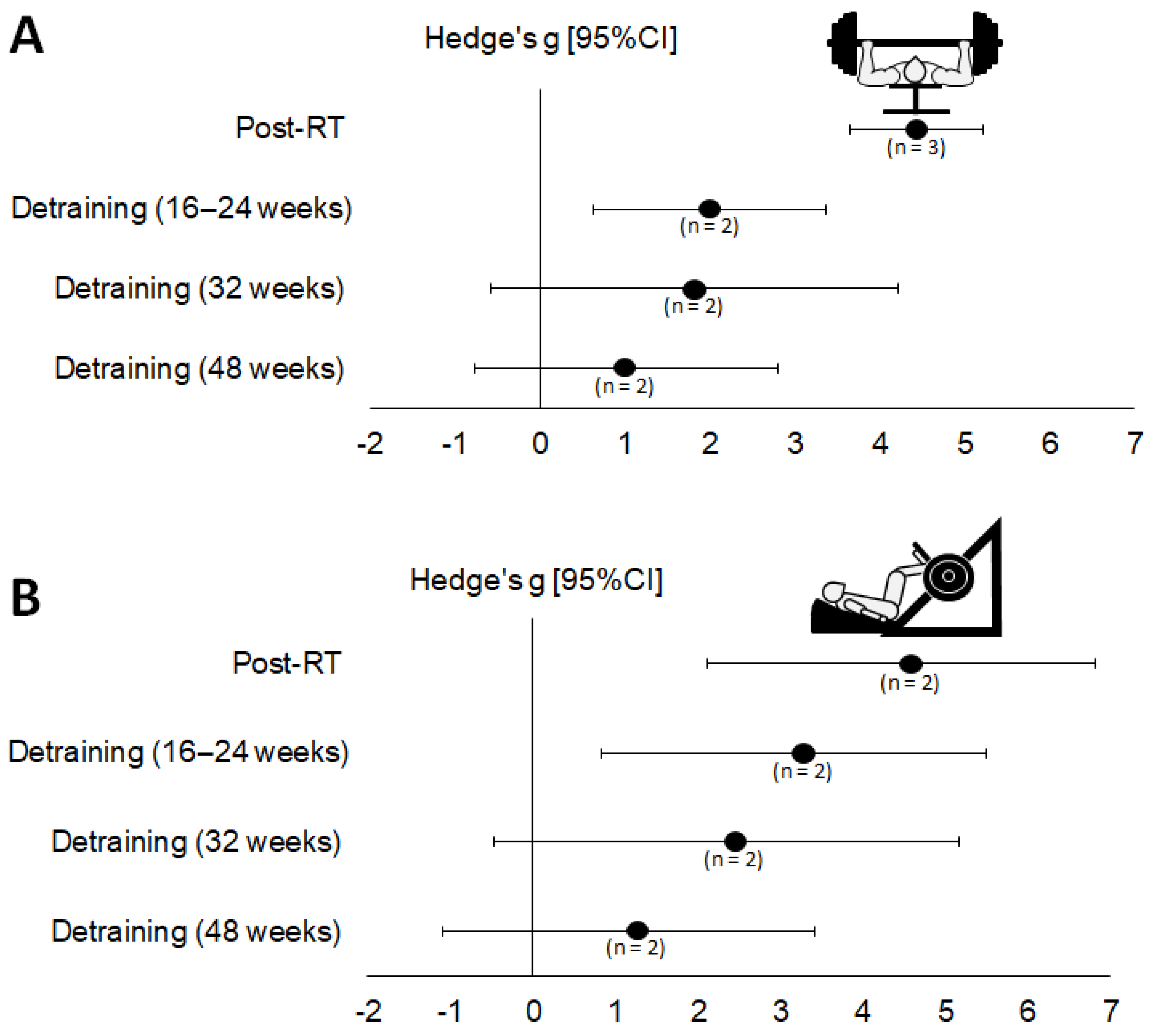
Period Analyzed and 1RM
Strength Test Used
r = 0.5r = 0.7r = 0.9
Hedges’ g (95% CI)pHedges’ g (95% CI)pHedges’ g (95% CI)p
Detraining period (16–24 weeks)
Chest press (n = 3)
Post-RT—Baseline3.57 [2.89; 4.24]<0.0014.43 [3.65; 5.22]<0.0016.68 [5.37; 8.00]<0.001
Detraining—Post-RT−1.67 [−2.16; −1.19]<0.001−2.10 [−2.62; −1.58]<0.001−3.29 [−3.94; −2.64]<0.001
Detraining—Baseline1.60 [0.49; 2.72]0.0051.99 [0.62; 3.36]0.0042.98 [0.98; 4.98]<0.001
Leg press (n = 2)
Post-RT—Baseline3.64 [1.81; 5.47]<0.0014.47 [2.12; 6.82]<0.0016.35 [2.55; 10.15]0.001
Detraining—Post-RT−1.12 [−1.64; −0.61]<0.001−1.43 [−2.05; −0.81]<0.001−2.33 [−3.22; −1.43]<0.001
Detraining—Baseline2.56 [0.61; 4.51]0.0103.16 [0.82; 5.50]0.0084.60 [1.55; 7.66]0.003
Detraining period (32 weeks)
Chest press (n = 2)
Post-RT—Baseline3.67 [2.88; 4.46]<0.0014.53 [3.48; 5.58]<0.0016.57 [4.62; 8.52]<0.001
Detraining—Post-RT−2.29 [−2.90; −1.68]<0.001−2.85 [−3.53; −2.16]<0.001−4.28 [−5.15; −3.41]<0.001
Detraining—Baseline1.43 [−0.39; 3.25]0.1241.81 [−0.59; 4.21]0.1392.94 [−1.40; 7.28]0.184
Leg press (n = 2)
Post-RT—Baseline3.64 [1.81; 5.47]<0.0014.47 [2.12; 6.82]<0.0016.35 [2.55; 10.15]<0.001
Detraining—Post-RT−2.00 [−2.59; −1.41]<0.001−2.52 [−3.15; −1.88]<0.001−3.96 [−4.79; −3.14]<0.001
Detraining—Baseline1.85 [−0.39; 4.09]0.1062.34 [−0.48; 5.16]0.1043.73 [−0.65; 8.12]0.095
Detraining period (48 weeks)
Chest press (n = 2)
Post-RT—Baseline3.67 [2.88; 4.46]<0.0014.53 [3.48; 5.58]<0.0016.57 [4.62; 8.52]<0.001
Detraining—Post-RT−2.79 [−3.46; −2.12]<0.001−3.51 [−4.28; −2.74]<0.001−5.47 [−6.66; −4.29]<0.001
Detraining—Baseline0.80 [−0.61; 2.21]0.2671.01 [−0.76; 2.79]0.2631.57 [−1.22; 4.35]0.270
Leg press (n = 2)
Post-RT—Baseline3.64 [1.81; 5.47]<0.0014.47 [2.12; 6.82]<0.0016.35 [2.55; 10.15]<.0001
Detraining—Post-RT2.34 [−2.97; −1.72]<0.001−3.01 [−3.72; −2.31]<0.001−5.12 [−6.11; −4.12]<0.001
Detraining—Baseline0.96 [−0.88; 2.80]0.3081.16 [−1.09; 3.42]0.3111.64 [−1.52; 4.79]0.309
1RM: one maximum repetition. CI, confidence interval; RT, resistance training.
Publisher’s Note: MDPI stays neutral with regard to jurisdictional claims in published maps and institutional affiliations.

