Article

Molecular Patterns and Antimicrobial Resistance Characterization of Salmonella enterica Non-Typhoidal from Human, Food, and Environment Samples Isolated in Luanda, Angola

by
Moisés Francisco
1,2,3,
Adriana Belas
4,5,6,7,
Sofia Santos Costa
3,
Juliana Menezes
6,7,
Jorge Ramos
3,
Isabel Couto
3,
Miguel Viveiros
3 and
Constança Pomba
6,7,*
1
Faculty of Medicine, University Agostinho Neto, Box 116, Luanda, Angola
2
National Institute of Public Health, Avenida Amilcar Cabral, 96 Maianga, Luanda, Angola
3
Global Health and Tropical Medicine, GHTM, LA-REAL, Instituto de Higiene e Medicina Tropical, IHMT, Universidade NOVA de Lisboa, 1500-210 Lisboa, Portugal
4
Veterinary and Animal Research Centre (CECAV), I-MVET-Faculty of Veterinary Medicine, Lusófona University, University Centre of Lisbon, 1749-024 Lisbon, Portugal
5
Polythecnic Institute of Lusofonia (IPLUSO), School of Health, Protection and Animal Welfare, 1700-098 Lisbon, Portugal
6
CIISA—Centre for Interdisciplinary Research in Animal Health, Faculty of Veterinary Medicine, University of Lisbon, 1300-477 Lisbon, Portugal
7
Associate Laboratory for Animal and Veterinary Sciences (AL4AnimalS), 1300-477 Lisbon, Portugal
*
Author to whom correspondence should be addressed.
Zoonotic Dis. 2024, 4(4), 259-270; https://doi.org/10.3390/zoonoticdis4040022
Submission received: 6 August 2024 / Revised: 12 October 2024 / Accepted: 15 October 2024 / Published: 21 October 2024

Simple Summary

Salmonella spp. are common in Africa and amongst the main causes of morbidity and mortality in humans, with a public health impact. However. there is a lack of scientific record on the situation in Angola, despite the numerous reports of informal and clinical suspicions of these infections as the cause of enteric disease. This work involved the isolation and characterization of the different Salmonella serovars circulating in Luanda, Angola, focusing on their antimicrobial resistance patterns and epidemiological relationship, particularly between clinical, environmental and food isolates and their impact on public health in Angola. This study provides an initial microbiological and molecular characterization of the real epidemiological situation of Salmonella spp. occurrence in Luanda and demonstrates the need for continuous monitoring of this pathogenic agent at the clinical, food and environmental levels to implement epidemiological strategies for the control of salmonellosis in Angola.

Abstract

The aim of this study was to characterize the antimicrobial resistance phenotype and genotype of non-typhoidal Salmonella spp. isolated in Luanda, Angola. Between 2013 and 2015, human clinical samples, food, and environmental samples (n = 290) were collected at different regions of Luanda city and screened for the presence of Salmonella spp. Bacterial isolates were preliminarily identified using the API 20E Kit, and their identification was confirmed using PCR and serotyping. All Salmonella spp. isolates were tested by minimum inhibitory concentration against 19 antimicrobials. The isolates were also screened using PCR for the presence of resistance genes (blaOXA-1, blaSHV, blaTEM, sul1, sul2, sul3, qnrA, qnrB, qnrS, qnrC, qnrD, aac(6′)-Ib, dfrIa [targeting dfrA1, dfrA5, dfrA15, dfrA15b, dfrA16, dfrA16b] and dfrA12, cmlA, and floR) and typed using pulsed-field gel electrophoresis (PFGE) and multilocus sequence typing (MLST). Salmonella enterica non-typhoidal was detected in 21.3% of the clinical samples (n = 32/150), 11.1% of the food samples (n = 10/90), and 26% of the environmental samples (n = 13/50). Serotyping revealed that the monophasic variant of Salmonella Typhimurium (Salmonella enterica serovar 4,[5],12:i:-) was detected in 38.1% of the samples. Moreover, serovar Salmonella Enteritidis was the second most frequent. Only 7.3% of the isolates were resistant to at least one antimicrobial. Furthermore, isolates from different origins (clinical, environmental, and food) were associated with the same lineages, Salmonella Enteritidis ST11 and S. enterica ser. Typhimurium ST313. The detection of S. enterica serovar 4,[5],12:i:- in different settings reinforces the need for a One Health approach to control this zoonosis in Angola.

1. Introduction

Salmonella spp. is a ubiquitous pathogen frequently transmitted through contaminated food, causing a spectrum of illnesses ranging from gastroenteritis to life-threatening bacteremia [1]. With an estimated 94 million cases of gastroenteritis and 155,000 deaths annually worldwide attributed to Salmonella infections, its impact on public health is profound [1,2]. Combined with this challenge is the escalating rate of antimicrobial resistance observed in certain Salmonella serovars, such as the emergence of an epidemic multidrug-resistant clonal lineage of Salmonella enterica serovar 4,[5],12:i:-, a monophasic variant of Salmonella Typhimurium that has been implicated in several outbreaks in Europe [3,4,5].
The presence of S. enterica non-typhoidal isolates in different types of food, such as vegetables, chicken, fish, and seafood, constitutes a public health issue [1,6]. The lack of hygienic practices in the food chain (conservation, transportation, processing, and commercialization) has been associated as a preponderant factor for Salmonella spp. contamination [6,7]. The morbidity associated with highly pathogenic clones, the emergence of non-typhoid invasive strains, and the rising rates of antimicrobial resistance, coupled with globalization, are key aspects requiring a multisectoral approach to combat salmonellosis [8].
The reporting rate for salmonellosis cases varies from country to country, depending on the policies and programs adopted to control the disease. In the African region, surveillance, documentation, and reporting of salmonellosis are often insufficient and fragmented despite the increasing reports of non-typhoidal Salmonella, a particularly invasive, multidrug-resistant strain in Sub-Saharan Africa [7,9,10]. Understanding the molecular epidemiology and antimicrobial resistance profiles of Salmonella isolates is paramount for devising effective control strategies, especially in countries with a high incidence of salmonellosis, such as Angola [9,10]. Hence, this study aimed to characterize retrospectively the molecular epidemiology, as well as the antimicrobial resistance phenotype and genotype of non-typhoidal S. enterica isolated from clinical, food, and environmental samples collected by the National Institute of Public Health of Angola between August 2013 and November 2015.

2. Results

2.1. Sample Processing, Isolation, and Serovar Identification

A total of 290 biological samples from different regions of Luanda, Angola, were processed (Figure 1); 19% (n = 55) of the samples were positive for non-typhoidal S. enterica. A single Salmonella isolate was isolated from each sample. Of these 55 isolates, 58.0% (n = 32) were of clinical origin (all from fecal samples), 22.4% (n = 13) of environmental origin (water sewers) and 18.2% (n = 10) of food origin (fish, chicken, and corn flour). Statistical analysis revealed significant differences between the frequency of non-typhoidal S. enterica isolation in clinical, environmental, and food samples (p < 0.001).
Among the non-typhoidal S. enterica isolates, the serovar Salmonella Enteritidis, the monophasic variant of S. Typhimurium (S. enterica serovar 4,[5],12:i:-), the serovar S. Typhimurium and others that belonged to unknown serovars (non-typeable), were identified (Table 1).

2.2. Antimicrobial Resistance

Among the 55 non-typhoidal S. enterica isolates recovered, only four isolates presented resistance to at least one antimicrobial from the 19 tested. The most common antimicrobial resistance detected was to ampicillin (7.3%, n = 4/55) and piperacillin (7.3%, n = 4/55), followed by the combination ampicillin/sulbactam (5.5%, n = 3/55), chloramphenicol (5.5%, n = 3/55), and trimethoprim/sulfamethoxazole (5.5%, n = 3/55) (Table 2). Moreover, three of these isolates (n = 3/55, 5.5%) presented a multidrug-resistant profile, all of clinical origin (Table 2). MIC values (minimum inhibitory concentration) for each isolate are shown in Table S1.
Regarding the presence of antimicrobial resistance genes, blaTEM-1 gene was the most frequent (7.3%, n = 4/55), followed by sul1 and dfrIa gene (5.5%, n = 3/55, for both genes) (Table 2). Fluoroquinolones and phenicols resistance genes (cmlA and floR genes) were not detected in this study (Table 2).

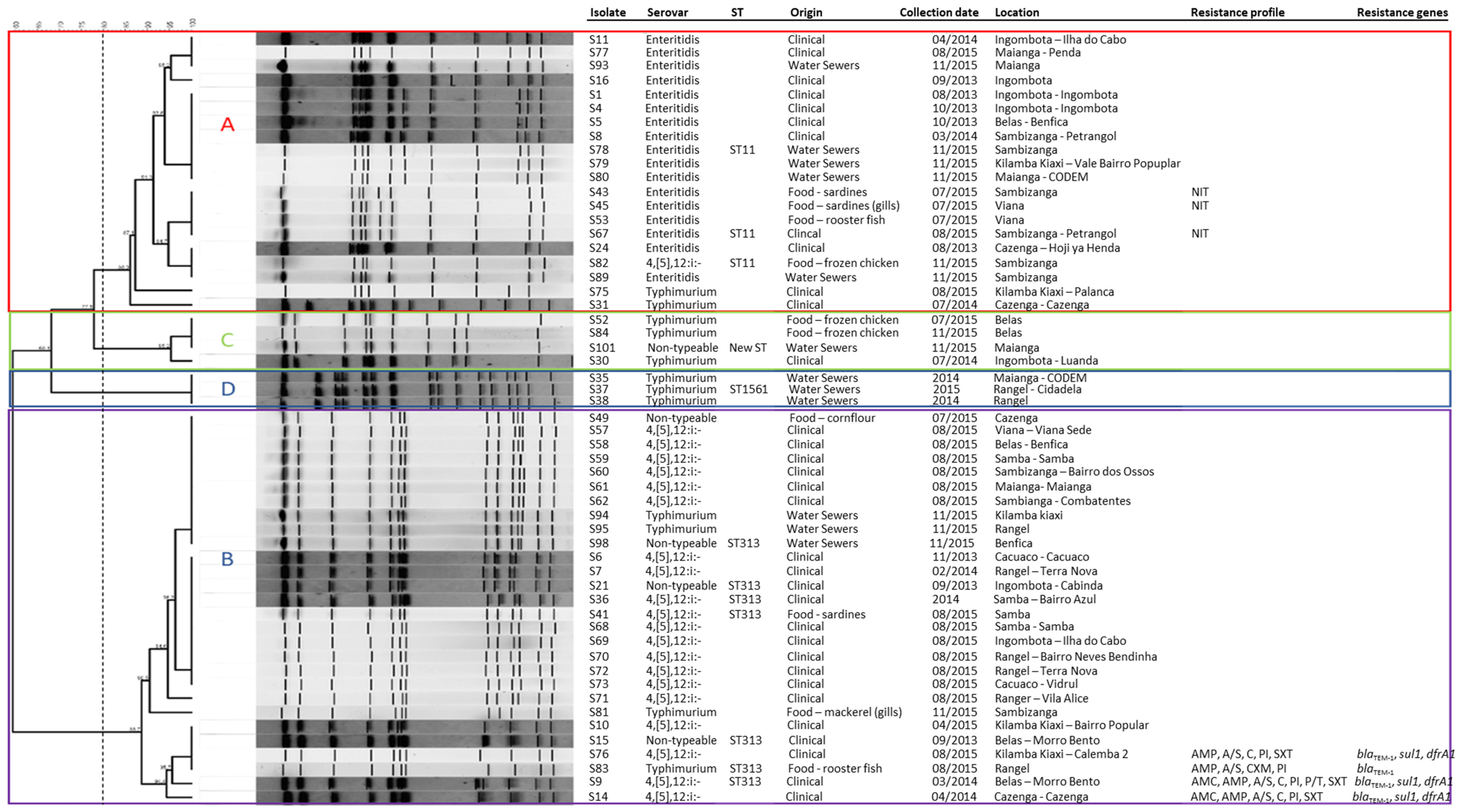
2.3. Clonal Relationship and MLST Analysis

The clonal relationship between the S. enterica non-typhoidal isolates, whether of clinical, food or wastewater origin, was tested using XbaI-PFGE. This molecular typing technique allowed the discrimination of four pulsotypes (A to D) among the 55 isolates. The most frequent pulsotype was the pulsotype B (n = 28); 20 of them belonged to Salmonella enterica serovar 4,[5],12:i-, four to the serovar Typhimurium and the other four to non-typeable serovar (Figure 2).
Multilocus sequence typing (MLST) of seven representative isolates from pulsotype B, including those of unknown serovar (non-typeable), revealed that all belong to ST313. Interestingly, three of the isolates were detected in fish samples. Furthermore, pulsotype A was the second most frequent pulsotype, represented by 20 isolates, mainly belonging to the serovar Enteritidis. MLST typing of three representative isolates of this pulsotype demonstrated that they belong to ST11. Pulsotype C was composed of four isolates, three from serovar Typhimurium and one from a non-typeable serovar. MLST typing revealed that one of the representative isolates of the C pulse type belongs to a new ST. Regarding pulsotype D, one (n = 1/3) of the S. Typhimurium isolates belongs to ST1561. All pulsotypes harbored strains from clinical, food, and environmental origin, except for pulsotype D, which was composed only of strains from environmental origin (Figure 2).
Most of the human salmonellosis cases detected in this study corresponded to a restricted set of serotypes/pulsotype/MSLT strains, with special relevance to the highly invasive ST313 strain.

3. Discussion

There are only a few studies on the incidence of S. enterica non-typhoidal in Angola. Recently, a high occurrence and unusual serotype diversity of non-typhoidal Salmonella in non-clinical niches was described in the country [11]. In our study, we demonstrated for the first time the existence of a clinical human reservoir of S. enterica non-typhoidal alongside its presence in food-borne and environmental settings.
In this study, S. enterica non-typhoidal was detected in 11.1% of the food samples (n = 10/90), in 21.3% of the clinical ones (n = 32/150) and in 26% of the environmental samples (n = 13/50). The majority of these S. enterica non-typhoidal isolates belonged to the Typhimurium monophasic variant S. enterica serovar 4,[5],12:i:- (38%, n = 21/55), a multidrug-resistant serovar widespread across Europe [12,13]. Considering that Angola is not an industrialized country, with low levels of animal production and most of the food being imported from Europe [14], the detection of S. enterica serovar 4,[5],12:i:- strains may be associated with the trade of food products, that can easily spread through the community. The basic sanitation conditions in the city of Luanda, associated with the disorderly growth of the city and, in many cases, the lack of reasonable habitable living conditions, as well as the lack of health education in the communities, are crucial aspects for the dissemination of any infectious disease and in particular salmonellosis.
The prevalence of S. enterica serovar 4,[5],12:i:- has been increasing among salmonellosis since the mid-1990s [15]. Studies carried out in Germany and Luxembourg on this variant coming from pigs and humans showed profiles that were identical or closely related when analyzed by PFGE and Multiple Locus Variable-Number Tandem Repeat Analysis (MLVA) [12,13]. This suggests a link between infections in humans and animal reservoirs [11,12]. Furthermore, infection by S. enterica serovar 4,[5],12:i:- is of great public health concern due to the high rates of resistance associated with this serovar. This epidemic lineage, known as the European clone, is resistant to ampicillin, streptomycin, sulphonamides and tetracycline (R-type ASSuT) [4] and has been implicated in several outbreaks across Europe [13,16]. Additionally, isolates of S. enterica serovar 4,[5],12:i:- isolated in Italy, Luxemburg, and Canada have shown further resistance to ampicillin, chloramphenicol, streptomycin, sulphonamides, and tetracycline (R-type ACSSuT) [13,16,17]. In this study, the presence of serovars Enteritidis, Typhimurium, and its monophasic variant S. enterica 4,[5],12:i:- in clinical, environmental, and food samples is noteworthy. However, most isolates of the various serovars showed susceptibility to all antimicrobials tested by broth microdilution, which is reassuring. Nevertheless, these results show that depending on the lifestyle conditions of specific communities, the S. enterica non-typhoidal transmission may occur in different ways. In the specific case of Angola, as expected, the study results seem to indicate that the transmission of this pathogenic agent to humans occurs through different routes, including contaminated food or environment or due to person-to-person contact.
In the case of Africa, these concerns are even greater due to the increase in cases of non-typhoid invasive salmonellosis and the countless risk factors that contribute to the morbidity of the disease [18]. Numerous outbreaks due to food contamination by S. enterica non-typhoidal have been reported in several African countries [7,18,19]. Isolates of S. enterica non-typhoidal in these countries have been isolated in cattle, goats, pigs and other production animals [18,19,20]. The lack of potable drinking water in many communities, malnutrition, human immunodeficiency virus (HIV), malaria, direct and indirect contact with animals, intra-hospital infections, transmission between humans, and other endemic diseases provide a favorable environment for the dissemination of any transmissible disease [1,17,21]. Additionally, street food contamination plays an important role in the epidemiological gastrointestinal disease chain [22]. These conditions indicate that health authorities and the scientific community should perform constant monitoring of salmonellosis, reporting clinical, epidemiological, and laboratory information.
Analysis using XbaI-PFGE allowed for the discrimination of the S. enterica non-typhoidal isolates into four pulsotypes. The most frequent pulsotype, B (n = 28), encompasses most isolates belonging to S. enterica serovar 4,[5],12:i:-, serovar Typhimurium, and non-typeable serovar isolates, mostly from clinical origin (n = 20). All these isolates belong to the clonal lineage ST313, which has been described in many countries across sub-Saharan Africa, contributing to invasive infections in regions such as Mozambique, Nigeria, the Democratic Republic of Congo, Kenya, and Malawi [9,23,24,25].
The second most frequent pulsotype (pulsetype A, n = 20) belongs to the ST11 clonal lineage, and it was represented by isolates from clinical, food, and environmental origin. This pulsotype had previously been described as the cause of invasive infections in Brazil and in the United Kingdom [13,26]. ST11 clonal lineage, associated with serovar Enteritidis, has been reported in the African continent (such as Mozambique, Tunisia, and Kenya), Europe, Asia, and the American continent [3,24,27,28,29,30,31,32]. The pulsetype D was associated with the ST1561 lineage and was characterized by serovar Typhimurium isolates from the wastewater samples.
The identification of identical pulsotypes in samples from different origins highlights the need for a multi-actor approach for the quick detection and successful control of salmonellosis. Routine comparison of food chain, environmental and human Salmonella spp. isolates using molecular typing tools would be helpful to establish transmission routes.
This study provides, for the first time in the literature, a retrospective preliminary description of S. enterica non-typhoidal isolates circulating in Luanda, Angola, and emphasizes the need for continuous prospective monitoring of this pathogen to provide information that will help to build effective epidemiological strategies for the salmonellosis control in Angola.

4. Materials and Methods

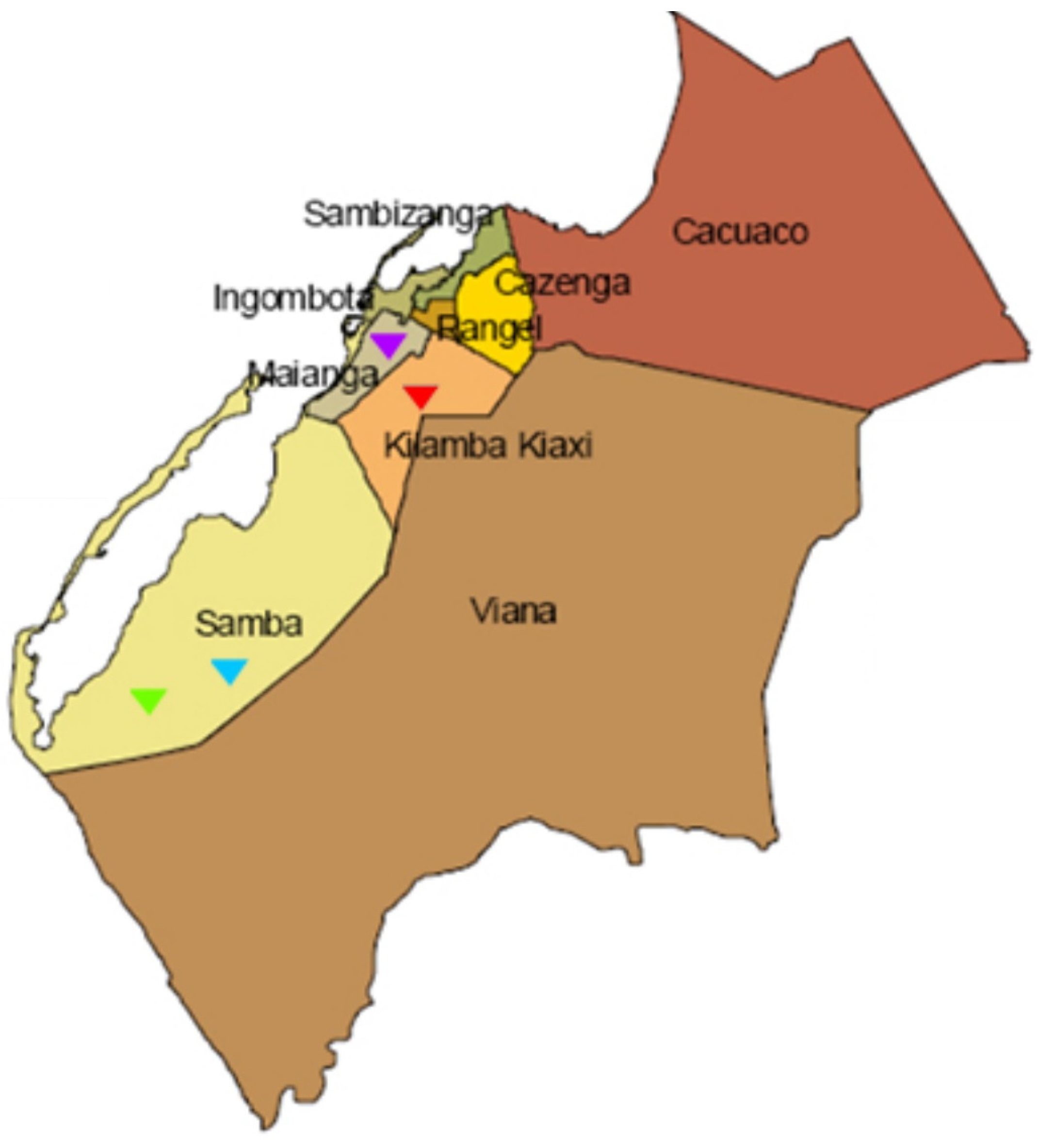
4.1. Study Area and Sample Collection

The Salmonella isolates included in this study were obtained from different regions of Luanda between 2013 and 2015 (Figure 1). Samples were sent to the National Institute of Public Health and to the Microbiology Laboratory of Agostinho Neto University. A total of 290 samples were collected, including the following:
(i) Clinical samples: A total of 150 human clinical samples (blood and feces) were obtained from both adults and children admitted to health unit services with suspected typhoid fever at Clínica Sagrada Esperança and the National Institute of Public Health in Luanda, Angola. Approximately 25 g of feces were collected in sterile plastic containers, and 2–8 mL of blood were collected in bottles containing culture medium for blood cultures.
(ii) Food Samples: A total of 90 food samples (including chicken, fish, corn flour, and vegetables) were collected randomly from various commercial establishments and informal market stalls across different regions of Luanda. These samples were maintained in their commercial packaging and transported immediately to the laboratory under refrigeration in isothermal boxes.
(iii) Environmental Samples: A total of 50 environmental samples (wastewater) were obtained from different points in the city of Luanda. One liter of wastewater was collected at a depth of approximately 30 cm using sterile amber glass bottles, which were then placed in isothermal boxes for transportation.

4.2. Bacteria Isolation, Identification, and DNA Extraction

All clinical, food, and environmental samples were processed following the recommendations of the ISO 6579:2002 Annex D guidelines [33]. Briefly, samples were pre-enriched with buffered peptone water (Biokar Diagnostics, Beauvais, France) in a tenfold dilution and incubated for 18–24 h at 35–37 °C. Then, 0.1 mL of the pre-enriched cultures were transferred to 10 mL of Rappaport-Vassiliadis soya broth (Oxoid, Thermofisher Scientific, Dardilly Cedex, France), and 1 mL of pre-enriched cultures was transferred to 10 mL of Tetrathionate broth (Oxoid, Thermofisher Scientific, France) and incubated for 24 h at 41.5 °C and 37 °C, respectively, for selective enrichment. After 24 h of incubation, 10 μL of culture from each enriched broth was seeded onto Xylose Lysine Deoxycholate agar (Biokar Diagnostics, Beauvais, France) and Hektoen Enteric agar (Biokar Diagnostics, Beauvais, France) and incubated for 24 h at 37 °C. The plates were examined for the presence of colonies typical of Salmonella, and for each sample, one presumptive Salmonella colony was selected to be further studied.
The identification of the genus Salmonella was performed with the API 20E kit, software APIWEBTM (https://apiweb.biomerieux.com/login, accessed on 19 September 2024, BioMérieux, Marcy-l’Étoile, France), and confirmed using PCR amplification of the invA gene [34].
All Salmonella spp. isolates (n = 55) were serotyped based on the Kauffmann–White–Le Minor scheme. Briefly, the isolates were tested serologically using polyvalent O antiserum, monovalent O:9 antiserum, and type-specific H antiserum (Bio-Rad Laboratories, Inc. Richamond, CA, USA) [35,36]. Initially, the isolates were screened with polyvalent O antiserum. For those yielding a positive reaction, further testing was conducted with specific O sera (OMA, OMB) to narrow down the spectrum of somatic antigens using monovalent sera.
The genus and the absence of the second-phase flagellar antigen fljB were confirmed by PCR, as recommended by the European Food Safety Authority (EFSA) Panel on Biological Hazards using DreamTaq DNA Polymerase (Thermo Fisher Scientific Inc, Waltham, MA, USA) [37,38]. Total DNA extraction was performed using a rapid boiling procedure [39].

4.3. Antimicrobial Susceptibility Testing

Antimicrobial susceptibility phenotypes were evaluated by determination of minimum inhibitory concentrations (MICs) using the microdilution system MicroScan®—Neg MIC panel Type 44 (Siemens Healthcare Diagnostics, West Sacramento, CA, USA). Susceptibility testing was performed for the following antimicrobials, according to the manufacturer’s instructions: amikacin, amoxicillin/clavulanic acid, ampicillin, ampicillin/sulbactam, aztreonam, cefotaxime, cephalothin, ceftazidime, cefuroxime, cefepime, cefoxitin, chloramphenicol, ciprofloxacin, gentamicin, nalidixic acid, nitrofurantoin, piperacillin, piperacillin/tazobactam, tetracycline, and trimethoprim/sulfamethoxazole. Details of the different antimicrobial dilutions used are provided in Supplementary Table S2. Antimicrobial susceptibility phenotypes were interpreted according to the European Committee on Antimicrobial Susceptibility Testing (EUCAST) clinical breakpoints 2024 [40] and, except for amoxicillin/clavulanic acid, ampicillin/sulbactam and nitrofurantoin, for which criteria from the Clinical and Laboratory Standards Institute (CLSI) were used [41]. In addition, the Escherichia coli ATCC 25922 strain was used as a quality control organism.
Isolates showing resistance to three or more antibiotic classes were classified as multidrug-resistant bacteria [42].

4.4. Detection of Antimicrobial Resistance Genes

All 55 Salmonella spp. isolates were screened by PCR and sequencing for the presence of beta-lactamase resistance genes (blaTEM, blaSHV, and bla OXA-1) using specific primers and conditions described previously [43,44,45]. Antimicrobial resistant/intermediate isolates were further screened using PCR for the presence of genes encoding for resistance against trimethoprim/sulphamethoxazole (sul1, sul2, sul3, dfrIa [targeting dfrA1, dfrA5, dfrA15, dfrA15b, dfrA16, dfrA16b], and dfrA12) [46], chloramphenicol (cmlA and floR) [45], and the plasmid-mediated quinolone resistance (PMQR) genes (qnrA, qnrB, qnrS, qnrC, qnrD, aac(6′)-Ib) [47]. Negative and previously sequenced positive controls were included in all PCR reactions using DreamTaq DNA Polymerase (Thermo Fisher Scientific Inc, Waltham, MA, USA) in all reactions.

4.5. Typing and Subtyping Using Pulsed-Field Gel Electrophoresis (PFGE) and Multilocus Sequence Type (MLST)

In all Salmonella spp. isolates clonality was assessed using PFGE with XbaI restriction (New England Biolabs, Ipswich, MA, USA) according to the Pulsenet protocol and using Salmonella serotype Braenderup H9812 as the reference strain [48]. The patterns were analyzed using BioNumerics v 6.6 software (Applied Maths, Kortrijk, Belgium) and the unweighted pair group method with arithmetic mean and the Dice similarity coefficient. The definition of a pulsed-field type cluster was based on a similarity cut-off value of ≥80% [49].
Twelve representative strains from different pulsotype groups were further characterized by the PCR/sequencing of seven housekeeping genes aroC (chorismate synthase), dnaN (DNA polymerase III beta subunit), hemD (uroporphyrinogen III cosynthase) hisD (histidinol dehydrogenase), purE (phosphoribosylaminoimidazole carboxylase), sucA (alpha-ketoglutarate dehydrogenase), and thrA (aspartokinase+homogenize dehydrogenase) using NZYTaq II DNA polymerase (NZYtech, Lisbon, Portugal). Internal fragments of the seven housekeeping genes were amplified using specific primers and conditions described previously [50,51]. PCR products were sequenced, and the sequences obtained were submitted to the MLST database to retrieve an allelic profile and sequence type for each isolate.

4.6. Statistical Analysis

Statistical analysis was performed using the SAS statistical software package for Windows, version 9.4 (SAS Institute, Cary, NC, USA). Fisher’s Exact test was used for comparisons between groups; the results were considered statistically significant when p < 0.05.

5. Conclusions

This study evidenced a high occurrence of S. enterica non-typhoidal isolates in patients, food, and environment in Luanda, Angola. Our data showed the presence of the epidemic monophasic Salmonella European clone, as well as other diverse Salmonella enterica serotypes. The findings of the current research showed the need for continuous surveillance on salmonellosis in Angola, within well-established epidemiological surveillance programs.

Supplementary Materials

The following supporting information can be downloaded at https://www.mdpi.com/article/10.3390/zoonoticdis4040022/s1, Table S1: Antimicrobial resistance patterns of Salmonella enterica non-typhoidal isolates, Luanda Angola, 2013–2015 (n = 55 isolates); Table S2: Dilution ranges for minimum inhibitory concentration (MIC) testing of antimicrobial agents.

Author Contributions

Conceptualization, C.P., I.C. and M.V.; methodology, A.B. and S.S.C.; formal analysis, M.F., A.B., J.R. and S.S.C.; investigation, M.F., A.B. and S.S.C.; resources, M.F., M.V. and C.P.; data curation, A.B. and S.S.C.; writing—original draft preparation, M.F.; writing—review and editing, A.B., J.M. and C.P.; funding acquisition, C.P. All authors have read and agreed to the published version of the manuscript.

Funding

This research was supported by Fundação para a Ciência e a Tecnologia (FCT, Portugal) through funds to GHTM—UIDP/04413/2020, LA-REAL—LA/P/0117/2020, CIISA and AL4AnimalS (UIDB/00276/2020 and LA/P/0059/2020, respectively). J.M. was supported by a PhD fellowship (2020.07562.BD). S.S.C. was supported by FCT through CEECINST/00042/2021/CP1773/CT0009 (https://doi.org/10.54499/CEECINST/00042/2021/CP1773/CT0009).

Institutional Review Board Statement

Not applicable.

Informed Consent Statement

Not applicable.

Data Availability Statement

The original contributions presented in the study are included in the article, further inquiries can be directed to the corresponding author.

Acknowledgments

We would like to acknowledge the National Institute of Public Health, Angola, the Faculty of Medicine, University Agostinho Neto, Angola, and the Clínica Sagrada Esperança, Luanda, Angola. We thank Vicent Perreten for providing the positive controls for the sul3 PCR screening.

Conflicts of Interest

The authors declare no conflicts of interest.

References

  1. Ananchaipattana, C.; Hosotani, Y.; Kawasaki, S.; Pongsawat, S.; Mdlatiful, B.; Isobe, S.; Inatsu, Y. Prevalence of Foodborne Pathogens in Retailed Foods in Thailand. Foodborne Pathog. Dis. 2012, 9, 835–840. [Google Scholar] [CrossRef] [PubMed]
  2. Majowicz, S.E.; Musto, J.; Scallan, E.; Angulo, F.J.; Kirk, M.; O’Brien, S.J.; Jones, T.F.; Fazil, A.; Hoekstra, R.M. The Global Burden of Nontyphoidal Salmonella Gastroenteritis. Clin. Infect. Dis. 2010, 50, 882–889. [Google Scholar] [CrossRef] [PubMed]
  3. Bado, I.; García-Fulgueiras, V.; Cordeiro, N.F.; Betancor, L.; Caiata, L.; Seija, V.; Robino, L.; Algorta, G.; Chabalgoity, J.A.; Ayala, J.A.; et al. First Human Isolate of Salmonella enterica Serotype Enteritidis Harboring Bla CTX-M-14in South America. Antimicrob. Agents Chemother. 2012, 56, 2132–2134. [Google Scholar] [CrossRef] [PubMed]
  4. Hopkins, K.L.; Kirchner, M.; Guerra, B.; Granier, S.A.; Lucarelli, C.; Porrero, M.C.; Jakubczak, A.; Threlfall, E.J.; Mevius, D.J. Multiresistant Salmonella enterica Serovar 4,[5],12:I:- In Europe: A New Pandemic Strain? Eurosurveillance 2010, 15, 19580. [Google Scholar] [CrossRef]
  5. Gallati, C.; Stephan, R.; Hächler, H.; Malorny, B.; Schroeter, A.; Nüesch-Inderbinen, M. Characterization of Salmonella enterica Subsp. Enterica Serovar 4,[5],12:I:- Clones Isolated from Human and Other Sources in Switzerland between 2007 and 2011. Foodborne Pathog. Dis. 2013, 10, 549–554. [Google Scholar] [CrossRef]
  6. Elhadi, N. Prevalence and Antimicrobial Resistance of Salmonella spp. In Raw Retail Frozen Imported Freshwater Fish to Eastern Province of Saudi Arabia. Asian Pac. J. Trop. Biomed. 2014, 4, 234–238. [Google Scholar] [CrossRef]
  7. Tack, B.; Vanaenrode, J.; Verbakel, J.Y.; Toelen, J.; Jacobs, J. Invasive Non-Typhoidal Salmonella Infections in Sub-Saharan Africa: A Systematic Review on Antimicrobial Resistance and Treatment. BMC Med. 2020, 18, 212. [Google Scholar] [CrossRef] [PubMed]
  8. Hodges, J.R.; Kimball, A.M. The Global Diet: Trade and Novel Infections. Glob. Health 2005, 1, 1–7. [Google Scholar] [CrossRef]
  9. Akullian, A.; Montgomery, J.M.; John-Stewart, G.; Miller, S.I.; Hayden, H.S.; Radey, M.C.; Hager, K.R.; Verani, J.R.; Ochieng, J.B.; Juma, J.; et al. Multi-Drug Resistant Non-Typhoidal Salmonella Associated with Invasive Disease in Western Kenya. PLoS Neglected Trop. Dis. 2018, 12, e0006156. [Google Scholar] [CrossRef]
  10. Feasey, N.A.; Dougan, G.; Kingsley, R.A.; Heyderman, R.S.; Gordon, M.A. Invasive Non-Typhoidal Salmonella Disease: An Emerging and Neglected Tropical Disease in Africa. Lancet 2012, 379, 2489–2499. [Google Scholar] [CrossRef]
  11. Antunes, P.; Campos, J.; Mourao, J.; Ribeiro, T.G.; Novais, C.; Peixe, L. High Occurrence and Unusual Serotype Diversity of Non-Typhoidal Salmonella in Non-Clinical Niches, Angola. Epidemiol. Infect. 2017, 145, 883–886. [Google Scholar] [CrossRef] [PubMed]
  12. Hauser, E.; Tietze, E.; Helmuth, R.; Junker, E.; Blank, K.; Prager, R.; Rabsch, W.; Appel, B.; Fruth, A.; Malorny, B. Pork Contaminated with Salmonella Enterica Serovar 4,[5],12:I:-, An Emerging Health Risk for Humans. Appl. Environ. Microbiol. 2010, 76, 4601–4610. [Google Scholar] [CrossRef] [PubMed]
  13. Mossong, J.; Marques, P.; Ragimbeau, C.; Huberty-Krau, P.; Losch, S.; Meyer, G.; Moris, G.; Strottner, C.; Rabsch, W.; Schneider, F. Outbreaks of Monophasic Salmonella Enterica Serovar 4,[5],12:I:- in Luxembourg, 2006. Eurosurveillance 2007, 12, 11–12. [Google Scholar] [CrossRef] [PubMed]
  14. Ricardo Dias Poultry and Products Annual. Available online: https://apps.fas.usda.gov/newgainapi/api/Report/DownloadReportByFileName?fileName=Poultry%20and%20Products%20Annual_Luanda_Angola_09-01-2021 (accessed on 19 September 2024).
  15. Moreno Switt, A.I.; Soyer, Y.; Warnick, L.D.; Wiedmann, M. Emergence, Distribution, and Molecular and Phenotypic Characteristics of Salmonella enterica Serotype 4,5,12:I:-. Foodborne Pathog. Dis. 2009, 6, 407–415. [Google Scholar] [CrossRef]
  16. Barco, L.; Ramon, E.; Cortini, E.; Longo, A.; Dalla Pozza, M.C.; Lettini, A.A.; Dionisi, A.M.; Olsen, J.E.; Ricci, A. Molecular Characterization of Salmonella enterica Serovar 4,[5],12:I:- DT193 ASSuT Strains from Two Outbreaks in Italy. Foodborne Pathog. Dis. 2014, 11, 138–144. [Google Scholar] [CrossRef]
  17. Mulvey, M.R.; Finley, R.; Allen, V.; Ang, L.; Bekal, S.; el Bailey, S.; Haldane, D.; Hoang, L.; Horsman, G.; Louie, M.; et al. Emergence of Multidrug-Resistant Salmonella enterica Serotype 4,[5],12:I:- Involving Human Cases in Canada: Results from the Canadian Integrated Program on Antimicrobial Resistance Surveillance (CIPARS), 2003–10. J. Antimicrob. Chemother. 2013, 68, 1982–1986. [Google Scholar] [CrossRef]
  18. Uche, I.V.; MacLennan, C.A.; Saul, A. A Systematic Review of the Incidence, Risk Factors and Case Fatality Rates of Invasive Nontyphoidal Salmonella (INTS) Disease in Africa (1966 to 2014). PLoS Neglected Trop. Dis. 2017, 11, e0005118. [Google Scholar] [CrossRef]
  19. Morpeth, S.C.; Ramadhani, H.O.; Crump, J.A. Invasive Non-Typhi Salmonella Disease in Africa. Clin. Infect. Dis. 2009, 49, 606–611. [Google Scholar] [CrossRef] [PubMed]
  20. Gelaw, A.K.; Nthaba, P.; Matle, I. Detection of Salmonella from Animal Sources in South Africa between 2007 and 2014. J. S. Afr. Vet. Assoc. 2018, 89, 1–10. [Google Scholar] [CrossRef]
  21. World Health Organization. Foodborne Disease Burden Epidemiology Reference Group 2007–2015. In WHO Estimates of the Global Burden of Foodborne Diseases; World Health Organization: Geneva, Switzerland, 2015. [Google Scholar]
  22. World Health Organization & Food and Agriculture Organization of the United Nations. The International Food Safety Authorities Network (INFOSAN) Progress Report 2004–2010; World Health Organization: Geneva, Switzerland, 2011.
  23. Mandomando, I.; Bassat, Q.; Sigaúque, B.; Massora, S.; Quintó, L.; Ácacio, S.; Nhampossa, T.; Vubil, D.; Garrine, M.; Macete, E.; et al. Invasive Salmonella Infections Among Children From Rural Mozambique, 2001–2014. Clin. Infect. Dis. 2015, 61, S339–S345. [Google Scholar] [CrossRef]
  24. Leekitcharoenphon, P.; Friis, C.; Zankari, E.; Svendsen, C.A.; Price, L.B.; Rahmani, M.; Herrero-Fresno, A.; Fashae, K.; Vandenberg, O.; Aarestrup, F.M.; et al. Genomics of an Emerging Clone of Salmonella Serovar Typhimurium ST313 from Nigeria and the Democratic Republic of Congo. J. Infect. Dev. Ctries. 2013, 7, 696–706. [Google Scholar] [CrossRef] [PubMed]
  25. Kingsley, R.A.; Msefula, C.L.; Thomson, N.R.; Kariuki, S.; Holt, K.E.; Gordon, M.A.; Harris, D.; Clarke, L.; Whitehead, S.; Sangal, V.; et al. Epidemic Multiple Drug Resistant Salmonella typhimurium Causing Invasive Disease in Sub-Saharan Africa Have a Distinct Genotype. Genome Res. 2009, 19, 2279–2287. [Google Scholar] [CrossRef] [PubMed]
  26. Ashton, P.M.; Owen, S.V.; Kaindama, L.; Rowe, W.P.M.; Lane, C.R.; Larkin, L.; Nair, S.; Jenkins, C.; de Pinna, E.M.; Feasey, N.A.; et al. Public Health Surveillance in the UK Revolutionises Our Understanding of the Invasive Salmonella typhimurium Epidemic in Africa. Genome Med. 2017, 9, 92. [Google Scholar] [CrossRef] [PubMed]
  27. Zhao, X.; Gao, Y.; Ye, C.; Yang, L.; Wang, T.; Chang, W. Prevalence and Characteristics of Salmonella Isolated from Free-Range Chickens in Shandong Province, China. Biomed. Res. Int. 2016, 2016, 8183931. [Google Scholar] [CrossRef]
  28. Ktari, S.; Ksibi, B.; Gharsallah, H.; Mnif, B.; Maalej, S.; Rhimi, F.; Hammami, A. Molecular Epidemiological Characteristics of Salmonella enterica Serovars Enteritidis, Typhimurium and Livingstone Strains Isolated in a Tunisian University Hospital. APMIS 2016, 124, 194–200. [Google Scholar] [CrossRef]
  29. Antunes, P.; Mourão, J.; Machado, J.; Peixe, L. First Description of QnrS1-IncN Plasmid in a ST11 Salmonella enteritidis Clinical Isolate from Portugal. Diagn. Microbiol. Infect. Dis. 2011, 69, 463–465. [Google Scholar] [CrossRef]
  30. Papadopoulos, T.; Petridou, E.; Zdragas, A.; Mandilara, G.; Nair, S.; Peters, T.; Chattaway, M.; de Pinna, E.; Passiotou, M.; Vatopoulos, A. Comparative Study of All Salmonella enterica Serovar Enteritidis Strains Isolated from Food and Food Animals in Greece from 2008 to 2010 with Clinical Isolates. Eur. J. Clin. Microbiol. Infect. Dis. 2016, 35, 741–746. [Google Scholar] [CrossRef]
  31. Kim, Y.; Bae, I.K.; Jeong, S.H.; Lee, C.H.; Lee, H.K.; Ahn, J.; Lee, M.K.; Lee, S.; Lee, K. Occurrence of IncFII Plasmids Carrying the BlaCTX-M-15 Gene in Salmonella enterica Serovar Enteritidis Sequence Type 11 in Korea. Diagn. Microbiol. Infect. Dis. 2011, 71, 171–173. [Google Scholar] [CrossRef]
  32. Ghaderi, R.; Tadayon, K.; Khaki, P.; Mosavari, N. Iranian Clonal Population of Salmonella enterica Serovar Enteritidis, Characterized by Multi-Locus Sequence Typing (MLST) Method. Iran. J. Microbiol. 2015, 7, 251–259. [Google Scholar]
  33. ISO 6579. Microbiology of food and animal feeding stuffs—Horizontal method for the detection of Salmonella spp. 2002. Available online: https://www.iso.org/obp/ui/#iso:std:iso:6579:ed-4:v1:en (accessed on 19 September 2024).
  34. Malorny, B.; Hoorfar, J.; Bunge, C.; Helmuth, R. Multicenter Validation of the Analytical Accuracy of Salmonella PCR: Towards an International Standard. Appl. Environ. Microbiol. 2003, 69, 290–296. [Google Scholar] [CrossRef]
  35. Grimont, P.A.D.; Weill, F.X. Antigenic Formulae of the Salmonella Serovars. In WHO Collaborating Center for Reference and Research on Salmonella, 9th ed.; Institute Pasteur: Paris, France, 2007. [Google Scholar]
  36. Koneman, E.W.; Allen, S.D.; Janda, W.M.; Schreeckenberger, P.C. Color Atlas and Textbook of Diagnostic Microbiology, 5th ed.; Lippincott Williams & Wilkins: Philadelphia, PA, USA, 1997. [Google Scholar]
  37. Tennant, S.M.; Diallo, S.; Levy, H.; Livio, S.; Sow, S.O.; Tapia, M.; Fields, P.I.; Mikoleit, M.; Tamboura, B.; Kotloff, K.L.; et al. Identification by PCR of Non-Typhoidal Salmonella enterica Serovars Associated with Invasive Infections among Febrile Patients in Mali. PLoS Neglected Trop. Dis. 2010, 4, e621. [Google Scholar] [CrossRef] [PubMed]
  38. EFSA Panel on Biological Hazards (BIOHAZ). Scientific Opinion on Monitoring and Assessment of the Public Health Risk of “Salmonella typhimurium-like” Strains. EFSA J. 2010, 8, 1826. [Google Scholar] [CrossRef]
  39. Féria, C.; Machado, J.; Correia, J.D.; Gonçalves, J.; Gaastra, W. Virulence Genes and P Fimbriae PapA Subunit Diversity in Canine and Feline Uropathogenic Escherichia coli. Vet. Microbiol. 2001, 82, 81–89. [Google Scholar] [CrossRef]
  40. The European Committee on Antimicrobial Susceptibility Testing Breakpoint Tables for Interpretation of MICs and Zone Diameters Version 14.0. Available online: http://www.eucast.org/fileadmin/src/media/PDFs/EUCAST_files/Breakpoint_tables/v_5.0_Breakpoint_Table_01.pdf (accessed on 8 April 2023).
  41. CLSI. Performance Standards for Antimicrobial Susceptibility Testing, 30th ed.; Clinical and Laboratory Standards Institute: Wayne, PA, USA, 2020; ISBN 9781684400324. [Google Scholar]
  42. Magiorakos, A.P.; Srinivasan, A.; Carey, R.B.; Carmeli, Y.; Falagas, M.E.; Giske, C.G.; Harbarth, S.; Hindler, J.F.; Kahlmeter, G.; Olsson-Liljequist, B.; et al. Multidrug-Resistant, Extensively Drug-Resistant and Pandrug-Resistant Bacteria: An International Expert Proposal for Interim Standard Definitions for Acquired Resistance. Clin. Microbiol. Infect. 2012, 18, 268–281. [Google Scholar] [CrossRef]
  43. Pomba, C.; Mendonça, N.; Costa, M.; Louro, D.; Baptista, B.; Ferreira, M.; Correia, J.D.; Caniça, M. Improved Multiplex PCR Method for the Rapid Detection of β-Lactamase Genes in Escherichia coli of Animal Origin. Diagn. Microbiol. Infect. Dis. 2006, 56, 103–106. [Google Scholar] [CrossRef]
  44. Woodford, N.; Fagan, E.J.; Ellington, M.J. Multiplex PCR for Rapid Detection of Genes Encoding CTX-M Extended-Spectrum β-Lactamases. J. Antimicrob. Chemother. 2006, 57, 154–155. [Google Scholar] [CrossRef] [PubMed]
  45. Pérez-Pérez, F.J.; Hanson, N.D. Detection of Plasmid-Mediated AmpC β-Lactamase Genes in Clinical Isolates by Using Multiplex PCR. J. Clin. Microbiol. 2002, 40, 2153–2162. [Google Scholar] [CrossRef]
  46. Sáenz, Y.; Briñas, L.; Domínguez, E.; Ruiz, J.; Zarazaga, M.; Vila, J.; Torres, C. Mechanisms of Resistance in Multiple-Antibiotic-Resistant Escherichia coli Strains of Human, Animal, and Food Origins. Antimicrob. Agents Chemother. 2004, 48, 3959–3967. [Google Scholar] [CrossRef]
  47. Cattoir, V.; Poirel, L.; Rotimi, V.; Soussy, C.J.; Nordmann, P. Multiplex PCR for Detection of Plasmid-Mediated Quinolone Resistance Qnr Genes in ESBL-Producing Enterobacterial Isolates. J. Antimicrob. Chemother. 2007, 60, 394–397. [Google Scholar] [CrossRef]
  48. Centers for Disease Control and Prevention (CDC). Standard Operating Procedure for PulseNet PFGE of Escherichia coli O157:H7, Escherichia coli Non-O157 (STEC), Salmonella serotypes, Shigella sonnei and Shigella flexneri. Available online: https://www.pulsenetinternational.org/assets/PulseNet/uploads/pfge/PNL05_Ec-Sal-ShigPFGEprotocol.pdf (accessed on 23 February 2023).
  49. Carriço, J.A.; Pinto, F.R.; Simas, C.; Nunes, S.; Sousa, N.G.; Frazão, N.; de Lencastre, H.; Almeida, J.S. Assessment of Band-Based Similarity Coefficients for Automatic Type and Subtype Classification of Microbial Isolates Analyzed by Pulsed-Field Gel Electrophoresis. J. Clin. Microbiol. 2005, 43, 5483–5490. [Google Scholar] [CrossRef]
  50. Alikhan, N.-F.; Zhou, Z.; Sergeant, M.J.; Achtman, M. A Genomic Overview of the Population Structure of Salmonella. PLoS Genet. 2018, 14, e1007261. [Google Scholar] [CrossRef] [PubMed]
  51. Achtman, M.; Zhou, Z.; Alikhan, N.-F.; Tyne, W.; Parkhill, J.; Cormican, M.; Chiou, C.-S.; Torpdahl, M.; Litrup, E.; Prendergast, D.M.; et al. Genomic Diversity of Salmonella enterica—The UoWUCC 10K Genomes Project. Wellcome Open Res. 2021, 5, 223. [Google Scholar] [CrossRef] [PubMed]
Figure 1. Map of Luanda showing sample collection points. Water sewage sampling points are represented in the pictures on the map.
Zoonoticdis 04 00022 g001
Figure 2. Clonal relationship between the Salmonella enterica non-typhoidal isolates from clinical, food, and environmental samples, Luanda, Angola (n = 55). The dendrogram was inferred within the Bionumerics program according to the unweighted pair group method based on Dice similarity coefficients. A–D represents different pulsed-field type clusters based on a similarity cut-off value of ≥80%. Legend: ST, Sequence typing. AMC, amoxicillin/clavulanic acid; AMP, ampicillin; A/S, ampicillin/sulbactam; CXM, cefuroxime; C, chloramphenicol, ciprofloxacin; NIT, nitrofurantoin; PI, piperacillin; P/T, piperacillin/tazobactam; SXT, trimethoprim/sulfamethoxazole.
Zoonoticdis 04 00022 g002
Table 1. Serovar distribution of Salmonella enterica non-typhoidal isolates according to the sample origin, Luanda, Angola, 2013–2015 (n = 55 isolates).
Salmonella SerovarIsolates
n (%)
Origin
Clinical
n
Food
n
Environment
n
Enteritidis17 (30.9)935
4,[5],12:i:-21(38.1)1821
Typhimurium12 (21.8)345
Non-typeable5 (9.0)212
Total55 (100)321013
Table 2. Antimicrobial resistance patterns of Salmonella enterica non-typhoidal isolates and the presence of resistance genes, Luanda Angola, 2013–2015 (n = 4 isolates).
Sample IDSample OriginSalmonella SerovarAntimicrobial Resistance Phenotypes 1 and MIC Values (mg/L)Resistance Genes Pattern
S9Human fecesEnteritidisAMP (>16), A/S (>16/8), PI (>64), C (>16), SXT (>4/76)blaTEM-1-sul1-dfrIa
S14Human feces4,[5],12:i:-AMP (>16), A/S (>16/8), PI (>64), C (>16), SXT (>4/76)blaTEM-1-sul1-dfrIa
S76Human feces4,[5],12:i:-AMP (>16), PI (>64), C (>16), SXT (>4/76)blaTEM-1-sul1-dfrIa
S83FishTyphimuriumAMP (>16), A/S (>16/8), PI (>64)blaTEM-1
1 AMP: ampicillin, A/S: ampicillin/sulbactam; C: chloramphenicol; PI: piperacillin; SXT: sulfamethoxazole/trimethoprim. MIC: minimum inhibitory concentration.
Disclaimer/Publisher’s Note: The statements, opinions and data contained in all publications are solely those of the individual author(s) and contributor(s) and not of MDPI and/or the editor(s). MDPI and/or the editor(s) disclaim responsibility for any injury to people or property resulting from any ideas, methods, instructions or products referred to in the content.

