Article

Genomic Analysis of Novel Bacterial Species Corynebacterium ramonii ST344 Clone Strains Isolated from Human Skin Ulcer and Rescued Cats in Japan

1
Toxin and Biologicals Research Laboratory, Kumamoto Health Science University, 325 Izumi-machi, Kita-ku, Kumamoto 861-5598, Japan
2
The Chemo-Sero-Therapeutic Research Institute, Kumamoto 860-0806, Japan
3
Kumamoto Rosai Hospital Clinical Laboratory Center, Yatsushiro 866-0826, Japan
4
Kumamoto University Hospital Clinical Laboratory Center, Kumamoto 860-8556, Japan
5
Kumamoto Prefectural Institute of Public Health and Environmental Science, Uto 869-0425, Japan
6
Pathogen Genomics Center, National Institute of Infectious Diseases, Tokyo 162-8640, Japan
7
Department of Medical Technology, Faculty of Health Sciences, Kumamoto Health Science University, 325 Izumi-machi, Kita-ku, Kumamoto 861-5598, Japan
*
Authors to whom correspondence should be addressed.
Zoonotic Dis. 2024, 4(4), 234-244; https://doi.org/10.3390/zoonoticdis4040020
Submission received: 16 July 2024 / Revised: 24 September 2024 / Accepted: 9 October 2024 / Published: 11 October 2024

Abstract

:

Simple Summary

Corynebacterium ramonii, which is linked to zoonotic diseases, was isolated from a human ulcer (ST344) and oral cavity of a cat (ST337/ST344) in Japan. The close genetic ties between human and cat strains suggest endemicity. The number of ST344 cases may increase, warranting ongoing genomic surveillance for infection control.

Abstract

Some Corynebacterium strains produce toxins that are similar to those produced by Corynebacterium diphtheriae, leading to human infections that are often transmitted through zoonotic diseases. A novel species, which is formerly classified as Corynebacterium ulcerans lineage II, was recently re-evaluated and renamed “Corynebacterium ramonii sp. nov.”. We isolated C. ramonii from a human skin ulcer in Japan in 2023 (KCU0303-001) and identified it as ST344 using a genomic analysis. In addition, C. ramonii KPHES-18084 (ST344) and six strains of C. ulcerans (ST337/ST1011) were isolated from the oral cavities of 7/208 rescued cats (3.4%). The human ulcer strain KCU0303-001 and the rescued cat strain KPHES-18084 were found to be ST344 and closely related clones by core-genome and pan-genome analyses, suggesting that ST344 may be endemic to both clinical and companion animals in Japan. In support of this finding, another clinical isolate of ST344 (TSU-28 strain) was reported in Japan in 2019. Although ST337 is the most common C. ulcerans infection, the second most recent clinical isolate of C. ramonii, ST344, might be increasing; therefore, further genomic surveillance is required to monitor C. ramonii and C. ulcerans infections.

1. Introduction

Diphtheria toxin (DT) is mainly produced by Corynebacterium diphtheriae, which causes pseudomembrane formation in the pharynx in the respiratory tract, and characteristic symptoms such as hoarseness are observed. Some Corynebacterium strains produce DT, which leads to human infections that are often transmitted through zoonotic diseases. Thus far, a DT-positive Corynebacterium diphtheriae complex, including C. ulcerans and C. pseudotuberculosis, has been reported. Among C. ulcerans strains, a novel species, which is formerly classified as C. ulcerans lineage 2, was recently re-evaluated and renamed “Corynebacterium ramonii sp. nov.”.
Some strains of C. ramonii and C. ulcerans show DT-positive PCR, causing diphtheria-like symptoms due to respiratory tract infections and skin ulcers [1]. It causes zoonotic diseases of animal origins. It was first isolated from the pharynx of a patient in 1927 [2], and a wide range of infected hosts has been reported worldwide, including companion animals (cats and dogs) [3,4,5,6,7,8,9], domestic animals (cattle and horses) [10,11,12,13], non-domestic animals (hedgehogs, penguins, ibis, and ferrets) [14,15,16,17,18], and other animals (wild boar and deer) [19,20,21], and isolates from the same zoo (killer whales and lions) [22].
In Japan, 34 cases were reported from 2001 to 2020 [23], mostly from dogs and cats. Thus far, two deaths have been reported in the past 20 years. The virulence factor DT is encoded by the tox gene located on the lysogenized prophage in C. diphtheriae [24,25]. C. ulcerans has similar characteristics, but possesses a gene encoding diphtheria toxin on a different prophage than the diphtheria prophage [26]. Previously reported DTs have also revealed that C. ulcerans harbors two toxins isolated from patients with diphtheria [27].
However, last year, the Pasteur Institute proposed a novel toxigenic member of the C. diphtheriae species complex, Corynebacterium ramonii sp. nov., as a new diphtheria-causing organism [28]. Previously, one case of C. ramonii (ST344) was reported in Japan [27]. We isolated eight strains of C. ramonii and C. ulcerans from different periods and origins, including the novel strains ST344 and ST1011, which are members of the same group of characterized strains reported in Japan.
In this study, complete genome sequences of these strains (KCU0303-001, KPHES-18088, and KPHES-18084) were determined and characterized for the feature of clinical isolate KCU0303-001 using a molecular phylogenetic analysis.

2. Materials and Methods

2.1. Isolation and Identification of C. ramonii from Samples

Swabs from the patient’s foot skin ulcer area were inoculated on a sheep blood agar medium at 35 °C and 5% carbon dioxide for 12 h. For single-colony isolation, the observed colony was subsequently cultivated on a sheep blood agar medium at 37 °C for 24 h, and the developing colonies were subjected to Gram staining. The DT gene was confirmed using Tox 1 (5′-ATCCACTTTTAGTGCGAGAACCTTCGTCA-3′) and Tox 2 (5′-GAAACTTCTTCGTACCACGGGGACTAA-3′) primers from the method of H. Nakao et al. [29]. The PCR conditions were 95 °C for 2 min, 95 °C for 20 s, and 55 °C for 30 s, followed by 35 cycles of amplification at 95 °C for 2 min, 95 °C for 20 s, 55 °C for 30 s, and 72 °C for 1 min, and extension at 72 °C for 10 min. Positive PCR amplicons for the DT gene were confirmed using agarose gel electrophoresis. The bacterial species were determined using a VITEK MS: MYLA 4.7.1 (bioMerieux, Marcy-l’Etoile, Lyon, France) mass spectrometer.
Oral swabs were collected from 208 rescued cats and incubated for 24 h at 37 °C in Arakawa’s modified medium (500 mL of heart extract, 10 g peptone, 2 g dextrose, 5 g NacL, 0.5 g activated charcoal powder, and 15 g agar in 1 L purified water, adjust pH to 7.6, and sterilize at 121 °C for 15 min; perform separate supplementation of 40 mL pH-adjusted and sterilized 1% potassium tellurite solution). Black colonies were subjected to biochemical characterization using API Coryne V4.0 (bioMerieux, Tokyo, Japan) and glycolytic tests (glucose, maltose, sucrose, and glycogen). The DT gene PCR was performed as described above, and direct sequencing of the rpoB gene was performed using the method described by Khamis et al. [30].
Toxin production was tested in accordance with the diphtheria prevention manual of the National Institute of Infectious Diseases using the agar gel sedimentation reaction test (Elek’s method) [31] and Vero cells to detect toxin activity [32].

2.2. Whole-Genome C. ramonii and C. ulcerans Isolate Sequencing and Analysis

Whole-genome sequencing was performed as previously described [33]. Briefly, the isolates were incubated in 10 mL of a Brain–Heart Infusion medium at 37 °C for 24 h, and centrifuged at 8000 rpm for 20 min, and the recovered cells were inactivated with phenol-chloroform. The bacterial cell suspension was beaded using 0.1/0.5 mm beads in ZR BashingBead Lysis Tubes (Zymo Research, Irvine, CA 92602 USA), and DNA was purified using a MinElute PCR Purification Kit (QIAGEN, Hilden, Germany). Next-Generation Sequencing DNA-seq libraries were prepared using QIAseq FX ((QIAGEN, Hilden, Germany) and sequenced using NextSeq 1000/2000 P1 reagents (150 mer paired ends, 300 cycles) (Illumina, San Diego, CA 92110, USA) and NextSeq 2000 sequencers (Illumina, San Diego, CA 92110, USA). The diphtheria group was identified by multilocus sequence typing (MLST) [34] using draft genome information from de novo assembly using SPAdes version 3.15.2 [35] and genome annotation using the DFAST website [36]. Furthermore, a core-genome phylogenetic tree and pairwise single-nucleotide variations (SNVs) were determined using Parsnp v1.7.4. [37]. A maximum-likelihood phylogenetic analysis was performed using iQtree version 2.2.5 with [38] 1000-fold bootstrapping with the following parameters (iqtree2-s parsnp.snps.mblocks–alrt 1000-B 1000-nt 6).

2.3. Complete Genome Sequence Determination

To complete a circularized genome sequence from the draft genome sequence, long-read sequencing of KCU0303-001, KPHES-18084, and KPHES-18088 was performed on a Nanopore ONT GridION (Oxford, UK) using R10.4.0 flow cells, followed by hybrid de novo assembly using Unicycler v0.4.8 [39]. Genome annotation was performed using DFAST [36] and a circular genome map was generated using Prokse [40]. Structural genome comparisons were performed using the DiGAlign version 2.0 web tool [41]. A pan-genome analysis was performed using the Roary v3.13.0 program with [42] default settings, and the phylogenetic tree and pan-genome were visualized by the Phandango website (https://jameshadfield.github.io/phandango/, accessed on: 13 September 2024).

3. Results

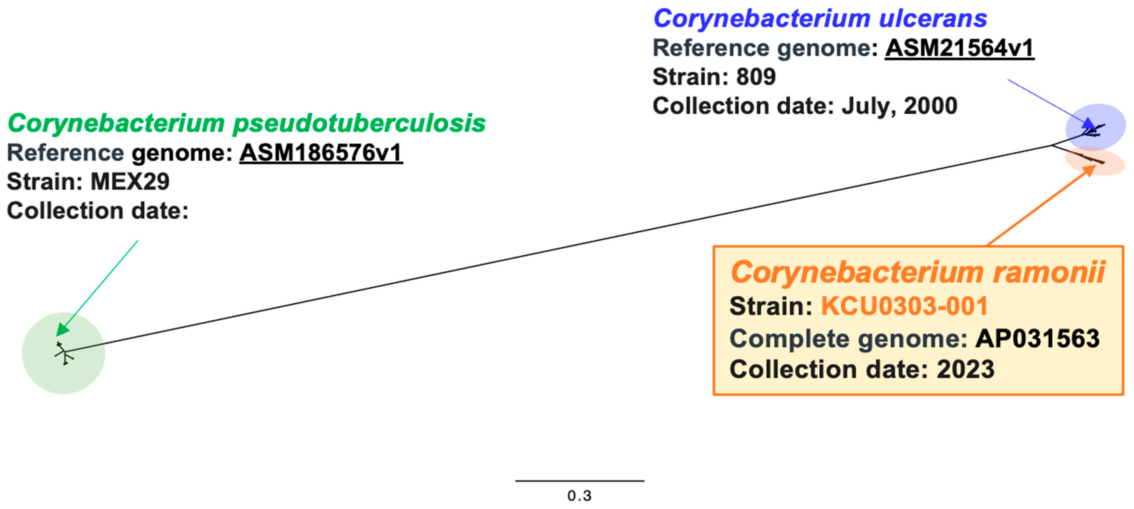
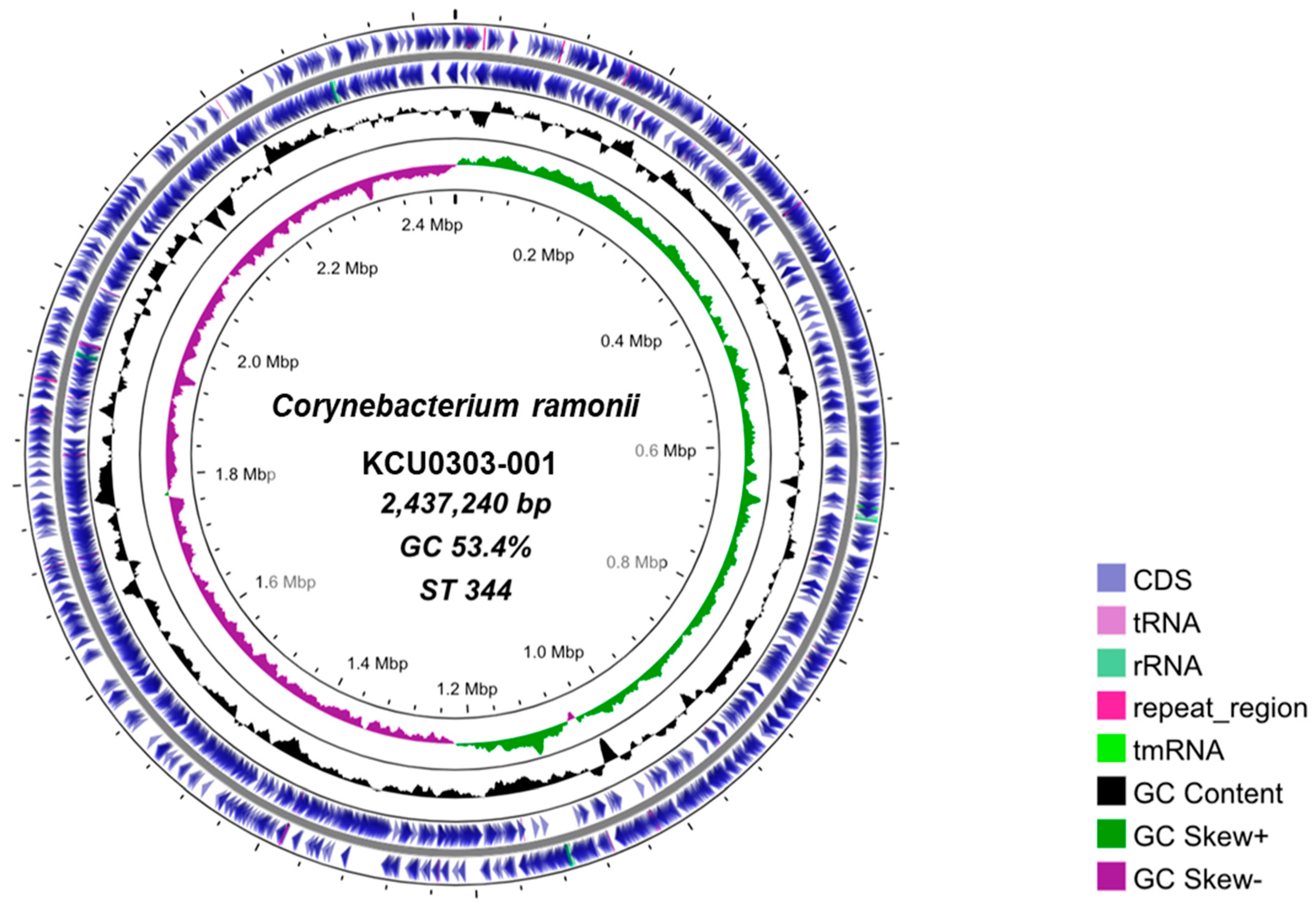
3.1. C. ramonii Clinical Isolate KCU0303-001

The potential bacterial pathogen KCU0303-001 was isolated from a patient’s skin ulcer. Short Gram-positive rods were observed, and DT-PCR was positive. However, MALDI-TOF-MS (VITEK-MS) could not correctly identify the bacterial species, showing either 50% C. ulcerans or 50% C. pseudotuberculosis.
To determine the correct genetic features, whole-genome sequencing of KCU0303-001 was performed, suggesting that the most similar bacterial species among the type strains were the C. ulcerans NCTC7910-type strain, with a 95.9% average nucleotide identity using the DFAST annotation program. The Pasteur MLST analysis showed ST344 C. ramonii of the C. diphtheriae complex.
The comparative core-genome analysis of C. ulcerans/C. ramonii strains with C. pseudotuberculosis revealed that they are widely separated species of the genus Corynebacterium (Figure 1), although MALDI-TOF MAS showed ambiguous reports for species identification, as described above.

3.2. C. ramonii and C. ulcerans Rescued Cat Isolates

Seven strains were isolated from oral swabs of 208 rescued cats using Arakawa’s modified medium. DT PCR testing was positive and API Coryne testing showed that the seven isolates were C. ulcerans, followed by species confirmation with rpoB sequence typing. Regarding the toxin-producing properties of seven isolates, the Elek test was negative (−), but Vero cell activity was positive (+) (Table 1).
In addition to the clinical isolate KCU0303-001, the whole-genome sequence of the rescued cat isolates were obtained. The MLST analysis revealed that KPHES-18084 and KPHES-19104 were ST344 and ST337, respectively. The other five isolates were likely novel STs because of mutations in the fusA gene among the seven gene alleles. We registered the novel isolates on cgMLST at the Pasteur Institute and obtained novel ST1011 for five isolates (Table 1).

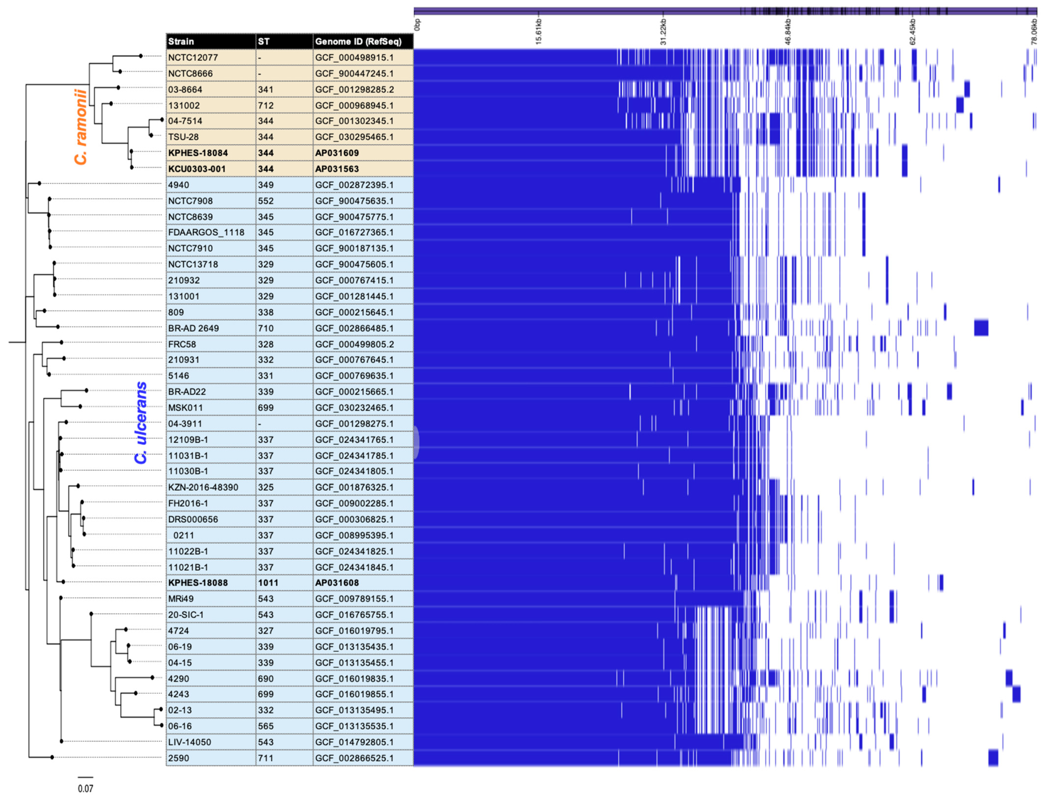
3.3. Comparative Genomic Analysis of Clinical and Rescued Cat Strains

A comparative core-genome analysis was performed to determine the phylogeny of the eight isolates in this study compared with the publicly available 74 strains, including the reference strain 809 [43]. The phylogenetic tree showed two main clades, one of which included five strains of ST337 and ST1011. Five strains, ST337 and ST1011, were likely clonal (Figure 2) instead of fusA gene mutations. ST337 has been previously isolated from cats and humans in Japan. The second clade C. ramonii, which included ST344 (KCU0303-001 and KPHES-18084), was phylogenetically distant from the main clade.
There were 81 pairwise SNVs between KCU0303-001 and KPHES-18084 (total coverage among all sequences: 90.9% genomic region). These ST344 isolates were very similar to the 04-7514 strain from lip ulcers in canines in 2004 in France [3] and the clinical isolate TSU-28 carrying two tox genes in Japan [27].
In addition to the core-genome analysis, the pan-genome analysis also supported the notable difference between C. ramonii and C. ulcerans (Figure 3); sixty-eight genes were extracted for potential C. ramonii-shared genes in this study.

3.4. Structural Comparison of Complete Genome Sequences for C. ramonii ST344

The complete genome sequences of ST344 (KCU0303-001 and KPHES-18084) and ST1011 (KPHES-18088) were determined (Table 2).
A circular genome map of KCU0303-001 was generated (Figure 4), with a genome size of 2,437,240 bp and 53.4% GC.
In general, the C. diphtheriae complex, including C. ulcerans, carries a single DT gene (tox) in its genome. However, the two-tox-gene-positive C. ramonii strain, TSU-28, was isolated in 2019 in Japan, and the complete genome sequence of TSU-28 is available; therefore, pairwise structural genome comparisons were performed, including the clinical isolate KCU0303-001 and veterinary isolate KPHES-18084 (Figure 5).
KCU0303-001 and KPHES-18084 were identical clones and carried one tox gene, whereas TSU-28 had two tox genes in an additional tox phage.

4. Discussion

Here, we obtained a novel clinical isolate, C. ramonii KCU0303-001, and potential zoonotic sources of C. ramonii KPHES-18084 and C. ulcerans isolates from rescued cats. KCU0303-001 exhibited DT gene positivity, but MALDI-TOF-MS suggested partial identification with low accuracy for C. ulcerans/pseudotuberculosis; therefore, further genetic testing, including whole-genome sequencing, is required to achieve its correct identification. Core-genome and pan-genome analyses identified KCU0303-001 as C. ramonii and ST344 (Figure 2 and Figure 3), which has been recently proposed to distinguish the second lineage of C. ulcerans as a novel species, “Corynebacterium ramonii sp.”, from a team at the Pasteur Institute. The team remarked that ST344 is of this type and has the potential for human-to-human transmission; therefore, it should be monitored as closely as C. ulcerans in the future [28].
DT-positive C. ulcerans infections are considered zoonotic diseases; therefore, we confirmed the isolation status of C. ulcerans from domestic companion animals around the South Kyusyu area, and found seven isolates of C. ulcerans/ramonii in this study (Table 1). Phylogenomic analyses demonstrated that C. ramonii KPHES-18084 from rescued cats was a potentially originated clone with KCU0303-001, whereas there is no evidence of contact between the patient and C. ramonii-positive companion animals. Furthermore, there were 81 pairwise SNVs between KCU0303-001 and KPHES-18084 (total coverage among all sequences: 90.9% genomic region) (Figure 2). A genomic epidemiological study of the C. diphtheriae outbreak in Yemen estimated its evolutionary rate to be ~4 SNVs/genome/year [44]. If applicable to this C. ramonii case, the evolutionary gap between these two isolates could be approximately 20 years. In addition, pairwise alignment of the complete genome sequences indicated that these two genomes have a highly conserved structure (Figure 5); therefore, the KCU0303-001-relative C. ramonii clone may be disseminated in veterinary and companion animals in Japan. Indeed, TSU-28 [27] is also closely related to the ST344 strain, but additional horizontal gene acquisition was found in this structural comparison (Figure 5).
Our study suggests that clonal ST344 strains were detected at least three times in Japan between 2018 and 2023, and may have spread in Japan over a five-year period. In addition, 04-7514, which has been reported overseas, is a similar clone isolated from canines in 2004 [3]. As C. ramonii may be widely disseminated in animals as a source of infection in humans, extreme caution should be exercised when in contact with suspected canines and felines. ST344, ST337, and ST1011 were also detected in rescued cats (Table 1). Cats are suspected hosts for ST337 and ST344 infections, which are likely to be endemic nationwide [7,45]. The positive detection rate (7/208 cats; 3.4%) in this study showed a trend similar to previous epidemiological surveys of canine and feline carriers (5–8%) in Japan [46,47]; therefore, monitoring networks for the clinical and veterinary fields will be indispensable to avoid increasing detection.
In Europe, the management of C. ulcerans infections is the same as that of C. diphtheriae, including the prevention, diagnosis, and treatment of patients presenting with diphtheria-like symptoms [48]. In Japan, only C. diphtheriae is defined as a “Class II Infectious Disease” under the Japanese Infectious Disease Control Law, whereas DT-positive C. ulcerans (and C. ramonii) has not been documented in the above law in Japan. Thus far, 34 clinical cases of C. ulcerans infection were reported in Japan from 2001 to 2020, with a marked increase in incidence during the most recent decade and an overall mortality rate of 5.9% [23]. Mortality due to C. diphtheriae has been reported to be 5–10% [49], and the severity of domestic cases of C. ulcerans, with a mortality rate of approximately 6%, is similar to that reported above. Therefore, conducting epidemiological studies on the prevalence and severity of C. ulcerans (and C. ramonii) infections as a basis for administrative decisions is important. Additionally, establishing appropriate legal support for diphtheria toxoid immunization in adults at high risk of infection is crucial for controlling infection.

5. Conclusions

We isolated C. ramonii from a limited area, suggesting that C. ramonii may be endemic in both clinical and companion animals, and the possibility of human-to-human transmission as well as zoonosis cannot be ruled out. Although we were unable to determine the background of C. ramonii infection in the patient, we suggest the careful observation of diphtheria-like symptoms caused by C. ramonii and C. ulcerans, including the investigation of companion animals and environmental routes of infection.

Author Contributions

Conceptualization, M.T. and M.K.; methodology, C.S., K.H., T.Y. and M.K.; validation, C.S., M.M. (Mikoto Moriguchi), H.H., K.M., M.M. (Misato Mori). and M.K.; formal analysis, M.K.; investigation, C.S., M.M. (Mikoto Moriguchi), H.H., K.M. and M.M. (Misato Mori); resources, M.M. (Mikoto Moriguchi), H.H., K.M., M.M. (Misato Mori), E.T., S.Y. and S.G.; data curation, C.S. and M.K.; writing—original draft preparation, C.S., M.K. and M.T.; writing—review and editing, M.K. and M.T.; visualization, C.S. and M.K.; supervision, M.K. and M.T.; project administration, M.T.; funding acquisition, M.K. and M.T. All authors have read and agreed to the published version of the manuscript.

Funding

This research was funded by the Research Program on Emerging and Reemerging Infectious Diseases of the Japan Agency for Medical Research and Development, grant number JP22fk0108636j0001.

Institutional Review Board Statement

The study was conducted in accordance with the Declaration of Helsinki and approved by the Institutional Review Board (or Ethics Committee) of the Kumamoto Health Science University (approval number: 210230) and by the Ethics Review of Kumamotorosai Hospital (approval number: 24-1).

Informed Consent Statement

Written informed consent was not obtained from individual patients. The Ethics Committee at KHSU and Kumamotorosai Hospital waived the requirement for written consent for research on bacterial genome sequences.

Data Availability Statement

The reported nucleotide sequence data are available in the DDBJ Sequenced Read Archive under the accession numbers PRJDB18028 and DRR551240-DRR551250. Complete and draft genome sequences are available in the DDBJ, as shown in Table 1 and Table 2.

Acknowledgments

We gratefully acknowledge the staff of the Laboratory of Bacterial Genomics, Pathogen Genomics Center, National Institute of Infectious Diseases, Tokyo, Japan. We thank the Pasteur Institute teams for the curation and maintenance of the BIGSdb-Pasteur databases at http://bigsdb.pasteur.fr/.

Conflicts of Interest

Motohide Takahashi has received an advisory contract from The Chemo-Sero-Therapeutic Research Institute. The other authors declare no conflicts of interest.

References

  1. Pappenheimer, A.M., Jr.; Gill, D.M. Diphtheria. Science 1973, 182, 353–358. [Google Scholar] [CrossRef] [PubMed]
  2. Gilbert, R.; Stewart, F.C. Corynebacterium ulcerans: A pathogenic microorganism resembling Corynebacterium diphtheriae. J. Lab. Clin. Med. 1927, 12, 756–761. [Google Scholar]
  3. Lartigue, M.F.; Monnet, X.; Le Fleche, A.; Grimont, P.A.; Benet, J.J.; Durrbach, A.; Fabre, M.; Nordmann, P. Corynebacterium ulcerans in an immunocompromised patient with diphtheria and her dog. J. Clin. Microbiol. 2005, 43, 999–1001. [Google Scholar] [CrossRef]
  4. Katsukawa, C.; Kawahara, R.; Inoue, K.; Ishii, A.; Yamagishi, H.; Kida, K.; Nishino, S.; Nagahama, S.; Komiya, T.; Iwaki, M.; et al. Toxigenic Corynebacterium ulcerans isolated from the domestic dog for the first time in Japan. Jpn. J. Infect. Dis. 2009, 62, 171–172. [Google Scholar] [CrossRef]
  5. Sekizuka, T.; Katsukawa, C.; Kuroda, M.; Shibayama, K.; Otsuji, K.; Saito, M.; Yamamoto, A.; Iwaki, M. Limitations of Ribotyping as Genotyping Method for Corynebacterium ulcerans. Emerg. Infect. Dis. 2020, 26, 2457–2459. [Google Scholar] [CrossRef]
  6. Abbott, Y.; Efstratiou, A.; Brennan, G.; Hallanan, S.; Leggett, B.; Leonard, F.C.; Markey, B.K.; Tuite, C.; Fry, N.K. Toxigenic Corynebacterium ulcerans associated with upper respiratory infections in cats and dogs. J. Small Anim. Pract. 2020, 61, 554–560. [Google Scholar] [CrossRef]
  7. Wake, K.; Kikuchi, J.; Uchida, M.; Nemoto, M.; Kaji, Y.; Yokoyama, T.; Suzuki, H.; Fukushima, A.; Yamamoto, A.; Iwaki, M.; et al. Transmission of toxigenic Corynebacterium ulcerans infection with airway obstruction from cats to a human. Acute Med. Surg. 2021, 8, e705. [Google Scholar] [CrossRef]
  8. Hoefer, A.; Herrera-Leon, S.; Dominguez, L.; Gavin, M.O.; Romero, B.; Piedra, X.B.A.; Calzada, C.S.; Uria Gonzalez, M.J.; Herrera-Leon, L.; Case Study Investigation Group. Zoonotic Transmission of Diphtheria from Domestic Animal Reservoir, Spain. Emerg. Infect. Dis. 2022, 28, 1257–1260. [Google Scholar] [CrossRef]
  9. Museux, K.; Arcari, G.; Rodrigo, G.; Hennart, M.; Badell, E.; Toubiana, J.; Brisse, S. Corynebacteria of the diphtheriae Species Complex in Companion Animals: Clinical and Microbiological Characterization of 64 Cases from France. Microbiol. Spectr. 2023, 11, e0000623. [Google Scholar] [CrossRef]
  10. Bostock, A.D.; Gilbert, F.R.; Lewis, D.; Smith, D.C. Corynebacterium ulcerans infection associated with untreated milk. J. Infect. 1984, 9, 286–288. [Google Scholar] [CrossRef]
  11. Higgs, T.M.; Smith, A.; Cleverly, L.M.; Neave, F.K. Corynebacterium ulcerans infections in a dairy herd. Vet. Rec. 1967, 81, 34–35. [Google Scholar] [CrossRef] [PubMed]
  12. Hommez, J.; Devriese, L.A.; Vaneechoutte, M.; Riegel, P.; Butaye, P.; Haesebrouck, F. Identification of nonlipophilic corynebacteria isolated from dairy cows with mastitis. J. Clin. Microbiol. 1999, 37, 954–957. [Google Scholar] [CrossRef] [PubMed]
  13. Zendri, F.; Isgren, C.M.; Sinovich, M.; Richards-Rios, P.; Hopkins, K.L.; Russell, K.; Groves, N.; Litt, D.; Fry, N.K.; Timofte, D. Case Report: Toxigenic Corynebacterium ulcerans Diphtheria-Like Infection in a Horse in the United Kingdom. Front. Vet. Sci. 2021, 8, 650238. [Google Scholar] [CrossRef] [PubMed]
  14. Berger, A.; Dangel, A.; Peters, M.; Muhldorfer, K.; Braune, S.; Eisenberg, T.; Szentiks, C.A.; Rau, J.; Konrad, R.; Hormansdorfer, S.; et al. Tox-positive Corynebacterium ulcerans in hedgehogs, Germany. Emerg. Microbes Infect. 2019, 8, 211–217. [Google Scholar] [CrossRef]
  15. Martel, A.; Boyen, F.; Rau, J.; Eisenberg, T.; Sing, A.; Berger, A.; Chiers, K.; Van Praet, S.; Verbanck, S.; Vervaeke, M.; et al. Widespread Disease in Hedgehogs (Erinaceus europaeus) Caused by Toxigenic Corynebacterium ulcerans. Emerg. Infect. Dis. 2021, 27, 2686–2690. [Google Scholar] [CrossRef]
  16. Saunderson, S.C.; Nouioui, I.; Midwinter, A.C.; Wilkinson, D.A.; Young, M.J.; McInnes, K.M.; Watts, J.; Sangal, V. Phylogenomic Characterization of a Novel Corynebacterium Species Associated with Fatal Diphtheritic Stomatitis in Endangered Yellow-Eyed Penguins. mSystems 2021, 6, e0032021. [Google Scholar] [CrossRef]
  17. Busse, H.J.; Kleinhagauer, T.; Glaeser, S.P.; Spergser, J.; Kampfer, P.; Ruckert, C. Classification of three corynebacterial strains isolated from the Northern Bald Ibis (Geronticus eremita): Proposal of Corynebacterium choanae sp. nov., Corynebacterium pseudopelargi sp. nov., and Corynebacterium gerontici sp. nov. Int. J. Syst. Evol. Microbiol. 2019, 69, 2928–2935. [Google Scholar] [CrossRef]
  18. Marini, R.P.; Cassiday, P.K.; Venezia, J.; Shen, Z.; Buckley, E.M.; Peters, Y.; Taylor, N.; Dewhirst, F.E.; Tondella, M.L.; Fox, J.G. Corynebacterium ulcerans in ferrets. Emerg. Infect. Dis. 2014, 20, 159–161. [Google Scholar] [CrossRef]
  19. Contzen, M.; Sting, R.; Blazey, B.; Rau, J. Corynebacterium ulcerans from diseased wild boars. Zoonoses Public Health 2011, 58, 479–488. [Google Scholar] [CrossRef]
  20. Eisenberg, T.; Kutzer, P.; Peters, M.; Sing, A.; Contzen, M.; Rau, J. Nontoxigenic tox-bearing Corynebacterium ulcerans infection among game animals, Germany. Emerg. Infect. Dis. 2014, 20, 448–452. [Google Scholar] [CrossRef]
  21. Rau, J.; Blazey, B.; Contzen, M.; Sting, R. [Corynebacterium ulcerans infection in roe deer (Capreolus capreolus)]. Berl. Munch. Tierarztl. Wochenschr. 2012, 125, 159–162. [Google Scholar] [PubMed]
  22. Seto, Y.; Komiya, T.; Iwaki, M.; Kohda, T.; Mukamoto, M.; Takahashi, M.; Kozaki, S. Properties of corynephage attachment site and molecular epidemiology of Corynebacterium ulcerans isolated from humans and animals in Japan. Jpn. J. Infect. Dis. 2008, 61, 116–122. [Google Scholar] [CrossRef]
  23. Yamamoto, A.; Hifumi, T.; Ato, M.; Iwaki, M.; Senoh, M.; Hatanaka, A.; Nureki, S.; Noguchi, Y.; Hirose, T.; Yoshimura, Y.; et al. Clinical Characteristics of Corynebacterium ulcerans Infection, Japan. Emerg. Infect. Dis. 2023, 29, 1505–1515. [Google Scholar] [CrossRef] [PubMed]
  24. Meinel, D.M.; Margos, G.; Konrad, R.; Krebs, S.; Blum, H.; Sing, A. Next generation sequencing analysis of nine Corynebacterium ulcerans isolates reveals zoonotic transmission and a novel putative diphtheria toxin-encoding pathogenicity island. Genome Med. 2014, 6, 113. [Google Scholar] [CrossRef] [PubMed]
  25. Sing, A.; Hogardt, M.; Bierschenk, S.; Heesemann, J. Detection of differences in the nucleotide and amino acid sequences of diphtheria toxin from Corynebacterium diphtheriae and Corynebacterium ulcerans causing extrapharyngeal infections. J. Clin. Microbiol. 2003, 41, 4848–4851. [Google Scholar] [CrossRef]
  26. Sekizuka, T.; Yamamoto, A.; Komiya, T.; Kenri, T.; Takeuchi, F.; Shibayama, K.; Takahashi, M.; Kuroda, M.; Iwaki, M. Corynebacterium ulcerans 0102 carries the gene encoding diphtheria toxin on a prophage different from the C. diphtheriae NCTC 13129 prophage. BMC Microbiol. 2012, 12, 72. [Google Scholar] [CrossRef]
  27. Kawase, J.; Sekizuka, T.; Sakai, T.; Fujisawa, N.; Iwaki, M.; Kimura, M.; Kuroda, M. Complete Genome Sequence of Corynebacterium ulcerans Strain TSU-28, Harboring Two Diphtheria Toxin Genes, Isolated from a Patient with Diphtheria-Like Symptoms. Microbiol. Resour. Announc. 2023, 12, e0007223. [Google Scholar] [CrossRef]
  28. Crestani, C.; Arcari, G.; Landier, A.; Passet, V.; Garnier, D.; Bremont, S.; Armatys, N.; Carmi-Leroy, A.; Toubiana, J.; Badell, E.; et al. Corynebacterium ramonii sp. nov., a novel toxigenic member of the Corynebacterium diphtheriae species complex. Res. Microbiol. 2023, 174, 104113. [Google Scholar] [CrossRef]
  29. Nakao, H.; Popovic, T. Development of a direct PCR assay for detection of the diphtheria toxin gene. J. Clin. Microbiol. 1997, 35, 1651–1655. [Google Scholar] [CrossRef]
  30. Khamis, A.; Raoult, D.; La Scola, B. Comparison between rpoB and 16S rRNA gene sequencing for molecular identification of 168 clinical isolates of Corynebacterium. J. Clin. Microbiol. 2005, 43, 1934–1936. [Google Scholar] [CrossRef]
  31. Engler, K.H.; Glushkevich, T.; Mazurova, I.K.; George, R.C.; Efstratiou, A. A modified Elek test for detection of toxigenic corynebacteria in the diagnostic laboratory. J. Clin. Microbiol. 1997, 35, 495–498. [Google Scholar] [CrossRef] [PubMed]
  32. Hoy, C.S.; Sesardic, D. In vitro assays for detection of diphtheria toxin. Toxicol. In Vitro 1994, 8, 693–695. [Google Scholar] [CrossRef] [PubMed]
  33. Shitada, C.; Sekizuka, T.; Yamamoto, A.; Sakamoto, C.; Hashino, M.; Kuroda, M.; Takahashi, M. Comparative pathogenomic analysis reveals a highly tetanus toxin-producing clade of Clostridium tetani isolates in Japan. mSphere 2023, 8, e0036923. [Google Scholar] [CrossRef]
  34. Jolley, K.A.; Bray, J.E.; Maiden, M.C.J. Open-access bacterial population genomics: BIGSdb software, the PubMLST.org website and their applications. Wellcome Open Res. 2018, 3, 124. [Google Scholar] [CrossRef]
  35. Bankevich, A.; Nurk, S.; Antipov, D.; Gurevich, A.A.; Dvorkin, M.; Kulikov, A.S.; Lesin, V.M.; Nikolenko, S.I.; Pham, S.; Prjibelski, A.D.; et al. SPAdes: A new genome assembly algorithm and its applications to single-cell sequencing. J. Comput. Biol. 2012, 19, 455–477. [Google Scholar] [CrossRef]
  36. Tanizawa, Y.; Fujisawa, T.; Nakamura, Y. DFAST: A flexible prokaryotic genome annotation pipeline for faster genome publication. Bioinformatics 2018, 34, 1037–1039. [Google Scholar] [CrossRef]
  37. Treangen, T.J.; Ondov, B.D.; Koren, S.; Phillippy, A.M. The Harvest suite for rapid core-genome alignment and visualization of thousands of intraspecific microbial genomes. Genome Biol. 2014, 15, 524. [Google Scholar] [CrossRef]
  38. Minh, B.Q.; Schmidt, H.A.; Chernomor, O.; Schrempf, D.; Woodhams, M.D.; von Haeseler, A.; Lanfear, R. IQ-TREE 2: New Models and Efficient Methods for Phylogenetic Inference in the Genomic Era. Mol. Biol. Evol. 2020, 37, 1530–1534. [Google Scholar] [CrossRef]
  39. Wick, R.R.; Judd, L.M.; Gorrie, C.L.; Holt, K.E. Unicycler: Resolving bacterial genome assemblies from short and long sequencing reads. PLoS Comput. Biol. 2017, 13, e1005595. [Google Scholar] [CrossRef]
  40. Grant, J.R.; Enns, E.; Marinier, E.; Mandal, A.; Herman, E.K.; Chen, C.Y.; Graham, M.; Van Domselaar, G.; Stothard, P. Proksee: In-depth characterization and visualization of bacterial genomes. Nucleic Acids Res. 2023, 51, W484–W492. [Google Scholar] [CrossRef]
  41. Nishimura, Y.; Yamada, K.; Okazaki, Y.; Ogata, H. DiGAlign: Versatile and Interactive Visualization of Sequence Alignment for Comparative Genomics. Microbes Environ. 2024, 39, ME23061. [Google Scholar] [CrossRef] [PubMed]
  42. Page, A.J.; Cummins, C.A.; Hunt, M.; Wong, V.K.; Reuter, S.; Holden, M.T.; Fookes, M.; Falush, D.; Keane, J.A.; Parkhill, J. Roary: Rapid large-scale prokaryote pan genome analysis. Bioinformatics 2015, 31, 3691–3693. [Google Scholar] [CrossRef] [PubMed]
  43. Trost, E.; Al-Dilaimi, A.; Papavasiliou, P.; Schneider, J.; Viehoever, P.; Burkovski, A.; Soares, S.C.; Almeida, S.S.; Dorella, F.A.; Miyoshi, A.; et al. Comparative analysis of two complete Corynebacterium ulcerans genomes and detection of candidate virulence factors. BMC Genom. 2011, 12, 383. [Google Scholar] [CrossRef] [PubMed]
  44. Badell, E.; Alharazi, A.; Criscuolo, A.; Almoayed, K.A.A.; Lefrancq, N.; Bouchez, V.; Guglielmini, J.; Hennart, M.; Carmi-Leroy, A.; Zidane, N.; et al. Ongoing diphtheria outbreak in Yemen: A cross-sectional and genomic epidemiology study. Lancet Microbe 2021, 2, e386–e396. [Google Scholar] [CrossRef]
  45. Umeda, K.; Hirai, Y. Comparative whole-genome analysis of diphtheria toxin-producing Corynebacterium ulcerans isolates from sheltered cats in Osaka, Japan. J. Vet. Med. Sci. 2023, 85, 735–738. [Google Scholar] [CrossRef]
  46. Saeki, J.; Katsukawa, C.; Matsubayashi, M.; Nakanishi, H.; Furuya, M.; Tani, H.; Sasai, K. The detection of toxigenic Corynebacterium ulcerans from cats with nasal inflammation in Japan. Epidemiol. Infect. 2015, 143, 2660–2665. [Google Scholar] [CrossRef]
  47. Katsukawa, C.; Komiya, T.; Yamagishi, H.; Ishii, A.; Nishino, S.; Nagahama, S.; Iwaki, M.; Yamamoto, A.; Takahashi, M. Prevalence of Corynebacterium ulcerans in dogs in Osaka, Japan. J. Med. Microbiol. 2012, 61, 266–273. [Google Scholar] [CrossRef]
  48. Public Health Control and Management of Diphtheria in England. Available online: https://assets.publishing.service.gov.uk/media/654944a9bdb7ef000d4af91c/diphtheria-guidelines-version19-November2023.pdf (accessed on 3 July 2024).
  49. Diphtheria Nigeria. Available online: https://www.who.int/emergencies/disease-outbreak-news/item/2023-DON485 (accessed on 3 July 2024).
Figure 1. Unrooted core-genome phylogenetic tree of C. ramonii, C. ulcerans, and C. pseudotuberculosis.
Zoonoticdis 04 00020 g001
Figure 2. Comparative phylogenetic tree based on core-genome analysis. Reference genome: C. ulcerans 809, Number of samples: 49 strains, Total coverage among all sequences: 75.6%. Maximum-likelihood phylogenetic tree was analyzed by iQ-Tree 2 with 1000-fold bootstrapping. Eight strains newly obtained in this study were highlighted in the strain information table.
Zoonoticdis 04 00020 g002
Figure 3. Pan-genome analyses between C. ramonii and C. ulcerans strains were analyzed using the Roary program, and the phylogeny and pan-genome were visualized by the phandango website. With a 99% threshold of amino acid identity, total pan-genomes (identity 0% ≤ strains ≤ 100%) were cumulated to 3901 genes. The pan-genome was summarized by 1272 core genes (99% ≤ strains ≤ 100%), 360 soft core genes (95% ≤ strains < 99%), 928 shell genes (15% ≤ strains < 95%), and 1341 cloud gene (0% ≤ strains < 15%). Sixty-eight genes were extracted for potential C. ramonii shared genes among all C. ramonii strains (highlighted in brown).
Zoonoticdis 04 00020 g003
Figure 4. Circular genome map of C. ramonii KCU0303-001 was generated using Proksee [40]. From outside, circle 1 and 2: genetic features including coding sequence (CDS) and RNAs; circle 3: GC content; and circle 4: GC skew, (G − C)/(G + C).
Zoonoticdis 04 00020 g004
Figure 5. The structural genomic comparison of the ST344 strains was performed using the DiGAlign web tool [41]. Prior to the analysis, complete genome sequences of KCU0303-001 (human skin ulcer) and KPHES-18084 (rescued cat) were obtained by hybrid de novo assembly, followed by pairwise genome alignment of the two strains with the ST344 strain TSU-28. Nucleotide %-identity (BLASTn %-identity, color scale at top left) is shown between pairwise alignments of two strains. There were 81 pairwise SNVs between KCU0303-001 and KPHES-18084 (total coverage among all sequences: 90.9% genomic region), suggesting that these two are closely related clonal strains. TSU-28 is also closely related to the two strains, but additional horizontal gene acquisition was found in this structural comparison.
Zoonoticdis 04 00020 g005
Table 1. Biochemical properties and detection of toxin activity.
StrainIsolation YearSourceAPI CorynerpoB * DT-PCR †Elek ‡Vero §MLST ||
KCU0303-0012023HumanC. ulceransC. ulcerans+NDND344
KPHES-180882018CatC. ulceransC. ulcerans++1011
KPHES-180892018CatC. ulceransC. ulcerans++1011
KPHES-180902018CatC. ulceransC. ulcerans++1011
KPHES-180872018CatC. ulceransC. ulcerans++1011
KPHES-180842018CatC. ulceransC. ulcerans++344
KPHES-180912018CatC. ulceransC. ulcerans++1011
KPHES-191042019CatC. ulceransC. ulcerans++337
* Sequencing of the rpoB RNA polymerase beta subunit gene. † PCR testing for the tox diphtheria toxin gene. ‡ The Elek plate test is an in vitro test of virulence performed on specimens of Corynebacterium diphtheriae. § Vero cell assays for causing cell death of diphtheria toxin. || Multilocus sequence typing for Corynebacterium diphtheria complex. http://bigsdb.pasteur.fr/ (accessed on: 21 May, 2024). ND: not determined.
Table 2. Complete genome sequences of C. ramonii and C. ulcerans in this study.
KCU0303-001KPHES-18084KPHES-18088
Total Length (bp)2,437,2402,438,5742,490,749
No. of Sequences111
GC Content (%)53.4%53.4%53.3%
No. of CDSs216621672215
No. of rRNA121212
No. of tRNA515153
Coding Ratio (%)88.3%88.3%88.3%
MLSTST344ST344ST 1011
GenBank IDAP031563AP031609AP031608
Disclaimer/Publisher’s Note: The statements, opinions and data contained in all publications are solely those of the individual author(s) and contributor(s) and not of MDPI and/or the editor(s). MDPI and/or the editor(s) disclaim responsibility for any injury to people or property resulting from any ideas, methods, instructions or products referred to in the content.

