2. Materials and Methods
2.1. Study Design and Ethical Approval
This retrospective single-center cohort study analyzed de-identified clinical records from patients treated between February 2019 and June 2026 at a specialty pediatric dental practice. The study was designed to derive a measurement-based intraoperative surgical adequacy framework for lingual frenotomy under general anesthesia. Clinical procedures, intraoperative observations, and tongue mobility measurements were completed by the author, a single experienced clinician-surgeon, using a consistent operative and measurement protocol. The examiner was not blinded to patient status because measurements were obtained as part of real-time intraoperative decision-making. Use of a single-examiner protocol supported internal consistency during derivation of the Tool for Optimal Tongue Rest Posture (TOTRP) and avoided introducing inter-examiner variability before the measurement framework had been established. Reporting followed the Strengthening the Reporting of Observational Studies in Epidemiology (STROBE) guidelines.
Ethics approval was obtained from Advarra, an independent institutional review board (Protocol #00091702; approved 12 December 2025). Because the study involved the retrospective analysis of de-identified clinical data, the institutional review board granted a waiver of informed consent. The study was conducted in accordance with the Declaration of Helsinki (1975, revised 2013).
2.2. Study Cohort and Analytic Subsets
Patients were eligible for inclusion if they underwent lingual frenotomy for the removal of a lingual mechanical restriction (ankyloglossia/tongue tie), under general anesthesia, and had intraoperative documentation of tongue protrusion, tongue vertical extent, and tongue resting posture before and after the procedure. Unrestricted tongue mobility was characterized by the tongue lifting vertically and separating from the floor of the mouth while the underlying floor remained flat. A lingual mechanical restriction/tongue tie was clinically identified when the lingual frenum limited or prevented this natural separation from the floor of the mouth. Post-release assessment included documentation of whether complete passive palatal suction was achieved. Complete passive palatal suction was defined as full-length passive contact of the tongue against the hard palate.
Because available measurements varied across the study period, additional analytic subsets were evaluated according to measurement completeness. The primary Tool for Optimal Tongue Rest Posture (TOTRP) derivation subset included patients who had hard palate length measured at the time of lingual frenotomy, in addition to pre-release and post-release tongue protrusion and vertical extent measurements under general anesthesia. A secondary comparison subset included patients who also had tongue protrusion measured and documented during their awake new-patient examinations, allowing for comparison between awake clinical protrusion and asleep intraoperative protrusion measurements.
2.3. Surgical Setting and Intraoperative Conditions
In this practice, lingual frenotomy for patients aged ≥3 years is performed under general anesthesia to allow controlled release of lingual mechanical restriction and standardized intraoperative assessment. This approach removed conscious voluntary participation from the assessment, and controlled the influence of gag reflex, guarding, muscular fatigue, and patient discomfort. In pediatric patients, additional considerations included age, cooperation, ability to tolerate intraoral manipulation, erupted dentition, and severity or complexity of lingual restriction.
All patients were positioned supine with the head maintained in a neutral position. General anesthesia was administered and monitored by a medical anesthetist, with two registered nurses present for intraoperative monitoring and clinical support. Patients were nasally intubated, and neuromuscular blocking agents were not administered at any time during the procedure.
The usual anesthetic protocol included oral acetaminophen premedication dosed by weight. In pediatric patients, inhalational induction with sevoflurane was used to establish intravenous access, followed by propofol and fentanyl to facilitate nasal intubation. In adult patients, anesthesia was induced intravenously, and inhalational/gas induction was not used. After intubation, dexamethasone, ondansetron, and ketorolac were administered when clinically appropriate for anti-inflammatory prophylaxis, nausea prophylaxis, and analgesia. Sevoflurane was used for the maintenance of anesthesia in pediatric patients, while adult patients were maintained with intravenous anesthesia without inhalational gases. Morphine was titrated as needed for additional analgesia. For patients with a history of postoperative nausea and vomiting, total intravenous anesthesia using a propofol infusion was used following induction. All patients were extubated while deeply anesthetized and spontaneously breathing.
General anesthesia was not considered equivalent to awake function, natural sleep, or active functional tongue use. Rather, it provided a controlled intraoperative condition for assessing passive tongue mobility and tongue–palate contact before and after release. Because pre-release and post-release assessments were performed under the same anesthetic conditions within each patient, each patient served as their own comparison for evaluating change in passive tongue resting configuration after the removal of lingual mechanical restriction.
Local anesthetic was not used in order to avoid the potential influence of local tissue anesthesia on immediate postoperative tongue sensation, tongue movement, oral awareness, and early functional use. This decision was not made at the expense of patient comfort, pain control, or safety. Pain management was provided through the general anesthetic protocol, with systemic analgesia administered and titrated by the anesthetist as clinically indicated. Intraoperative anesthetic depth, analgesia, and physiologic status were managed continuously by the anesthetist. Any signs of discomfort, if present, were managed intraoperatively while the patient remained under general anesthesia using the titrated analgesic regimen. No intraoperative behavioral responses or physiologic concerns attributable to the absence of local anesthetic were documented. A McKesson mouth prop was placed to maintain mouth opening during measurement acquisition and surgical release.
The general anesthesia setting permitted standardized measurement of passive tongue mobility, functional fascial release, and immediate assessment of tongue resting posture before and after release under the same anesthetic conditions.
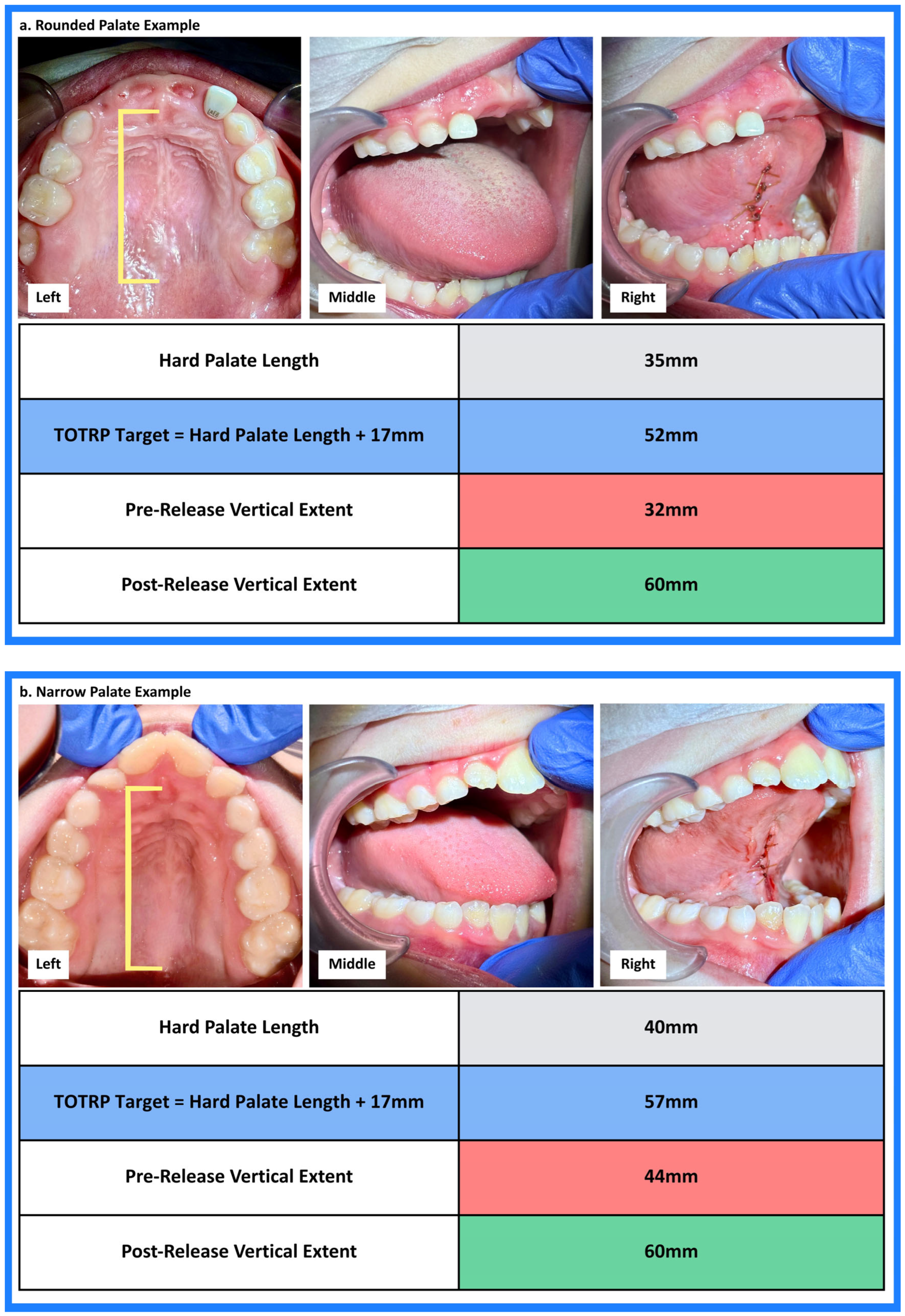
Palatal morphology was assessed intraoperatively and categorized as wide, rounded, or narrow. Classification was based on palpation, clinical observation of dental spacing and/or crowding, and inter-cuspid and inter-molar measurements at the level of the second primary molars or first permanent molars, when applicable.
Post-release surgical closure with sutures was performed to support wound stabilization, optimize healing, and reduce the risk of postoperative adherence between the ventral tongue and the floor of the mouth.
2.4. Assessment of Tongue Resting Posture
Tongue resting posture was assessed intraoperatively immediately before and after the removal of lingual mechanical restriction. Assessment was performed while patients remained under the same anesthetic conditions.
This assessment was used to document the tongue’s passive intraoperative resting configuration under controlled anesthetic conditions. The observation was not intended to represent awake active tongue posture, speech, swallowing, or functional performance. Instead, it was used to determine whether the removal of lingual mechanical restriction changed the tongue’s passive ability to achieve full-length contact against the hard palate within the same anesthetic episode.
Tongue position was visualized using gentle mandibular guidance. If the mouth was closed, light pressure was applied to the chin to lower the mandible enough to observe the tongue–palate relationship. If the mouth was open, the mandible was first guided toward passive closure and then gently lowered to permit visualization of the resting configuration. The tongue itself was not contacted or stimulated during this assessment. Each observation was documented photographically and/or by video, and the maneuver was repeated three times to confirm that the observed resting configuration was consistent.
During visualization, mandibular opening was adjusted to the minimum degree needed to observe whether full-length passive tongue contact against the hard palate was maintained. This adjustment was made with an awareness of palatal morphology because the amount of opening compatible with preserved intraoral suction may vary between patients. When complete passive palatal suction was present, the tongue could be seen maintaining full-length contact against the hard palate and was often followed by an audible suction release as the mandible opened further (
Figure 1).
Because the pre-release and post-release assessments were performed within the same anesthetic setting, each patient served as their own control for evaluating whether the removal of the lingual mechanical restriction changed the tongue’s passive ability to achieve full-length palatal contact.
Resting posture was categorized into four mutually exclusive configurations: inferior tongue position toward the floor of mouth, anterior tongue position between the teeth, partial anterior or blade-only palatal contact, and full-length palatal contact extending along the hard palate. Full-length palatal contact was defined as complete passive palatal suction and was considered the clinical endpoint of optimal tongue rest posture. For analytic purposes, resting posture categories were collapsed into binary variables: full-length palatal contact versus non-palatal resting configuration.
2.5. Measurement Protocols
2.5.1. Maximal Mouth Opening Measurement
Maximal mouth opening was measured intraoperatively under general anesthesia before and after lingual release. A round millimeter ruler was positioned between the incisal edges of the maxillary and mandibular central incisors, and the maximum interincisal distance was recorded in millimeters.
This measurement was obtained as part of the standardized surgical measurement protocol to document baseline and post-release oral opening. Maximal mouth opening was recorded for completeness of the intraoperative assessment but was not included as a candidate TOTRP formulation in the present analysis. The candidate TOTRP formulations evaluated in this study were based on tongue protrusion and tongue vertical extent relative to measured hard palate length.
2.5.2. Hard Palate Length Measurement
Hard palate length measurement was introduced after the operative protocol had already included routine intraoperative assessment of tongue protrusion, tongue vertical extent, and complete passive palatal suction. The surgical technique, pre-release and post-release mobility assessments, and clinical endpoint did not change during the study period. Rather, hard palate length was added as a stable, patient-specific anatomic reference after it became clinically and analytically apparent that this measurement could quantify the relationship between post-release tongue vertical extent and the consistently observed endpoint of complete passive palatal suction.
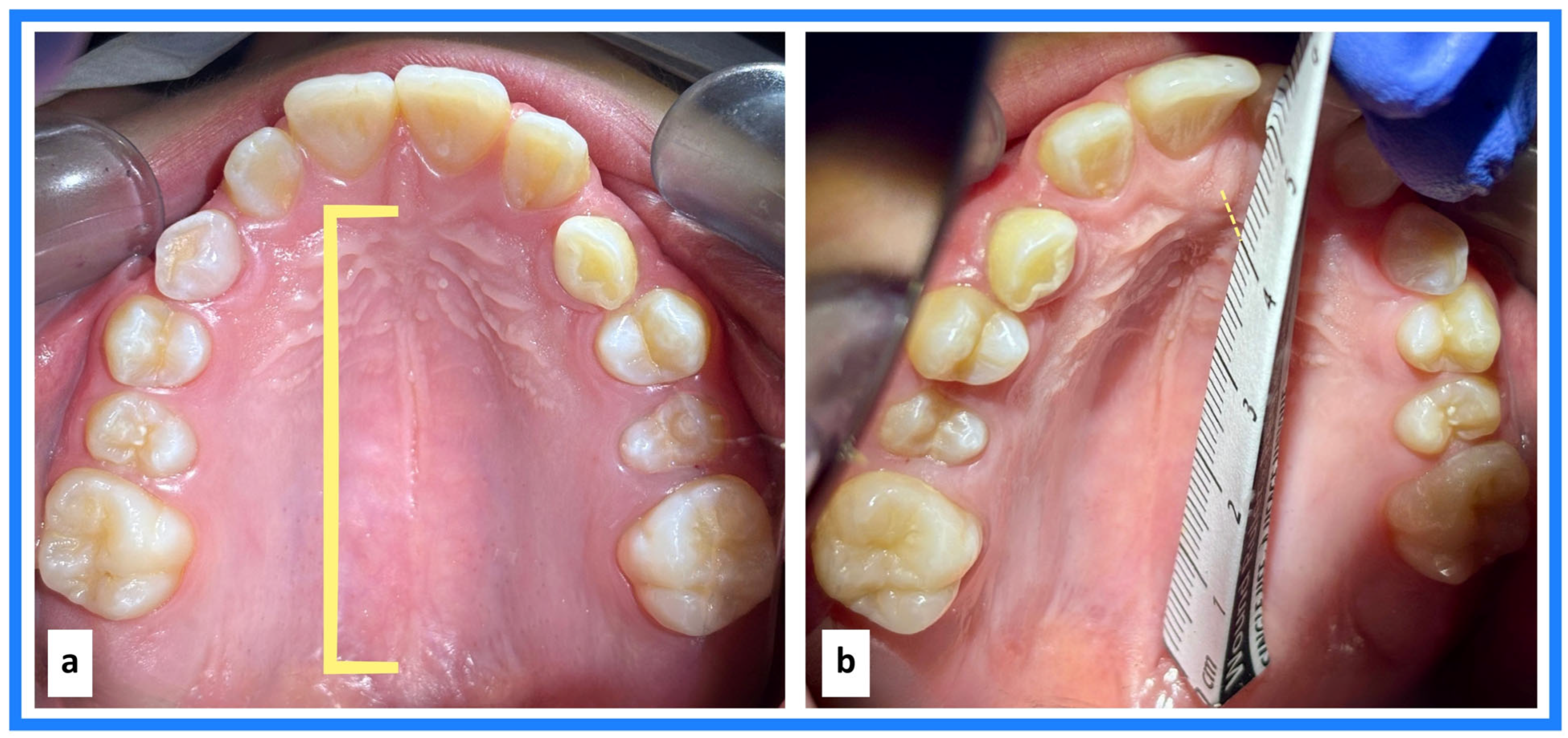
Hard palate length was measured intraoperatively under general anesthesia using a rigid millimeter ruler. The posterior landmark was defined as the junction of the hard and soft palate, and the anterior landmark was defined as the midpoint of the incisive papilla. The ruler was positioned along the midline of the hard palate, and the linear distance between these two landmarks was recorded in millimeters (
Figure 2).
This measurement was used as each patient’s individualized anatomic reference for TOTRP derivation. Direct measurement of hard palate length was used rather than estimated palate length to account for inter-individual anatomic variability.
Hard palate length was selected because it corresponds to the observed endpoint being measured. The TOTRP was not intended to model palatal vault shape, palatal width, palatal height, or total palatal volume. Rather, it was designed to relate post-release tongue vertical extent to the anterior–posterior hard palate distance over which the tongue must achieve full-length contact for complete passive palatal suction. Measuring from the midpoint of the incisive papilla to the junction of the hard and soft palate provided reproducible intraoperative landmarks and an endpoint-driven anatomic reference.
2.5.3. Tongue Protrusion Measurement
Tongue protrusion was measured using a rigid millimeter ruler positioned flush against the buccal surface of the mandibular central incisor and extending anteriorly beyond the oral cavity. The linear distance from the incisal edge of the mandibular central incisor to the most anterior point of the tongue was recorded in millimeters.
General Anesthesia Measurement
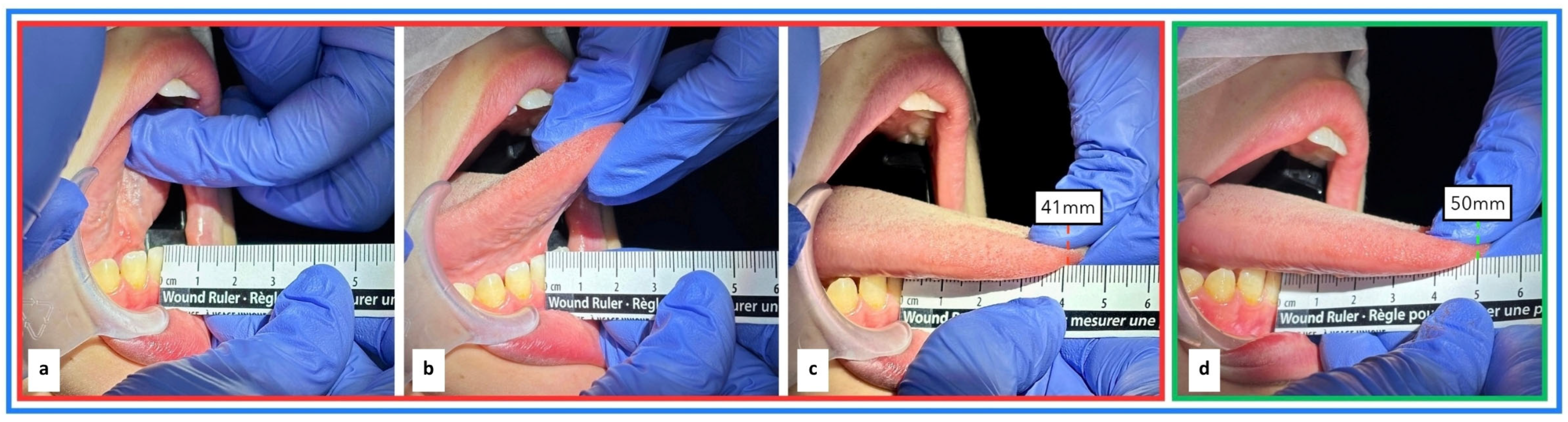
Under general anesthesia, patients were positioned supine and a McKesson mouth prop was placed to maintain mouth opening. The apex of the tongue was gently grasped and stabilized. Standardized anterior traction was applied by the examiner to extend the tongue maximally beyond the mandibular incisors, generating examiner-assisted passive protrusion. The measurement was obtained before release and repeated after release using the same anatomical landmarks, ruler position, head orientation, and examiner-applied traction (
Figure 3).
Awake Clinical Measurement
During the pre-release awake new-patient examination, patients were seated upright for protrusion measurement. The ruler was positioned using the same mandibular incisor landmark. The patient was instructed to protrude their tongue as far as possible, and the examiner gently assisted by grasping and stabilizing the tongue apex when tolerated. The linear distance from the incisal edge of the mandibular central incisor to the most anterior point of the tongue was recorded in millimeters.
Because awake protrusion was obtained in an upright, voluntary clinical setting, whereas intraoperative protrusion was obtained supine under general anesthesia using examiner-assisted passive traction, the awake and asleep protrusion measurements were analyzed separately and assessed for agreement rather than assumed to be interchangeable.
2.5.4. Tongue Vertical Extent Measurement
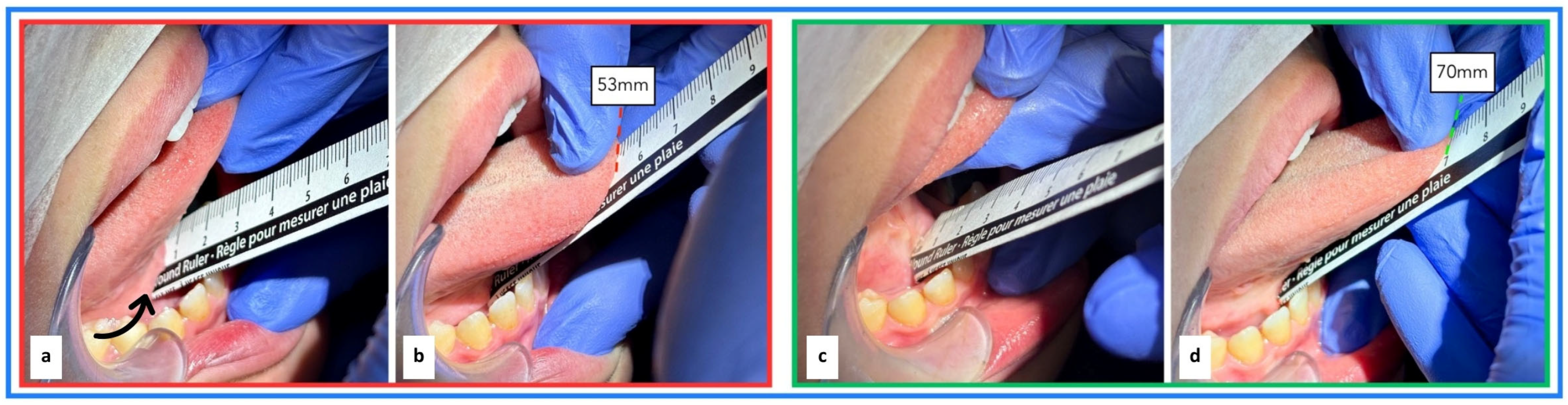
Tongue vertical extent, clinically described as vertical lift, was measured intraoperatively under general anesthesia before and after lingual release using a rigid millimeter ruler. A McKesson mouth prop was used to maintain mouth opening. The apex of the tongue was gently grasped and stabilized, and standardized examiner-applied vertical traction was used to elevate the tongue passively toward the palate.
The tongue was elevated to expose the sublingual caruncles, which served as the consistent inferior anatomic landmark for measurement. In patients with greater lingual restriction, visualization of the floor of the mouth may be limited before release; however, the sublingual caruncles remained identifiable. The inferior end of the ruler was positioned gently at the floor-of-mouth region immediately below the sublingual caruncles, at the base of the tongue. The ruler was aligned vertically, and the linear distance from this inferior reference point to the apex of the elevated tongue was recorded in millimeters.
The measurement was obtained before release and repeated after release using the same anatomical reference point, ruler position, mouth opening, and examiner-applied vertical traction (
Figure 4).
2.6. Functional Lingual Frenotomy Technique
After completion of the pre-release maximal mouth opening measurement, tongue mobility measurements, and resting posture assessment, a functional lingual frenotomy was performed under general anesthesia. A McKesson mouth prop was used to maintain mouth opening throughout the procedure.
Although some clinicians may describe similar sutured releases as a frenuloplasty, the term functional lingual frenotomy is used here because the primary intervention was the release of restrictive lingual tissue, with mucosal approximation performed for wound stabilization and wound healing.
Using a grooved director, the tongue was elevated and stabilized in a posterior-superior direction to permit visualization of the ventral tongue surface, sublingual caruncles, and floor-of-mouth region.
The initial release was performed superior to the sublingual caruncles, at the midpoint of the visible lingual restriction between the caruncles and the superior insertion of the restriction into the ventral tongue. Restrictive tissue at this site was clamped with a hemostat, followed by scissor incision through the clamped region.
Straight iris scissors were used in this cohort according to the surgical protocol; however, the objective checkpoints described in this study are intended to evaluate release adequacy rather than to compare surgical instruments.
The grooved director was then repositioned to continue posterior–superior retraction of the tongue, separating the tongue from the floor of the mouth under tension and allowing sequential visualization of the remaining restrictive lingual fibers. Additional restrictive fibers were identified, clamped with a hemostat, and released with scissors in an incremental fashion. This process was repeated until visual separation of the tongue from the floor of mouth was achieved.
After the sequential hemostat-assisted scissor incisions were completed, a functional fascial release maneuver was performed. A rolled 4 × 4 cotton gauze was placed beneath the tongue over the incision site. With the examiner’s fingers positioned over the gauze and index fingers together, controlled pressure was applied downward toward the floor of the mouth, followed by posterior pressure toward the oropharynx. The maneuver was held until residual submucosal fascial tension relaxed, and a broad, flat floor-of-mouth configuration was visualized.
Release adequacy was then assessed by direct visualization and palpation. The first surgical checkpoint was defined as ease of tongue separation from the floor of the mouth, with confirmation of a flat floor-of-mouth configuration. If this endpoint was not achieved, the release was considered incomplete. The ventral tongue–floor-of-mouth relationship was reassessed by visualization and palpation to identify residual restrictive tension. Persistent restriction localized to the ventral tongue was addressed with additional targeted hemostat-assisted scissor release. Whereas, any residual tension appreciated through the floor-of-mouth soft tissues was addressed with additional functional fascial stretching.
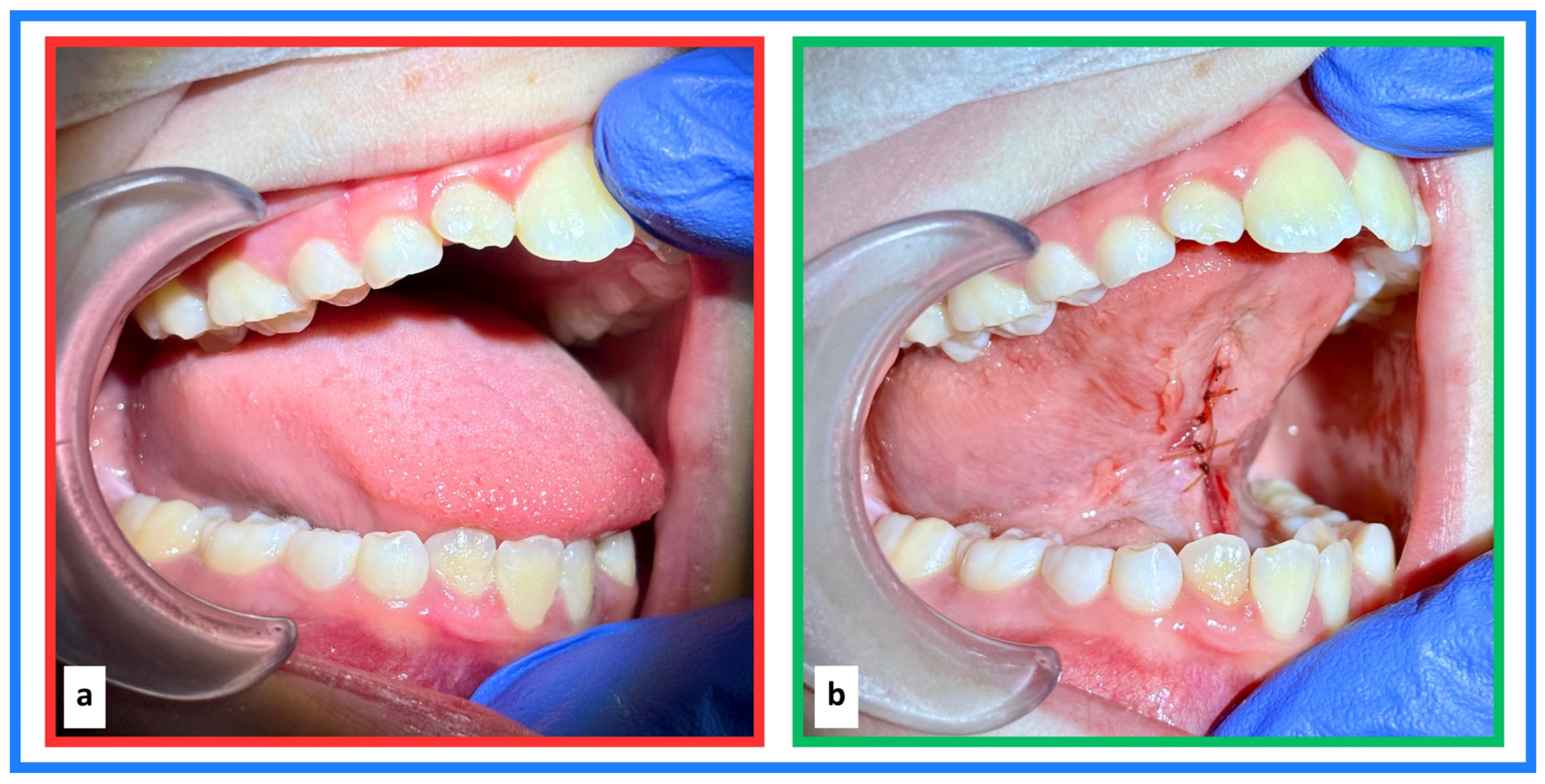
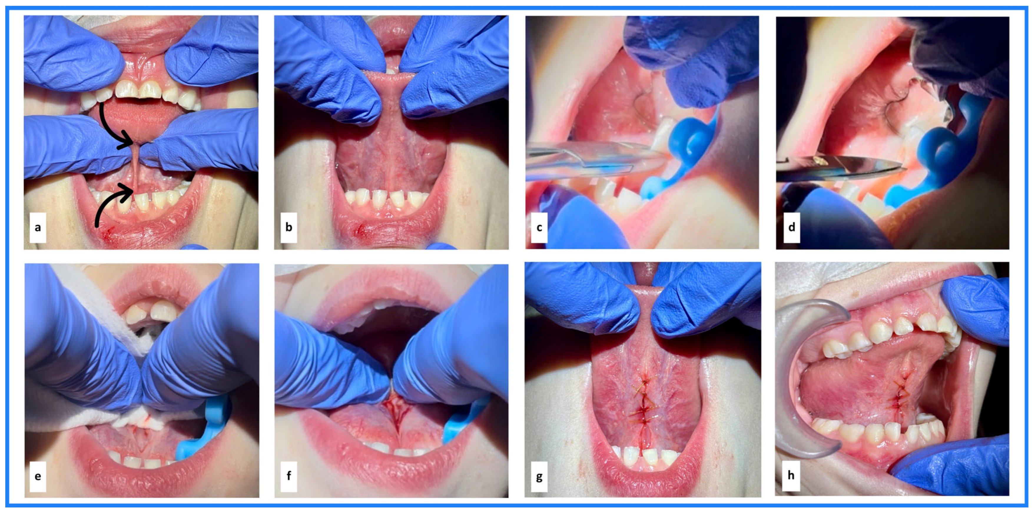
After the first surgical checkpoint was achieved, post-release tongue protrusion and tongue vertical extent measurements were obtained using the protocols described above. The incision site was then approximated using single interrupted 5-0 chromic gut sutures placed within the outer mucosa of the ventral tongue to achieve primary intention closure. Suturing was limited to the ventral tongue mucosa and did not incorporate deeper soft tissue or musculature. Sutures were not placed along the floor of the mouth; this region was allowed to heal by secondary intention. Suture closure was stopped at the junction where the ventral tongue meets the floor of the mouth to support wound stabilization, reduce the risk of postoperative adherence between the ventral tongue and floor of the mouth, and minimize disruption of floor-of-mouth soft tissues, including salivary structures. Following closure, tongue resting posture was reassessed to confirm complete passive palatal suction, representing the second and final surgical checkpoint. The functional lingual frenotomy sequence, including identification of the lingual mechanical restriction, hemostat-assisted scissor release, functional fascial stretching, confirmation of the first surgical checkpoint, mucosal approximation, and post-release complete passive palatal suction, is illustrated in
Figure 5.
2.7. Statistical Analysis
Demographic, clinical history, measurement, and clinical outcome data were summarized descriptively. Categorical variables were reported as counts and percentages. Continuous variables were summarized using means and standard deviations (SDs) when approximately normally distributed, and medians with interquartile ranges (IQRs) when distributions were skewed.
2.7.1. Derivation of the TOTRP Constant
To derive the Tool for Optimal Tongue Rest Posture (TOTRP), post-release tongue mobility measurements were evaluated relative to each patient’s measured hard palate length. All included patients demonstrated complete passive palatal suction following lingual release, confirming attainment of the clinical endpoint of optimal tongue rest posture.
Two candidate formulations were assessed.
For the vertical extent formulation, the mobility constant was defined as:
For the protrusion formulation, the mobility constant was defined as:
The distribution of each candidate constant was summarized using descriptive statistics, including mean, SD, range, and selected lower-bound percentile estimates. The 5th and 10th percentiles were used to characterize lower-bound mobility margins after confirmed attainment of complete passive palatal suction. Confidence intervals (CIs) for percentile estimates were obtained using nonparametric bootstrap resampling with 1000 iterations, with a seed set for replicability.
2.7.2. Comparison of TOTRP Candidate Formulations
The vertical extent-based and protrusion-based TOTRP formulations were compared by examining the distribution, variability, and lower-bound percentile stability of their respective constants. For each formulation, dispersion was assessed using the SD and range, and lower-bound stability was evaluated using the 5th and 10th percentile estimates with corresponding bootstrap CIs. The formulation demonstrating lower variability, narrower bootstrap CIs around lower-bound percentile estimates, and greater consistency of the derived constant across patients was considered more suitable for clinical derivation of the TOTRP threshold.
2.7.3. Relationship Between Age, Hard Palate Length, and the Vertical Extent-Derived TOTRP Constant
The relationship between age and measured hard palate length was initially assessed graphically using scatterplots. Given evidence of a nonlinear developmental relationship, analyses were stratified using the conventional pediatric/adult distinction of <18 years and ≥18 years. Within each age group, the association between age and hard palate length was evaluated using linear regression. Regression coefficients, 95% confidence intervals, R2 values, and residual standard deviations were reported. Residual variability was used to assess the potential precision of age-based substitution for direct hard palate measurement.
Given the broad age range of the cohort, the association between age and the vertical extent-derived TOTRP constant was additionally evaluated using linear regression, with as the dependent variable and age as the independent variable. LOWESS smoothing was used descriptively to assess whether an obvious developmental breakpoint or age-related discontinuity was present in the vertical extent-derived constant.
2.7.4. Comparison of Awake and Asleep Pre-Release Protrusion Measurements
Exploratory analyses comparing pre-release tongue protrusion measured during the awake clinical examination with pre-release protrusion measured intraoperatively under general anesthesia were performed using paired t-tests. Agreement between awake and asleep protrusion measurements was assessed using the Bland–Altman method. The mean difference, or bias, and 95% limits of agreement, calculated as the mean difference ±1.96 SD, were used to evaluate interchangeability at the individual level.
Complete-case analyses were conducted for all aims. Statistical analyses were performed using Stata MP version 19.0 (StataCorp, College Station, TX, USA). Statistical significance was defined as a two-sided p < 0.05.
4. Discussion
The present study introduces the Tool for Optimal Tongue Rest Posture (TOTRP) as a patient-specific, measurement-based surgical guide and intraoperative checkpoint for lingual frenotomy under general anesthesia. Building on the previously established clinical endpoint of complete passive palatal suction [
5], this study evaluated whether post-release tongue mobility could be quantified relative to each patient’s measured hard palate length and used to define a reproducible intraoperative target for surgical completeness. The principal finding was that post-release tongue vertical extent, interpreted relative to hard palate length, provided a more stable and clinically meaningful formulation than tongue protrusion for derivation of the TOTRP.
This distinction is clinically important. Lingual frenotomy is often described according to release location, wound appearance, or general improvement in tongue mobility; however, these features alone do not confirm whether the tongue has gained sufficient mobility to achieve complete passive palatal suction. The TOTRP reframes surgical adequacy around the functional endpoint of optimal tongue rest posture. In this framework, measured hard palate length provides the patient-specific anatomic reference, post-release vertical extent provides the mobility measurement most directly related to palatal resting posture, and complete passive palatal suction provides the observed endpoint confirming that the surgical objective has been achieved.
The vertical extent-based formulation demonstrated greater stability than the protrusion-based formulation, with a narrower bootstrap confidence interval and lower standard error around the 5th percentile threshold. This supports the anatomic premise that the mobility required for optimal tongue rest posture is not primarily anterior movement beyond the mandibular incisors, but sufficient vertical elevation from the floor-of-mouth/base-of-tongue region to permit full-length passive contact against the hard palate. In contrast, protrusion is measured from the mandibular incisal edge and reflects a different anatomic vector. These findings further support that protrusion and vertical extent are not interchangeable mobility constructs when interpreted relative to hard palate length.
Together, these findings suggest that the TOTRP provides a clinically useful framework for guiding lingual frenotomy toward a measurable anatomic and functional target. Rather than relying on visual release alone, the clinician can measure hard palate length, assess post-release vertical extent relative to that patient-specific reference, and confirm complete passive palatal suction under standardized intraoperative conditions. In this way, the TOTRP functions not only as a post-release measurement but as a surgical guide for achieving the mobility required for optimal tongue rest posture.
4.1. Relationship to Complete Passive Palatal Suction and Muscular Hydrostat Behavior
The TOTRP should be interpreted within the context of complete passive palatal suction as the clinical endpoint of optimal tongue rest posture. In the prior muscular hydrostat study, removal of lingual mechanical restriction was associated with a reproducible shift from non-palatal resting configurations to full-length palatal contact, including 99.5% full-length palatal contact in the awake infant cohort and 100% full-length palatal contact in the general anesthesia cohort following the restoration of unrestricted mobility [
5]. The present study builds on that observation by translating the endpoint of complete passive palatal suction into a measurable intraoperative framework.
The intraoperative endpoint should also be interpreted within the controlled anesthetic conditions in which it was observed. General anesthesia does not replicate awake function, natural sleep, or active functional tongue use, and anesthetic agents may influence upper-airway muscle tone and passive tissue compliance. However, the present study used a within-subject pre-release/post-release design under the same anesthetic conditions. Before release, all patients demonstrated non-palatal tongue resting posture; after release, all patients demonstrated complete passive palatal suction. This pre/post change under the same anesthetic conditions supports the interpretation that complete passive palatal suction reflected the restoration of sufficient mechanical tongue mobility following release, rather than an effect of general anesthesia alone.
In this context, complete passive palatal suction may be interpreted as an intraoperative passive expression of optimal tongue rest posture, demonstrating that the released tongue had sufficient structural freedom to elevate, broaden, and achieve full-length palatal contact.
The prior awake infant cohort is particularly relevant to the interpretation of the first surgical checkpoint. In infancy, passive resting states can be observed clinically without general anesthesia, allowing the muscular hydrostatic behavior of the tongue to be assessed in an awake setting. The reproducible shift from non-palatal resting configurations to full-length palatal contact in that cohort supports the concept that restoration of the tongue’s natural separation from the floor of mouth is a foundational requirement for complete lingual release, regardless of procedural setting [
5]. In the present study, this principle was operationalized under general anesthesia as the first surgical checkpoint: ease of tongue separation from the floor of the mouth with confirmation of a broad, flat floor-of-mouth configuration.
This distinction is important. Complete passive palatal suction represents the observed functional endpoint, whereas the TOTRP provides a patient-specific measurement target to help guide the surgical release toward that endpoint. In this study, all patients demonstrated non-palatal tongue resting posture before release and complete passive palatal suction after release. Therefore, the derived vertical extent-based constant was not based on an arbitrary mobility target, but on post-release measurements obtained only after the clinical endpoint of optimal tongue rest posture had been confirmed.
The relationship between vertical extent and complete passive palatal suction is also consistent with the tongue’s behavior as a muscular hydrostat. The tongue must have sufficient freedom to deform, elevate, broaden, and distribute along the hard palate within the spatial boundaries of the oral cavity. When lingual mechanical restriction is present, inferior, anterior, or partial palatal resting configurations may reflect tethering that prevents the tongue from assuming its stable palatal equilibrium. Following adequate release, the tongue’s ability to achieve full-length passive contact against the hard palate suggests restoration of the mobility required for this hydrostatic resting configuration.
The present findings therefore extend the muscular hydrostat framework from observation to surgical measurement. Rather than only documenting that the unrestricted tongue can assume complete passive palatal suction, this study identifies a measurable vertical mobility relationship associated with that outcome. In this way, the TOTRP provides a practical bridge between biologic endpoint, anatomic measurement, and intraoperative surgical decision-making.
Importantly, complete passive palatal suction was achieved regardless of palatal morphology and without dependence on awake cooperation or performance. While adjunctive myofunctional therapy may remain important for active functional outcomes, compensatory patterns, wound mobility, and long-term neuromuscular adaptation [
13,
14,
15], the present findings suggest that the immediate intraoperative endpoint of complete passive palatal suction reflects the restoration of sufficient mechanical tongue mobility. Under general anesthesia, this endpoint can be assessed independent of patient effort, guarding, voluntary motor control, or variable compliance with preoperative exercises.
4.2. Clinical Interpretation of the 17 mm Vertical Extent Margin
The vertical extent-based TOTRP formula identified a 17 mm margin beyond measured hard palate length as the lower-bound threshold associated with confirmed complete passive palatal suction. This threshold should be interpreted as a minimum vertical mobility margin rather than an average postoperative value. In the complete-measurement subset, the mean vertical extent-based constant was 24.20 mm, indicating that most patients exceeded the 17 mm margin after adequate release. The 17 mm value therefore represents a conservative lower-bound estimate derived from patients who had already achieved the clinical endpoint of optimal tongue rest posture. It should therefore be interpreted as a derived intraoperative surgical adequacy margin associated with confirmed complete passive palatal suction in this cohort, rather than as a diagnostic classifier.
This interpretation is clinically important because the TOTRP is intended to guide surgical adequacy, not to define maximal mobility. A post-release vertical extent equal to hard palate length alone would not account for the additional mobility required for the tongue to elevate, broaden, deform, and maintain full-length passive contact against the hard palate. The additional 17 mm margin reflects the functional mobility reserve required for the tongue to reach and sustain complete passive palatal suction within the three-dimensional oral cavity.
The use of a lower-bound percentile threshold also supports clinical applicability. A mean-based threshold would describe the central tendency of the cohort but could overestimate the minimum mobility required for some patients. In contrast, the 5th percentile threshold provides a conservative estimate of the lower mobility margin observed among patients who successfully achieved complete passive palatal suction. This approach is appropriate for developing a surgical checkpoint because the clinical question is not how much mobility the average patient achieved, but how much vertical extent appears necessary to reliably permit the endpoint.
Accordingly, the TOTRP formula should be applied as a patient-specific intraoperative guide: measured hard palate length plus 17 mm represents the minimum post-release tongue vertical extent target associated with optimal tongue rest posture in this cohort. If the measured post-release vertical extent does not meet this threshold, the release should be reassessed for residual restriction, persistent floor-of-mouth tension, or incomplete functional fascial mobility. In the present study setting, direct observation of complete passive palatal suction confirmed attainment of the intraoperative endpoint from which the TOTRP threshold was derived.
4.3. Why Vertical Extent Outperformed Protrusion
The superiority of the vertical extent-based formulation is anatomically, surgically, and functionally coherent. Complete passive palatal suction requires the tongue to separate from the floor of the mouth, elevate toward the palate, broaden, and maintain full-length passive contact along the hard palate. Therefore, a measurement that captures vertical extent from the floor-of-mouth/base-of-tongue region toward the palate is more directly aligned with the clinical endpoint than a measurement of anterior projection beyond the mandibular incisors.
Vertical extent is also directly related to the first surgical checkpoint of release adequacy. Across all lingual release procedures, restoration of the tongue’s natural separation from the floor of the mouth is a fundamental requirement for complete release. In the present general anesthesia technique, this principle was defined intraoperatively as the first surgical checkpoint: the tongue could be separated from the floor of the mouth with confirmation of a broad, flat floor-of-mouth configuration. The post-release vertical extent measurement then quantifies the amount of free tongue available to innately elevate from the floor-of-mouth/base-of-tongue region toward the hard palate. In this way, vertical extent does not simply measure tongue movement; it captures the functional availability of the released tongue to reach, adapt to, and suction against the palate.
Tongue protrusion remains clinically useful, but it reflects a different movement pattern. Protrusion measures anterior movement beyond the mandibular dentition and is influenced by the relationship between the tongue, mandibular incisors, anterior dentition, jaw position, and examiner traction. Although restricted protrusion may indicate lingual mechanical restriction, improved protrusion alone does not confirm that the tongue has separated sufficiently from the floor of the mouth or gained the vertical freedom required to achieve complete passive palatal suction. This distinction may explain why the protrusion-based constant demonstrated greater variability and less stable lower-bound percentile estimates than the vertical extent-based constant.
In contrast, vertical extent and hard palate length are functionally linked. Hard palate length defines the anatomic region over which full-length palatal contact must occur, and vertical extent reflects the amount of released tongue mobility available to reach and maintain that contact. The tighter distribution and greater lower-bound stability of the vertical extent-based constant therefore support its use as the preferred TOTRP formulation.
4.4. Awake and Asleep Protrusion Measurements Are Not Interchangeable
The comparison between awake and asleep protrusion measurements provides additional clinical context for applying a measurement-based tongue mobility assessment. Awake pre-release protrusion was significantly lower than pre-release protrusion measured under general anesthesia, and Bland–Altman analysis demonstrated wide limits of agreement. These findings indicate that awake clinical protrusion and asleep examiner-assisted protrusion under general anesthesia should not be considered interchangeable at the individual-patient level.
This lack of interchangeability is clinically expected. Awake tongue protrusion depends on voluntary effort, comprehension, cooperation, neuromuscular coordination, guarding, discomfort, gag reflex, and tolerance of examiner assistance. In contrast, protrusion under general anesthesia reflects examiner-assisted passive mobility under controlled intraoperative conditions, without conscious voluntary motor control. Anesthetic agents may also influence muscle tone and passive tissue compliance. Although both measurements describe tongue protrusion, they are obtained under fundamentally different physiologic and procedural conditions. Therefore, awake protrusion and asleep examiner-assisted protrusion should be interpreted as distinct measurement conditions rather than interchangeable measures of the same construct.
This distinction supports the use of intraoperative measurements for derivation of the TOTRP. The purpose of the awake/asleep comparison was not to validate asleep protrusion as a direct substitute for awake functional mobility, but to determine whether awake clinical protrusion could be used interchangeably with passive intraoperative protrusion. The wide Bland–Altman limits of agreement indicate that it cannot. Accordingly, the TOTRP was derived from post-release intraoperative measurements obtained under controlled general anesthesia conditions, rather than from awake clinical measurements.
This finding has practical implications for clinical use of the TOTRP. The TOTRP derived in the present study is an intraoperative tool based on measurements obtained under general anesthesia, after release, and after confirmation of complete passive palatal suction. Therefore, the present TOTRP formula should not be directly applied to awake clinical measurements without further validation. If measurement-based tongue mobility assessment is adapted for awake office-based examinations, a separate awake-specific threshold or conversion framework would be required.
Importantly, the lack of interchangeability does not reduce the value of awake clinical assessment. Awake examination remains essential for evaluating symptoms, functional patterns, oral habits, speech, swallowing, breathing, compensatory movement, cooperation, and baseline mobility. Rather, these findings clarify that awake protrusion and asleep passive protrusion represent different measurement conditions. Each may be clinically useful, but thresholds derived under one condition should not be assumed to apply directly to the other without validation.
4.5. Age Cannot Substitute for Direct Hard Palate Measurement
The relationship between age and hard palate length further supports the need for patient-specific anatomic measurement. In this cohort, age was significantly associated with measured hard palate length, which is consistent with expected craniofacial growth and development. However, age explained only a portion of the variability in hard palate length, and residual inter-individual variation remained substantial.
This finding is clinically important because the TOTRP formula depends on the relationship between tongue vertical extent and each patient’s measured hard palate length. Although older patients generally demonstrated greater hard palate length than younger patients, the scatterplot demonstrated broad dispersion within age groups and relative plateauing at older ages. Therefore, age-based estimation would not provide the same precision as direct intraoperative measurement.
Direct hard palate measurement avoids assuming that patients of the same age have equivalent palatal dimensions. This is particularly relevant in a cohort spanning early childhood through adulthood, where craniofacial growth, palatal morphology, dental development, arch form, and individual anatomic variation may all influence measured hard palate length. By anchoring the TOTRP to each patient’s measured anatomy, the formula remains individualized rather than relying on age-based prediction.
The selection of hard palate length should also be interpreted in relation to the endpoint being measured. The TOTRP is not intended to provide a global three-dimensional model of palatal morphology or to estimate palatal vault height. In this cohort, complete passive palatal suction was achieved across narrow, rounded, and wide palatal morphologies, indicating that palatal morphology did not preclude attainment of the endpoint when adequate tongue mobility was achieved. The observed endpoint was complete passive palatal suction, defined as full-length passive tongue contact along the hard palate. Therefore, hard palate length was selected as the endpoint-matched anatomic reference because it represents the anterior–posterior distance over which full-length tongue–palate contact must be achieved. Palatal width, height, arch form, and vault shape may remain relevant for broader craniofacial description, but they were not required to derive the intraoperative TOTRP target in this cohort.
These findings support direct hard palate measurement as a necessary component of the TOTRP framework. While age may provide general developmental context, it should not replace patient-specific measurement when determining the minimum post-release vertical extent required to achieve complete passive palatal suction.
The same principle applies to interpretation of the 17 mm vertical extent margin. The TOTRP does not assign the same absolute tongue vertical extent target to all patients across age groups. Instead, each patient’s target is calculated from that individual’s measured hard palate length, and the 17 mm margin is applied only after the patient-specific anatomic reference has been measured. In the present analysis, age explained only 5% of the variability in , and LOWESS smoothing did not demonstrate an obvious developmental breakpoint. These findings support interpretation of the derived margin as part of a patient-specific measured-anatomy framework rather than as an age-based formula.
4.6. Clinical Application of the TOTRP as a Surgical Guide and Intraoperative Checkpoint
The TOTRP has practical application as both a surgical guide and an intraoperative checkpoint. In the present technique, release adequacy is first assessed by direct visualization and palpation of tongue separation from the floor of the mouth, with confirmation of a broad, flat floor-of-mouth configuration. Post-release vertical extent is then measured to determine whether sufficient free tongue mobility has been achieved relative to the patient’s measured hard palate length. In the present study setting, complete passive palatal suction provided confirmation that the functional endpoint had been attained.
This sequence is clinically important because surgical completeness is not defined by wound appearance alone, but by restoration of the tongue’s natural separation from the floor of the mouth, confirmation of a broad, flat floor-of-mouth configuration, and achievement of the patient-specific TOTRP target. When these checkpoints are achieved, the release has restored the mobility associated with complete passive palatal suction and optimal tongue rest posture in this cohort. A visible release confirms that tissue has been incised, but does not necessarily confirm that the tongue has gained the functional mobility required to separate from the floor of the mouth and elevate toward the palate. By combining the first surgical checkpoint, patient-specific hard palate measurement, and post-release vertical extent, the TOTRP provides a structured framework for intraoperative decision-making. Within this framework, meeting or exceeding the patient-specific TOTRP target functions as the necessary second and final surgical checkpoint for complete release, before final confirmation of complete passive palatal suction where directly visible.
If post-release vertical extent remains below the patient-specific TOTRP target, the surgeon can reassess for residual ventral tongue restriction, persistent floor-of-mouth tension, or incomplete functional fascial mobility. Additional targeted release or functional fascial stretching can then be performed before closure and final resting posture confirmation. This stepwise approach allows residual restriction to be addressed within the same procedure, rather than relying on postoperative revision to achieve the intended mobility endpoint.
The TOTRP therefore supports a shift from the subjective assessment of release adequacy toward a reproducible, measurement-based process that can be documented, audited, and evaluated in future studies. This may be particularly valuable in complex or severe restrictions, where visual release alone may not reliably indicate whether sufficient functional mobility has been achieved.
This framework may also be useful in operative settings where complete tongue passive palatal suction cannot be directly visualized because of airway instrumentation, oral intubation, a laryngeal mask airway, or concurrent procedures limiting access to resting posture assessment. In these settings, the first surgical checkpoint and the patient-specific TOTRP target may provide an objective method for evaluating release completeness when direct confirmation of tongue passive palatal suction is not feasible.
4.7. Procedural Safety and Technique Considerations
The measurement-based framework described in this study should also be considered within the context of procedural safety. In the prior muscular hydrostat study, the removal of lingual mechanical restriction was associated with a reproducible shift toward full-length palatal contact without clinically significant postoperative complications documented in either the awake infant cohort or the pediatric general anesthesia cohort [
5]. Reported outcomes included no excessive bleeding, infection, clinically significant postoperative swelling, wound dehiscence, suture failure, or behavioral trauma, such as tongue biting [
5].
This safety context is relevant to the present study because the TOTRP framework depends on achieving a complete functional release sufficient to permit tongue separation from the floor of the mouth, adequate vertical extent, and complete passive palatal suction. A measurement-based surgical target should not be interpreted as encouragement for indiscriminate tissue release; rather, it provides a structured method for determining whether the release has restored the mobility required for the intended functional endpoint.
Importantly, the TOTRP may also support procedural restraint. Once the first surgical checkpoint has been achieved and the patient-specific vertical extent target has been met, further incision or excision is not required. In this way, the framework helps identify both incomplete release and sufficient release. Rather than relying on wound appearance alone, the surgeon can determine whether the tongue has regained the mobility associated with complete passive palatal suction and optimal tongue rest posture in this cohort. This may reduce the tendency to continue cutting based on wound appearance alone and may help protect adjacent floor-of-mouth structures, vascular tissues, salivary tissues, and healing mucosa.
In this cohort, complete passive palatal suction was achieved after a single lingual release procedure in all patients, despite variation in the severity and complexity of lingual mechanical restriction. This finding is clinically relevant because severe tongue ties, particularly those with marked restriction or near-complete adherence of the tongue to the floor of the mouth, are sometimes described as requiring staged procedures, multiple releases, or later revision to obtain adequate mobility. In the present framework, surgical completeness was not determined by apparent severity alone, but by the achievement of the first surgical checkpoint and attainment of the patient-specific TOTRP target. Together, these objective endpoints indicate that the tongue has regained the mobility associated with complete passive palatal suction and optimal tongue rest posture. These findings support the concept that complete functional release can be achieved in a single procedure when residual restriction is systematically reassessed and addressed before closure.
The surgical approach described here uses hemostat-assisted scissor release, functional fascial mobilization, direct visualization and palpation, and mucosal approximation with sutures. These features permit real-time adaptation to patient-specific anatomy, including variable floor-of-mouth tension, vascular anatomy, salivary tissues, and the relationship between the ventral tongue and floor of the mouth. By emphasizing targeted release, functional fascial mobility, objective measurement, and endpoint confirmation before closure, the technique aims to restore tongue mobility while avoiding unnecessary disruption of adjacent soft tissues.
Published safety data for lingual frenuloplasty with myofunctional therapy have similarly reported no major complications in a 348-patient cohort, although minor complications occurred in fewer than 5% of cases and included prolonged bleeding, temporary tongue-tip numbness, salivary gland issues, minor wound infection or inflammation, and revision to excise restrictive scar tissue [
13]. Differences in patient populations, surgical technique, wound management, anesthetic approach, postoperative protocols, and complication capture limit direct comparison across studies. Nevertheless, these data provide useful context for interpreting surgical safety when lingual release is performed with attention to anatomy, function, and postoperative wound stability.
In the present framework, procedural completeness and procedural restraint are not opposing goals. The TOTRP is intended to support complete functional release while maintaining surgical precision. By linking release adequacy to the first surgical checkpoint and measurable post-release vertical mobility relative to the patient-specific TOTRP target, the technique provides a reproducible method for determining when sufficient mobility has been achieved, rather than relying on wound appearance or additional tissue release alone.
4.8. Implications for Future Research
The present study establishes a measurement-based framework for applying the TOTRP under general anesthesia across a broad age range of patients who achieved complete passive palatal suction following adequate release. A major next step is to determine how this framework can be adapted for awake clinical assessment beyond infancy. Because the present findings demonstrate that awake and asleep protrusion measurements are not interchangeable, future research should evaluate whether a separate awake TOTRP threshold can be derived using awake measurements that reflect the same functional principle: restoration of natural tongue separation from the floor of the mouth and sufficient free tongue mobility to permit complete palatal suction.
Prospective paired awake/asleep measurement studies are currently underway. In this ongoing work, postoperative awake tongue vertical extent is being measured at 1-week follow-up in patients whose intraoperative muscular hydrostat function, complete passive palatal suction, and TOTRP attainment were confirmed under general anesthesia. These paired data will allow for a direct comparison of postoperative awake vertical extent with intraoperative asleep vertical extent and may support derivation of an awake TOTRP constant for clinical follow-up assessment. This represents an important next step in determining how intraoperative TOTRP attainment relates to postoperative awake tongue mobility, tongue resting posture, and functional outcomes.
Additional future work will evaluate how intraoperative complete passive palatal suction relates to objective or imaging-based tongue-palate posture, sleep-state posture, patient-reported symptoms, and longitudinal functional outcomes. These studies are necessary to determine how the intraoperative endpoint relates to function outside the operating room.
A particularly valuable next study would be to evaluate patients who have already undergone lingual frenotomy under general anesthesia, achieved the first surgical checkpoint, met their patient-specific asleep TOTRP target, and demonstrated complete passive palatal suction intraoperatively. After healing, these same patients could undergo standardized awake assessment of vertical extent, functional tongue–palate contact, floor-of-mouth mobility, and hard palate length. This design would allow postoperative awake measurements to be interpreted against a known intraoperative reference standard, rather than attempting to derive awake thresholds without confirmation that the surgical endpoint had been achieved. Because awake measurements may be influenced by facial tension, guarding, compensatory floor-of-mouth recruitment, limited mandibular opening, or tolerance for intraoral manipulation, future awake protocols may require adapted measurement methods or complementary clinical markers.
Measurement reproducibility should also be evaluated as the TOTRP framework is adopted beyond a single-practitioner setting. In the present study, measurements were obtained by one practitioner using a standardized protocol, supporting procedural consistency. Future multi-operator studies could assess inter-rater and intra-rater reliability for hard palate length, tongue vertical extent, floor-of-mouth release adequacy, and complete passive palatal suction to support broader clinical standardization.
Future investigations may also examine the relationship between TOTRP achievement and patient-centered clinical outcomes. The present study focused on defining a measurable intraoperative mobility target and confirming the immediate resting posture endpoint, rather than evaluating longitudinal symptom change. Subsequent studies can build on this framework by examining whether attainment of the TOTRP target and complete passive palatal suction are associated with improvements in nasal breathing, sleep symptoms, oral resting posture stability, speech, swallowing, compensatory floor-of-mouth recruitment, occlusal development, and need for adjunctive myofunctional therapy. These domains have previously been linked to tongue posture, lingual restriction, airway behavior, dentofacial development, and orofacial myofunctional therapy [
2,
3,
4,
14,
15]. This direction is also consistent with the broader literature identifying ongoing uncertainty in ankyloglossia diagnosis and treatment while reporting associations or outcome changes involving speech, feeding, sleep-disordered breathing, orofacial function, and craniofacial morphology following the assessment or treatment of restricted tongue mobility [
16,
17,
18,
19,
20].
Finally, objective imaging or biomechanical assessment may help refine our understanding of the relationship between tongue vertical extent, floor-of-mouth release, palatal contact, and hydrostatic resting behavior. Ultrasound, MRI, pressure mapping, or three-dimensional surface analysis could be used to characterize tongue–palate contact patterns and determine whether the measured vertical extent target corresponds to reproducible changes in tongue shape, elevation, and palatal distribution [
10,
11,
12].
4.9. Strengths
This study presents an objective, measurement-based framework for guiding the assessment and surgical completeness of lingual frenotomy using quantifiable anatomic relationships. By deriving the TOTRP constant from post-release measurements obtained after confirmed achievement of complete passive palatal suction, the framework is grounded in an observed functional endpoint rather than subjective visual assessment alone.
A major strength of this study is the integration of anatomy, surgical technique, functional endpoint confirmation, and statistical threshold derivation. Hard palate length provided the patient-specific anatomic reference, tongue vertical extent quantified the amount of released tongue mobility available to elevate toward the palate, and complete passive palatal suction confirmed attainment of the clinical endpoint of optimal tongue rest posture. This linkage between measurement and function supports the clinical relevance of the derived TOTRP formula.
The analytic approach incorporated descriptive statistics, percentile-based threshold derivation, and bootstrap resampling to assess lower-bound stability. Comparison of vertical extent-based and protrusion-based formulations allowed the preferred measurement approach to be selected based on variability, confidence interval width, and clinical interpretability. Bland–Altman analysis further provided practical insight into the lack of interchangeability between awake and asleep protrusion measurements.
Procedural consistency is another important strength. All assessments and measurements were performed by a single practitioner using standardized intraoperative protocols, supporting the uniformity of data acquisition across the cohort. The findings were observed across a broad age range, from early childhood through adulthood, and complete passive palatal suction was achieved regardless of palatal morphology following adequate release.
Together, these strengths support the internal consistency and clinical coherence of the present work. The study provides a reproducible foundation for measurement-based lingual frenotomy under general anesthesia and establishes a framework that can be further evaluated in awake assessment protocols, multi-operator settings, and longitudinal outcome studies.
4.10. Limitations
This study should be interpreted within the context of its design. The analysis was retrospective and based on clinical observations obtained from existing records. Although the measurement and assessment protocols were standardized within the practice, the retrospective design may introduce documentation variability and limits the ability to control all measurement conditions prospectively.
All evaluations, measurements, and procedures were performed by a single experienced clinician-surgeon at a single center using a standardized intraoperative measurement protocol. For a derivation study, this provided procedural consistency and internal standardization, and avoided introducing inter-examiner variability before the TOTRP measurement framework had been established. However, formal intra-examiner and inter-examiner reliability testing was not performed, and reproducibility across clinicians has not yet been established. Ruler-based intraoperative measurements of mobile tissue require careful standardization, and in the present protocol, measurement fidelity was supported by consistent anatomic landmarks, a rigid millimeter ruler, standardized head and mouth positioning, consistent examiner-applied traction, repeated pre-release and post-release measurement within the same operative setting, and direct endpoint confirmation through complete passive palatal suction. Future validation should evaluate reproducibility across clinicians using methods that preserve patient safety, surgical efficiency, and measurement fidelity. Potential approaches include prospective repeated measurements within a defined protocol, secondary intraoperative confirmation of the measurement recorded by the surgeon, standardized photographic or video-based measurement training, and multi-operator assessment when feasible and ethically appropriate.
Hard palate length was available for 192 out of 390 patients because this measurement was introduced after the operative protocol had already included the routine assessment of tongue protrusion, tongue vertical extent, and complete passive palatal suction. The absence of hard palate length measurements in earlier cases reflects evolution of the measurement framework, not a change in surgical technique, clinical endpoint, pre-release or post-release mobility assessment, or selective inclusion based on outcome. Complete passive palatal suction was the clinical endpoint across the cohort and was achieved after release in all patients. Future prospective studies should include hard palate length measurement from the outset in all patients.
Although the cohort included patients aged 3.0 to 52.2 years, the TOTRP is not applied as an age-based formula. It is anchored to each patient’s directly measured hard palate length, with the 17 mm margin applied after the patient-specific anatomic reference has been obtained. In the present analysis, age explained only 5% of the variability in , and LOWESS smoothing did not demonstrate an obvious developmental breakpoint. Future studies with larger age-stratified cohorts may further evaluate whether subgroup-specific refinements are warranted.
The TOTRP constant was derived from patients who achieved complete passive palatal suction following adequate release. Because there was no unsuccessful comparison group, the threshold was derived using distributional and lower-bound percentile analysis rather than formal diagnostic performance metrics such as sensitivity, specificity, or receiver operating characteristic analysis. This approach is appropriate for deriving a measurement-based surgical target from patients who achieved the clinical endpoint, but it does not directly evaluate diagnostic prediction in patients who fail to achieve complete passive palatal suction. Future studies including incomplete-release, persistent-restriction, or non-endpoint cases will be needed to evaluate discriminative performance, including sensitivity, specificity, ROC characteristics, and predictive accuracy.
The present TOTRP formula was also derived from measurements obtained under general anesthesia. These conditions allowed for standardized passive assessment without voluntary motor control, guarding, gag reflex, fatigue, or cooperation-related limitations. Therefore, the findings should not be directly extrapolated to awake clinical measurements without further study. Awake measurements may be influenced by effort, cooperation, facial tension, compensatory floor-of-mouth recruitment, limited mouth opening, and tolerance for intraoral manipulation.
The intraoperative measurements should therefore be interpreted within the controlled anesthetic context in which they were obtained. Patients were under general anesthesia, not light sedation, and neuromuscular blocking agents were not administered. General anesthesia does not replicate awake functional tongue use; however, it provided a controlled intraoperative state for assessing passive tongue mobility before and after release. Because pre-release and post-release measurements were obtained within the same anesthetic episode for each patient using the same measurement protocol, the study design supports within-subject comparison of passive tongue mobility under controlled surgical and anesthetic conditions.
Future work is underway to evaluate how intraoperative complete passive palatal suction relates to postoperative awake tongue vertical extent, objective or imaging-based tongue-palate posture, sleep-state posture, patient-reported symptoms, and longitudinal functional outcomes. These studies are necessary to determine how the intraoperative endpoint relates to function outside the operating room.
Finally, the present study focused on intraoperative mobility, measurement-based surgical adequacy, and immediate attainment of complete passive palatal suction. Longitudinal symptom change, stability of tongue resting posture over time, and broader patient-centered functional outcomes were not evaluated in this analysis.