Protocol

Effect of Orofacial Myofunctional Therapy with Auto-Monitoring on the Apnea–Hypopnea Index and Secondary Outcomes in Treatment-Naïve Patients with Mild to Moderate Obstructive Sleep Apnea (OMTaOSA): A Multicenter Randomized Controlled Trial Protocol

1
Department of Otorhinolaryngology, Division of Surgery, Akershus University Hospital, Sykehusveien 25, 1478 Lørenskog, Norway
2
Clinic of Surgery, Institute for Clinical Medicine, Faculty of Medicine, University of Oslo, Campus Akershus University Hospital, Sykehusveien 25, 1478 Lørenskog, Norway
3
Institute of Clinical Medicine, Medical Faculty, University of Oslo, 0372 Oslo, Norway
4
Institute of Dentistry, University of Tartu, 50405 Tartu, Estonia
5
North Estonia Medical Centre, University of Tartu, 13419 Tallinn, Estonia
6
Department of Otorhinolaryngology, Fertilitas Private Hospital, 10614 Tallinn, Estonia
7
Department of Research and Innovation, Division of Mental Health and Addiction, Oslo University Hospital, 0424 Oslo, Norway
8
Physical Activity, Physical Education, Sport and Health Research Centre (PAPESH), Sports Science Department, School of Social Sciences, Reykjavik University, 101 Reykjavik, Iceland
9
Department of Nursing and Health Promotion, Faculty of Health Sciences, Oslo Metropolitan University, 0166 Oslo, Norway
*
Author to whom correspondence should be addressed.
Int. J. Orofac. Myol. Myofunct. Ther. 2025, 51(2), 8; https://doi.org/10.3390/ijom51020008
Submission received: 25 June 2025 / Revised: 26 August 2025 / Accepted: 26 August 2025 / Published: 9 September 2025

Abstract

Background: The aim of this article is to describe the protocol of a large, multicenter randomized controlled trial evaluating the effects of orofacial myofunctional therapy with auto-monitoring (OMTa) versus auto-monitoring alone on obstructive sleep apnea (OSA) assessed by the apnea–hypopnea index and other pre-specified outcomes. Method: The OMTaOSA study protocol was registered at ClinicalTrials.gov (NCT06079073) in August 2023, and data collection ended in January 2025. One hundred and four participants with mild and moderate OSA were included. Randomization was conducted in a 1:1 ratio, using sex-stratified blocks. The intervention was a standardized protocol of OMT exercises previously shown to be effective, auto-monitoring with a Withings scan watch, and feedback from self-reports. Controls received watches and access to the same application without the exercise module. Sleep was measured over three nights at baseline and after three months. The sleep scorer and researchers evaluating other outcomes were blinded to the treatment allocation. Change in the apnea–hypopnea index was defined as the primary outcome. Secondary outcomes are published on Clinicaltrials.gov. Results: The results of the trial are still in preparation. Conclusions: By addressing the limitations of previous OMT studies, this trial may clarify the effectiveness of digitally delivered OMT for patients with mild to moderate OSA.

1. Introduction

Obstructive sleep apnea (OSA) is a common disorder in the Norwegian adult population and worldwide. A Norwegian study has estimated the prevalence of OSA defined by an apnea–hypopnea index (AHI) of ≥15 to be 8% among adults aged 30–65 years [1]. Globally, the disease is a public health concern estimated to affect 936 million (95% CI 903–970) people [2]. If untreated, OSA is associated with increased risks of cognitive impairment, cardiovascular morbidity, mortality, and daytime sleepiness [3].
Treatment with positive airway pressure (PAP) has been shown to reduce the AHI and, subsequently, cardiovascular risk; to alleviate symptoms; and to be cost-effective in patients with moderate to severe OSA [4]. However, approximately 25–50% do not adhere to PAP treatment, and patients who adhere use the machine less than prescribed and thus render the treatment less effective [5]. Complementary, less efficient treatment modalities for OSA are lifestyle changes [6], positional therapy [7], mandibular advancement devices [8], and surgery [9]. Novel treatment approaches such as the use of mobile health (Mhealth) are a promising strategy to increase participatory health in all chronic diseases [10,11].
OSA develops over time, and PAP treatment is often perceived as cumbersome. Consequently, participatory strategies aimed at strengthening the genioglossus muscle are promising [12]. Among such strategies, orofacial myofunctional therapy (OMT) has been demonstrated to be effective in mild to moderate OSA, as reported in a Cochrane report [13]. However, additional randomized controlled trials (RCTs) with blinded outcome assessments and an intention-to-treat design are required to substantiate these findings. OMT and other participatory strategies delivered via Mhealth, if effective, may also facilitate an active approach to managing mild and moderate OSA. Combining OMT and auto-monitoring (OMTa) by Mhealth may potentially also motivate treatment adherence, potentially mitigating the progression to severe disease and subsequent end-organ damage.
The overall aim of this article is to describe the design and methods of a large, multicenter RCT evaluating the effect of OMTa compared to auto-monitoring alone on change in the AHI and other pre-specified outcomes.

2. Materials and Methods

2.1. Design

The OMTa for OSA (OMTaOSA) RCT is an assessor-blinded parallel-group randomized controlled trial. It is a sub-study of the “Sleep Revolution” project funded by the Horizon 2020 program (grant number 965417). The study was accepted by the Regional South-Eastern Committee for research ethics (522434), 16 August 2023 and the Akershus University Hospital person security officer (2023_61). The study protocol was registered on ClinicalTrials.gov (NCT06079073) in August 2023, and the study is being conducted in accordance with the Declaration of Helsinki. Data collection was completed in January 2025. The statistical analysis plan (SAP) was finalized in March 2025 prior to any analysis on the effect of the primary outcome variable. The full SAP is available upon request. De-identified participant data, statistical code, and any other materials are all stored in a secure cluster located in Reykjavik, Iceland. A patient representative was involved in the design of the trial, and its conduct and reporting will be in accordance with the “Consolidated Standards of Reporting Trials (CONSORT) 2025 guidelines” [14].

2.2. Sample Size

The number in each group was calculated based on the effect of OMT on change in the AHI reported by Guimaraes and co-workers [15]. With an anticipated effect size (Cohen’s d) of 0.8 (high), power ≥ 80%, and probability level of 5%, we estimated a minimal sample size of 26 per group. Further, we expected the dropout of patients during the OMTa trial to be 25%, corresponding to a total of 20 participants. Moreover, to accommodate for this potential dropout and to have adequate power for subpopulation analysis for sex, we aimed for a sample size of 100 (50 in each group).

2.3. Participant Recruitment and Eligibility Criteria

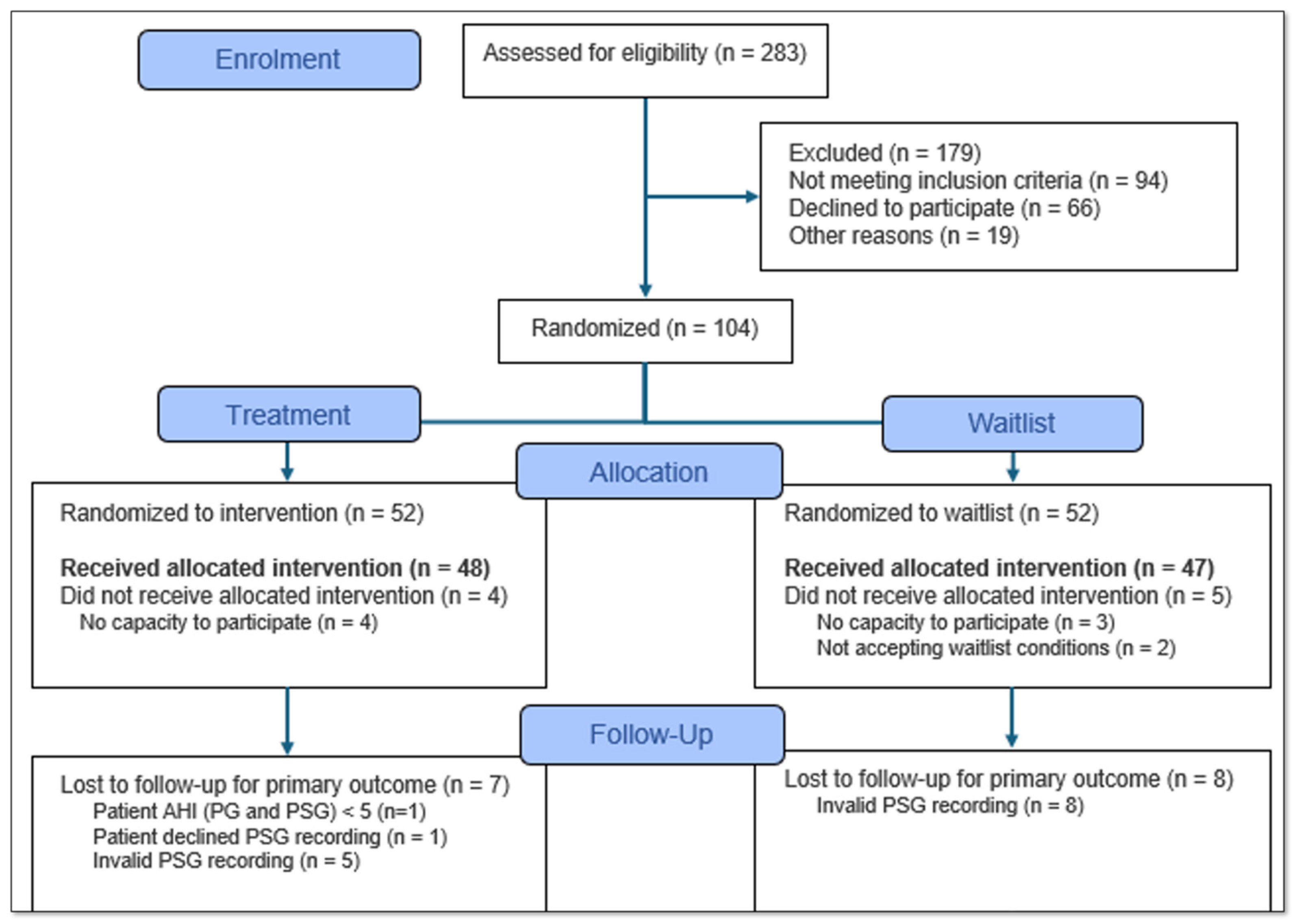
Participant recruitment is summarized in a CONSORT flow chart in Figure 1. Potential participants were invited at the otorhinolaryngology department at Akershus University Hospital (Ahus), Norway; at Fertilitas Private Hospital (FPH) in Tallinn, Estonia; and by otorhinolaryngologists practicing near these institutions. After performing a full otorhinolaryngology examination, doctors were asked to refer patients fulfilling the following inclusion criteria: 1: Suspected or confirmed mild or moderate OSA with a respiratory event index < 30 based on respiratory polygraphy. A diagnosis of OSA was expected in all participants after polysomnography described below. 2: A body mass index (BMI) < 30 based on self-reported height and weight. 3: No prior or current treatment with an oral device or positive airway pressure. 4: Ownership of a mobile phone compatible with the study software application. 5: Age ≥ 18 years. 6: The ability to breathe through the nose. 7: Ability to read and understand the Norwegian or Estonian language. 8: All frontal teeth present from second premolar to second premolar in both the upper and lower dental arches. 9: No administration of botulinum toxin in the facial muscles within the past three months. In total, 283 patients were invited to participate.
All potential eligible participants received brief information about the study along with an informed consent form at the time of referral. A researcher contacted potential participants, scheduled a baseline consultation, and gave information about the use of a mobile application as a pre-screening tool. A username (in the form of an email address) and password were delivered by email at first login. The tool consisted of an electronic sleep diary based on the consensus sleep diary developed by Carney and co-workers [16]. Support on using the application was available by telephone. Subjective, quantitative self-reported data including sleep efficiency were calculated based on the formulas proposed by Reed and co-workers and provided as auto-monitoring [17]. Details of the application have been published previously [18].

2.4. Baseline Final Inclusion

Inclusion criteria were re-assessed at the baseline consultation, including the BMI calculated by the standard formula (weight (kg) divided by height squared (m2) (kg/m2)) obtained from measurements. The exclusion criteria were 1: not completing at least 70% of days in the electronic sleep diary provided; 2: medical or psychiatric conditions which could interfere with the study protocol in the opinion of the investigators; and 3: not being able to open the mouth 50% with the maximal interincisal opening (MIO) compared to the interincisal opening with tongue tip to maxillary incisive papillae at the roof of the mouth (MOTTIP) [19]. Signed, informed consent was finally collected from 104 participants (Figure 1).

2.5. Randomization and Allocation

Participants were randomized in blocks to ensure an even distribution throughout the study period and to obtain a female-to-male ratio of approximately 40–60. Participants were randomized to either the OMTa intervention group or the auto-monitoring-only control group. Block randomization was conducted with 10 lots per block, per study site, and a 1:1 allocation ratio. The group-specific randomization codes were printed on paper and concealed in opaque envelopes, then kept and opened by the non-blinded therapists after the finalized baseline assessment. Codes were delivered by telephone to participants who entered them in the application and activated the exercise module or not.

2.6. Measurements

All measurements were registered in a “Redcap” case report form. In addition to the BMI, the waist-to-hip ratio was determined by dividing waist circumference by hip circumference. The neck circumference was measured with the participant’s head in the Frankfort Horizontal plane, immediately superior to the thyroid cartilage. Hand grip strength was assessed by hand dynamometry of the dominant hand with a Jamar hand dynamometer (Jamar Hydraulic Hand Dynamometer (5030J1), JLW Instruments, Chicago, IL, USA) [20]. Participants were seated upright in a chair with back support and fixed armrests; they squeezed maximally for 2–3 s in three alternating trials per hand, with 30 s rests between measurements. A standardized chewing efficiency test was performed using the CHEW Box Digital Reporting services (DRS CHEW box) from Orehab Minds (Stuttgart, Germany). The chewing test consisted of nine 30 s sequences with 30 s pauses, using gelatin units of soft, medium, and hard consistency (right, left, and bilateral). Chewed material was collected and imaged under standardized conditions in the DRS CHEW box and centrally analyzed for particle number and size. The IOPI (IOPI Medical, Woodinville, Washington, DC, USA), a plastic connecting tube, and standard 35 mm tongue bulbs were used to measure both maximal tongue and buccinator muscle strength. Measurements were taken with participants sitting in an upright position, three times, with a 30 s break between each measurement. The bulb was held by the examiner and positioned longitudinally on the anterior and posterior parts of the palate. The participants’ mandibles were unrestrained. Participants were asked to raise their tongue and squeeze the bulb against the palate as hard as they could for approximately 3 s. The highest of the three measurements was recorded as the participant’s tongue strength. In addition, the same procedure was repeated with the bulb placed between lateral teeth and the cheek. Lateral cephalograms were obtained for participants in Norway, while cone beam computer tomograms were obtained for participants in Estonia. Lateral cephalograms were taken in natural head position with the sagittal plane parallel to the film and the Frankfort plane parallel to the floor, at 73 kVp and 15 mA, with patients in centric occlusion and tongue at rest after swallowing. All the images were stored for future analysis using Planmeca Romexis® software version 6.4.5.202 (Planmeca Romexis® Cephalometric Analysis) in Estonia. Digital impressions of the maxillary and mandibular arches were obtained using a 3D intraoral scanner, TRIOS 3 (3Shape, Copenhagen, Denmark). Scans were conducted by un-blinded researchers trained according to manufacturer guidelines and were used for morphological analysis and occlusal evaluation.
Participants were also evaluated by the standardized Orofacial Myofunctional Evaluation with Scores protocol and additional tongue mobility assessments [19,21]. The MIO and MOTTIP were measured by calipers, and the tongue range-of-motion ratio was calculated [19]. Functional assessments were video-recorded with two cameras positioned at each side of the face at a 45-degree angle to facilitate inter-rater reliability analyses. All participants were seated in a chair without arm- or backrests for standardization purposes. The appearance of the face (symmetry), cheeks, mandible, lips, tongue, and hard palate was evaluated prior to the assessment of mobility of the same structures. Furthermore, breathing mode, deglutition, and mastication were evaluated.
Participants also completed self-reported questionnaires including a questionnaire assessing acceptance and control [22], the Epworth Sleepiness Scale [23], the patient health questionnaire-9 [24], the generalized anxiety disorder 7-item questionnaire [25], and the DS-14 questionnaire assessing type D personality [26].
Finally, participants were asked to wear a Withings Scanwatch, 1250, Boston, MA, USA, https://www.withings.com/be/en/scanwatch, programmed in research mode. The research mode setting implies that the watch operates as a sensor with increased sampling rate, while limited auto-monitoring from the device was available in the Withing’s mobile application.

2.7. Sleep Studies

All participants underwent three nights of self-applied polysomnography (PSG) (Nox Medical, Reykjavik, Iceland). The electroencephalography from the self-applied PSG is located only on the forehead (F-channels), with no electrodes located on the top of the scalp (C-channels) or back of the head (O-channels) [27]. The self-applied setup facilitates the use of multi-night studies to capture night-to-night variability. The participants applied the sensors themselves, using an instructional video.
All sleep registrations were scored by one sleep technologist at Reykjavik University Sleep Institute, using Noxturnal Research (version 6.1.0.30257, Nox Medical, Reykjavik, Iceland). Sleep efficiency (SE) was calculated as the scored total sleep time divided by the total sleep period (TST). TST was defined as the time from “lights out” to “lights on” divided by the scored total sleep time. Sleep was scored in accordance with the current version of the American Academy of Sleep Medicine (AASM) manual (version 2.6, 2020) [28]. The scoring was performed mainly by one expert sleep technologist. The EEG signal was modified from 0.3 Hz to 0.5 Hz to be compared with the recommended PSG scoring criteria. Moreover, the peak-to-peak amplitude of the slow wave activity was changed from 75 µV to 50 µV as traditional PSG signals are measured over the frontal regions and referenced to the contralateral ear or mastoid (F4-M1, F3-M2). In the self-applied PSG, the amplitude is referenced to the average of E4 and E3.

2.8. The Auto-Monitoring OMT Intervention

Features of the application have been published previously [29,30]. In addition to the sleep diary and additional self-reporting functions, the intervention group received access to an exercise module that was unlocked by a code in the randomization envelope. The module contained videos describing the exercises and a plan for biweekly video consultations with an Academy of Orofacial Myofunctional Therapy-certified myofunctional therapist in Norway and Estonia. In addition to video consultations, we scheduled one physical startup session at both study sites. This 60 min session was used to instruct participants in OMT exercises and use of the application. All participants received a disposable toothbrush and standardized balloons. After that, biweekly video sessions (time 20–30 min) were scheduled. The following exercises were presented in the treatment module:
Tongue
  • Tongue brushing: Brush the superior and lateral surfaces of the tongue while fixing the tongue in different positions of the mouth. Repeat each movement five times, three times a day.
  • Tongue sliding: Place the tip of the tongue against the hard palate behind your upper front teeth. Slide the tongue backwards along the hard palate. Repeat 20 times, three times a day.
  • Tongue suction: Suck the entire tongue upwards against your hard palate and press upwards with both short and long pressure intervals. Repeat 20 times short, 20 times long, three times a day. Short repetitions were defined as 1 s hold, with long intervals as 10 s.
  • Tongue down: Force the back of the tongue down against the floor of the mouth for one second. Keep the tip of the tongue in contact with the lower front teeth. Repeat 20 times, three times a day.
Soft palate
  • Elevate the soft palate and uvula whilst saying “ah” both intermittently (“a-a-a”) for one second and continuously (“aaa”) for five seconds. Repeat 20 times, three times a day.
  • Balloon blow: Inhale through your nose and blow into a balloon with force. Do not take the balloon off your mouth and repeat blowing five times, three times a day. If it is not socially acceptable to blow into the balloon, blow air through pressed lips five times.
Facial
  • Put your finger in the oral cavity against your cheek. Pull against your finger with the cheek muscles. Repeat 10 times on each side, three times a day. If it is not socially acceptable to put a finger in the mouth during daytime, please use a finger in the mornings and evenings.
  • Air pump: Trap air in your cheek while alternating sides. Keep the lips closed. Repeat 10 times on each side, three times a day.
The list of exercises was a revised version of the protocol published by Guimaraes et al. in 2009 [15]. All exercises were piloted and considered for social acceptance. A revised version of the “finger hook” exercise was introduced as an alternative to the original version. Participants performed these exercises at home three times daily for a total of 30–40 min per day over 12 weeks. Instructional videos describing the exercises were made by our research group and were provided through the exercise module in the application.
Progression in exercises, subjective sleep parameters, and treatment adherence were evaluated in video sessions with the myofunctional therapists at the respective locations. Training and calibration prior to the start of the study and during the study were solved with joint video or physical sessions with the therapist and a senior OMT therapist.

2.9. The Auto-Monitor Control Condition

Participants not receiving a code to unlock the treatment module had full access to all other parts of the mobile application.

2.10. Outcome Evaluation

At the outcome evaluation examination, all assessments from the baseline evaluation were repeated by the same researchers, blinded for randomization results. Finally, each participant was given five feedback questions to be completed in free text. The brief interview was audio-taped for later transcription.
The primary end point of the RCT is change in the AHI measured by self-applied PSG. The index represents the number of apneas or hypopneas per hour from 0/h. Higher values represent more severe disease. All other end points were pre-defined but regarded as exploratory in nature.
Pre-defined secondary endpoints:
  • Change in the Epworth Sleepiness scale and PROMs in the European Sleep Questionnaire. The scale is a validated tool measuring sleepiness between 0 and 24. Higher values represent more sleepiness.
  • Orofacial myofunctional therapy adherence measured by application registration, between 1 and 3 per day. Three exercises per day is the maximum score.
  • Change in desaturation severity parameter measured by medical device through photoplethysmography obtained by self-applied PSG. Greater severity represents worse disease.
  • Change in desaturation duration measured by medical device through photoplethysmography obtained by self-applied PSG. A longer duration represents worse disease.
  • Change in objective sleep quality measured by self-applied PSG. Sleep quality is the ratio between the total sleep time and time in bed. A higher ratio is better.
  • Change in desaturation severity parameter measured by photoplethysmography obtained by wearable Withings Scan Watch. Greater severity represents worse disease.
  • Change in desaturation duration measured by photoplethysmography obtained by wearable Withings Scan Watch. A longer duration represents worse disease.
  • Change in Stroop test measured by flexibility game in application. More correct answers is better.
  • Change in reaction test measured by reaction game in application. A shorter reaction time is better.
  • Change in memory test measured by memory game in application. Longer sequences memorized are better.
  • Change in perception test measured by perception game in application. More correct answers is better.
  • Change in general health status measured by a visual analogue scale.
  • Changes in the Orofacial Myofunctional Evaluation with Scores measured by a scorer blinded for randomization. Range 37–103. A lower score represents greater dysfunction.
  • Changes in tongue and cheek strength measured by the IOPI. A higher score represents greater strength.
  • Changes in hyoid position as assessed by a lateral cephalogram.
  • Changes in upper airway volume as assessed by cone beam computer tomography (sub-study).
Adverse events were registered systematically.

3. Results

The results of the study are under preparation.
Figure 1. CONSORT flow chart prior to analysis. PG = polygraphy; PSG = polysomnography.
Ijom 51 00008 g001

4. Discussion

Exercise-based treatment is of great clinical and public health relevance given the high prevalence and potential impacts to the health and well-being of patients with OSA. We recently published the effect of a generalized exercise protocol and a lifestyle app on OSA [31]. The OMTaOSA RCT will clarify whether exercises targeting the upper airways are also effective in reducing the AHI in newly diagnosed patients with mild and moderate OSA. Previous studies have reported promising effects of OMT, but methodological limitations such as non-standardized treatment protocols, low sample sizes, no blinding in outcome assessments, and no intention-to-treat analyses limit the value of these results [13]. Accordingly, the effect of OMT in AHI reduction and secondary outcomes is still debated.
Several challenges are present in the design of the OMTaOSA RCT. 1: The targeted study population originates from multiple clinicians referring patients to the two sites, potentially affecting the final study sample. 2: Screening by a sleep diary may lead to a high adherence to treatment but may also exclude participants regarding the sleep diary as irrelevant. 3: We regarded double blinding as both unethical and not possible due to the massive media interest after the first results of OMT were published 16 years ago. Accordingly, single blinding was chosen. 4: As in any clinical trial, cross-over and loss to follow-up are inevitable despite attempts to prevent this. We therefore chose to double the number of participants proposed by the power calculation. 5: Patients with a short lingual frenulum were excluded to avoid participants not being able to complete parts of the protocol. We did not select participants based on muscle tone because validated measurements on muscle tone in OMT studies could not be identified and because properties have been tested and found to be of little clinical value in other samples [32]. Finally, we emphasize that patients with severe OSA were excluded from participation so as to not delay treatment with positive airway pressure. The OMTaOSA RCT will be analyzed with a rigorous intention-to-treat statistical design according to an SAP locked in in March 2025. We waited to prepare this protocol article until after this milestone to be sure that a comprehensive protocol article could be published in a journal read by specialists in the field. Accordingly, this article will hopefully be a source of reference for future, more focused articles reporting results for a broader audience.
Regarding the design of the study, the sample size only allows for analysis of the effect of a standardized OMT protocol on change in the AHI. Other pre-specified secondary outcomes and sub-group analyses are planned but must be regarded as exploratory in nature. We therefore believe that the findings on change in the AHI will have key implications for the future use of a standardized Mhealth OMTa protocol as a primary treatment option for mild to moderate OSA. However, we emphasize that the current study will not address the effect of other, more personalized protocols or the use of OMT in combination with other treatment modalities. The decision to focus on newly diagnosed patients will also preclude conclusions on the effect of OMT in patients already treated with PAP, oral appliances, and surgical interventions. We also acknowledge that the choice not to systematically select patients by muscle tone or pharyngeal obstruction is a limitation that may increase the risk of a negative result. Moreover, by including patients with AHI < 5 with suspected OSA, expecting diagnostic criteria to be fulfilled after PSG, patients without OSA may have been randomized. In that case, such participants will be excluded from the per protocol analysis according to the statistical analysis plan.
The exercises selected for the OMTaOSA protocol were carefully evaluated and tested in a pilot study with face-to face administration of exercises. Only exercises previously published and believed to be effective regardless of anatomical differences were considered and discussed with experienced OMT providers. The final intervention is mainly based on the original standardized protocol introduced by Guimaraes and co-workers in 2009 and later modified [15,33]. We also chose to restrict our certified OMT therapist from making adjustments other than omitting painful exercises. This was decided to minimize inter-therapist differences. Without this rigorous approach, the study would have tested only the skills of the therapists involved in the study [15,33]. Finally, we had a particular focus on socially acceptable exercises suitable for delivery by Mhealth to enhance eventual future use in established health systems. Exercises involving tongue protrusion against a mobile screen, previously found to be highly effective, were therefore not selected [34]. This decision prevented us from collecting data on objective adherence. After designing our study, we also became aware of another study demonstrating the effect of an OMT application with another method for objective adherence monitoring [35]. Kim and co-workers discuss the benefits of feedback based on such measures in detail, but we are not aware of external validation studies of this system. On the contrary, information about adherence reported to our therapists during video consultations was checked against registration in the application and possibly supplemented in case of inconsistency. This is an advantage compared to unsupervised applications that can also be manipulated, for example by finger pressure rather than tongue pressure.

5. Conclusions

A rigorously designed RCT with standardized exercises and intention-to-treat analysis is crucial to determine the effect of OMT on changes in the AHI and secondary variables in newly diagnosed patients with mild or moderate OSA. This protocol paper clarifies the design of the OMTaOSA multicenter RCT. The future results should, if positive, foster the integration of OMT into routine sleep medicine practice, potentially revolutionizing the management of OSA for motivated patients. On the contrary, a negative result should lead to a critical discussion about the methodological weaknesses of previous studies reporting an effect of OMT in treatment-naïve mild to moderate OSA. The interpretation of a negative result may also stimulate further studies testing the effect of other treatment protocols and novel adherence measures, or validating tools to reliably identify subgroups defined by muscle tone, anatomical obstruction, or other still-unknown endo- or phenotypes.

Author Contributions

Conceptualization, H.H.-S., H.M., H.S., T.D. and T.J.; methodology, H.H.-S., D.D.H., X.F., H.M., U.T., A.K., H.V., O.K., H.S., T.D. and T.J.; software, H.H.-S., D.D.H., X.F., H.M., U.T., A.K., H.V., H.S., T.D. and T.J.; validation, H.H.-S., D.D.H., X.F., H.M., U.T., A.K., H.V., O.K., J.M.S., H.S., T.D. and T.J.; formal analysis, H.H.-S., D.D.H., X.F., H.M., U.T., A.K., H.V., O.K., H.S., T.D. and T.J.; investigation, H.H.-S., D.D.H., X.F., H.M., U.T., A.K., H.V., H.S., T.D. and T.J.; resources, H.H.-S., T.D. and T.J.; data curation, H.H.-S., D.D.H., X.F., H.M., U.T., A.K., H.V., O.K., J.M.S., H.S., T.D. and T.J.; writing—original draft preparation, H.H.-S., D.D.H., X.F., H.M., U.T., A.K., H.V., O.K., J.M.S., H.S., T.D. and T.J.; writing—review and editing, H.H.-S., D.D.H., X.F., H.M., U.T., A.K., H.V., O.K., J.M.S., H.S., T.D. and T.J.; visualization, U.T.; supervision, H.H.-S., H.S., T.D. and T.J.; project administration, H.H.-S., T.D. and T.J.; funding acquisition, H.H.-S., T.D., H.S. and T.J. All authors have read and agreed to the published version of the manuscript.

Funding

This research was funded by Horizon 2020, grant number 965417.

Institutional Review Board Statement

This study was conducted in accordance with the Declaration of Helsinki and approved by the Norwegian Regional South-Eastern Committee for research ethics (522434), 16 August 2023 and the Akershus University Hospital person security officer (2023_61).

Informed Consent Statement

Informed consent was obtained from all subjects involved in the study.

Data Availability Statement

Data are unavailable due to privacy or ethical restrictions.

Acknowledgments

We thank the Academy of Orofacial Myofunctional Therapy (AOMT) for their training and certification of study personnel, free of charge. We also thank their staff for providing valuable feedback on the final exercise protocol. Moreover, we sincerely thank the staff at Akershus University Hospital and the Fertilitas Clinic for their contribution to the study. Finally, we would like to sincerely thank all participants for their time and effort going through this extensive protocol.

Conflicts of Interest

The authors declare no conflicts of interest. The funders had no role in the design of the study; in the collection, analyses, or interpretation of data; in the writing of the manuscript; or in the decision to publish the results.

Abbreviations

The following abbreviations are used in this manuscript:
OMTOrofacial myofunctional therapy
OSAObstructive sleep apnea
AHIApnea–hypopnea index
PAPPositive airway pressure
MhealthMobile Health
RCTRandomized controlled trial
AhusAkershus University Hospital
FPHFertilitas Private Hospital
BMIBody mass index
MIOMaximal interincisal opening
MOTTIPMaxillary incisive papillae at the roof of the mouth
PSGPolysomnography

References

  1. Hrubos-Strom, H.; Randby, A.; Namtvedt, S.K.; Kristiansen, H.A.; Einvik, G.; Benth, J.; Somers, V.K.; Nordhus, I.H.; Russell, M.B.; Dammen, T.; et al. A Norwegian population-based study on the risk and prevalence of obstructive sleep apnea. The Akershus Sleep Apnea Project (ASAP). J. Sleep. Res. 2011, 20 Pt 2, 162–170. [Google Scholar] [CrossRef] [PubMed]
  2. Benjafield, A.V.; Ayas, N.T.; Eastwood, P.R.; Heinzer, R.; Ip, M.S.M.; Morrell, M.J.; Nunez, C.M.; Patel, S.R.; Penzel, T.; Pepin, J.L.; et al. Estimation of the global prevalence and burden of obstructive sleep apnoea: A literature-based analysis. Lancet Respir. Med. 2019, 7, 687–698. [Google Scholar] [CrossRef]
  3. Kendzerska, T.; Mollayeva, T.; Gershon, A.S.; Leung, R.S.; Hawker, G.; Tomlinson, G. Untreated obstructive sleep apnea and the risk for serious long-term adverse outcomes: A systematic review. Sleep Med. Rev. 2013, 18, 49–59. [Google Scholar] [CrossRef]
  4. Giles, T.L.; Lasserson, T.J.; Smith, B.H.; White, J.; Wright, J.; Cates, C.J. Continuous positive airways pressure for obstructive sleep apnoea in adults. Cochrane Database Syst. Rev. 2006, 25, CD001106. [Google Scholar]
  5. Kribbs, N.B.; Pack, A.I.; Kline, L.R.; Smith, P.L.; Schwartz, A.R.; Schubert, N.M.; Redline, S.; Henry, J.N.; Getsy, J.E.; Dinges, D.F. Objective measurement of patterns of nasal CPAP use by patients with obstructive sleep apnea. Am. Rev. Respir. Dis. 1993, 147, 887–895. [Google Scholar] [CrossRef]
  6. Thomasouli, M.A.; Brady, E.M.; Davies, M.J.; Hall, A.P.; Khunti, K.; Morris, D.H.; Gray, L.J. The impact of diet and lifestyle management strategies for obstructive sleep apnoea in adults: A systematic review and meta-analysis of randomised controlled trials. Sleep Breath. Schlaf Atm. 2013, 17, 925–935. [Google Scholar] [CrossRef]
  7. Ravesloot, M.J.; van Maanen, J.P.; Dun, L.; de Vries, N. The undervalued potential of positional therapy in position-dependent snoring and obstructive sleep apnea—A review of the literature. Sleep Breath. Schlaf Atm. 2013, 17, 39–49. [Google Scholar]
  8. Lim, J.; Lasserson, T.J.; Fleetham, J.; Wright, J. Oral appliances for obstructive sleep apnoea. Cochrane Database Syst. Rev. 2004, 2006, CD004435. [Google Scholar] [CrossRef]
  9. Sundaram, S.; Bridgman, S.A.; Lim, J.; Lasserson, T.J. Surgery for obstructive sleep apnoea. Cochrane Database Syst. Rev. 2005, 19, CD001004. [Google Scholar] [CrossRef] [PubMed]
  10. Alonso, S.G.; de la Torre Díez, I.; Zapiraín, B.G. Predictive, Personalized, Preventive and Participatory (4P) Medicine Applied to Telemedicine and eHealth in the Literature. J. Med. Syst. 2019, 43, 140. [Google Scholar] [PubMed]
  11. Wannheden, C.; Åberg-Wennerholm, M.; Dahlberg, M.; Revenäs, Å.; Tolf, S.; Eftimovska, E.; Brommels, M. Digital Health Technologies Enabling Partnerships in Chronic Care Management: Scoping Review. J. Med. Internet Res. 2022, 24, e38980. [Google Scholar] [CrossRef]
  12. Mediano, O.; Romero-Peralta, S.; Resano, P.; Cano-Pumarega, I.; Sanchez-de-la-Torre, M.; Castillo-Garcia, M.; Martinez-Sanchez, A.B.; Ortigado, A.; Garcia-Rio, F. Obstructive Sleep Apnea: Emerging Treatments Targeting the Genioglossus Muscle. J. Clin. Med. 2019, 8, 1754. [Google Scholar] [CrossRef]
  13. Rueda, J.R.; Mugueta-Aguinaga, I.; Vilaró, J.; Rueda-Etxebarria, M. Myofunctional therapy (oropharyngeal exercises) for obstructive sleep apnoea. Cochrane Database Syst. Rev. 2020, 11, CD013449. [Google Scholar] [CrossRef]
  14. Hopewell, S.; Chan, A.W.; Collins, G.S.; Hróbjartsson, A.; Moher, D.; Schulz, K.F.; Tunn, R.; Aggarwal, R.; Berkwits, M.; Berlin, J.A.; et al. CONSORT 2025 statement: Updated guideline for reporting randomised trials. Lancet 2025, 405, 1633–1640. [Google Scholar] [CrossRef] [PubMed]
  15. Guimaraes, K.C.; Drager, L.F.; Genta, P.R.; Marcondes, B.F.; Lorenzi-Filho, G. Effects of oropharyngeal exercises on patients with moderate obstructive sleep apnea syndrome. Am. J. Respir. Crit. Care Med. 2009, 179, 962–966. [Google Scholar] [CrossRef] [PubMed]
  16. Carney, C.E.; Buysse, D.J.; Ancoli-Israel, S.; Edinger, J.D.; Krystal, A.D.; Lichstein, K.L.; Morin, C.M.J.S. The consensus sleep diary: Standardizing prospective sleep self-monitoring. Sleep 2012, 35, 287–302. [Google Scholar] [CrossRef] [PubMed]
  17. Reed, D.L.; Sacco, W.P. Measuring Sleep Efficiency: What Should the Denominator Be? J. Clin. Sleep Med. JCSM Off. Publ. Am. Acad. Sleep Med. 2016, 12, 263–266. [Google Scholar] [CrossRef]
  18. Schmitz, L.; Sveinbjarnarson, B.F.; Gunnarsson, G.N.; Davíðsson, Ó.A.; Davíðsson, Þ.B.; Arnardottir, E.S.; Óskarsdóttir, M. Towards a Digital Sleep Diary Standard. In Proceedings of the Americas Conference on Information Systems (AMCIS), Minneapolis, MN, USA, 10–14 August 2022. [Google Scholar]
  19. Yoon, A.; Zaghi, S.; Weitzman, R.; Ha, S.; Law, C.S.; Guilleminault, C.; Liu, S.Y.C. Toward a functional definition of ankyloglossia: Validating current grading scales for lingual frenulum length and tongue mobility in 1052 subjects. Sleep Breath. Schlaf Atm. 2017, 21, 767–775. [Google Scholar]
  20. Albrecht, B.M.; Stalling, I.; Bammann, K. Sex- and age-specific normative values for handgrip strength and components of the Senior Fitness Test in community-dwelling older adults aged 65–75 years in Germany: Results from the outdoor active study. BMC Geriatr. 2021, 21, 273. [Google Scholar] [CrossRef]
  21. Felício, C.M.; Ferreira, C.L. Protocol of orofacial myofunctional evaluation with scores. Int. J. Pediatr. Otorhinolaryngol. 2008, 72, 367–375. [Google Scholar] [CrossRef]
  22. van Erp, L.W.; van Gerven, J.; Bloem, S.; Groenen, M.J.M.; Wahab, P.J. Acceptance and Perceived Control are Independently Associated with Quality of Life in Inflammatory Bowel Disease: Introduction of a New Segmentation Model. J. Crohns Colitis 2021, 15, 1837–1845. [Google Scholar] [PubMed]
  23. Johns, M.W. A new method for measuring daytime sleepiness: The Epworth sleepiness scale. Sleep 1991, 14, 540–545. [Google Scholar] [CrossRef] [PubMed]
  24. Kroenke, K.; Spitzer, R.L.; Williams, J.B. The PHQ-9: Validity of a brief depression severity measure. J. Gen. Intern. Med. 2001, 16, 606–613. [Google Scholar] [PubMed]
  25. Spitzer, R.L.; Kroenke, K.; Williams, J.B.; Löwe, B. A brief measure for assessing generalized anxiety disorder: The GAD-7. Arch. Intern. Med. 2006, 166, 1092–1097. [Google Scholar]
  26. Denollet, J. DS14: Standard Assessment of Negative Affectivity, Social Inhibition, and Type D Personality. Psychosom. Med. 2005, 67, 89–97. [Google Scholar] [CrossRef]
  27. Arnardottir, E.S.; Islind, A.S.; Óskarsdóttir, M. The Future of Sleep Measurements: A Review and Perspective. Sleep Med. Clin. 2021, 16, 447–464. [Google Scholar] [CrossRef]
  28. Berry, R.; Quan, S.; Abreu, A. The AASM manual for the scoring of sleep and associated events: Rules, terminology and technical specifications, version 2.6. Health 2020, 12. [Google Scholar]
  29. Kristbergsdottir, H.; Schmitz, L.; Arnardottir, E.S.; Islind, A.S. Evaluating User Compliance in Mobile Health Apps: Insights from a 90-Day Study Using a Digital Sleep Diary. Diagnostics 2023, 13, 2883. [Google Scholar] [CrossRef]
  30. Arnardottir, E.S.; Islind, A.S.; Oskarsdottir, M.; Olafsdottir, K.A.; August, E.; Jonasdottir, L.; Hrubos-Strom, H.; Saavedra, J.M.; Grote, L.; Hedner, J.; et al. The Sleep Revolution project: The concept and objectives. J. Sleep. Res. 2022, 31, e13630. [Google Scholar]
  31. Fridgeirsdottir, K.Y.; Murphy, C.J.; Islind, A.S.; Árnadóttir, B.S.; Hrubos-Strøm, H.; Arnardottir, E.S.; Saavedra, J.M. Effects of Exercise and a Lifestyle App on Sleep-Disordered Breathing, Physical Health, and quality of Life. ERJ Open Res. 2024, 11, 01134–02024. [Google Scholar]
  32. Dietsch, A.M.; Solomon, N.P.; Sharkey, L.A.; Duffy, J.R.; Strand, E.A.; Clark, H.M. Perceptual and instrumental assessments of orofacial muscle tone in dysarthric and normal speakers. J. Rehabil. Res. Dev. 2014, 51, 1127–1142. (In English) [Google Scholar] [CrossRef]
  33. de Felicio, C.M.; da Silva Dias, F.V.; Trawitzki, L.V.V. Obstructive sleep apnea: Focus on myofunctional therapy. Nat. Sci. Sleep 2018, 10, 271–286. [Google Scholar] [CrossRef]
  34. O’Connor-Reina, C.; Ignacio Garcia, J.M.; Rodriguez Ruiz, E.; Morillo Dominguez, M.D.C.; Ignacio Barrios, V.; Baptista Jardin, P.; Casado Morente, J.C.; Garcia Iriarte, M.T.; Plaza, G. Myofunctional Therapy App for Severe Apnea-Hypopnea Sleep Obstructive Syndrome: Pilot Randomized Controlled Trial. JMIR Mhealth Uhealth 2020, 8, e23123. [Google Scholar] [CrossRef]
  35. Kim, H.; Cho, N.B.; Kim, J.; Kim, K.M.; Kang, M.; Choi, Y.; Kim, M.; You, H.; Nam, S.I.; Shin, S. Implementation of a Home-Based mHealth App Intervention Program with Human Mediation for Swallowing Tongue Pressure Strengthening Exercises in Older Adults: Longitudinal Observational Study. JMIR Mhealth Uhealth 2020, 8, e22080. (In English) [Google Scholar] [CrossRef]
Disclaimer/Publisher’s Note: The statements, opinions and data contained in all publications are solely those of the individual author(s) and contributor(s) and not of MDPI and/or the editor(s). MDPI and/or the editor(s) disclaim responsibility for any injury to people or property resulting from any ideas, methods, instructions or products referred to in the content.