Next Article in Journal
Introducing Food Practice
Previous Article in Journal
Editor's Corner: Guest Editor
 
 
International Journal of Orofacial Myology and Myofunctional Therapy is published by MDPI from Volume 51 Issue 1 (2025). Previous articles were published by another publisher in Open Access under a CC-BY (or CC-BY-NC-ND) licence, and they are hosted by MDPI on mdpi.com as a courtesy and upon agreement with the previous journal publisher.
Brief Report

Poster 4: Multitasking Properties of Some Orofacial Muscles

by
Lucia Coceani Paskay
2226 Greenfield Avenue, Los Angeles, CA 90064, USA
Int. J. Orofac. Myol. Myofunct. Ther. 2016, 42(1), 61-65; https://doi.org/10.52010/ijom.2016.42.1.8
Submission received: 1 November 2016 / Revised: 1 November 2016 / Accepted: 1 November 2016 / Published: 1 November 2016

Abstract

:
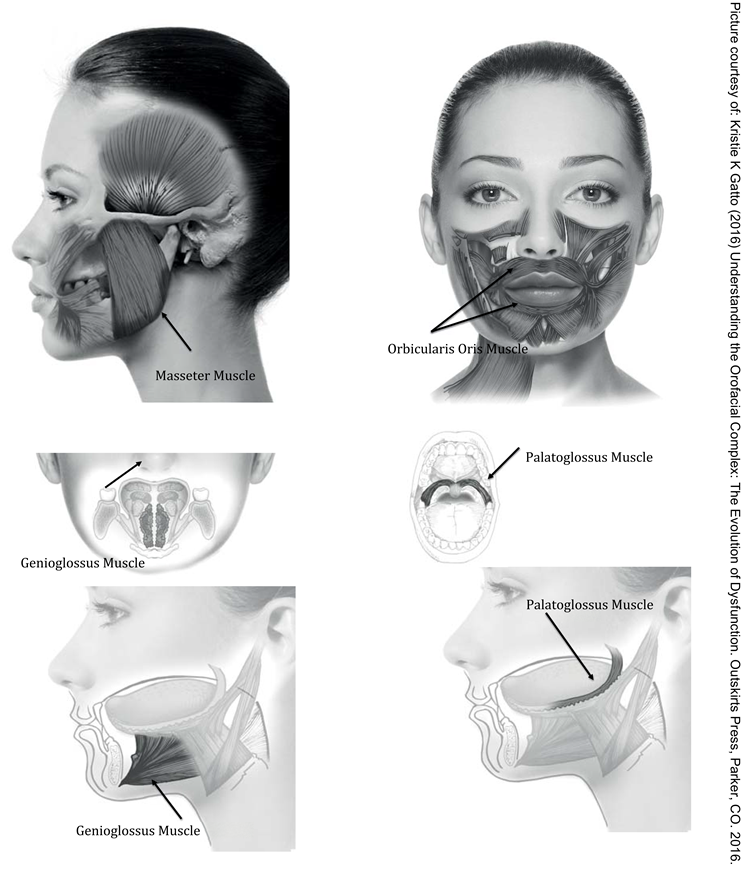
In recent years many studies on the anatomy and neurophysiology of muscles of the face and mouth have revealed a much more complex ability of muscles to “multitask”. Apart from being involved in sucking, swallowing, chewing and speaking, some muscles are also involved in the patency of the airways during sleep, the postural position of the jaw during physical activities, the promotion of nasal breathing, or stress relief, just to name a few functions. In this brief review, some additional properties of the genioglossus (GG), the palatoglossus (PG), the masseters (MM) and the orbicularis oris (OO) will be reviewed.

INTRODUCTION

Speech-language pathologists and myofunctional therapists usually consider orofacial muscles from the point-of-view of sucking, swallowing, chewing and speaking, but several orofacial muscles have other interesting purposes. This article will focus only on 4 muscles: The genioglossus, the palatoglossus, the masseters and the orbicularis oris. It should come to no surprise that these four muscles in particular adapt their function to “serve” breathing first and foremost. Therefore, considering these muscles through the lens of breathing helps the clinician identify and appreciate the common function that ties these (and other) orofacial muscles.

Genioglossus (GG)

The genioglossus (GG) is an extrinsic single tongue muscle, mainly responsible for tongue thrust, as it moves the tongue toward the origin of the muscle, which is the genial tubercle in the lingual face of the mandible. The GG is regulated by the 12th pair of cranial nerves, in plexus with the Vagus nerve (10th), receiving information from both the swallowing and the breathing central pattern generators in the brain and making the GG both a swallowing and a breathing muscle (Sawczuck & Mosier, 2001; Saboisky, Butler, Fogel, Taylor, Trinder, White, & Gandevia, 2006; McFarland & Lund, 1994; McFarland & Lund, 1995). Its purpose is to assist the propulsion of food into the esophagus but most importantly to keep the upper airway open at all times. The GG has sensors for negative pressure (such as during snoring or sleep apnea) and excessive CO2, which triggers the anterior thrust of the tongue to increase airway space (Fogel, Malhotra, & White, 2004, Mortimore, Mathur, & Douglas, 1995; Napadow, Chen, Weeden, & Gilbert, 1999). It is considered a “dilator muscle”, that is active in the supine position and during sleep (Arens & Marcus, 2003; Mathur, Mortimore, Jan, & Douglas,1995; Malhotra, Pillar, Fogel, Edwards, Ayas, Akahoshi, Hess, & White, 2002; Jordan, White, Lo, Wellman, Eckert, Yim-Yeh, Eikermann, Smith, Stevenson, & Malhotra, 2009; Popovic & White, 1995).
During evaluation, it’s important to distinguish between a tongue thrust due to a restricted lingual frenum or a tongue thrust due to breathing issues, during the day and during the night.
A tongue thrust behavior that originate in poor nasal breathing will surely require a multidisciplinary approach to address the primary dysfunction, in which myofunctional therapy is one of the modalities to be implemented.
Ijom 42 00061 i001
A therapeutic approach is to promote the stable resting position of the tongue against the palate achieved by creating an area of negative pressure during swallowing that can be maintained even for prolonged periods (Engelke, Jung, & Knosel, 2011) daytime and nighttime. Other exercises for the genioglossus are described in Guimarães, Drager, Genta, Marcondes, & Lorenzi-Filh, 2009.

Palatoglossus (PG)

The palatoglossus (PG) are paired muscles that originate from the raphe between the hard and soft palate and inserts in the posterior lateral sides of the tongue. When activated it pulls the back of the tongue up and the soft palate forward, therefore increasing the free space in the airway (Patil, Schneider, Marx, Gladmon, Schwartz, & Smith, 2006; Sant’Ambrogio, Tsubone, & Sant’Ambrogio,1995; Shea, Edwards, & White, 1999). Although it functions as an extrinsic tongue muscle, usually it’s not considered as such, as it is innervated by the Vagus nerves (10th), making it a breathing muscle. This pair of muscles is more active when laying supine and breathing nasally, and it’s also considered a “dilator” muscle which is crucial for optimal sleep breathing (Arens & Marcus, 2003; Mathur et al.,1995; Malhotra et al., 2002; Jordan et al., 2009; Popovic & White, 1995).
During the evaluation of a patient, the ability to create a suction with the tongue within the oral cavity would guide the therapy. The negative pressure created in the oral cavity, will maintain the palatoglossus and the soft palate in a physiologically optimal position for nasal breathing.

Masseters (MM)

Clearly the masseters (MM) are very important for chewing, being the “crushers” of food. In fact, if the masseters are not activated in biting, that indicates posterior dental malocclusion. However, the MM (like the temporalis muscles) are the only orofacial muscles that have a crisp “stretch reflex” similar to skeletal muscles (Clark, 2003; Miles, Flavel, & Nordstrom, 2004), making them an important postural system, in which the tone of the MM allows for optimal jaw position during activities such as running, jumping or walking (Miles, 2007). Thanks to the stretch reflex, and unlike other orofacial muscles, their repeated activity in chewing and clenching causes an increase in the muscle mass. The MM are active in chewing and clenching, which are functions considered natural stress relievers (Gómez, Ortega, Horrillo, & Meana, 2010; Hori et al., 2005). They are also active in bruxing (Gallo, Gross & Palla, 1999), which is involved in micro-arousals to limit the damage of sleep apnea, as the function of the MM depends on, and it is influenced by breathing (Hyama, Ono, Ishiwata, Kuroda, & Ohyama, 2003). Finally, the MM are involved with strength exertion (as in weight lifting or bearing down) and with expression of aggression. Excessive chewing and clenching may become a parafunction with deleterious effects on oral structures and functions.
During evaluation, the MM can be assessed with just clenching, chewing on various foods with increasingly harder consistency or, in research, chewing on “colorimetric beads” that allows us to visually measure the pressure of the masseters (Escudiero-Santos, 2006).
Prevention of the masseters’ dysfunction should be a therapeutic goal facilitated by encouraging breastfeeding, which promotes optimal muscle function and development, and by encouraging the introduction of appropriate chewable foods in weaning. If therapy is required, it may address the hyper-functions (such as chewing gum, clenching when stressed, or sleep bruxing) or the hypo-functions (such as dental malocclusion, habitual soft textured diet, or open mouth posture), in a multidisciplinary manner. Therapy may focus on the masseters’ function in cases of TMJ rehabilitation, facial pain or post-surgery resection, which is best accomplished within a multidisciplinary treating team (De Felício, Ferreira, Medeiros, Rodrigues Da Silva, Tartaglia, & Sforza, 2011; Tartaglia, Lodetti, Paiva, De Felicio, & Sforza, 2011). Chewing proper food and sham chewing with silicon wafers or latex/rubber tubes are also strategies used to recover masseters’ function.

Orbicularis Oris

Apart from the orbicularis oris’ fundamental purpose of sealing off the mouth during sucking, suckling, chewing and swallowing and in speech articulation, it is involved in providing optimal breathing when the nasal passages are somewhat congested (Busanello-Stella, Blanco- Dutra, Corrêa, & Silva, 2015) and during sleep (Eastwood, Curran, Smith, & Dempsey, 1998; Fitzpatrick, McLean, Urton, Tan, O’Donnell, & Driver, 2003). Also, when closed, lips aide in the proper positioning of the jaw, and therefore of the tongue. When the lips are open, the jaw is open which results in excessive freeway space that may be damaging for the temporomandibular joints. The somatosensory cortex of the brain (as well as the somatosensory motor cortex) has connections from individual lips (upper and lower) but also from lips together (Toda & Taoka, 2002). The latter projection is as large as the projected connection from the whole face (Baumgartner et al, 1992), thus neurologically dictating the importance of keeping the lips closed. Moreover, the orbicularis oris has some stretch reflex (Clark, 2003) suggesting that closed lips are also part of the postural system that keeps the jaw in the proper position.
During evaluation, the ability to breathe nasally, normally, should be assessed and mouth opening at night should be inquired. The ability to keep the tongue up against the palate in a suction should also be assessed, as the proper tongue position aids the proper closure of the orbicularis oris (Engelke, Jung, & Knosel, 2011). Multidisciplinary therapy for lip closure involves addressing possible allergies and some significant malocclusions, proper nasal breathing training, possible sensory-kinesio-taping, and myofunctional therapy. Myofunctional therapy has been expanding its applications as the intricate functions (and dysfunctions) of orofacial muscles has become more and more apparent, either through publication of recent studies or through novel connections between well know articles published long time ago. A new appreciation of the multiple functions of orofacial muscles may assist the clinician in considering additional variables when assessing patients; when determining a course of action; when coaching preventative strategies, and when making a referral. Above all, the clinician should assess nasal breathing, compatibly within his or her scope of practice, in order to identify and/or rule out breathing difficulties that may impact orofacial muscles. Current standard of care requires the clinician to consider all of these variables, because muscle physiology is an ever expanding field and therapists can now find so many more ways to help their patients than ever before.

References

  1. Arens, R.; Marcus, C. L. Pathophysiology of upper airway obstruction: A developmental perspective. Sleep 2003, 27, 997–1019. [Google Scholar]
  2. Baumgartner, C.; Barth, D. S.; Levesque, M. F.; Sutherling, W. W. Human hand and lip sensorimotor cortex as studied on electrocorticography. Electroencephalography Clinical Neurophysiology 1992, 84(2), 115–126. [Google Scholar] [CrossRef] [PubMed]
  3. Busanello-Stella, A. R.; Blanco-Dutra, A. P.; Corrêa, E. C.; Silva, A. M. Electromyographic fatigue of orbicular oris muscles during exercises in mouth and nasal breathing children. CoDAs 2015, 27(1), 80–88, [Article in English, Portuguese]. [Google Scholar] [CrossRef] [PubMed]
  4. Clark, H. M. Neuromuscular treatments for speech and swallowing: A tutorial. American Journal of Speech Language Pathology 2003, 12, 400–415. [Google Scholar]
  5. De Felício, C. M.; Ferreira, C. L.; Medeiros, A. P.; Rodrigues Da Silva, M. A.; Tartaglia, G. M.; Sforza, C. Electromyographic indices, orofacial myofunctional status and temporomandibular disorders severity: A correlation study. Journal of Electromyography Kinesiology 2012, 22(2), 266–272. [Google Scholar]
  6. Eastwood, P. R.; Curran, A. K.; Smith, C. A.; Dempsey, J. A. Effect of upper airway negative pressure on inspiratory drive during sleep. Journal of Applied Physiology 1998, 84, 1063–1075. [Google Scholar]
  7. Engelke, W.; Jung, K.; Knosel, M.W.E. Intra-oral compartment pressures: A biofunctional model and experimental measurements under different conditions of posture. Journal of Clinical Oral Investigations 2011, 15(2), 165–176. [Google Scholar]
  8. Escudiero Santos, C.; de Freitas, O.; Spadaro, A. C. C.; Mestriner-Junior, W. Development of a Colorimetric System for Evaluation of the Masticatory Efficiency. Brazilian Dental Journal 2006, 17(2), 9599. [Google Scholar]
  9. Fitzpatrick, M. F.; McLean, H.; Urton, A. M.; Tan, A.; O’Donnell, D.; Driver, H. S. Effect of nasal or oral breathing route on upper airway resistance during sleep. European Respiratory Journal 2003, 22, 827–832. [Google Scholar]
  10. Fogel, R. B.; Malhotra, A.; White, P. Pathophysiology of obstructive sleep Apnoea/Hypopnoea Syndrome. Thorax 2004, 59, 159–163. [Google Scholar] [CrossRef]
  11. Gallo, L. M.; Gross, S. S.; Palla, S. Nocturnal masseter activity of healthy subjects in a natural environment. Journal of Dental Research 1999, 78, 1436–1444. [Google Scholar] [PubMed]
  12. Gatto, K.K. Understanding the orofacial complex: The evolution of dysfunction; Outskirts Press, 2016. [Google Scholar]
  13. Gómez, F.M.; Ortega, J.E.; Horrillo, I.; Meana, J.J. Relationship between non-functional masticatory activity and central dopamine in stressed rats. Journal of Oral Rehabilitation 2010, 37, 827–833. [Google Scholar] [PubMed]
  14. Guimarães, K.C.; Drager, L.F.; Genta, P.R.; Marcondes, B.F.; Lorenzi-Filho, G. Effects of Oropharyngeal Exercises on Patients with Moderate Obstructive Sleep Apnea Syndrome. American Journal of Respiratory and Critical Care Medicine 2009, 15, 179 & 962–966. [Google Scholar]
  15. Hyama, S.; Ono, T.; Ishiwata, Y.; Kuroda, T.; Ohyama, K. Effects of experimental nasal obstruction on human masseter and suprahyoid muscle activities during sleep. Angle Orthodontist 2003, 73(2), 151–157. [Google Scholar]
  16. Hori, N.; Lee, M. C.; Sasaguri, K.; Ishii, H.; Kamei, M.; Kimoto, K.; Toyoda, M.; Sato, S. Suppression of Stress-induced nNOS Expression in the Rat Hypothalamus by Biting. Journal of Dental Research 2005, 84(7), 624–628. [Google Scholar]
  17. Jordan, A. S.; White, D. P.; Lo, Y. L.; Wellman, A.; Eckert, D. J.; Yim-Yeh, S.; Eikermann, M.; Smith, S. A.; Stevenson, K. E.; Malhotra, A. Airway Dilator Muscle Activity and Lung Volume During Stable Breathing in Obstructive Sleep Apnea. Sleep 2009, 32(3), 361–368. [Google Scholar]
  18. Malhotra, A.; Pillar, G.; Fogel, R. B.; Edwards, J. K.; Ayas, N.; Akahoshi, T.; Hess, D.; White, D. P. Pharyngeal pressure and flow effects on genioglossus activation in normal subjects. American Journal of Respiratory Critical Care and Medicine 2002, 165, 71–77. [Google Scholar]
  19. Mathur, R.; Mortimore, I. L.; Jan, M. A.; Douglas, N. J. Effect of breathing, pressure and posture on palatoglossal and genioglossal tone. Clinical Science 1995, 89, 441–445. [Google Scholar]
  20. McFarland, D. H.; Lund, J. P. Modification of mastication and respiration during swallowing in the adult human. Journal of Neurophysiology 1995, 74, 1509–1517. [Google Scholar]
  21. McFarland, D. H.; Lund, J. P.; Gagner, M. Effects of posture on the coordination of respiration and swallowing. Journal of Neurophysiology 1994, 72, 2431–2437. [Google Scholar]
  22. Miles, T. S.; Flavel, S. C.; Nordstrom, M. A. Stretch reflexes in the human masticatory muscles: A brief review and a new functional role. Human Movement Science 2004, 40, 337–349. [Google Scholar] [CrossRef] [PubMed]
  23. Miles, T. S. Postural control of the human mandible. Archives of Oral Biology 2007, 52(4), 347–352. [Google Scholar] [CrossRef] [PubMed]
  24. Mortimore, I. L.; Mathur, R.; Douglas, N. J. Effect of posture, route of respiration, and negative pressure on palatal muscle activity in humans. Journal of Applied Physiology 1995, 79(2), 448–454. [Google Scholar] [CrossRef] [PubMed]
  25. Napadow, V. J.; Chen, Q.; Weeden, V. J.; Gilbert, R. J. Intramural mechanics of the human tongue in association with physiological deformation. Journal of Biomechanics 1999, 32, 1–12. [Google Scholar] [CrossRef]
  26. Patil, S.P.; Schneider, H.; Marx, J.J.; Gladmon, E.; Schwartz, A.R.; Smith, P.L. Neuromechanical Control of Upper Airway Patency During Sleep. Journal of Applied Physiology 2006, 102(2), 547–556. [Google Scholar] [CrossRef]
  27. Popovic, R.M.; White, D.P. Influence of waking genioglossal electromyogram and upper airway resistance. American Journal of Respiratory Critical Care and Medicine 1995, 152, 725–731. [Google Scholar] [CrossRef]
  28. Saboisky, J. P.; Butler, J. E.; Fogel, R. B.; Taylor, J. L.; Trinder, J. A.; White, D. P.; Gandevia, S. C. Tonic and Phasic Respiratory Drives to Human Genioglossus Motoneurons During Breathing. Journal of Neurophysiology 2006, 95(4), 2213–2221. [Google Scholar] [CrossRef]
  29. Sant’Ambrogio, G.; Tsubone, H.; Sant’Ambrogio, F. B. Sensory information from the upper airway: Role in the control of breathing. Respiratory Physiology 1995, 1021-16. [Google Scholar] [CrossRef]
  30. Sawczuk, A.; Mosier, K. M. Neural Control of Tongue Movement With Respect To Respiration and Swallowing. Critical Reviews in Oral Biology & Medicine 2001, 12(l), 18–37. [Google Scholar]
  31. Shea, S. A.; Edwards, J. K.; White, D. P. Effect of wake-sleep transitions and rapid eye movement sleep on pharyngeal muscle response to negative pressure in humans. Journal of Physiology 1999, 520 Pt 3, 897–908. [Google Scholar] [CrossRef]
  32. Tartaglia, G. M.; Lodetti, G.; Paiva, G.; De Felicio, C. M.; Sforza, C. Surface electromyographic assessment of patients with long lasting temporomandibular joint disorder pain. Journal of Electromyography and Kinesiology 2011, 21(4), 659–64. [Google Scholar] [CrossRef]
  33. Toda, T.; Taoka, M. Integration of the upper and lower lips in the postcentral area 2 of conscious macaque monkeys (Macaca fuscata). Archives of Oral Biology 2002, 47(6), 449–456. [Google Scholar] [CrossRef]