Next Article in Journal
Negative Pressure and Muscle Activity During Discrete Sips from High Resistance Straws
Previous Article in Journal
Oral and Oral Preparatory Phase of Swallowing in Children with Tonsil Hypertrophy: Videofluoroscopy Study
 
 
International Journal of Orofacial Myology and Myofunctional Therapy is published by MDPI from Volume 51 Issue 1 (2025). Previous articles were published by another publisher in Open Access under a CC-BY (or CC-BY-NC-ND) licence, and they are hosted by MDPI on mdpi.com as a courtesy and upon agreement with the previous journal publisher.
Article

Maximum Protrusive Tongue Force in Healthy Young Adults

by
Monalise Costa Batista Berbert
1,
Vivian Garro Brito
2,
Renata Maria Moreira Moraes Furlan
3,
Tatiana Vargas de Castro Perilo
4,
Amanda Freitas Valentim
4,
Marcio Falcao Santos Barroso
5,
Estevam Barbosa de Las Casas
6 and
Andréa Rodrigues Motta
7
1
Universidade Federal do Rio Grande do Sul (UFRGS), R. Felipe Camarão, 712, Apart 21, Porto Alegre 90035140, Brazil
2
Graduate Program in Electrical Engineering, Universidade Federal de Minas Gerais (UFMG), Av. Antônio Carlos, 6627, Escola de Engenharia, Bloco I, Sala 4205, Belo Horizonte 31210-000, Brazil
3
Graduate Program in Structural Engineering, Universidade Federal de Minas Gerais (UFMG), Av. Antônio Carlos, 6627, Escola de Engenharia, Bloco I, Sala 4205, Belo Horizonte 31210-000, Brazil
4
Universidade Federal de Minas Gerais (UFMG), Av. Antônio Carlos, 6627, Escola de Engenharia, Bloco I, Sala 4205, Belo Horizonte 31210-000, Brazil
5
Electrical Engineering Departamento de Engenharia Elétrica, Sala 2.24-E, Universidade Federal de São João del-Rei, Praça Frei (UFSJ), Orlando 170, Centro, São João del-Rei 36307-352, Brazil
6
Department of Structural Engineering, Universidade Federal de Minas Gerais (UFMG), Av. Antônio Carlos, 6627, Escola de Engenharia, Bloco I, Sala 4205, Belo Horizonte 31210-000, Brazil
7
Department of Speech Language Pathology and Audiology, Universidade Federal de Minas Gerais (UFMG), Rua João Chagas, 333/1002, União, Belo Horizonte 31170-370, Brazil
Int. J. Orofac. Myol. Myofunct. Ther. 2014, 40(1), 56-63; https://doi.org/10.52010/ijom.2014.40.1.5
Submission received: 1 November 2014 / Revised: 1 November 2014 / Accepted: 1 November 2014 / Published: 1 November 2014

Abstract

:
In clinical speech-language pathology practice, tongue force is usually evaluated qualitatively. Perception and practical experience are used to classify this force. The Biomechanical Engineering Group from the Federal University of Minas Gerais developed an instrument to quantify tongue force. The purposes of this study were to quantify maximum tongue protrusion force in Brazilian subjects with normal tongue strength and to compare force values between gender groups. In total, 105 subjects, 43 men and 62 women, aged from 18 to 29 years, with normal tongue strength according to qualitative evaluation, underwent quantitative evaluation by using the instrument. The mean of the maximum tongue force values of all participants was 17.58 ± 7.95 N. There were significant differences in the median values for maximum tongue forces between the genders, with higher values observed for men. In intersubject comparisons, high variation coefficients were evident due to the variability among individuals. However, the study suggested that the instrument could be an interesting tool for intrasubject comparisons, especially during the follow-up.

INTRODUCTION

Poor tongue strength has been related to disturbances in swallowing (Yoshida, Kikutani, Tsuga, Utanohara, Hayashi & Akagawa, 2006) and speech articulation (Dworkin, Aronson & Mulder, 1980). In clinical speech-language pathology practice, tongue strength is evaluated in a subjective way by asking the patient to press the tip of their tongue against a tongue depressor, with resistance provided by the examiner (Clark, Henson, Barber, Stierwalt & Sherril, 2003; Solomon, 2004; Solomon, Clark, Makashay & Newman, 2008). This kind of evaluation is influenced by the experience of the professional and it hinders diagnosis and follow-up.
Several methods of quantifying tongue strength have been reported. Some instruments have a small pressure sensor placed in a palatal appliance or a replica denture (Robbins, Gangnon, Theis, Kays, Hewitt & Hind, 2005; Ruan, Chen, Gu, Lu, Su & Guo, 2005; Hewitt, Hind, Kays, Nicosia, Doyle, Tompkins, Gangnon & Robbins, 2008; Kieser, Singh, Swain, Ichim, Waddell, Kennedy, Foster & Livingstone, 2008; Hori, Ono, Tamine, Kondo, Hamanaka, Maeda, Dong & Hatsuda, 2009). Other instruments have a balloon which is filled with air and connected to a pressure sensor (Clark et al., 2003; McAuliffe, Ward, Murdoch & Farrel, 2005; Ball, Idel, Cotton & Perry, 2006; Utanohara, Hayashi, Yoshikawa, Yoshida, Tsuga & Akagawa, 2008). Some researchers have developed adapted dynamometers (Posen, 1972; Trawitzki, Borges, Giglio & Silva, 2011). Despite the reports of these instruments, there is currently no instrument commercially available in Brazil.
Recognizing the need for an objective method of measuring tongue protrusion force, the Biomechanical Engineering Group of the Federal University of Minas Gerais has developed the FORLING, the first such device developed in Brazil (Motta, Perim, Perilo, Las Casas, Costa, Magalhaes & Saffar, 2004; Barroso, Costa, Saffar, Las Casas, Motta, Perilo, Batista & Britto, 2009; Furlan, Valentim, Perilo, Costa, Barroso, Las Casas & Motta, 2010; Furlan, Motta, Valentim, Barroso, Costa & Las Casas, 2013). One advantage of this device is that it measures tongue protrusion force in a way that is similar to what is done in the clinical qualitative evaluation, but it does not measure tongue force during elevation or lateralization.
In the primary tests using this instrument, the values obtained in the quantitative assessment were compatible with those reported by other researchers and there was agreement between qualitative and quantitative evaluations (Motta et al., 2004; Barroso et al., 2009; Furlan et al., 2013). However, standard values or normal ranges of tongue pressure according to gender have not yet been established for the Brazilian population. The present study aimed to quantify maximum and average tongue forces of subjeets with normal tongue strength and to compare these force values between gender groups.

MATERIALS AND METHODS

A cross-sectional study was developed at the Federal University of Minas Gerais (UFMG) Clinics Hospital, after approval from the University Ethics Committee (registration number 498/05). Informed consent was obtained from the subjects.
  • Subjects
The sample consisted of 43 men and 62 women, with ages ranging from 18 to 29 years, and with a diagnosis of normal tongue strength as determined by two speech language pathologists via qualitative evaluation. All subjects were students or employees of the UFMG Medical School. Subjects were also excluded from the study if they had severe occlusion problems that made it impossible to fit the device correctly into the mouth, hearing impairment, or any physical or physiological disturbance that could interfere with the results of this research.
  • Subjective (Qualitative) Evaluation
Subjects underwent clinical qualitative evaluation of tongue strength by protruding the tip of their tongue while using maximal effort against a tongue depressor and against the finger of the examiner when placed in front of the mouth for approximately 10 s, with resistance provided by the examiner (Clark et al., 2003; Furlan et al., 2010). The evaluation was performed by two speech language pathologists and only individuals who were classified as having normal tongue strength by both examiners were selected. In this 10-second evaluation the therapist is able to observe the behavior of tongue protrusion force against resistance during isometric contraction in an amount of time similarly to those recommended in some isometric exercises (Felicio, 2009), and also see whether there is presence of fatigue in this situation.
  • Quantitative Evaluation
Quantitative, measurable, and recordable evaluation of tongue strength was accomplished using the FORLING (Motta et al., 2004; Barroso et al., 2009; Furlan et al., 2010; Furlan et al., 2013). This device, shown in Figure 1, consists of a piston/cylinder set attached to a double silicone protector and to a head that connects it to the cylinder that hydraulically transmits the force exerted to a pressure sensor. The pressure sensor measurements are transmitted through a data acquisition device to a personal computer. Data processing is performed by software that provides auditory cues to the subject at the beginning and end of each test and records the tongue force exerted by subjects over time, continuously throughout the test (Motta et al., 2004). Instrument calibration was performed at the Department of Physical Tests at the Minas Gerais Technology Foundation Center (Cetec), and instrument uncertainty was determined to be below 0.18% of the lingual force (Barroso et al., 2009).
During quantitative evaluation, the instrument was fitted into the participant's mouth so that the teeth remained in the mouthguard. The mouthguard is used to keep the instrument attached to the mouth and to control the level of mouth opening. The extent of jaw separation was reported by Solomon and Munson (2004) to affect the pressure the tongue is able to exert in an elevation task. In a protrusion task, jaw position might be also important as the tongue is coupled with the mandible and could be moved forward by the mandible which would potentially inflate the results.
A period of 15 s after the initation of the test was considered as an adjustment time. Each subject was then instructed to push the cylinder head with their tongue, using the maximum force they could exert, and to maintain that force for 10 s. Three trials were conducted and recorded, with 1-min intervals between each trial. A sound alarm signaled the beginning and the end of each trial.
  • Data Analysis
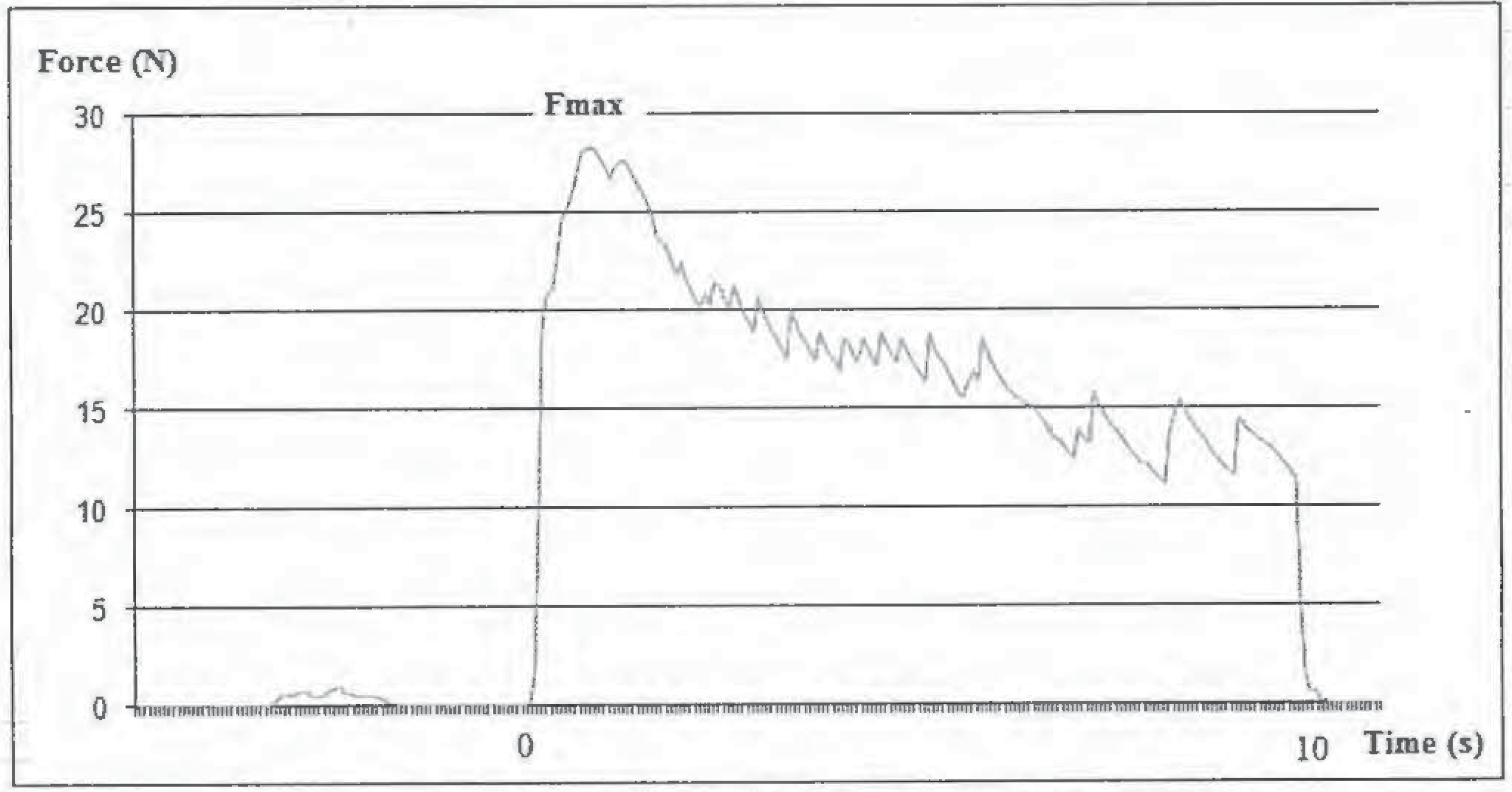
Signals were acquired and processed by a microcomputer by using software developed specifically for this purpose. The software records the force exerted throughout the trial and graphs it in real-time, as depicted in Figure 2.
The parameter analyzed was maximum force, defined as the highest peak force and recorded in Newtons (N).
  • Statistical Analysis
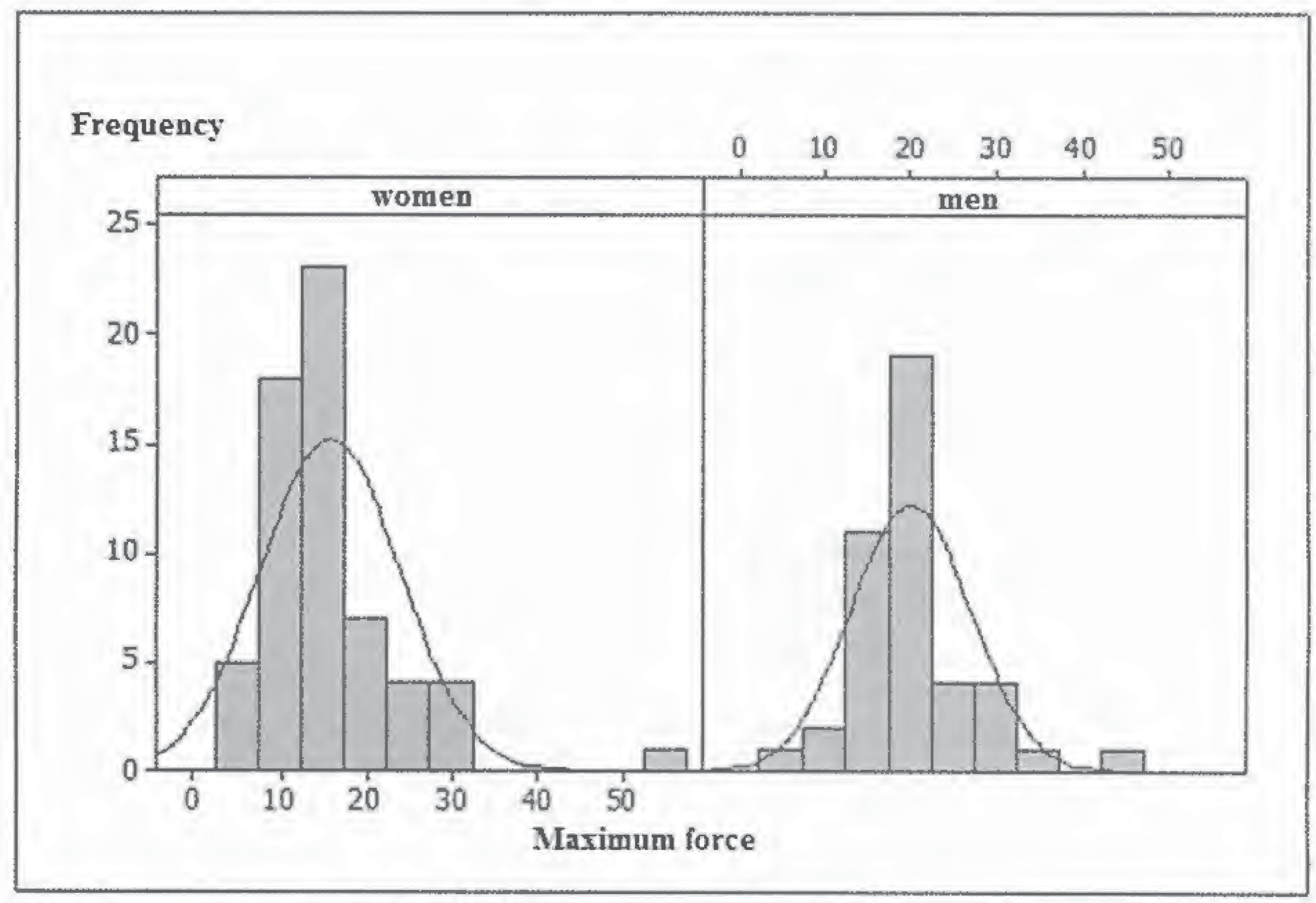
Analysis of the data distribution was accomplished using histograms. A descriptive analysis of the data was performed and tests of hypotheses were undertaken to investigate potential differences between groups. Anderson-Darling test was applied to determine normality of the data. The normality test suggested that the distribution of the data was not normal. Thus, the average was not considered to be the best descriptive parameter of the data, as averages are sensitive to extreme values; therefore, the median was the parameter chosen as the central value. The Mann-Whitney non parametric test was used to analyze differences between the medians of the groups. The Friedman test was conducted to compare force values of the same participant in different trials. The level of statistical significance was set at p < 0.05. Additionally, variation coefficients were used to analyze homogeneity of the results.

RESULTS

Maximum tongue force of each group is shown in Table 1. Coefficients of variation were considered high for both genders, even if the outliers were eliminated (42.8 N and 57.18 N by one male and one female, respectively). Women presented higher coefficient of variation than men.
There were significant differences in the medians of maximum forces by gender, and the median values for men were higher than those for women. A p value of less than 0.001 was obtained.
Comparing the maximum force values produced across three trials by the same subject, the coefficients of variation were lower than those yielded by intersubject comparisons, averaging 14.88%.
There were significant differences in the medians of maximum forces comparing trials 1 and 2 or 1 and 3 (p < 0.05). The first trial was higher than trial 2 and trial 3, and there was no difference between trials 2 and 3. For most of the participants the first trial presented the highest maximum force value.

DISCUSSION

Data from this research were obtained through a quantitative method for the measurement of tongue protrusion force, in an effort to minimize the subjectivity of the evaluation. In this method, tongue force is evaluated during protrusion, as it is in the subjective (qualitative) evaluation.
Motta et al. (2004) have hypothesized that based on the tongue protrusion force capacity of a subject, it is possible to infer their capacity to accomplish other tasks. This is because the muscles responsible for tongue protrusion, the genioglossus, verticalis and transversus muscles (Pittman & Bailey, 2008), participate actively in multiple functions. There is constant interaction between extrinsic and intrinsic tongue muscles in all of these functions.
The values obtained in this study for maximum tongue force (average of 17.58 ± 7.95 N) were comparable to those reported by other authors who have measured tongue protrusion force in normal subjects: Kydd (1956) (maximum force 23.13 N); Posen (1972) (maximum force between 6 N and 25 N); Dworkin et al. (1980) (maximum force 32.9 N for men and 27.5 N for women); Mortimore, Fiddes, Stephens & Douglas (1999) (maximum force of 26 ± 8 N for men and 20 ± 7 N for women); Motta et al. (2004) (maximum force between 21.1 N and 25.7 N); and Furlan et al. (2013) (maximum force 18.91 ± 7.95).
Force measurements were compared between genders. Maximum force was significantly lower in women compared to men. This may be explained by the presence of greater muscular mass in men (Stierwalt & Youmans, 2007). Examinations of sex differences in tongue protrusion strength are mixed. Some studies have found that men exhibit greater tongue protrusion strength than women (Dworkin et al., 1980; Mortimore et al., 1999), others have reported no differences between men and women on this measure (Clark & Solomon, 2012).
When comparing tongue force between trials, it was noted that the strength decreased systematically after the first trial reflecting the effect of fatigue due to the 10-second contraction at maximum effort levels. Generally, the highest value was produced in the first trial. Figure 2 shows the presence of fatigue even in the same trial. It was observed for participants with normal tongue strength, that the maximum value is obtained in the first seconds of the measurement, after that the force produced by the tongue decreases. This suggests that, for those participants, a short time contraction during evaluation is preferable to avoid or reduce the effects of fatigue.
While all participants had been classified as having normal tongue strength by two examiners via a qualitative evaluation, high variation in force values was observed among the subjects. This evaluation showed that normal tongue force encompasses a wide range. Even when the subjects were divided by gender, high variation coefficients were observed. Data derived from women had higher variability. Other studies have also reported substantial variability in tongue strength measures across participants (Clari & Solomon, 2012). A likely explanation for these discrepancies is the biological variability of individuals , as the values obtained are influenced directly by each individual’s anatomical and physiological constitution. Differences in motivation can also influence the measures. Other sources of variability may relate to the method used. Clark and Solomon (2012) found that variability is higher for tongue protrusion than for tongue elevation force.
Due to individual variability, a comparison of tongue force values among patients is not efficient, even if they have been classified as having normal tongue force in a qualitative evaluation. However, intrasubject comparisons can be interesting and useful as they allow the monitoring of progress during treatment and follow-up of patients. Force values and graph shape can be used to motivate the patient during treatment and also to facilitate a patient’s understanding of their condition.
Maximum force was adequate to characterize tongue force (Motta et al., 2004; Barroso et al., 2009). Even though mastication, sucking, swallowing, and speech functions performed by the tongue do not require maximum force, subjects deficient in maximum tongue strength experience less functionalreserve in proportion to the force needed to perform an activity and the muscles tire more quickly (Burkhead, Sapienza & Rosenbek, 2007). Robbins et al. (2005) measured tongue force in the elderly and verified that the higher the force they were able to exert in isometric contraction, the higher the force was that they exhibited during swallowing. Yoshida et al. (2006) observed that tongue pressure is related to clinical signs of dysphagia and suggested that measurement of tongue pressure is useful for the evaluation of swallowing function.
The device developed by the Biomechanical Engineering Group from UFMG was a good tool to quantify tongue force. Its use in an orofacial myology evaluation can assist speech-language pathologists during the assessment of tongue force, and facilitate more effective follow-up of patients by quantifying the gain in force resulting from treatment. There is a lack of studies of tongue force in Brazilians, and up to now the largest examined sample is the one described herein. These results may have significant implications in clinical practice for professionals responsible for the evaluation and rehabilitation of oral forces.

CONCLUSIONS

Overall, a maximum tongue force value of 17.58 ± 7.95 N measured across a 10-s maximum-effort protrusive task was observed. There were significant differences in tongue force between genders, with the highest values observed in men. In intersubject comparisons, high variation coefficients were evident. This suggests that these data are of little clinical relevance, due to the variability among individuals. However, the instrument could be a valuable tool for intrasubject comparisons, facilitating more efficient patient follow up. Using this device the speech language pathologist could monitor the force values for each patient and quantify their progress as a result of treatment over time, as well as monitoring decreases in force in the case of degenerative diseases.

Acknowledgments

The authors acknowledge support from the Brazilian research funding agencies Fundayao de Amparo a Pesquisa do Estado de Minas Gerais (FAPEMIG), Coordenayao de Aperfeir;:oamento de Pessoal de Nlvel Superior (CAPES) e Conselho Nacional de Desenvolvimento Cientifico e Tecnol6gico (CNPq).

References

  1. Ball, S., O. Idel, S. Cotton, and A. Perry. 2006. Comparison of two methods for measuring tongue pressure during swallowing in people with head and neck cancer. Dysphagia 21: 28–37. [Google Scholar]
  2. Barroso, M. F. S., C. G. Costa, J. M. E. Saffar, E. B. Las Casas, A. R. Motta, T. V. C. Perilo, M. C. Batista, and V. G. Brito. 2009. Development of a prototype system for the objective measurement of tongue forces. SBA Controle & Automacao 20, 2: 156–163. (In Portuguese). [Google Scholar]
  3. Burkhead, L. M., C. M. Sapienza, and J. C. Rosenbek. 2007. Strength-training exercise in dysphagia rehabilitation: Principles, procedures, and directions for future research. Dysphagia 22: 251–265. [Google Scholar]
  4. Clark, H. M., P. A. Henson, W. D. Barber, J. A. G. Stierwalt, and M. Sherrill. 2003. Relationsh ips among subjective and objective measures of tongue strength and oral phase swallowing impairments. American Journal of Speech-Language Pathology 12: 40–50. [Google Scholar]
  5. Clark, H. M., and N. P. Solomon. 2012. Age and sex differences in orofacial strength. Dysphagia 27: 2–9. [Google Scholar] [PubMed]
  6. Dworkin, J. P., A. E. Aronson, and D. W. Mulder. 1980. Tongue force in normals and in dysarthric patients with amyotrophic lateral sclerosis. Journal of Speech and Hearing Research 23, 4: 828–837. [Google Scholar] [PubMed]
  7. Felicio, C. M. 2009. Edited by C. M. Felicio and L. W. Trawitzki. Desordens temporomandibulares: terapia fonoaudiol6gcia. In Interfaces da Medicina, Odontologia e Fonoaudiologia no complexo cervico-craniofacial. Barueri: Pro-Fono: Vol. 1. [Google Scholar]
  8. Furlan, R. M. M. M., A. R. Motta, A. F. Valentim, M. F. S. Barroso, C. G. Costa, and E. B. Las Casas. 2013. Protrusive tongue strength in people with severely weak tongues. International Journal of Speech-Language Pathology 1: 1–8. [Google Scholar]
  9. Furlan, R. M. M. M., A. F. Valentim, T. V. C. Perilo, C. G. Costa, M. F. S. Barroso, E. B. Las Casas, and A. R. Motta. 2010. Quantitative evaluation of tongue protrusion force. International Journal of Orofacial Myology 36: 33–43. [Google Scholar]
  10. Hewitt, A., J. Hind, S. Kays, M. Nicosia, J. Doyle, W. Tompkins, R. Gangnon, and J. Robbins. 2008. Standardized Instrument for Lingual Pressure Measurement. Dysphagia 23: 16–25. [Google Scholar]
  11. Hori, K., T. Ono, K. Tamine, J. Kondo, S. Hamanaka, Y. Maeda, J. Dong, and M. Hatsuda. 2009. Newly developed sensor sheet for measuring tongue pressure during swallowing. Journal of Prosthodontic Research 53: 28–32. [Google Scholar]
  12. Kieser, J., B. Singh, M. Swain, I. Ichim, J. N. Waddell, D. Kennedy, K. Foster, and V. Livingstone. 2008. Measuring intraoral pressure: Adaptation of a dental appliance allows measurement during function. Dysphagia 23: 237–243. [Google Scholar] [PubMed]
  13. Kydd, W. 1956. Quantitative analyses offorce of the tongue. Journal of Dental Research 35: 171–174. [Google Scholar]
  14. McAuliffe, M. J., E. C. Ward, B. E. Murdoch, and A. M. Farrell. 2005. A nonspeech investigation of tongue function in Parkinson’s disease. The Journals of Gerontology Series A Biological Sciences and Medical Sciences 60: 667–674. [Google Scholar] [PubMed]
  15. Mortimore, I. L., P. Fiddes, S. Stephens, and N. J. Douglas. 1999. Tongue protrusion force and fatiguability in male and female subjects. European Respiratory Journal 14: 191–195. [Google Scholar]
  16. Motta, A. R., J. V. Perim, T. V. C. Perilo, E. B. Las Casas, C. G. Costa, F. E. Magahlaes, and J. M. E. Saffar. 2004. Method for the measurement of axial forces produced by the human tongue. Revista CEFAC 6, 2: 164–169. (In Portuguese). [Google Scholar]
  17. Pittman, L. J., and E. F. Bailey. 2008. Genioglossus and intrinsic electromyographic activities in impeded and unimpeded protrusion tasks. Journal of Neurophysiology 101: 276–282. [Google Scholar]
  18. Posen, A. L. 1972. The influence of maximum perioral and tongue force on the incisor teeth. Angle Orthodonicts 42, 4: 285–309. [Google Scholar]
  19. Robbins, J., R. E. Gangnon, S. M. Theis, S. A. Kays, M. S. Hewitt, and J. A. Hind. 2005. The effects of lingual exercise on swallowing in older adults. Journal of the American Geriatrics Society 53: 1483–1489. [Google Scholar]
  20. Ruan, W., M. Chen, Z. Gu, Y. Lu, J. Su, and Q. Guo. 2005. Muscular forces exerted on the normal deciduous dentition. Angle Orthodontics 75, 5: 785–790. [Google Scholar]
  21. Solomon, N. P. 2004. Assessment of tongue weakness and fatigue. International Journal of Orofacial Myology 30: 8–19. [Google Scholar]
  22. Solomon, N. P., and B. Munson. 2004. The effect of jaw position on measures of tongue strength and endurance. Journal of Speech and Language Hearing Research 47: 584–594. [Google Scholar]
  23. Solomon, N. P., H. M. Clark, M. J. Makashay, and L. A. Newman. 2008. Assessment of orofacial strength in patients with dysarthria. Journal of Medical Speech-Language Pathology 16, 4: 251–258. [Google Scholar] [PubMed]
  24. Stierwalt, J. A. G., and S. R. Youmans. 2007. Tongue measures in individuals with normal and impaired swallowing. American Journal of Speech-Language Pathology 16, 2: 148–156. [Google Scholar]
  25. Trawitzki, L. V., C. G. Borges, L. O. Giglio, and J. B. Silva. 2011. Tongue strength of healthy young adults. Journal of Oral Rehabilitation 38: 482–486. [Google Scholar] [PubMed]
  26. Utanohara, Y., R. Hayashi, M. Yoshikawa, M. Yoshida, K. Tsuga, and Y. Akagawa. 2008. Standard values of maximum tongue pressure taken using newly developed disposable tongue pressure measurement device. Dysphagia 23: 286–290. [Google Scholar]
  27. Yoshida, M., T. Kikutani, K. Tsuga, Y. Utanohara, R. Hayashi, and Y. Akagawa. 2006. Decreased tongue pressure reflects symptom of dysphagia. Dysphagia 21: 61–65. [Google Scholar]
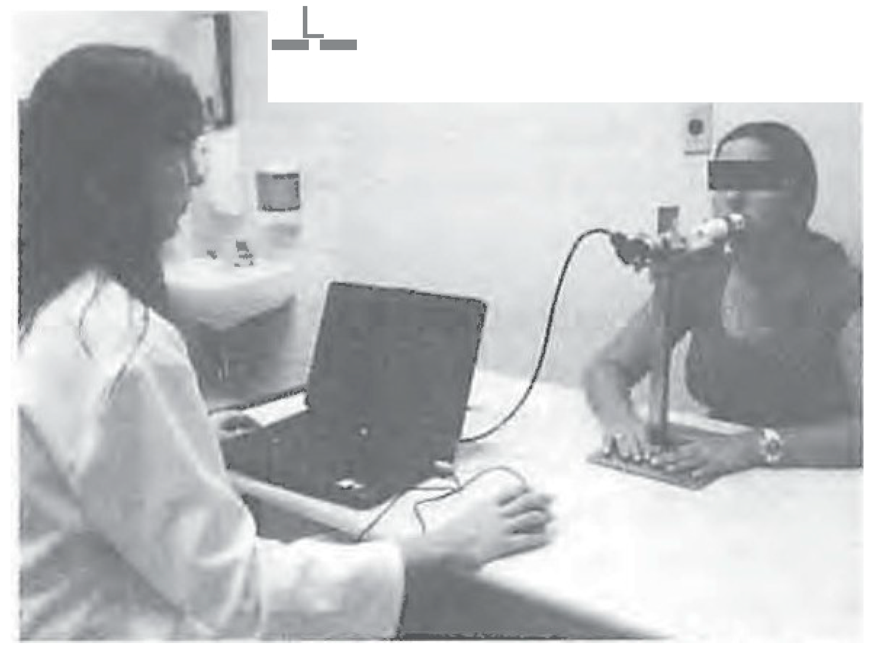
Figure 1. Quantitative evaluation of tongue force.
Ijom 40 00056 g001
Figure 2. Force time history graph example.
Ijom 40 00056 g002
Figure 3. Histogram of maximum force by gender.
Ijom 40 00056 g003
Table 1. Results by gender.
GroupsAverage (N)Median (N)SDMinimum (N)Maximum (N)Pearson’s Variation Coefficient
Male and Female17.5816.237.955.8457.1845%
Female15.7914.518.135.8457.1851%
Male20.1619.937.015.8442.8035%
SD—standard deviation, N—Newton.
Table 2. Presents the average and median values of maximum tongue protrusion force of the participants for each trial.
TrialAverage (N)Median (N)SD
Trial 119.7818.028.52
Trial 216.5715.657.86
Trial 316.3814.858.15