Next Article in Journal
Orthodontic Changes in Oral Dyskinesia and Malocclusion Under the Influence of Myofunctional Therapy
Previous Article in Journal
Assessment of Tongue Weakness and Fatigue
 
 
International Journal of Orofacial Myology and Myofunctional Therapy is published by MDPI from Volume 51 Issue 1 (2025). Previous articles were published by another publisher in Open Access under a CC-BY (or CC-BY-NC-ND) licence, and they are hosted by MDPI on mdpi.com as a courtesy and upon agreement with the previous journal publisher.
Article

Cephalometric Evaluation in Children Presenting Adapted Swallowing During Mixed Dentition

by
Milene Maria Bertolini
1,*,
Sérgio Vilhegas
,
Denise Yvonne Janovitz Norato
2,* and
Jorge Rizzato Paschoal
3,*
1
Service of Otolaryngology of FCM­UNICAMP, Cidade Universitaria, Campinas 13083-970, SP, Brazil
2
Clinical Orthodontist, Presidente Prudente, SP, Brazil
3
Medical Genetics Department, Medical Sciences School, State University of Campinas (UNICAMP), Campinas 13083-970, SP, Brazil
*
Authors to whom correspondence should be addressed.
Int. J. Orofac. Myol. Myofunct. Ther. 2003, 29(1), 29-41; https://doi.org/10.52010/ijom.2003.29.1.3
Submission received: 1 November 2003 / Revised: 1 November 2003 / Accepted: 1 November 2003 / Published: 1 November 2003

Abstract

:
The tongue trust and anterior projection through the dental arcades during swallowing is known as Adapted Swallowing (AS) and is a common finding during mixed dentition. The authors studied the morphologic dentofacial characteristics through cephalometric measures in 38 girls and 35 boys, aged seven to nine years, presenting AS, as determined by the speech therapist evaluation. Cephalometric evaluation was obtained by Radiocef 2.0 software. The measurements studied were the mandibular and maxillary relations to the cranial base, the nasolabial angle, and the upper airway patency, as proposed by McNamara Jr (1984), and the convexity and facial axis angles and maxillary height from Ricketts (1960; 1981). Results, analyzed in relation to sex and racial group, showed an association of Class II facial convexity with the African Brazilian children with dark black skin. This classification does not reflect an abnormality as all African Brazilians studied were classified as class II, due to the fact that the standard measurements are based in the white American population. These findings point to the need of specific racial normative standard for evaluating orofacial and dental structures.

INTRODUCTION

The tongue trust and anterior projection through the dental arcades during swallowing is known as Adapted Swallowing (AS) and is a common finding during mixed dentition (Bertolini & Paschoal, 2001), but is also seen when orofacial myofunctional disturbances are present (Hanson, 1979; 1988). Orthodontists see AS as an important factor determining the final results in orofacial corrections (Straub, 1951/1961; Graber, 1958; Andersen, 1963; Brauer & Holt, 1965; Proffit & Norton, 1970; Garliner, 1974; Silva Filho, Freitas & Cavassan, 1986; Van der Linden, 1990; Henriques et al., 2000).
Swallowing patterns show physiologic variations related to age and dentofacial configurations and are due to the interrelation of form, posture and function of the structures involved in the physiologic process. The tongue anterior projection during swallowing accompanies the morphologic and postural alterations of the cervical-cranio-facial segment. The same structures that participate in swallowing movements are also related to tongue. resting position and are associated to other oral functions as suction, chewing, speech, and breathing.
Morphologic dental and skeletal alterations impair orofacial functions and result in functional adaptations, but functional adaptation, can itself be responsible in altering normal anatomical structures. It is well known that soft tissue pressure and traction influence bone growth (Moss, 1962; Silling, 1978); Functional adaptation during mixed dentition can be transitory, but if definitive, will need specialized intervention.
Eruption of the definitive incisors happens before craniofacial bone growth is finished, and the space left by the loss of teeth is commonly occupied by the tongue during swallowing, speech or rest. Dente-facial alterations and muscle malfunction that occur during the dental transition period as a transitory functional adaptation can become permanent altering dental-skeletal configuration and function (Kohler, 1994). When the transitory functional adaptation doesn't disappear, altering dental-skeletal configuration, inadequate function will require intervention.
Cephalometric studies, used since 1931, when Broadbent in the United States and Hofrath (1931) in Germany developed the Cefalostat, are fundamental for the correct diagnosis of dental-skeletal relationship. The normal standards were proposed by several authors, for specific populations (Cotton, Takano & Wong, 1951; Chan, 1972; Garcia, 1975; Alexander, & Hitchcock, 1978; Jacobson, 1978; Moraes, Freitas & Henriques, 1988; Pinzan, 1994; Miyajima, 1996). The lateral craniofacial X-ray shows the relations that exist between dental skeletal structures and the soft tissues structures (tongue, palate and pharyngeal tonsils, nose and lips).
The Speech Therapist evaluation of function and posture takes into consideration the craniofacial morphology. The cephalometric analysis is an important tool to help determine if the observed alterations are compromising harmonic development and function. In this work, the authors describe the cephalometric findings during mixed dentition in a group of school children presenting Adapted Swallowing.

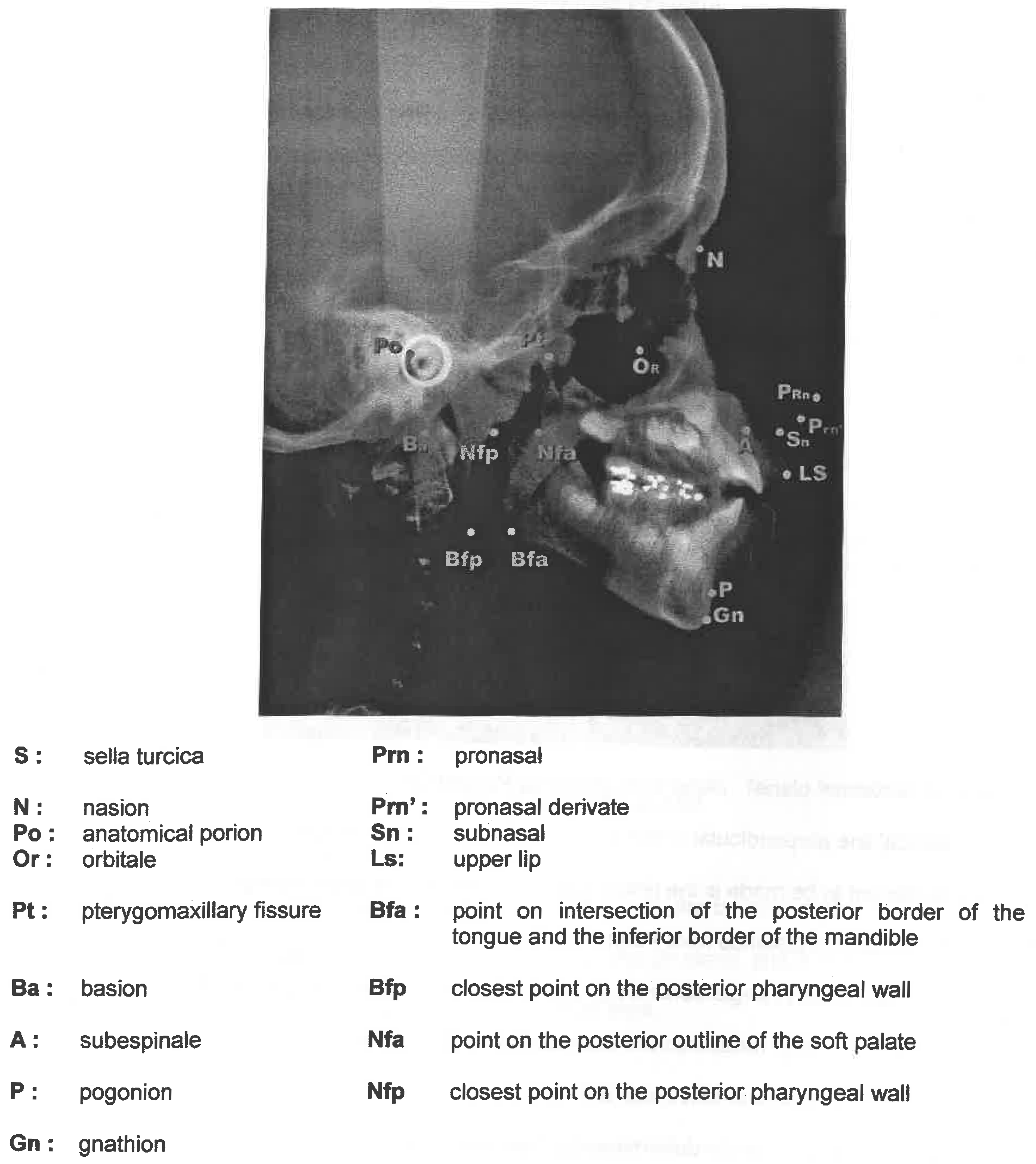
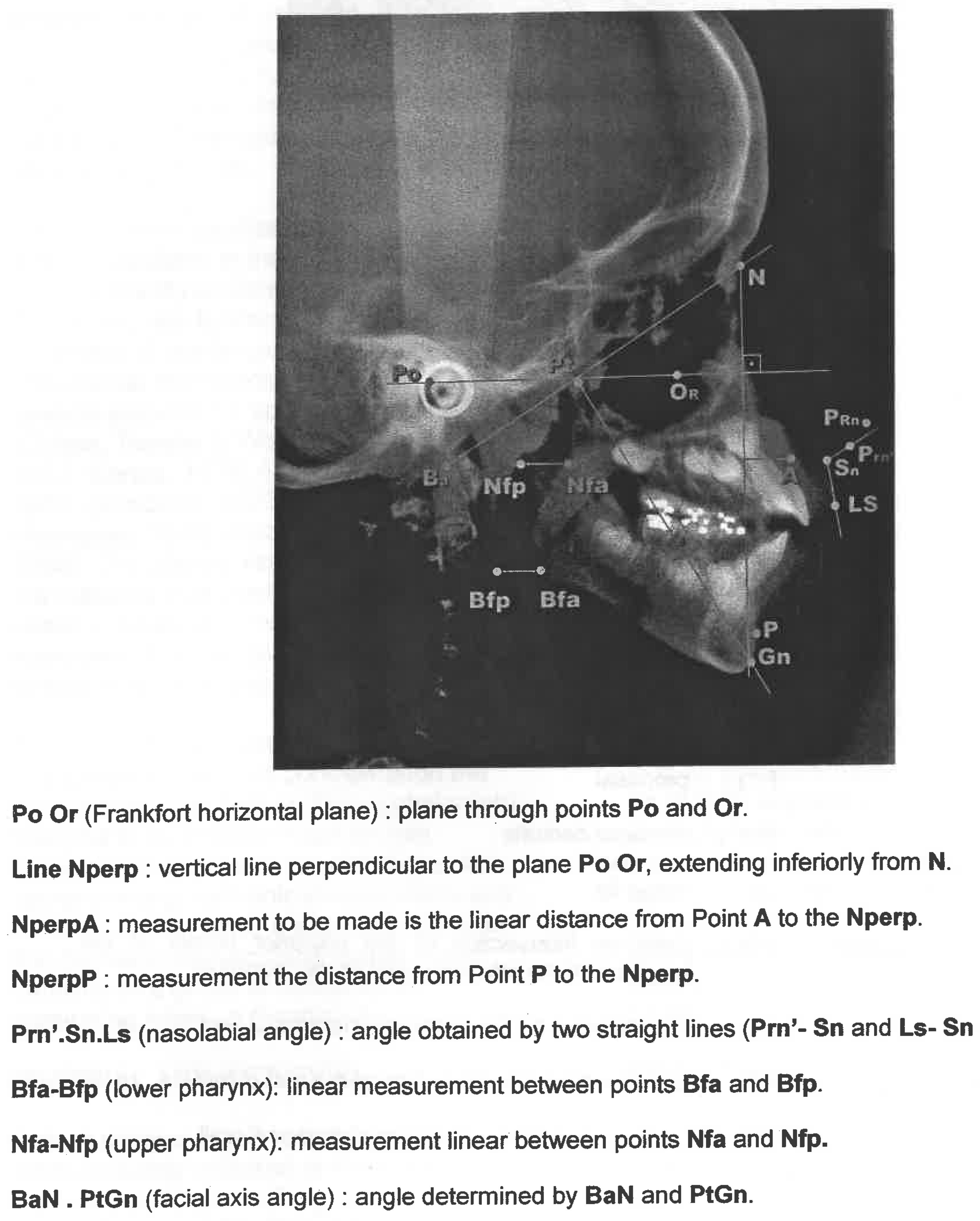
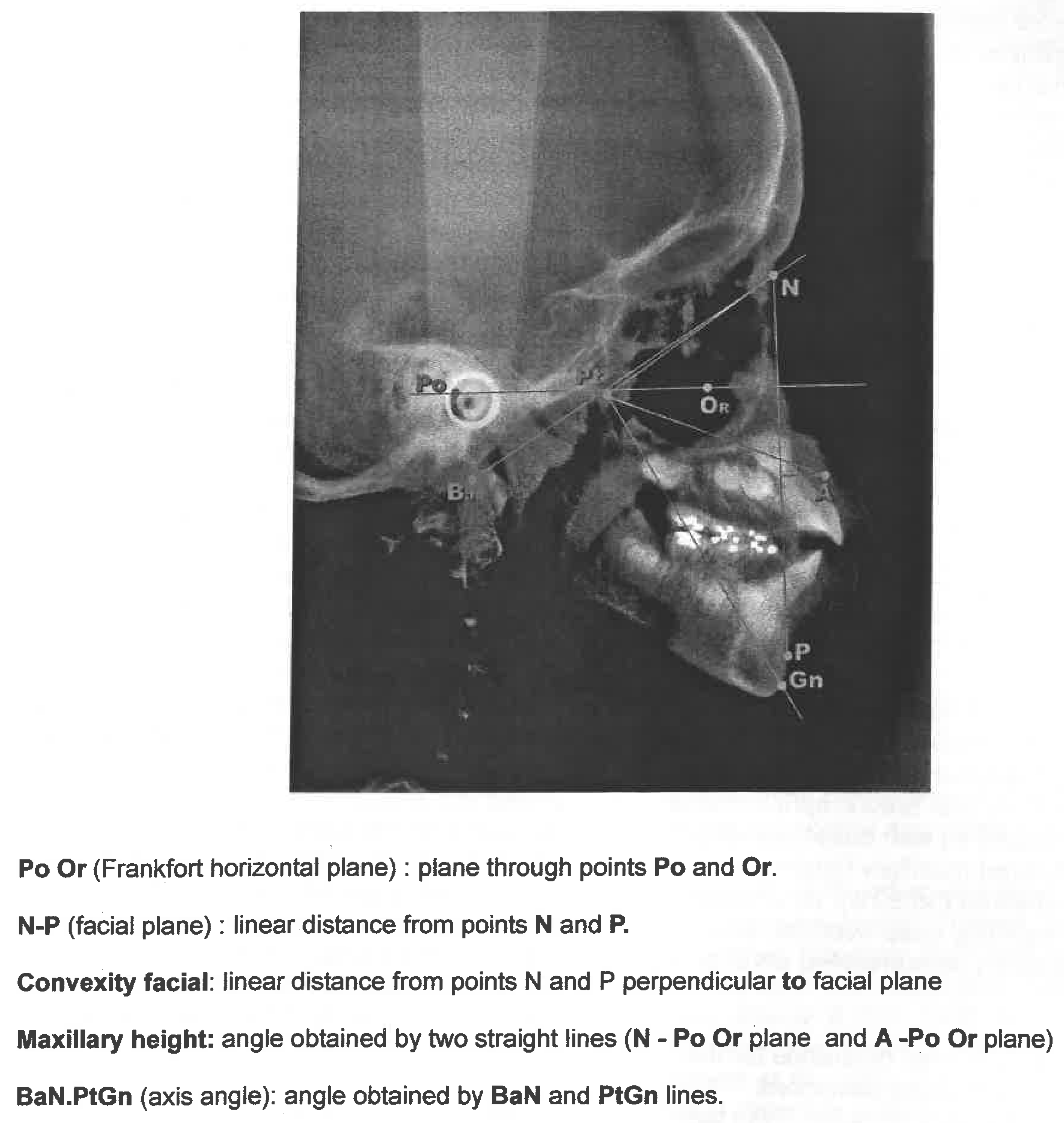
MATERIAL AND METHODS

School children aged seven to nine years were randomly selected at the Children and Adolescents' Support Program (PRODECAD) of the State University of Campinas. From 81 children evaluated, 73 were diagnosed as having AS. Cephalometric studies were conducted on 38 girls and 35 boys. Racial group definition is very difficult in the highly racially miscigenated Brazilian population, so skin color was used in order to classify children. The group thus consisted of 30 white and 43 African Brazilians, the latter consisting of 25 children with dark black skin and 18 with light black skin. South American Indians and oriental descendents were not included. AS was diagnosed when at least two of three criteria were associated: tongue adapted rest position, tongue adapted swallowing position and perioral muscle hypercontraction. All children were evaluated by an otorhinolaryngologist.
Orthoceph 5 equipment and a cefalostat were used for obtaining the lateral cephalometric roentgenograms, in normal occlusion, as described by Broadbent (1937). Cephalometric tracings were done by a technician using Radiocef 2.0 software and a Hewlett Packard – HP/4C/T scanner, 12 to 2250 dpi, 45s/1.100 dpi digitation speed, with a transparency reader. The measurements studied were the mandible and maxillary relations to the cranial base, the nasolabial angle, and the upper airway patency, as proposed by McNamara Jr, in 1984 (Figure 2), and, the linear measures of the facial convexity, facial axis angle and maxillary height from Ricketts (1960; 1981) (Figure 3). Measurements were approximated up to 0.25 degrees or 0.25 millimeters. Cephalometric tracings analysis was done by two observers, and the numerical results were used to classify type and intensity of morphologic deviations as proposed by the Orthodontic Service of the Bauru Odonthology School, Sao Paulo State University (USP).

RESULTS

AS evaluation considering three criteria showed that all 73 children had adapted tongue rest position, all also presenting adapted tongue position during swallowing and 25 (34.25%) hypercontraction of perioral muscles. The three criteria were present in 25 (34.25%) children.
The otorhinolaryngology (ORL) evaluation detected nasal mucous membrane pallor in 29 (39.73%) children, turbinate hyperplasia in two (2.74%), hypertrophic palatal tonsils in 6 (8.22%) and hypertrophic pharyngeal tonsils in 20 (27.40%). No ORL alterations were seen in 29 children. Summarizing parents' responses to a questionnaire, it was noted that parents fe3lt their children exhibited: oral breathing- present in 33 (45.20%) children, snoring -present in 29 (39.73%), nocturnal salivary incontinence present in 32 (43.84%) and swallowing problems -present in four (5.48%). Cephalometric evaluations detected hypertrophic palatal tonsils in 28 (38.36%) children, but in the otorhinolaryngologist evaluation, only 6 children were diagnosed as having hypertrophic palatal tonsils, two of these not confirmed by the cephalometry. No child was diagnosed with hypertrophic pharyngeal tonsils by cephalometry, although this was seen by the otorhinolaryngologist exam in 20 (27.40%) children. The frequency of type and intensity of morphologic deviations for the studied groups are shown in Tables 1 and 2.
Figure 1. Points In Lateral Cephalometric Roentgenograms.
Ijom 29 00003 g001
Figure 2. Point demarcation and tracing for linear and angular measures utilized in this study (modified from MacNamara Jr).
Ijom 29 00003 g002
Figure 3. Point demarcation and tracing for linear and angular measures utilized in this study (modified from Ricketts).
Ijom 29 00003 g003
The 31 (42.46%) children presenting facial axis deviations detected by the cephalometric evaluation consisted of five (16.13%) children with brachicephalic facial axis and 26 (83.87%) with dolicocephalic facial axis. Altered maxillary height was noted in 27 children (36.99%), 22 children (81.48%) presenting deep overbite, and five children (18.52%) demonstrated anterior open bite.
There was no significant difference for the frequency of morphologic deviations between boys and girls (p>0,05). With the exception of facial convexity no significant difference for the frequency of morphologic deviations was detected between white and African Brazilian·children. For facial convexity an association of Class 11 was seen with African Brazilian children with dark colored skin (p<0,01), as only one child of this group was not classified as class II (Table 3). The African Brazilian children with light colored skin differed from the latter group (p<0.05) and behaved as the white children group (p>0.05).

DISCUSSION

Oromyofunctional evaluation by the speech therapist must include the exam of the morphophysiological integrity of the cranio cervicofacial segment. In a previous report, Bertolini & Paschoal (2001) found that 57 % of school children examined presented morphological alterations, as diagnosed by clinical evaluation. The use of the cephalometric measures enables the professional to relate dentofacial aspects to posture and function, quantifying the carniocervicofacial complex, with data from maxillary-mandibular relationship and facial development tendencies, showing the patients' muscle pattern.
When evaluating AS in children during the mixed dentition phase it is difficult to determine if the morphological alteration is a transitory anomaly or if there will be definite changes in orofacial configurations. One important issue to be considered is the criteria utilized for defining if the child presents AS. Several of the myofunctional findings are commonly found during this phase and disappear when dentition is defined (Milne & Cleall, 1970; Cleall, 1972; Cleall & Bogole, 1982. Several authors include ORL alterations as a factor for orofacial development deviations (Ricketts, 1968; Trotman et al., 1997; Proffit, 1978; Linder-Aronson, 1979; Harvold et al., 1981, McNamara Jr., 1981; Mcloughlin, 1988).
In our sample, although the otorhinolaryngologist evaluation detected slight abnormalities as nasal mucosa pallor, turbinate hyperplasia, hypertrophic palatal and pharyngeal tonsils, this was not confirmed by the lateral roentograms and had no association with cephalometric deviations. The high frequency of positive symptoms suggestive of upper airway obstruction related by parents suggests that although there was no ORL clinical and radiological alterations, other aspects of the orofacial function including the swallowing pattern was compromised. This could, as indicated by Moss (1962), lead to permanent morphological alterations.
In this group of schoolchildren with mixed dentition presenting AS, almost half showed facial development deviation tendencies, which could be a sign that the myofunction had already influenced bone growth and dental occlusion as previously proposed by other authors (Brodie, 1953; Moss, 1962; McNamara Jr, 1973). Among the orofacial development deviations studied, it was detected that more than one-third of the children exhibited altered maxillary height. However, this .specific deviation is not to be considered indicative of an abnormal development in the mixed dentition initial phase as several authors have found (Diamond, 1944; Barrow & White, 1952; Björk, 1953; Moorrees, 1959; Fleming, 1961; Van der Linden, 1990).
The higher frequency of Class II morphologic deviations of facial convexity, seen in the African Brazilian children with dark colored skin, was due to orthodontists in Brazil using the cephalometric analysis standards of Ricketts (1960, 1981) and of McNamara Jr (1984) which were both based in a Caucasian population.
Maxilla and mandible relations to the cranial base, as defined by A-Nperp and P-Nperp measures showed that the vast majority of these children with AS had abnormal relations which were: protrusion, maxillary in 61 (83.56%) children, and mandibular in 53 children (72.60%). Skeletal protusion in the mixed dentition phase has also been reported as a common characteristic in this age by Nanja and Taneja (1972), who studied Downs' angles (1956) in six to 13 year old children. They found that children presented dental protrusion and a convex facial profile. Cephalometric measurements by McNamara Jr (1984) and McNamara Jr & Brudon (1995) during mixed dentition show that during this phase of craniofacial growth and development, from 6 to 12 years of age, the pogonion can be less the six to eight millimeters from the Nperp line. With age, the distance is reduced, and adults show a distance from less the two millimeters up to over four millimeters. These values are compatible with a protrusion, although normal for age. In our sample, we considered normal, i.e. without protrusion, up to the values referred by McNamara Jr (1984), so when the children were classified as a convex profile they had a more severe protrusion than expected for age. This was more evident for the children with dark black skin.
Nasolabial Angle configuration was normal in more than half the children; however, when the Nasolabial Angle was altered it was severe. Half of the children who had severely altered Nasolaboal Angle presented with a closed angle and half presented with an open angle. Janson (1990), McNamara Jr (1984) and McNamara Jr & Brudon (1995) refers that in children who have normal nasolabial angle configurations, the findings of altered A-Nperp measures should not be considered for diagnosis. The clinical evaluation should prevail. In our group, more than half of the children who had altered A-Nperp, presented normal nasiolabial angle.
There are no Brazilian cephalometric or myofunctional studies in black children in this phase. However, there are some studies done with teenagers (Altemus, 1963; Brauer & Holt, 1965; Altemus, 1968, Medeiros, Henriques & Freitas; 1988; Moraes et al., 1988, Bertoz & Martins, 1981). Altemus (1960) has attributed the prognathism observed in blacks to a dentoalveolar protusion. Cardim (1987) and Drumond (1968) reported that dental and skeletal protrusion of maxilla and mandible is characteristic of the black race.
In the current study dental positions were evaluated by radiograms but were not included as an objective. Reviewing the data further analysis was conducted for the group of 25 black children, for dental and skeletal relationships and lower lip hypotony, as shown in Table 4.
Moraes et al. (1988) reports that the racial differences in facial convexity are related to the relative positions of the points, N (nasion - in the cranial base), A (subspinale) e P (pogonion). N varies in accord to the size and inclination of the cranial base. A varies by the degree of maxillary protusion, which is greater in blacks (Cotton et al., 1951, Altemus, 1960/1968; Drumond, 1968; Alexander & Hitchcock, 1978). P which also shows racial characteristics, was retropositioned due to the greater inclination of the mandibular plane (Drumond, 1968), or, to the exaggerated aJveolar maxillary prognathism seen in blacks (Alexander & Hitchcock, 1978; Moraes et al., 1988). In this study racial differences referred to in the P point were not seen, as only three children had mandibular retrusion. As suggested by Moraes et al. (1988), consideration was also given to the rnaxillary and mandibular protusion observed in black children which must be related to the lower incisor dentoalveolar protusion and not to an skeletal component. Bertoz & Martins (1981), report that in black teenagers the incisor dentolalveolar protusion should be considered a racial characteristic.
Although all authors that studied non-white samples have called attention to the need for properly applying racial standards, it is not a common practice among orthodontist and speech therapists to consider this when planning therapy (Cotton, et al., 1951; Altemus, 1960/1968; Drumond, 1968; Silva, 1975; Alexander & Hitchcock, 1978; Jacobson, 1978; Bertoz & Martins, 1981; Medeiros et al., 1988, Moraes et al., 1988; Henriques & Freitas, 1990).
Another aspect discussed by Cardim (1987) is the soft tissue distribution in the black race in the lips, and periorbicular muscles, with black children showing hypotonic lip. Altemus (1963; 1968) comments that both lower and upper lips in blacks show an important protusion, and that these aspects are normal racial features. This should not be forgotten when planning orthodontic and myofunctional therapies. Although in the current study the frequency of hypotonic lower lip did not significantly differ between races, 64% of the black children presented hypotonic lower lip and concomitantly, in 32% of these children hypercontraction of perioral muscle during swallowing was observed. This could be an important aspect to consider when diagnosing AS in black children and perhaps should not be considered as a criteria for AS when isolated.

CONCLUSION

Cephalometric measurements in children presenting adapted swallowing during the mixed dentition phase showed that the vast majority presented maxillary and mandibular protrusion. There was rio significant difference for the frequency of morphologic deviations between boys and girls. However, between whites and non-whites an association of Class II was seen with the African-Brazilian children with dark black colored skin. Dental and skeletal protrusion of maxilla and mandible and differences in soft tissue distribution is characteristic of the black race. These aspects, that are normal racial features, should not be forgotten when planning orthodontic and myofunctional therapies. The necessity of applying.racial standards, not a common practice among orthodontist and speech therapists is to be considered when evaluating and planning therapy. It should also be considered that in Brazil, due to the racial mixture, standards should be reviewed, not only considering the influenqe of the black miscigenation, but in certain regions, also the Native and the Asiatic influence should be studied.

References

  1. Alexander, T. L., and H. P. Hitchcock. 1978. Cephalometric standards for American Negro children. American Journal of Orthodontics 74, 3: 298–304. [Google Scholar] [PubMed]
  2. Altemus, L.A. 1960. A comparison of cephalofacial relationships. Angle Orthodontist 30, 4: 223–40. [Google Scholar]
  3. Altemus, L.A. 1963. Comparative integumental relationships. Angle Orthodontist 33, 3: 217–21. [Google Scholar]
  4. Altemus, L. A. 1968. Cephalofacial relationships. Angle Orthodontist 38, 3: 175–84. [Google Scholar]
  5. Andersen, W. S. 1963. The relationship of the tongue thrust syndrome to maturation and other factors. American Journal of Orthodontics 49, 4: 264–75. [Google Scholar]
  6. Angle, E. H. Classification of malocclusion. Dental Cosmos 41, 18: 248–64.
  7. Barrow, G. V., and J. R. White. 1952. Developmental changes of the maxillary and mandibular dental arches. Angle Orthodontist 22, 1: 41–6. [Google Scholar]
  8. Bertolini, M. M. 1998. Prevalência da deglutição adaptada numa população de escolares. Campinas [Dissertação de mestrado, Universidade Estadual de Campinas]. [Google Scholar]
  9. Bertolini, M. M., and J. R. Paschoal. 2001. Prevalence of adapted swallowing in a population of school children. International Journal of Orofacial Myology 27: 33–43. [Google Scholar]
  10. Björk, A. 1953. Variability and changes in overjet and overbite. American Journal of Orthodontics 39, 10: 779–801. [Google Scholar]
  11. Bertoz, F. A., and D.R. Martins. 1981. Determinayao da linha "I "em melanodermas brasileiros, masculinos de 12 a 17 anos, com oclusao normal. Ortodontia 14, 3: 186–98. [Google Scholar]
  12. Brauer, J. S., and T. V. Holt. 1965. Tongue thrust classification. Angle Orthodontist 35, 2: 106–112. [Google Scholar] [PubMed]
  13. Broadbent, B. H. 1931. A new x-ray technique and its application to orthodontia. Angle Orthodontist 1: 45–66. [Google Scholar]
  14. Brodie, A. G. 1953. Muscular factors in the diagnosis and treatment of maloccusion. Angle Orthodontist 23, 2: 71–7. [Google Scholar]
  15. Cardim, V. L. N. 1987. Crescimento craniofacial. In Cirurgia craniomaxilofacial: osteotomias esteticas da face. Edited by J. M. PSILLAKIS. Rio de Janeiro, Medsi: pp. 25–41. [Google Scholar]
  16. Chan, G. K. H. 1972. A cephalometric appraisal of the Chinese (Cantonese). American Journal of Orthodontics 61, 3: 279–85. [Google Scholar]
  17. Cleall, J. F. 1972. Circumstances limiting the development and verification of a comprehensive theory of craniofacial morphogenesis. Acta Morphologica Neerlando Scandinavica 10, 1: 115–26. [Google Scholar]
  18. Cleall, J. F., and E. A. Begole. 1982. Diagnosis and treatment of class II division 2 malocclusion. Angle Orthodontist 52, 1: 38–60. [Google Scholar] [PubMed]
  19. Cotton, W. N., W. S. Takano, and W. M. W. Wong. 1951. The Downs analysis applied to three other ethnic groups. Angle Orthodontist 21, 4: 213–20. [Google Scholar]
  20. Diamond, M. 1944. The development of the dental height. American Journal of Orthodontics 30, 111: 589–605. [Google Scholar]
  21. Downs, W. B. 1956. Analysis of the dento-facial profile. Angle Orthodontist 26, 4: 191–212. [Google Scholar]
  22. Drumond, R. A. 1968. A determination of cephalometric norms for the Negro race. American Journal of Orthodontics 54, 9: 670–82. [Google Scholar]
  23. Fleming, H. B. 1961. An investigation of the vertical overbite during eruption of the permanent dentition. Angle Orthodontist 31, 1: 52–62. [Google Scholar]
  24. Garcia, C. J. 1975. Cephalometric evaluation of mexican americans using the Downs snd Steiner analyses. American Journal of Orthodontics 68, 1: 67–74. [Google Scholar] [PubMed]
  25. Garliner, D. 1974. Myofunctional therapy in dental practice, 2nd ed. Brooklyn: Coral Gables. 333p. [Google Scholar]
  26. Graber, T. M. 1958. The finger sucking habit and associated problems. Journal of Dentistry for Children 25: 145–51. [Google Scholar]
  27. Hanson, M. L. 1979. An introduction to oral myofunctional disorders. The International Journal of Oral Myology 5, 2: 5–9. [Google Scholar]
  28. Hanson, M. L. 1988. Orofacial myofunctional therapy: historical and physiological consideration. The International Journal of Oral Myology 14, 1: 3–10. [Google Scholar]
  29. Harvold, E. P., B. S. Tomer, and K. Vargervik. 1981. Primate experiments on oral respiration. American Journal of Orthodontics 79: 359–72. [Google Scholar]
  30. Henriques, J. F. C., and M. R. Freitas. 1990. Determina o da linha Wits em jovens brasileiros melanodermas com "oclusao normal "e comparayao com os jovens leucodermas. Ortodontia 23, 2: 4–10. [Google Scholar]
  31. Henriques, J. F. C., G. Janson, R. R. de Almeida Dainesi, and S. M. Hayasaki. 2000. Mordida aberta anterior: a importancia da abordagem multidisciplinar e considera oes sobre etiologia, diagn6stico e tratamento. Apresentayao de um caso clrnico. Revista Dental Press Ortodontia e Ortopedia Facial 5, 3: 29–36. [Google Scholar]
  32. Hofrath, H. 1931. Die Bedeutung der r"ontgenfern und abstandsaufnahme fur die diagnostik der kieferanomalien. Fortschrit Orthodontics 1: 232–58. [Google Scholar]
  33. Jacobson, A. 1978. The craniofacial skeletal pattern of south African Negro. American Journal of Orthodontics 73, 6: 681–91. [Google Scholar]
  34. Janson, G. R. P. 1990. Estudo longitudinal e comparative do crescimento facial - dos 13 aos 18 anos de idade - em jovens brasileiros leucodermas, utilizando a analise cefalometrica de McNAMARA JR. Bauru [Tese de doutorado - Universidade Estadual de Sao Paulo). [Google Scholar]
  35. Linder-Aronson, S. 1979. Respiratory function in relation to facial morphology and the dentition. British Journal of Orthodontics 6: 59–72. [Google Scholar] [PubMed]
  36. Kohler, G. I. 1994. Desenvolvimento da oclusao. In Ortodontia para fonoaudiologia. Edited by E. PETRELLI. Sao Paulo, Ed. Lovise: pp. 65–79. [Google Scholar]
  37. Mcl oughlin, P. J. 1988. Orofacial myology: current trends. The International Journal of Oral Myology 14, 1: 1–12. [Google Scholar]
  38. McNamara, J. A., Jr. 1973. Neuromuscular and skeletal adaptation to altered function in the orofacial region. American Journal of Orthodontics 64, 6: 578–606. [Google Scholar] [PubMed]
  39. McNamara, J. A., Jr. 1981. Influence of respiratory pattern on craniofacial growth. Angle Orthodontist 51, 4: 269–300. [Google Scholar]
  40. McNamara, J. A., Jr. 1984. A. A method of cephalometric evaluation. American Journal of Orthodontics 86, 6: 449–469. [Google Scholar]
  41. McNamara, J. A., Jr. 1995. Tratamiento ortod6ncico y ortopedico la dentiti6n mixta. Traducci6n Azucena Rivas de Montes. Ann Arbor, Needham Press: pp. 1–133, Original title: Orthodontic and orthopedic treatment in the mixed dentition. [Google Scholar]
  42. Medeiros, M. A. Q. B., J. F. C. Henriques, and M. R. Freitas. 1988. Estudo cefalometrico do padrao dentario de jovens brasileiros melanodermas do sexo feminine, com "oclusao normal". Ortodontia 21, 1: 34–48. [Google Scholar]
  43. Milne, I. M., and J. F. Cleall. 1970. [ELIMINATE. POINT 1970 BEFORE] Cinefluorographic study of functional adaptation of the oropharyngeal structures. Angle Orthodontist 40, 4: 267–83. [Google Scholar]
  44. Miyajima, K., and et al. 1996. Craniofacial structure of Japanese and European-American adults with occlusions and well-balanced faces. American Journal of Orthodontics 110, 4: 431–438. [Google Scholar]
  45. Moorrees, C. F. A. 1959. The dentition of growing child: a longitudinal study of dental development between 3 and 18 years of age. Cambridge: Harvard Press. 245p. [Google Scholar]
  46. Moraes, C., M. R. Freitas, and J. F. C. Henriques. 1988. Cefalometria-determina9ao do padrao esqueletico das adolescentes melanodermas brasileiras, com "oclusao normal". Ortodontia 22, 2: 4–14. [Google Scholar]
  47. Moss, M. L. 1962. The functional matrix. In Vistas in orthodontics. Edited by B. S. KRAUS and R. A. RIEDEL. Philadelphia: Lea & Febiger, pp. 36–63. [Google Scholar]
  48. Nanda, R. S., and R. C. Taneja. 1972. Growth of face during the transitional period. Angle Orthodont 42, 2: 165–71. [Google Scholar]
  49. Pinzan, A. 1994. Estudo cefalometrico longitudinal das medidas SNA Nperp-A SNB, SND, Nperp-P, AnB, Sn.GoGn, SN.Gn, PoOr.GoMe e BaN.ptGn, em jovens leucodermas brasileiros de ambos os sexos, com oclusao normal dos 5 aos 11 anos. Bauru [Tese de livre docencia - · Faculdade de Odontologia de Bauru, Universidade Estadual de Sao Paulo]. [Google Scholar]
  50. Proffit, W. R. 1978. Equilibrium theory revisted: factor influencing position of the teeth. Angle Orthodontist 48, 3: 175–86. [Google Scholar]
  51. Proffit, W. R., and L. A. Norton. 1970. The tongue and oral morphology: influences of tongue activity during speech and swallowing. In: WORKSHOP SPEECH AND THE DENTOFACIAL COMPLEX, 5, Washington, 1970. Proceeding. Washington, ASHA Reports. 106–15. [Google Scholar]
  52. Ricketts, R. M. 1960. The influence of orthodontic treatment on facial growth and development. Angle Orthodontist 30: 103–33. [Google Scholar]
  53. Ricketts, R. M. 1968. Forum on the tonsil and adenoid problem in orthodontics. Respiratory obstruction syndrome. American Journal of Orthodontics 54, 7: 495–514. [Google Scholar] [CrossRef] [PubMed]
  54. Ricketts, R. M. 1981. Perspectives in the clinical application of cephalometric-the 1St 50 years. Angle Orthodontist 51, 2: 115–150. [Google Scholar]
  55. Silling, G. 1978. Minor tooth movement made easier. The New York Journal of Dentistry 48, 5: 148–50. [Google Scholar]
  56. Silva Filho, O. G. da, S. F. de Freitas, and A. de O. Cavassan. 1986. Habitos de sucyao passiveis de intervenyao. Estomatologia e Cultura 16, 4: 61–71. [Google Scholar]
  57. Silva, A. N. 1975. O esqueleto cranio-facial de criancas melanodermas: estudo radiocefalometrico. Piracicaba [Dissertayao de mestrado - Faculdade de Odontologia de Piracicaba - Universidade Estadual de Campinas]. [Google Scholar]
  58. Straub, W. J. 1951. The etiology of the perverted swallowing habit. American Journal of Orthodontics 37, 8: 603–10. [Google Scholar] [CrossRef]
  59. Straub, W. J. 1961. Malfunction of the tongue. Part II. The abnormal swallowing habit: its causes, effects and results in relation to orthodontic treatment and speech therapy. American Journal of Orthodontics 47, 8: 596–617. [Google Scholar] [CrossRef]
  60. Trotman, C.-A., J. A. McNamara, Jr., J. M. H. Dibbets, and L. T. Van der Weele. 1997. Association of lip posture and the dimension of the tonsils and sagital airway with facial morphology. Angle Orthodontist 67, 6: 425–32. [Google Scholar]
  61. Van der Linden, F. P. G. M. 1990. Crescimento e ortopedia facial. Sao Paulo: Quintessence Books. 244p. [Google Scholar]

