Article

Comparative Antimicrobial Effects of Dimethylsulfoxide and Dimethylsulfone on the Planktonic Growth and Viability of Porphyromonas gingivalis and Their Cytotoxic Effects on Human Oral Epithelial Cells

1
DeBusk College of Osteopathic Medicine, Lincoln Memorial University, Harrogate, TN 37752, USA
2
DeBusk College of Osteopathic Medicine, Lincoln Memorial University, Knoxville, TN 37932, USA
3
Department of Biology, Muhlenberg College, Allentown, PA 18104, USA
*
Author to whom correspondence should be addressed.
Bacteria 2025, 4(4), 57; https://doi.org/10.3390/bacteria4040057
Submission received: 23 September 2025 / Revised: 25 October 2025 / Accepted: 28 October 2025 / Published: 1 November 2025

Abstract

Background: Past studies have documented the antimicrobial effects of dimethyl sulfoxide (D.M.SO). However, the side effects and toxicity profiles of DMSO in vivo have been a significant deterrent for its wide-ranging clinical use. Dimethyl sulfone (DMSO-2), a natural metabolite of DMSO, is currently used as a safe dietary supplement due to its antioxidant properties and multimodal mechanisms of action. While DMSO displays antimicrobial activity, little is known concerning DMSO-2’s antimicrobial effect. Thus, this investigation compares the antimicrobial effects of DMSO and DMSO-2 on the growth and viability of the pathogenic anaerobic bacteria, Porphyromonas gingivalis, and their cytotoxic effect on human oral epithelial (OKF6/TERT-2) cells. Methods: P. gingivalis was grown in TSBY media in the presence of DMSO or DMSO-2 (0–4%) for planktonic growth and viability determinations. OKF6/TERT-2 cells were expanded in vitro and similarly exposed to DMSO or DMSO-2 for viability studies. Results: After 24 h exposure to DMSO or DMSO-2, growth of P. gingivalis is inhibited by 57% and 77%, respectively, while viability is inhibited by 55% and 62%. In contrast, 24 h exposure to similar concentrations of DMSO or DMSO-2 induces 5% and 2% cytotoxicity in OKF6/TERT-2 cells, respectively. Conclusions: Both DMSO and DMSO-2 inhibit the growth and viability of P. gingivalis but show minimal toxic effect on OKF6/TERT-2 cells. Therefore, the utility of these two natural compounds as antimicrobial agents against anaerobic pathogens should be further investigated.

1. Introduction

Infections with pathogenic anaerobic bacteria continue to pose challenges in the elderly and immunocompromised and are a significant burden on healthcare costs globally [1,2]. Currently, effective management of anaerobic bacterial infections is complicated and requires targeted antibiotic therapy and, in some cases, surgical intervention [3]. Comprehensive understanding, timely diagnosis and appropriate treatment are crucial in mitigating anaerobic bacterial diseases, which often arise in sites with limited oxygen access, such as deep tissue wounds (e.g., Clostridium perfringens—gas gangrene), intra-abdominal spaces (e.g., Campylobacter jejuni—gastric enteritis) and dental abscesses (e.g., Porphyromonas gingivalis—periodontal disease) [4]. P. gingivalis is considered a keystone pathogen associated with periodontal disease [5]. If allowed to grow unchecked, this pathogen can render a normal microbiome into one of dysbiosis, leading to inflammatory disease and potentially systemic pathology [6]. Consequently, the discovery of safe and effective alternative antimicrobial therapies against anaerobic infections would be of significant translational value. In this study, we test the effects of dimethyl sulfoxide (DMSO) and its natural physiological metabolite, dimethyl sulfone (DMSO-2), on P. gingivalis and human oral epithelial cells.
To combat microbial diseases, especially in an age of enhanced microbial resistance, the search for newer and better antimicrobial agents remains ongoing. DMSO has long been debated as a prophylactic therapeutic and/or as an antimicrobial agent [7,8]. While DMSO has been documented to have significant antimicrobial activity against pathogenic facultative and anaerobic bacteria [9,10,11,12,13], its use has been mostly limited to that of a cryogenic reagent or as a drug delivery vehicle [7,8] due to negative reports of its possible side-effects. For example, DMSO was found to be neurotoxic in rats [14] and has been shown to induce drastic changes in human cellular and epigenetic processes [15]. A review by Kollerup Madsen et al. (2018) [16] lists the many adverse reactions associated with DMSO, ranging from mild to serious. Currently, the only FDA-approved clinical uses for DMSO are for the treatment of interstitial cystitis [16] and its use as a solvent in embolic systems [17]. On the other hand, DMSO-2 is a natural analog of DMSO and currently used as a supplement due to its antioxidant properties and multimodal mechanisms of action [18], but has not been thoroughly investigated for its antimicrobial effects. Figure 1 illustrates the bioconversion of DMS to DMSO-2. Williams et al. (1966) [19] described DMSO-2 as the stable end-product resulting from the oxidation of dimethyl sulfide (DMS) to DMSO and then the oxidation of DMSO to DMSO-2, which is ultimately excreted in urine. Furthermore, DMSO-2 in mammalian systems does not revert to DMSO in vivo. He and Slupsky [20] indicate the sources of mammalian DMSO-2 to be either direct dietary supplementation (fruits, vegetables and grains) or through microbial–mammalian co-metabolism of excess methionine in the gut. In either case, mammalian systems are capable of safely handling more than 4 g of DMSO-2 per day [18]. However, unlike DMSO, little is known about the antibacterial properties of DMSO-2 and its potential use as a safe antimicrobial alternative.
The oral cavity is generally lined with stratified squamous epithelial cells, which are closely associated with the oral microbiota, a diverse population of 834 microbial species [21]. A symbiotic balance between these microbes and the underlying mucosal epithelium is required to prevent oral and potentially systemic diseases [22]. This is to say that when microbial conditions shift from symbiosis to dysbiosis, there is a high correlation of damage to the underlying epithelium and subsequent risk of pathology [6]. P. gingivalis is an anaerobic Gram-negative opportunistic bacterium, primarily found in the deep pockets of the subgingival sulcus, and a primary culprit associated with periodontal disease [23]. OKF6/TERT-2 cells are oral mucosal epithelial cells that have been immortalized via telomerase 2 retroviral transduction and expression, as well as deletion of the p16INK4a regulatory protein [24]. Consequently, in using P. gingivalis as the test bacterium and OKF6/TERT-2 cells as the oral epithelium, the aim of this study is twofold: first, to compare and determine the optimal antimicrobial doses of DMSO and DMSO-2 that inhibit P. gingivalis growth and secondly, to determine the toxicity of these two natural compounds on OKF6/TERT-2 cells. We hypothesize that one or both reagents have sufficient antimicrobial activity against P. gingivalis at doses that are non-toxic or less toxic to OKF6/TERT-2 cells. Periodontal disease, as well as other anaerobic infections, are difficult to manage and require multiple targeted therapies. If DMSO-2 can achieve the antimicrobial effectiveness of DMSO with limited harm to the underlying epithelium, it could be added to the arsenal of antimicrobial agents to treat anaerobic infections.

2. Materials and Methods

2.1. Reagents and Supplies

All reagents and supplies for this study were obtained from ThermoFisher Scientific (Waltham, MA, USA), unless otherwise noted.

2.2. Preparation of Stock DMSO-2

DMSO and DMSO-2 [also known as methyl sulfonyl methane (MSM)] were purchased from Sigma-Aldrich (St. Louis, MO, USA). DMSO is 99.9% pure and comes in liquid form. DMSO-2 is 98.0% pure and comes in powder form. Stock DMSO-2 was prepared as a 33.3% solution in distilled water, autoclaved and stored at 37 °C for up to two weeks or until required.

2.3. Planktonic Growth of P. gingivalis

P. gingivalis W83 (from Dr. Ann Progulske-Fox at the University of Florida, Gainesville, FL, USA) was used in this study [25]. This gentamicin-resistant bacteria was stored at −80 °C in 25% glycerol stocks. For all experiments, P. gingivalis was initially streaked on sterile tryptic soy agar (TSA) fortified with 5% sheep blood, 1 µg/mL menadione (vitamin K) and 20 µg/mL gentamicin and incubated under anerobic conditions at 37 °C in a Bactron 300 anaerobic incubator (Sheldon Manufacturing, Cornelius, OR, USA), after which P. gingivalis was transferred to sterile tryptic soy broth fortified with 1 mg/mL yeast extract, 5 µg/mL porcine hemin, 1 µg/mL menadione and 20 µg/mL gentamicin (TSBY). Enough P. gingivalis was transfered to the TSBY to form a homogenous mixture with an optical density (OD) ≈ 0.4 at an absorbance wavelength of 595 nm after background subtraction. Since the stock DMSO-2 solution was 33.3% (as compared to 100% DMSO), three times more DMSO-2 was added to the TSBY than DMSO to make the working solution. Distilled water was added to the TSBY to make up the volume difference in the DMSO working solution. Solutions of 8% DMSO or 8% DMSO-2 in TSBY broth were prepared and serially diluted with TSBY to yield 4, 2 and 1% solutions. A control (0% DMSO or 0% DMSO-2) was prepared, where distilled water was again used to make up the volume difference. A positive control for growth inhibition was made using 10% hydrogen peroxide (Px) in TSBY. At the time of experiments, 100 µL of DMSO or DMSO-2 at concentrations of 1, 2, 4 or 8% and 100 µL of 10% Px, as well as 100 µL of TSBY broth (postive control for growth), were added to wells of five 96-well plates, where each plate represented a single time point and n = 6 for each treatment group. To each of these wells, 100 µL P. gingivalis in TSBY (OD ≈ 0.4) was added. The final P. gingivalis OD was reduced to ≈ 0.2 and the final DMSO and DMSO-2 working concentrations were 0.5%, 1.0%, 2.0% and 4.0%. The Px negative control was reduced to 5%. Note that the Px used was a 3% hydrogen peroxide (stabilized) solution purchased locally, making the final Px concentration 0.15%. Four of the five plates were placed in the Bactron 300 anaerobic incubator at 37 °C and absorbance readings where determined after 0, 6, 12, 24 and 48 h incubation. Absorbances readings were determined using a Synergy H1 (Bioteck, Winooski, VT, USA) microplate reader.

2.4. Viability of P. gingivalis

P. gingivalis was grown planktonically as previously discribed (see Section 2.3 above). Additional sets of control P. gingivalis were grown planktonically alongside the designated 96-well plates for the generation of Live/Dead standard curves. Immediately following absorbance readings after 0, 6, 12, 24 or 48 h of planktonic growth, P. gingivalis viability was determined using the Invitrogen™ LIVE/DEAD™ BacLight™ Bacterial Viability Kits, Cat No. L13152 (ThermoFisher Scientific, Waltham, MA, USA), according to instructions. From the additional sets of control P. gingivalis noted above, fifty percent were put on ice (live bacteria), and 50% were heat killed at 80 °C for 15 min (dead bacteria) using a Fisher Scientific heat/cool thermal mixer. The standards consisted of mixing 100%, 90%, 50%, 10% and 0% of live P. gingivalis with 0%, 10%, 50%, 90% and 100% of dead P. gingivalis, respectively. One hundred microliters of each standard (in quadriplicate) was placed in the wells of black 96-well plate with a clear flat bottom. The standard curve was generated from the ratio of live bacteria to dead bacteria. One hundred microliters of each sample from the original time-designated 96-well plates were transferred to another black 96-well plate with a clear flat bottom. Standards and samples were stained with 100 µL of the kit-provided Live/Dead stain (final volume = 200 µL for each well) and allowed to incubate in the dark at room temperature for 15 min before determining viability fluoremetrically. Fluorescence for live bacteria was determined at an excitation wavelength of 485 nm and an emission wavelength of 530 nm. Fluorescence for dead bacteria was determined at excitation wavelength of 485 nm and an emission wavelength of 630 nm.

2.5. CFU Analysis of P. gingivalis

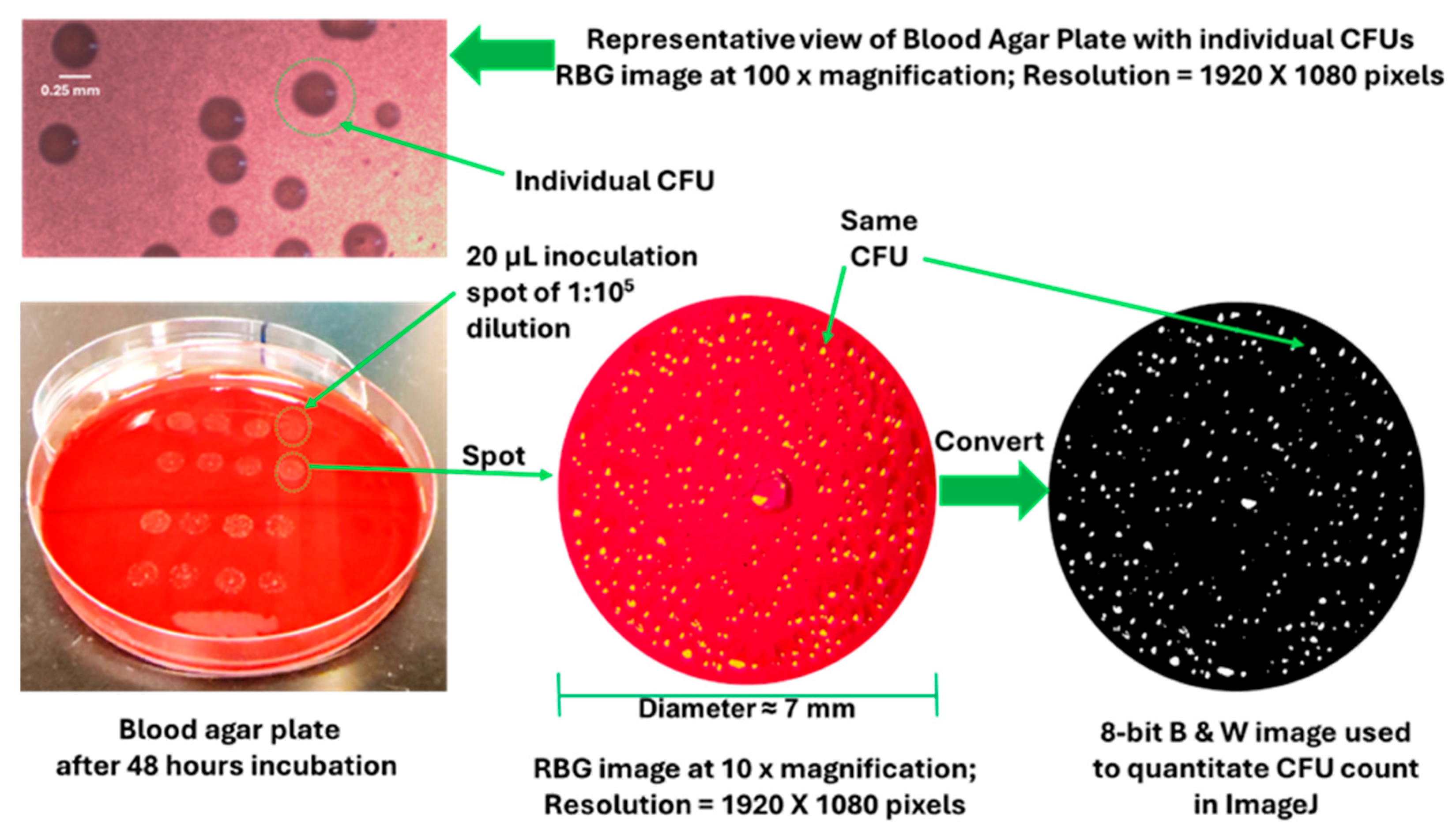
Immediately following 24 h absorbance readings (Section 2.3) of P. gingivalis cultures from two separate experiments, the original 96-well plates were serially diluted 1:106 (experiment 1) and 1:105 (experiment 2) with TSBY. Diluted samples were spot plated using 20 µL onto TSA blood agar plates (arranged by treatment group). Each blood agar plate contained two treatment groups with n = 6 or n = 8 for 1:106 and 1:105 dilutions, respectively, for a total n = 14. The blood agar plates were then placed in the Bactron 300 anaerobic incubator at 37 °C for 48 h. Quantification of the CFU number of P. gingivalis was carried out using a digital imaging technique, as outlined in Figure 2. Briefly, after 48 h anaerobic growth, each 20 µL spot on the blood agar plate (100 mm × 15 mm) was photographed using a Moticam 1080 HDMI and USB camera (Motic®, Richmond, BC, Canada) attached to a Fisher brand stereomicroscope. Photographic images were taken at 10× total magnification (resolution = 1920 × 1080 pixels) and converted to 8 bit black and white (B & W) images using the public domain software ImageJ 1.53t (National Institute of Health). ImageJ then tabulated the number of white particles (CFU count) in each spot [26]. These values were then used to calculate CFUs/mL as follows: CFU count × dilution factor × spot volume. For an enhanced view of individual P. gingivalis CFUs, representative images of CFUs on the blood agar plates (Figure 2) were taken using a Fisherbrand HDMI and USB camera (ThermoFisher Scientific, Waltham, MA, USA), attached to a Fisher brand light microscope at 100× total magnification (resolution = 1920 × 1080 pixels).

2.6. Morphology and Viability of OKF6/TERT-2 Cells

OKF6/TERT-2 cells were generously provided by Dr. Gill Diamond from the Louisville University School of Dentistry, but the cell line was originally established by Dickson et al. [24]. Growth and expansion of OKF6/TERT-2 cells is described in detail in Cuadra et al. (2023) [27]. Briefly, for the first 24 h, these cells were cultured in 24-well flat-bottom plates containing 1 mL Keratinocyte Serum-Free Medium (KSFM) supplemented with 30 μg/mL of bovine pituitary extract (BPE), 3 ng/mL epithelial growth factor, 0.3 mM calcium chloride, 2 mM glutamine, and 100 U/mL penicillin and streptomycin at standard conditions (37 °C, 5% CO2). The next day, the cells were switched to a 1:1 v/v ratio of KSFM and Dulbecco’s Modified Eagle’s Medium/Ham’s Nutrient Mixture F-12 (DMEM/F12) supplemented with the same nutrients as in KSFM. This combined media, referred to as DFK [27,28], bathed the OKF6/TERT-2 until they became >90% confluent, usually after three to four days. Once confluent, the DFK media was removed and replaced with 1 mL of DFK containing 0 (control), 1, 2, 3 or 4% DMSO or DMSO-2 and allowed to incubate at 37 °C, 5% CO2 for an additional 24 h. Cells treated with 5% Px in DFK served as a control indicating 100% cytotoxicity.
Twenty-four hours post-treatments, microscopic images of OKF6/TERT-2 cell monolayers were taken using a Nikon Eclipse TE2000-U inverted microscope with a Nikon Digital Sight DS-Fi1 camera and NIS Elements Imaging Software Version BR 3.1 (Nikon Instruments Inc., Melvin, NY, USA). All light microscopy images were captured at 100× magnification and evaluated for any treatment-induced morphological alterations. After imaging, the supernatants were collected and stored at −20 °C for subsequent lactate dehydrogenase (LDH) activity assays. An Invitrogen LDH cytotoxicity assay kit (Thermo Fisher, Waltham, MA, USA) was used according to the manufacturer’s instructions. Briefly, untreated OKF6/TERT-2 cell monolayers were lysed with a lysis buffer (provided with kit) and used to reference 100% LDH activity (an index of 0% viability). At the time of assay, 50 µL of thawed supernatants were added to 50 µL of the reaction mixture and incubated for 30 min in the dark at room temperature. In the final step, 50 µL of stopping solution was added to all reactions, and the absorbance was read at 595 nm.

2.7. Statistical Analysis

All data were expressed as means ± standard error of the mean (SEM) and all comparisons were made using either one-way ANOVA or two-way ANOVA followed by a Bonferroni multiple comparison test, where significance is achieved when p < 0.05. Additionally, Student t-tests were employed when comparing the effects of DMSO to the effects of DMSO-2 at a single treatment/time point. Version 5 of Prism (GraphPad Software, San Diego, CA, USA) was used to perform all statistical calculations and to generate all graphs.

3. Results

3.1. DMSO and DMSO-2 Inhibit Planktonic Growth of P. gingivalis

Figure 3 illustrates the inhibitory effect DMSO or DMSO-2 has on the 48 h planktonic growth of P. gingivalis, as indexed by absorbance at 595 nm (Figure 3A,B) and % change in absorbance (Figure 3C,D). The results indicate that both DMSO and DMSO-2 inhibit planktonic growth of P. gingivalis in a robust dose-dependent manner. The maximum inhibitory effect for DMSO appears to occur at 24 h for all doses, while the maximum inhibitory effect for DMSO-2 appears to occur at 12 h for the 0.5% and 1% doses and 24 h for the 2% and 4% doses. After 24 h exposure to 4% DMSO or DMSO-2, planktonic growth of P. gingivalis is inhibited by 57% and 77%, respectively. Additionally, the effectiveness of the 4% DMSO and DMSO-2 dose lingers on for up to 48 h.

3.2. DMSO and DMSO-2 Suppress Viability of P. gingivalis

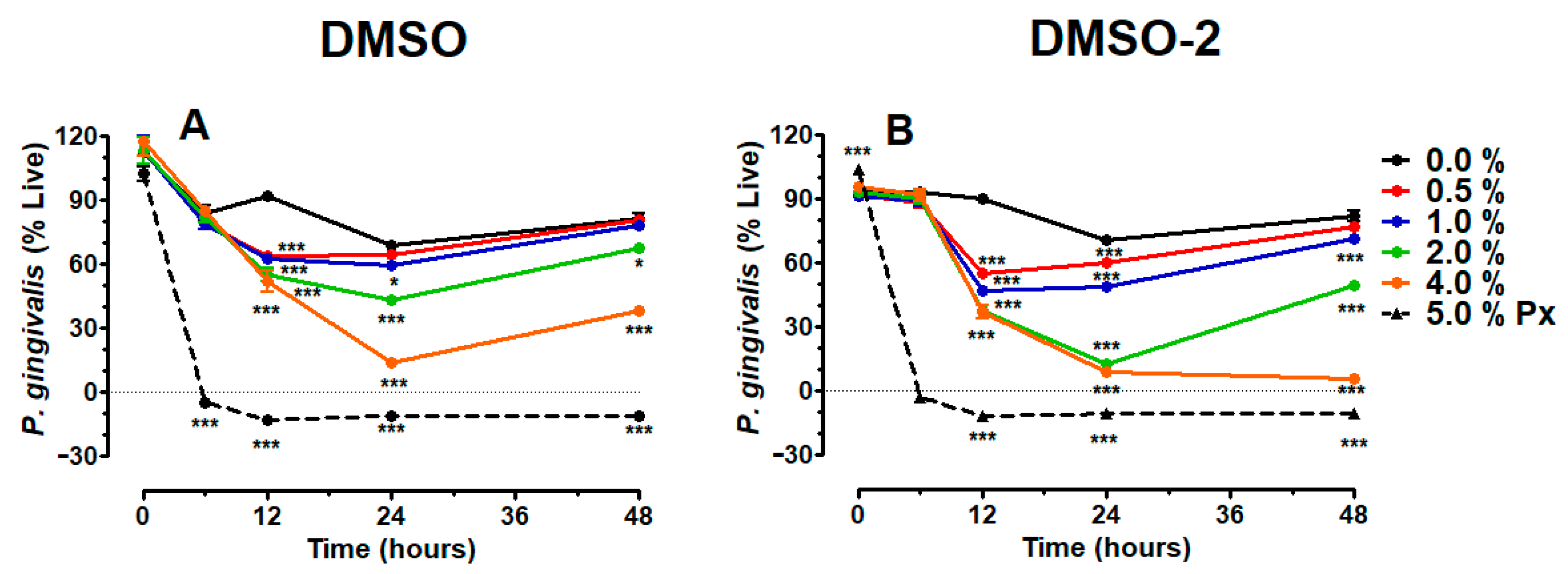
Figure 4 depicts the inhibitory effect DMSO (A) or DMSO-2 (B) has on the 48 h viability of P. gingivalis, as indexed by Live/Dead staining. The results indicate that DMSO and DMSO-2 suppress P. gingivalis viability in a dose-dependent manner. Except for the 0.5% dose, maximum suppression by DMSO occurs at 24 h for all other doses, while maximum suppression by DMSO-2 occurs at 12 h for the 0.5% and 1% doses and 24 h for the 2 and 4% doses. Furthermore, the suppression induced by the 4% DMSO and DMSO-2 dose lingers on for up to 48 h. After 24 h exposure to 4% DMSO or DMSO-2, the viability of P. gingivalis, relative to control, is suppressed by 56% and 61%, respectively. These results mirror DMSO- and DMSO-2-induced inhibition of the P. gingivalis planktonic growth shown above (Figure 3).

3.3. DMSO and DMSO-2 Reduce CFU Count of P. gingivalis

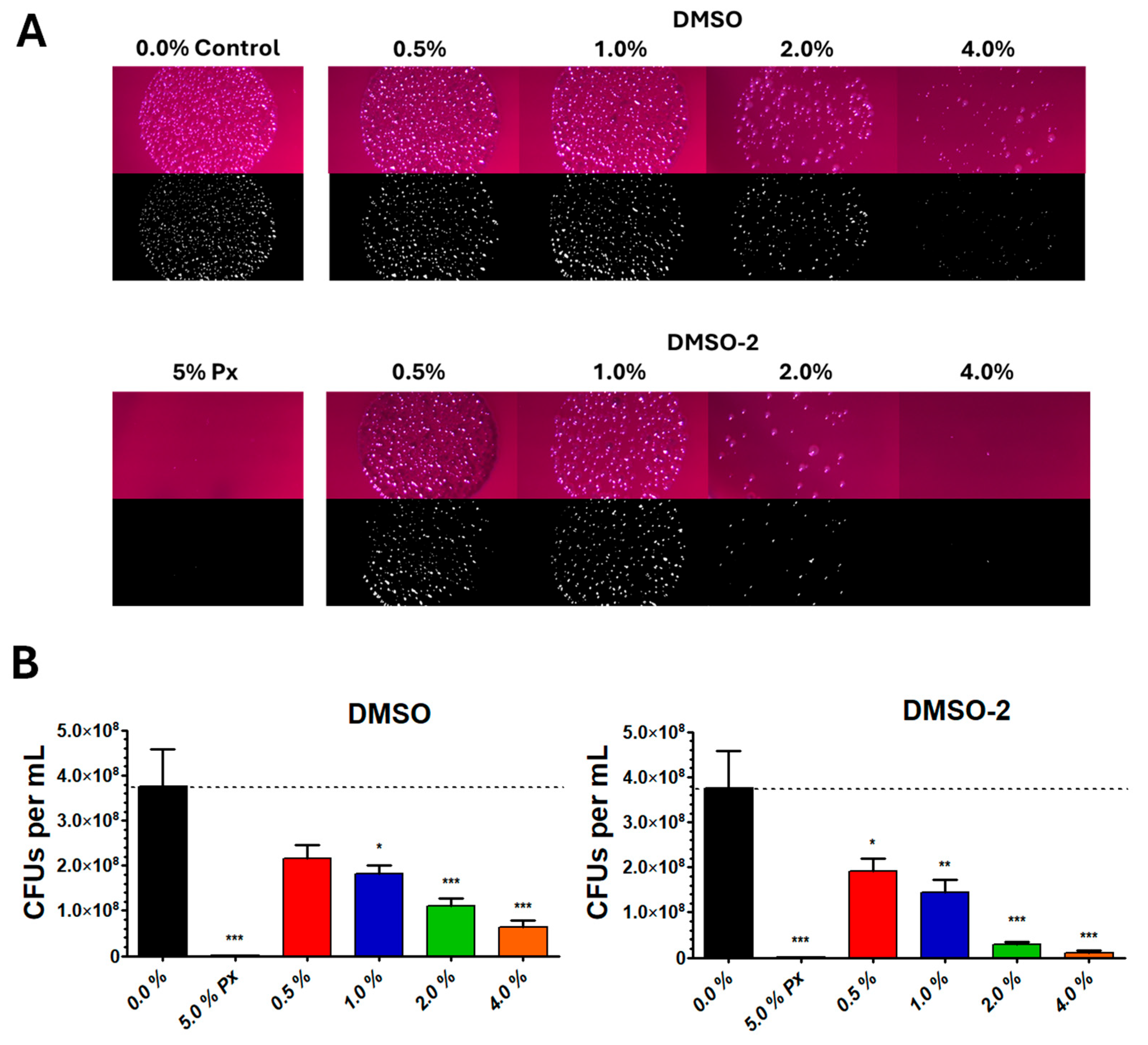
Figure 5A portrays representative images (original and ImageJ-transformed) of P. gingivalis CFUs generated from 20 µL spots from serial dilutions on blood agar after 24 h planktonic growth of DMSO or DMSO-2 treated cultures, while Figure 5B shows the CFU analysis. From visual inspection (Figure 5A), it is apparent that control P. gingivalis CFUs are more numerous than DMSO- or DMSO-2-treated CFUs at matching dilution factors. Furthermore, this effect is more pronounced for DMSO-2 than for DMSO. For enlarged views of each image shown in Figure 5A, see the Supplemental Data (Figure S1). Quantification of CFU counts (Figure 5B) confirm visual inspection. The CFU counts for P. gingivalis decrease from 3.8 × 10⁸ CFUs/mL in controls to 2.1 × 10⁸, 1.8 × 10⁸, 1.1 × 10⁸ and 6.4 × 10⁷ CFUs/mL after treatment with 0.5, 1, 2 and 4% DMSO, respectively. Similarly, CFU counts significantly decrease to 1.9 × 10⁸, 1.5 × 10⁸, 3.0 × 10⁷ and 1.1 × 10⁷ CFUs/mL after treatment with 0.5%, 1%, 2% and 4% DMSO-2, respectively. These results support DMSO- and DMSO-2-induced inhibition of P. gingivalis planktonic growth (Section 3.2) and suppression of P. gingivalis viability (Section 3.3), as described above.

3.4. Morphology and Viability of OKF6/TERT-2 Cells

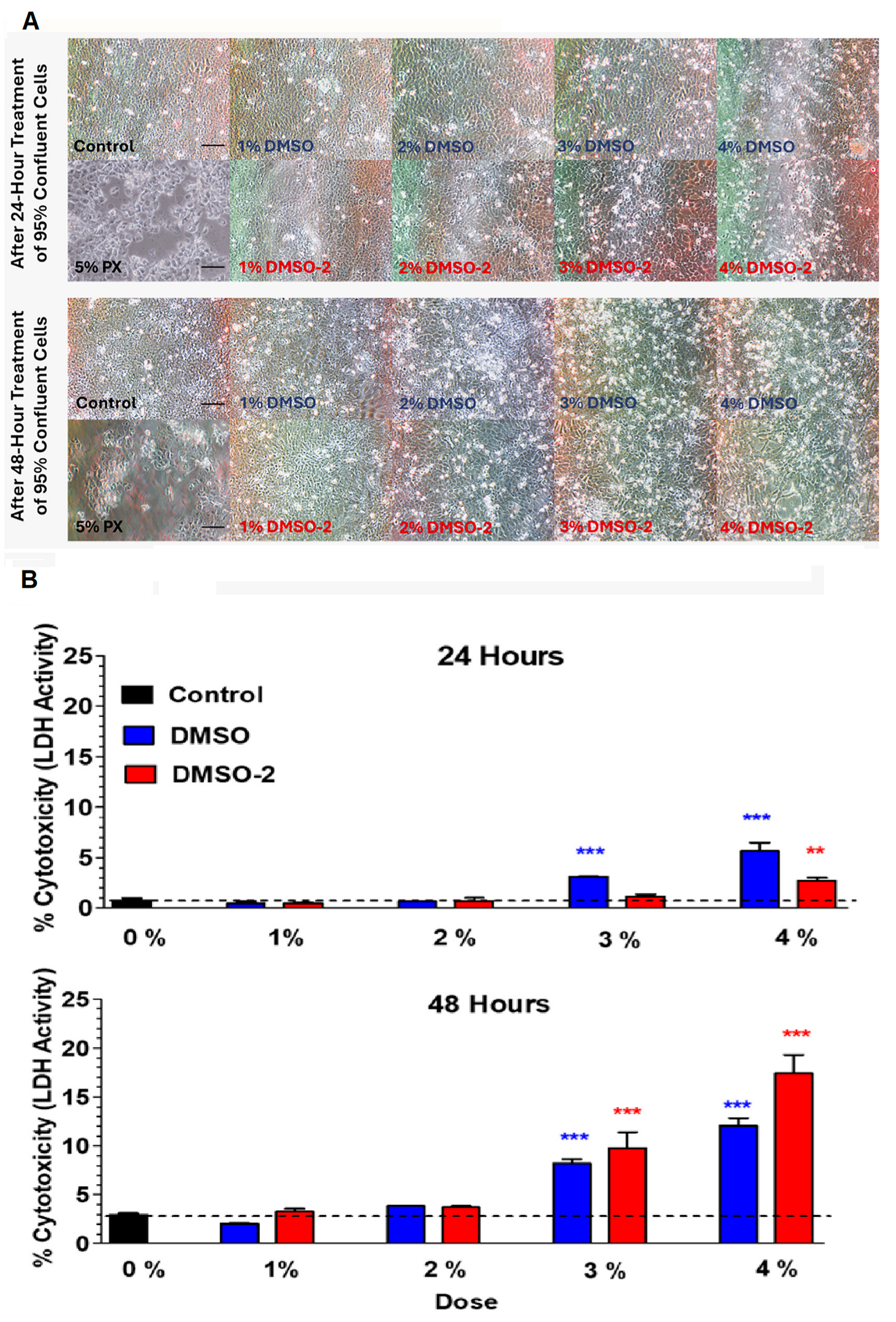
Figure 6A shows 24 and 48 h representative images of OKF6/TERT-2 cells exposed to DMSO or DMSO-2, while Figure 6B reports the % cytotoxicity of DMSO or DMSO-2, as indexed by LDH activity. After 24 h exposure to 1, 2 or 3% of DMSO or DMSO-2, the morphology of the OKF6/TERT-2 cells (Figure 6A) remains intact, retaining their typical cobblestone appearance. However, these cells begin to show signs of stress after exposure to 3% of both reagents after 48 h exposure. For enlarged views of each image shown in Figure 6A, see the Supplemental Data (Figure S2). LDH activity (Figure 6B) of OKF6/TERT-2 cells exposed to 24 h of 3% DMSO or DMSO-2 is increased by 2.3% and 0.3%, respectively, relative to the control, while 4% DMSO or DMSO-2 increases LDH activity by 4.8% and 1.9%, respectively. This effect is more pronounced after 48 h exposure to DMSO or DMSO-2. LDH activity of cells exposed to 48 h of 3% DMSO or DMSO-2 is increased by 5.1% and 6.8%, respectively, while 4% DMSO or DMSO-2 increases LDH activity by 9.1% and 14.4%, respectively.
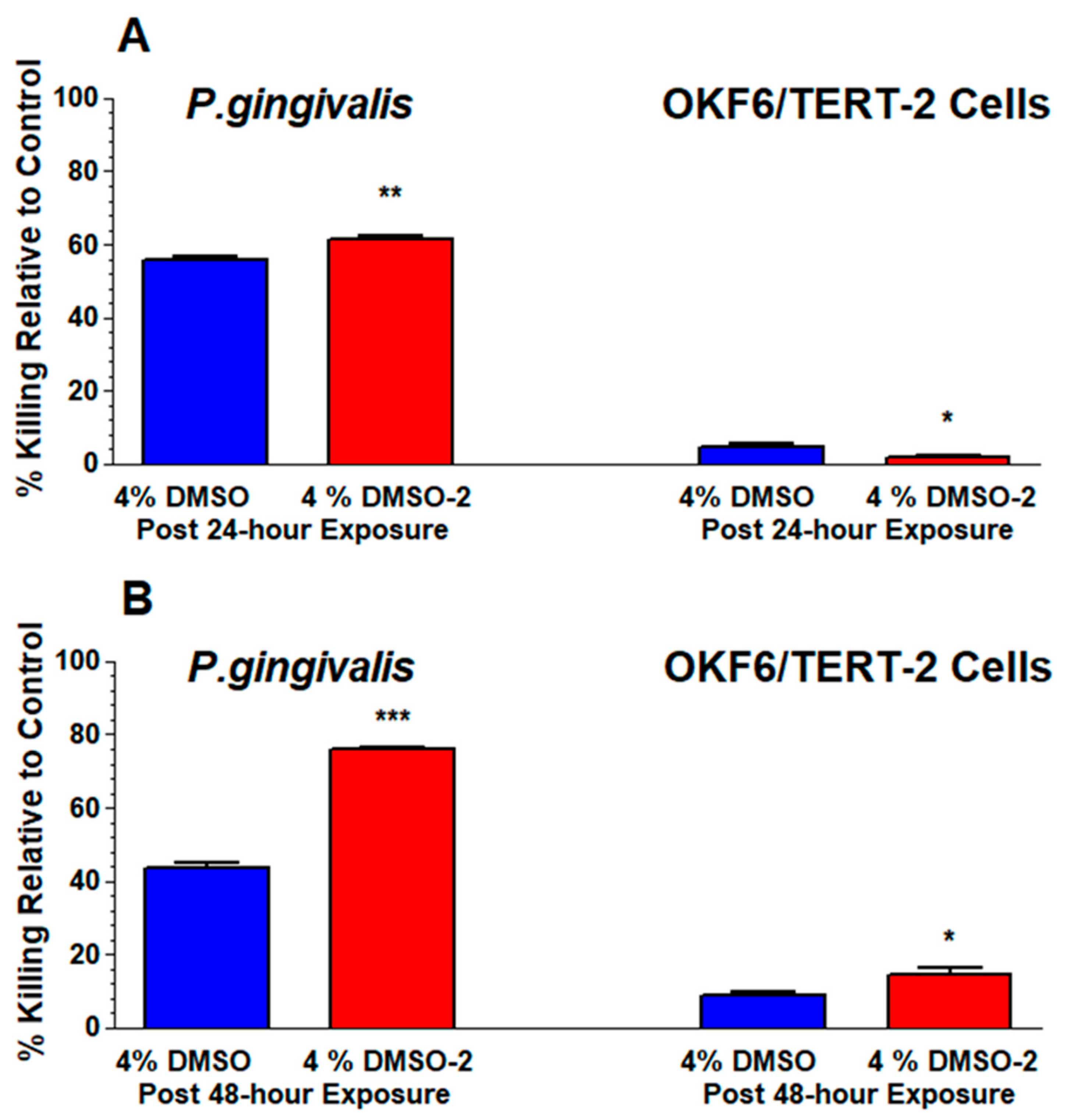
As a comparison, Figure 7 illustrates the relative differences in the viabilities of P. gingivalis and OKF6/TERT-2 cells exposed to 4% DMSO or DMSO-2 for 24 (Figure 7A) or 48 h (Figure 7B). From these data, it is evident that both DMSO and DMSO-2 are more effective in killing P. gingivalis than OKF6/TERT-2 cells at both time points. After the 24 h exposure of the 4% dose, the killing effectiveness of DMSO and DMSO-2 is 11.6 and 32.6 times greater, respectively, for P. gingivalis than for the OKF6/TERT-2 cells. After the 48 h exposure of the 4% dose, the killing effectiveness of DMSO and DMSO-2 is 4.8 and 5.3 times greater, respectively, for P. gingivalis than for the OKF6/TERT-2 cells. With respect to P. gingivalis, Student t-tests reveal that DMSO-2 has a greater killing effect than DMSO after a 24 h treatment (p < 0.01). This effect is reversed with the OKF6/TERT-2 cells, where DMSO has a modest but greater killing capacity (p < 0.05) than DMSO-2. After 48 h treatments with 4% DMSO or DMSO-2, the cytotoxicity induced by DMSO-2 is greater than DMSO for both P. gingivalis (p < 0.001) and OKF6/TERT-2 cells (p < 0.05). Overall, the 24 h data show that DMSO-2 has greater killing ability than its DMSO precursor on P. gingivalis, with only minimal killing effect on OKF6/TERT-2 cells. This makes DMSO-2 an effective, natural, antimicrobial candidate to treat pathologies such as gingivitis and periodontitis.

4. Discussion

This study shows that DMSO inhibits the planktonic growth and viability of P. gingivalis, which agrees with numerous past findings that DMSO has an antimicrobial effect on several facultative and anaerobic bacterial species [9,10,11,12,13]. Summer et al. (2022) [10] report that 1 to 10% DMSO in Mueller Hinton II broth exerts significant antimicrobial activity against Streptococcus pneumoniae and Pseudomonas aeruginosa. Wadhwani et al. (2009) [11] find that 4% DMSO in Mueller Hinton broth inhibits the growth of Salmonella paratyphi A, Staphylococcus epidermidis, Shigella flexneri, Vibrio cholerae and Pseudomonas oleovorans to 50%, 37%, 83%, 49% and 42%, respectively. Feldman et al. (1975) [12] and Ansel et al. (1969) [13] report antimicrobial activity of DMSO against Escherichia coli and Bacillus megaterium. Carrol et al. (2020) [9] provide the only report of DMSO-induced antimicrobial activity against P. gingivalis. They find that greater than 1% v/v DMSO in brain heart infusion media inhibits the growth of P. gingivalis.
Administration of DMSO to mammalian systems leads to mostly mild adverse reactions, although the severity of these reactions depends largely on the dose, route and location of administration [16]. The situation is different for cell cultures. Typically, 10% DMSO is used as a cryoprotective agent for cell storage at low temperatures [29]. However, Galvao et al. (2014) [14] report that 2–4% DMSO induces neuronal cell death in a caspase-3 independent manner. Verheijen et al. (2019) [15] claim that 3D cardiac and hepatic microtissues exposed to 0.1% DMSO induces more than 2000 differentially expressed genes. Singh et al. (2017) [30] find that 0.5 to 3% DMSO can reduce the viability of goat skin fibroblasts. Gallardo-Villagran et al. (2022) [31] report that a 24 h treatment with 10% DMSO reduces the viability of human fibroblast-like synoviocytes to 52.7% live cells, while 2.5% DMSO has no effect. Carrol et al. (2020) [9] find 1% DMSO to have no cytotoxic effect on the human keratinocyte HaCaT cells, but cytotoxicity increases slightly at doses greater than 2%. From these studies it is clear that the physiological effects of DMSO depend on cell type as well as its concentration. For example, Nguyen et al. (2015) [32] found that human oral squamous carcinoma SCC25 cells and non-cancerous human keratinocyte HaCaT cells respond differently when exposed to DMSO ranging from 0 to 1%. The cell viability was higher for the HaCaT cells (about 60% live with 1% DMSO) than for the SCC25 cells (about 30% live with 1% DMSO). From the Carrol et al. [9] and Nguyen et al. [32] studies, it is evident that changes in the cytotoxicity and/or viability of human keratinocyte begin to occur between 1% and 2% DMSO. In comparison, our study finds DMSO-induced cytotoxicity of OKF6/TERT-2 cells beginning at 3% by 24 h.
Despite the above findings using DMSO, only one investigation [33] demonstrates the antimicrobial activity of DMSO-2. Poole et al. [33] find that DMSO-2, between 0 and 16%, has a dose-dependent inhibitory effect on E. coli and Salmonella enterica grown in brain/heart infusion media. Our findings agree with Poole et al. [33] in that DMSO-2 induces antimicrobial activity towards P. gingivalis at concentrations as low as 0.5% for planktonic growth (Figure 3), viability (Figure 4) and colony formation on blood agar (Figure 5). To our knowledge, this is the first report showing that DMSO-2 exerts antimicrobial activity against P. gingivalis. Similarly, we are unaware of any findings investigating the effects of DMSO-2 on oral epithelial cells. In this study, we report that 4% DMSO-2 induces a slight cytotoxic effect on OKF6/TERT-2 after a 24 h exposure (Figure 6). On the other hand, it has been reported that 300 mmol/L DMSO-2 have protective effects against inflammation and epithelial barrier injury induced by lipopolysaccharide on porcine intestinal epithelial cells [34].
In this investigation we report that both DMSO and DMSO-2 inhibit the planktonic growth (Figure 3) and viability (Figure 4) of P. gingivalis in a dose-dependent manner, which is further supported by CFU analysis (Figure 5). Furthermore, our data supports a bactericidal mechanism of action for DMSO and DMSO-2, since Figure 4 shows a direct decrease in P. gingivalis viability and Figure 5 shows parallel results with CFU counts on blood agar. However, the killing induced by DMSO-2, as compared to DMSO, appears to have an earlier onset and a longer lasting effect. While this study also finds that DMSO and DMSO-2 inhibit the viability of OKF6/TERT-2 cells in a dose-dependent manner (Figure 6), the antimicrobial effect of both agents on P. gingivalis far exceeds the cytotoxic effect these agents have on OKF6/TERT-2 cells (Figure 7). In contrast, the bacteriostatic nature of DMSO and DMSO-2 is supported by other studies. Jacob’s group [8,35,36] find that a 20% solution of DMSO has bacteriostatic properties against E. coli, Staphylococcus aureus, and Pseudomonas bacilli, although at doses greater than 50% the bacteria was unable to recover. Similarly, Poole et al. (2019) [33] demonstrate the bacteriostatic nature of DMSO-2 as E. coli and S. enterica growth is halted by DMSO-2 but resumes when the bacteria are transferred to fresh growth media. However, recovery of E. coli and S. enterica was unattainable following long-term exposure from 10 to 16% DMSO-2 for 5 to 6 days, as shown by the lack of CFU presence.
The exact antimicrobial mechanism of action for DMSO and DMSO-2 is not clear. Ghajar and Harmon [37] find that treatment of Staphylococcus aureus with DMSO resulted in a greater rate of oxygen and lactose uptake, while the rate of glycine transport was reduced. Ansel et al. (1969) [13] speculate that DMSO can penetrate bacteria and alter cell division. More recently, using atomic-scale molecular dynamics simulations of lipid bilayers consisting of 128 dipalmitoyl-phosphatidylcholine lipids, Gurtovenko and Anwar [38] provided evidence that 10 to 15% v/v DMSO can induce water pores in the lipid bilayer, allowing for the diffusion of salt ions. Tuncer and Gurbanov [39] report that 1.0% and 2.5% v/v DMSO reduce intracellular levels of reactive oxygen species (ROS), change the content of cellular nucleic acid, affect the global 5-methylcytosine pattern of the genome, and modulate gene transcription in E. coli. In a study by Yahya et al. (2018) [40], 32% DMSO inhibits biofilm formation of E. coli, P. aeruginosa and Salmonella typhimurium, suggesting that chemical alterations in the extracellular polymeric substance matrix may be involved. Butawan et al. (2017) [18] suggest that since DMSO-2, like DMSO, easily penetrates membranes, it may involve the same types of activities noted for DMSO, such as reducing intracellular levels of ROS and modulating gene transcription. One difference between DMSO and DMSO-2 worth noting is that DMSO can methylate DNA in the presence of hydroxyl radicals [41]. In contrast, DMSO-2 does not methylate DNA [42]. Given the similar chemical structures of DMSO and DMSO-2 (Figure 1), it is reasonable to assume that the intracellular bacterial mechanisms leading to antimicrobial activity on P. gingivalis are similar, but not necessarily the same. This could explain the timing difference we observed in the planktonic growth and viability of P. gingivalis treated with DMSO or DMSO-2. Since DMSO-2 is a metabolite of DMSO [43], the possibility exists that at least some of the inhibitory effect of DMSO on the growth of P. gingivalis may be attributed to DMSO-2. This could explain the antimicrobial difference induced by DMSO-2 compared to DMSO. Consequently, the biochemical and physiological pathways of how P. gingivalis responds to DMSO or DMSO-2 in an anerobic environment need to be further elucidated.
The potential clinical applications for DMSO are several [7,44]; however, its use is controversial [45]. In 1965, the Food and Drug Administration (FDA) banned clinical trials with DMSO because it was found to cause eye damage in laboratory animals, but later lifted the ban when the public interest in DMSO subsided [46]. In 1978, DMSO received approval by the FDA for use in the palliative treatment of interstitial cystitis, but its use for other medical conditions has been limited by a lack of clinical evidence suggesting beneficial effects, incomplete clinical trials or unacceptable adverse reactions [16,47]. In a systematic review of 109 studies by Kollerup Madsen et al. (2018) [16], reported adverse reactions from the transdermal application of DMSO included halitosis/garlic breath (10%), nausea (5%), abdominal cramps/stomach ache (4%), diarrhea (3%) and vomiting (1%). As an alternative to DMSO, experimentation with DMSO-2 began in the 1970s [48], and shortly thereafter, a host of US patents were granted for DMSO-2 in order to smoothen and soften skin, strengthen nails, dilute blood, relieve stress, relieve pain, treat parasitic infections, increase energy, boost metabolism, enhance circulation and improve wound healing [18]. Notable is the lack of halitosis/garlic breath associated with DMSO-2 [49]. Between 1996 and 2016, a number of reports emerged suggesting that DMSO-2 may have clinical applications for arthritis and other inflammatory disorders such as interstitial cystitis, allergic rhinitis and acute exercise-induced inflammation [18]. In 2007, one product, OptiMSM® (Bergstrom Nutrition, Vancouver, WA, USA) received the “Generally Recognized as Safe (GRAS)” status by the FDA as a food ingredient and was reported to be safe at doses of less than 4845.6 mg/day [50]. There is scant primary literature regarding DMSO-2-related adverse reactions. Kim et al. (2005) [51] reported bloating, constipation, indigestion, fatigue, insomnia and headaches when oral doses of up to 6 g/day DMSO-2 were administered to a group of patients over a twelve-week period for the treatment of osteoarthritis. However, the occurrence of these adverse reactions did not differ from the placebo group. In a similar study by Debbi et al. (2011) [52], no clinical side effects were reported, presumably since the dose of DMSO-2 used in their study was half that used in the Kim et al. (2005) study [51].
Based on the limited information available, DMSO-2 may be a safe antimicrobial agent to use prophylactically against P. gingivalis for the treatment of gingivitis and periodontitis (or other diseases induced by aerobic or anaerobic pathogens) and should be rigorously explored. In fact, products containing DMSO-2 such as dietary supplements to rejuvenate joint and increase flexibility [53], creams to moisturize and enhance skin [54], shampoo bars to strengthen hair [55], nasal spray for sinusitis and snoring [56], toothpaste [57] and even mouthwash [58] can very easily be purchased through the internet, simply because DMSO-2 has the FDA GRAS designation and can be used liberally as a supplement. More than likely, however, the FDA has not evaluated any of the claims made by these companies. Prudence dictates that more stringent studies concerning the safety of DMSO-2 as a prophylactic agent against microbial infections be conducted. The primary literature clearly indicates that the cytotoxic effects of DMSO are a deterrent towards its utility as an antimicrobial agent. On the other hand, evidence of the safe use of DMSO-2 as a dietary supplement, coupled with our findings, endorses its potential as a novel antimicrobial agent in vivo.
Here we present evidence that DMSO and DMSO-2 are bactericidal to P. gingivalis and present minimal cytotoxicity to OKF6/TERT-2 cells. However, this study is not without limitations. First, P. gingivalis does not normally grow planktonically in the oral cavity, but rather in a community of microorganisms as a biofilm [59]. The possibility exists that the DMSO- and DMSO-2-induced inhibition in planktonic growth observed in this study may not translate to growth of P. gingivalis on a salivary pellicle in the presence of other bacteria. The optimal growth of P. gingivalis in a multispecies community depends on communication between nearby microbial species as well as with the underlying host epithelium, neither of which were present in this study. Consequently, the inhibitory effects induced by DMSO and DMSO-2 on the planktonic growth of P. gingivalis could over- or underestimate the actual growth of this opportunistic bacterium on oral biofilms. Similarly, the OKF6/TERT-2 cells used in this study represent an in vitro cell line. Oral epithelial cells do not normally exist by themselves in a closed system, but rather, they interact with a myriad of microbial species bathed in saliva within an open system. Second, it is evident from these experiments that DMSO and DMSO-2 inhibit P. gingivalis in a bactericidal manner, but the biochemical and physiological processes behind this effect remain unknown without further molecular studies. Third, we only investigated the effects of DMSO and DMSO-2 on P. gingivalis, a Gram-negative bacteria. Given the structural differences in the cell walls of Gram-negative (thinner peptidoglycan and outer lipid membrane) vs. Gram-positive bacteria (thicker peptidoglycan and no outer lipid membrane) [60], DMSO and DMSO-2 could have entirely different effects. While the cytotoxicity of DMSO and DMSO-2 on OKF6/TERT-2 cells is minimal, the actual cytotoxic mechanisms need to be investigated. Finally, it is not known whether DMSO is metabolized to DMSO-2 while incubated with P. gingivalis of the OKF6/TERT-2 cells. Hence, the stability of DMSO and DMSO-2 in the presence of P. gingivalis should be confirmed either through high performance liquid chromatographic or gas chromatographic methods [61,62].

5. Conclusions

DMSO and DMSO-2 have substantial inhibitory effects on the growth and viability of P. gingivalis. DMSO- and DMSO-2-mediated inhibition of P. gingivalis seems to occur via bactericidal mechanisms of action. While DMSO and DMSO-2 are minimally cytotoxic to OKF6/TERT-2 cells, the antimicrobial effect these agents have on P. gingivalis far exceeds the cytotoxic effect on these human oral epithelial cells. Based on our results and the safer nature of DMSO-2 (FDA GRAS designation), the use of DMSO-2 as a novel and natural antimicrobial agent should be further explored. Ultimately, our current findings could lead to an inexpensive therapeutic against anaerobic pathogens that would have profound translational potential.

Supplementary Materials

The following supporting information can be downloaded at https://www.mdpi.com/article/10.3390/bacteria4040057/s1, Figure S1: Enlargements of the original and ImageJ-transformed spot images of P. gingivalis CFUs; Figure S2: Original full-size images of OKF6/TERT-2 cells.

Author Contributions

Conceptualization, D.L.P., D.M. and G.A.C.; Methodology, D.L.P. and G.A.C.; Software, D.L.P.; Validation, D.L.P. and G.A.C.; Formal Analysis, D.L.P. and G.A.C.; Investigation, D.L.P., A.J., D.P., M.H., D.M., M.T., C.T., J.R.A. and G.A.C.; Resources, D.L.P., D.M. and G.A.C.; Data Curation, D.L.P.; Writing—original draft preparation, D.L.P.; Writing—review and editing, D.L.P., D.M. and G.A.C.; Visualization, D.L.P.; Supervision, D.L.P. and G.A.C.; Project Administration, D.L.P. and G.A.C.; Funding Acquisition, D.L.P., D.M. and G.A.C. All authors have read and agreed to the published version of the manuscript.

Funding

Internal funding by the DeBusk College of Osteopathic Medicine and Muhlenberg College. This research received no external funding.

Institutional Review Board Statement

Not applicable.

Informed Consent Statement

Not applicable.

Data Availability Statement

The raw data supporting the conclusions of this article will be made available by the authors on request.

Acknowledgments

This work was supported by the DeBusk College of Osteopathic Medicine and Muhlenberg College. Additionally, authors thank Mikaela Brown, Siara Minton, Kathy Christie and Sara Gill for technical support.

Conflicts of Interest

The authors declare no conflicts of interest.

References

  1. Clifford, K.; Dy-Boarman, E.; Haase, K.; Maxvill, K.; Pass, S.; Alvarez, C. Challenges with Diagnosing and Managing Sepsis in Older Adults. Expert Rev. Anti-Infect. Ther. 2016, 14, 231–241. [Google Scholar] [CrossRef] [PubMed]
  2. Church, D.L. Selected Topics in Anaerobic Bacteriology. Microbiol. Spectr. 2016, 4, 493–535. [Google Scholar] [CrossRef] [PubMed]
  3. Brook, I. Treatment of Anaerobic Infection. Expert Rev. Anti-Infect. Ther. 2007, 5, 991–1006. [Google Scholar] [CrossRef]
  4. Noor, A.; Khetarpal, S. Anaerobic Infections. In StatPearls; StatPearls Publishing: Treasure Island, FL, USA, 2024. [Google Scholar]
  5. Hajishengallis, G.; Darveau, R.P.; Curtis, M.A. The Keystone-Pathogen Hypothesis. Nat. Rev. Microbiol. 2012, 10, 717–725. [Google Scholar] [CrossRef]
  6. Cuadra, G.; Hanel, A.; Christian, N.; Pecorelli, S.; Ha, V.; Tomov, S.; Palazzolo, D. Perspective Chapter: The Impact of Electronic Cigarettes on the Oral Microenvironment. In Periodontal Frontiers [Working Title]; IntechOpen: London, UK, 2025. [Google Scholar]
  7. Madruga, G.M.; Crivellenti, L.Z.; Borin-Crivellenti, S.; Cintra, C.A.; Gomes, L.G.; Spiller, P.R. Comparative Use of Dimethyl Sulphoxide (DMSO) in Different Animal Species. Vet. Med. 2017, 62, 179–185. [Google Scholar] [CrossRef]
  8. Jacob, S.W.; Torre, J.C. Dimethyl Sulfoxide (DMSO) in Trauma and Disease; CRC press: Boca Raton, FL, USA, 2015; ISBN 978-1-4987-1467-9. [Google Scholar]
  9. Carrol, D.H.; Chassagne, F.; Dettweiler, M.; Quave, C.L. Antibacterial Activity of Plant Species Used for Oral Health against Porphyromonas Gingivalis. PLoS ONE 2020, 15, e0239316. [Google Scholar] [CrossRef]
  10. Summer, K.; Browne, J.; Hollanders, M.; Benkendorff, K. Out of Control: The Need for Standardised Solvent Approaches and Data Reporting in Antibiofilm Assays Incorporating Dimethyl-Sulfoxide (DMSO). Biofilm 2022, 4, 100081. [Google Scholar] [CrossRef]
  11. Wadhwani, T.; Desai, K.; Patel, D.; Lawani, D.; Bahaley, P.; Joshi, P.; Kothari, V. Effect of Various Solvents on Bacterial Growth in Context of Determining MIC of Various Antimicrobials. Int. J. Microbiol. 2008, 7, 1–6. [Google Scholar]
  12. Feldman, W.E.; Punch, J.D.; Holden, P.C. In Vivo and In Vitro Effects of Dimethyl Sulfoxide on Streptomycin-Sensitive and -Resistant Escherichia coli. Ann. N. Y. Acad. Sci. 1975, 243, 269–277. [Google Scholar] [CrossRef] [PubMed]
  13. Ansel, H.C.; Norred, W.P.; Roth, I.L. Antimicrobial Activity of Dimethyl Sulfoxide Against Escherichia Coli, Pseudomonas Aeruginosa, and Bacillus Megaterium. J. Pharm. Sci. 1969, 58, 836–839. [Google Scholar] [CrossRef]
  14. Galvao, J.; Davis, B.; Tilley, M.; Normando, E.; Duchen, M.R.; Cordeiro, M.F. Unexpected Low-dose Toxicity of the Universal Solvent DMSO. FASEB J. 2014, 28, 1317–1330. [Google Scholar] [CrossRef]
  15. Verheijen, M.; Lienhard, M.; Schrooders, Y.; Clayton, O.; Nudischer, R.; Boerno, S.; Timmermann, B.; Selevsek, N.; Schlapbach, R.; Gmuender, H.; et al. DMSO Induces Drastic Changes in Human Cellular Processes and Epigenetic Landscape in Vitro. Sci. Rep. 2019, 9, 4641. [Google Scholar] [CrossRef] [PubMed]
  16. Kollerup Madsen, B.; Hilscher, M.; Zetner, D.; Rosenberg, J. Adverse Reactions of Dimethyl Sulfoxide in Humans: A Systematic Review. F1000Research 2019, 7, 1746. [Google Scholar] [CrossRef]
  17. Vaidya, S.; Tozer, K.; Chen, J. An Overview of Embolic Agents. Semin. Interv. Radiol. 2008, 25, 204–215. [Google Scholar] [CrossRef] [PubMed]
  18. Butawan, M.; Benjamin, R.; Bloomer, R. Methylsulfonylmethane: Applications and Safety of a Novel Dietary Supplement. Nutrients 2017, 9, 290. [Google Scholar] [CrossRef]
  19. Williams, K.I.H.; Burstein, S.H.; Layne, D.S. Metabolism of Dimethyl Sulfide, Dimethyl Sulfoxide, and Dimethyl Sulfone in the Rabbit. Arch. Biochem. Biophys. 1966, 117, 84–87. [Google Scholar] [CrossRef]
  20. He, X.; Slupsky, C.M. Metabolic Fingerprint of Dimethyl Sulfone (DMSO2) in Microbial–Mammalian Co-Metabolism. J. Proteome Res. 2014, 13, 5281–5292. [Google Scholar] [CrossRef]
  21. HOMD: Human Oral Microbiome Database. Available online: https://www.homd.org/# (accessed on 15 August 2025).
  22. Gao, L.; Xu, T.; Huang, G.; Jiang, S.; Gu, Y.; Chen, F. Oral Microbiomes: More and More Importance in Oral Cavity and Whole Body. Protein Cell 2018, 9, 488–500. [Google Scholar] [CrossRef]
  23. Rafiei, M.; Kiani, F.; Sayehmiri, F.; Sayehmiri, K.; Sheikhi, A.; Zamanian Azodi, M. Study of Porphyromonas Gingivalis in Periodontal Diseases: A Systematic Review and Meta-Analysis. Med. J. Islam. Repub. Iran. 2017, 31, 62. Available online: https://pubmed.ncbi.nlm.nih.gov/29445691/ (accessed on 25 October 2025). [CrossRef] [PubMed]
  24. Dickson, M.A.; Hahn, W.C.; Ino, Y.; Ronfard, V.; Wu, J.Y.; Weinberg, R.A.; Louis, D.N.; Li, F.P.; Rheinwald, J.G. Human Keratinocytes That Express hTERT and Also Bypass a P16INK4a -Enforced Mechanism That Limits Life Span Become Immortal yet Retain Normal Growth and Differentiation Characteristics. Mol. Cell. Biol. 2000, 20, 1436–1447. [Google Scholar] [CrossRef]
  25. Werheim, E.R.; Senior, K.G.; Shaffer, C.A.; Cuadra, G.A. Oral Pathogen Porphyromonas Gingivalis Can Escape Phagocytosis of Mammalian Macrophages. Microorganisms 2020, 8, 1432. [Google Scholar] [CrossRef]
  26. Singer Instruments. How to Count Colonies with ImageJ (and Fiji); Singer Instruments: Roadwater, UK; Available online: https://www.singerinstruments.com/resource/counting-colonies-with-imagej/ (accessed on 25 October 2025).
  27. Cuadra, G.A.; Shamim, A.; Shah, R.; Morgan, J.; Palazzolo, D.L. Comparison of Culture Media for In Vitro Expansion of Oral Epithelial Keratinocytes. Appl. Biosci. 2023, 2, 308–327. [Google Scholar] [CrossRef]
  28. Shamim, A.; Herzog, H.; Shah, R.; Pecorelli, S.; Nisbet, V.; George, A.; Cuadra, G.A.; Palazzolo, D.L. Pathophysiological Responses of Oral Keratinocytes After Exposure to Flavored E-Cigarette Liquids. Dent. J. 2025, 13, 60. [Google Scholar] [CrossRef]
  29. Whaley, D.; Damyar, K.; Witek, R.P.; Mendoza, A.; Alexander, M.; Lakey, J.R. Cryopreservation: An Overview of Principles and Cell-Specific Considerations. Cell Transplant. 2021, 30, 096368972199961. [Google Scholar] [CrossRef] [PubMed]
  30. Singh, M.; McKenzie, K.; Ma, X. Effect of Dimethyl Sulfoxide on in Vitro Proliferation of Skin Fibroblast Cells. J. Biotech. Res. 2017, 8, 78–82. [Google Scholar]
  31. Gallardo-Villagrán, M.; Paulus, L.; Leger, D.Y.; Therrien, B.; Liagre, B. Dimethyl Sulfoxide: A Bio-Friendly or Bio-Hazard Chemical? The Effect of DMSO in Human Fibroblast-like Synoviocytes. Molecules 2022, 27, 4472. [Google Scholar] [CrossRef]
  32. Nguyen, T.; Parat, M.-O.; Hodson, M.; Pan, J.; Shaw, P.; Hewavitharana, A. Chemical Characterization and in Vitro Cytotoxicity on Squamous Cell Carcinoma Cells of Carica Papaya Leaf Extracts. Toxins 2015, 8, 7. [Google Scholar] [CrossRef]
  33. Poole, T.L.; Benjamin, R.; Genovese, K.J.; Nisbet, D.J. Methylsulfonylmethane Exhibits Bacteriostatic Inhibition of Escherichia Coli, and Salmonella Enterica Kinshasa, in Vitro. J. Appl. Microbiol. 2019, 127, 1677–1685. [Google Scholar] [CrossRef]
  34. Jiao, Y.; Li, H.; Ren, T.; Kim, I.H. Protective Effects of Methylsulfonylmethane (MSM) on Barrier Function Injury of Porcine Intestinal Epithelial Cells (IPEC-J2) Induced by Lipopolysaccharide (LPS). Can. J. Anim. Sci. 2023, 103, 262–272. [Google Scholar] [CrossRef]
  35. Jacob, S.W.; Bischel, M.; Herschler, R.J. Dimethyl Sulfoxide (DMSO): A New Concept in Pharmacotherapy. Curr. Ther. Res. Clin. Exp. 1964, 6, 134–135. [Google Scholar] [PubMed]
  36. Jacob, S.W.; Herschler, R. Pharmacology of DMSO. Cryobiology 1986, 23, 14–27. [Google Scholar] [CrossRef]
  37. Ghajar, B.M.; Harmon, S.A. The Effect of Dimethyl Sulfoxide (DMSO) on Permeability of Staphylococcusaureus. Biochem. Biophys. Res. Commun. 1968, 32, 940–944. [Google Scholar] [CrossRef]
  38. Gurtovenko, A.A.; Anwar, J. Ion Transport through Chemically Induced Pores in Protein-Free Phospholipid Membranes. J. Phys. Chem. B 2007, 111, 13379–13382. [Google Scholar] [CrossRef] [PubMed]
  39. Tunçer, S.; Gurbanov, R. Non-Growth Inhibitory Doses of Dimethyl Sulfoxide Alter Gene Expression and Epigenetic Pattern of Bacteria. Appl. Microbiol. Biotechnol. 2023, 107, 299–312. [Google Scholar] [CrossRef]
  40. Yahya, M.F.Z.R.; Alias, Z.; Karsani, S.A. Antibiofilm Activity and Mode of Action of DMSO Alone and Its Combination with Afatinib against Gram-Negative Pathogens. Folia Microbiol. 2018, 63, 23–30. [Google Scholar] [CrossRef] [PubMed]
  41. Kawai, K.; Li, Y.-S.; Song, M.-F.; Kasai, H. DNA Methylation by Dimethyl Sulfoxide and Methionine Sulfoxide Triggered by Hydroxyl Radical and Implications for Epigenetic Modifications. Bioorganic Med. Chem. Lett. 2010, 20, 260–265. [Google Scholar] [CrossRef]
  42. Cloutier, J.F.; Castonguay, A.; O’Connor, T.R.; Drouin, R. Alkylating Agent and Chromatin Structure Determine Sequence Context-Dependent Formation of Alkylpurines. J. Mol. Biol. 2001, 306, 169–188. [Google Scholar] [CrossRef] [PubMed]
  43. Kocsis, J.J.; Harkaway, S.; Snyder, R. Biological Effects of the Metabolites of Dimethyl Sulfoxide. Ann. N. Y. Acad. Sci. 1975, 243, 104–109. [Google Scholar] [CrossRef]
  44. Santos, N.C.; Figueira-Coelho, J.; Martins-Silva, J.; Saldanha, C. Multidisciplinary Utilization of Dimethyl Sulfoxide: Pharmacological, Cellular, and Molecular Aspects. Biochem. Pharmacol. 2003, 65, 1035–1041. [Google Scholar] [CrossRef]
  45. Muir, M. DMSO: Many Uses, Much Controversy. Altern. Complement. Ther. 1996, 2, 230–235. [Google Scholar] [CrossRef]
  46. Davis, P.W. An Incipient “Wonder Drug” Movement: DMSO and the Food and Drug Administrat. Soc. Probl. 1984, 32, 197–212. [Google Scholar] [CrossRef]
  47. Swanson, B.N. Medical Use of Dimethyl Sulfoxide (DMSO). Rev. Clin. Basic. Pharm. 1985, 5, 1–33. [Google Scholar]
  48. Jacob, S.W.; Appleton, J. MSM The Definitive Guide (A Comprehensive Review of a Science and Therapeutics of Methylsulfonylmethane), 1st ed.; Freedom Press: London, UK, 2003; ISBN 1-893910-21-0. [Google Scholar]
  49. Still, A.B.; Sellers, J.T.; Chrenek, M.; Nickerson, J.M.; Boatright, J.H. Systemic Treatment with Methylsulfonylmethane Protects against Light-Induced Retinal Degeneration. In Proceedings of the ARVO Annual Meeting, Denver, CO, USA, 1–4 May 2022. [Google Scholar]
  50. Borzelleca, J.; Spies, I.; Wallace, K. Dossier in Support of the Generally Recognized as Safe (Gras) Status of Optimsm (Methylsulfonylmethane; Msm) as a Food Ingredient; Food and Drug Administration: Vero Beach, FL, USA, 2007. [Google Scholar]
  51. Kim, L.S.; Axelrod, L.J.; Howard, P.; Buratovich, N.; Waters, R.F. Efficacy of Methylsulfonylmethane (MSM) in Osteoarthritis Pain of the Knee: A Pilot Clinical Trial. Osteoarthr. Cartil. 2006, 14, 286–294. [Google Scholar] [CrossRef] [PubMed]
  52. Debbi, E.M.; Agar, G.; Fichman, G.; Ziv, Y.B.; Kardosh, R.; Halperin, N.; Elbaz, A.; Beer, Y.; Debi, R. Efficacy of Methylsulfonylmethane Supplementation on Osteoarthritis of the Knee: A Randomized Controlled Study. BMC Complement. Altern. Med. 2011, 11, 50. [Google Scholar] [CrossRef] [PubMed]
  53. MSM JointGel. Available online: https://www.amazon.com/JointGel-Formula-Flavored-Combination-Bioactive/dp/B075FFBXD3/ref=sr_1_1_sspa?crid=1YGG5ONOPJSJR&dib=eyJ2IjoiMSJ9.zCJGi2t1lgay6SrpOWw641tPeE0vgaIHOMRpH4r6U_o7AKvm87XedNYNBVADhblbmfkhDVuAu2Za_v_1VPD53EOGK21xFW9AjCMopSsWUOUvh7-xj1cPxWMp49yrpQjOG__PGLvhVCgtnZDvSaQIL6SkbLLqCc5dvXIJqTLc8cZJYCLS96VeVn0xIDR8-1q9ZxEMt8qyT-stvGuHV73Yinz_qCdN-RaGVPx5m37_gFI-nnZwp3YowpYML4TvuXeyKTCInxoPHqYVGp9P9rmNnzVM0r1qs9amh66VxH7FZZ8U.mBaC6tv8wWlPayNmsf3soGnLMEuXAhzAou1zGCwlG7A&dib_tag=se&keywords=msm+products&qid=1711552097&s=beauty&sprefix=msm+products%2Cbeauty%2C95&sr=1-1-spons&sp_csd=d2lkZ2V0TmFtZT1zcF9hdGY&psc=1 (accessed on 25 October 2025).
  54. MSM Skin Enhance Cream. Available online: https://www.amazon.com/At-Last-Naturals-Enhance-Cream/dp/B00854H6GY/ref=sr_1_5?crid=1YGG5ONOP-JSJR&dib=eyJ2IjoiMSJ9.zCJGi2t1lgay6SrpOWw641tPeE0vgaIHOMRpH4r6U_o7AKvm87XedNYNBVADhblbmfkhDVuAu2Za_v_1VPD53KJH_x-a3Anu1R4nvhT3_uWU7s8mmpJb9A7tdc1kYjmUR1PMKvuQZLLGDx5rbS-pnaSkbLLqCc5dvXIJqTLc8cZJYCLS96VeVn0xIDR8-1q9ZxEMt8qyT-stvGuHV73YimIfDDaOwv8NyGPzXUxCYOKMnxWDEv-zUDJFW4BoiFVv-7pE2gDZEwzWY3vcou44QxlM0r1qs9amh66VxH7FZZ8U.qAisAHG8lNA5NHutHJ2Rzs6HAj0n6rsWmkOYMdW4QnE&dib_tag=se&keywords=msm+products&qid=1711552755&s=beauty&sprefix=msm+products%2Cbeauty%2C95&sr=1-5 (accessed on 25 October 2025).
  55. MSM Shampoo Bar. Available online: https://www.amazon.com/Buffalo-Gal-Grassfed-Beauty-Unscented/dp/B0BSTG6L6S/ref=sr_1_1_sspa?crid=1GFCKFTWDW5RU&dib=eyJ2IjoiMSJ9.6e0FqrxJUEIPa4qS97jewK_TYotOhD-fUQMuU9iK0JoDbeIngJMv55oDY-NAD6CPzq4zDTzNz8OvacBhQiDde_GPuiO9O3kqNVHOKXeQIDrbvcEx8aRg8HTQlbXqTSCcl8FHavilcyEFjRTffG4K91pvXea7YmYEZO4d9rFM8_1O7AnZO9nHptvF--yKK9JSLMDo9nqSCUujDG1OjvUGgeLoicS7p4DtD4IxGKPpi7iwnly_JJ6lt5o93BRjRtOw8S2_xLkXPcWLdcxWJs6S5dHi2zCIlaW7j5FozW1skrR0.lbFIwaV7xsbe1d0F-o0hKcFhDpUW-gaU8leVDFgSI22s&dib_tag=se&keywords=msm+shampoo&qid=1711552981&s=beauty&sprefix=msm+shampoo%2Cbeauty%2C82&sr=1-1-spons&sp_csd=d2lkZ2V0TmFtZT1zcF9hdGY&psc=1 (accessed on 25 October 2025).
  56. MSM Nasal Spray. Available online: https://www.socalhealthyhideaway.net/product/msm-nasal-spray/34 (accessed on 29 October 2025).
  57. MSM Toothpaste. Available online: https://www.amazon.com/Trimedica-MSM-Toothpaste-3-oz/dp/B0013QRZRQ/ref=cm_cr_arp_d_product_top?ie=UTF8 (accessed on 25 October 2025).
  58. MSM Mouthwash. Available online: https://store.turbify.net/drkowalski/coolmout16oz.html (accessed on 25 October 2025).
  59. Kolenbrander, P.E.; Andersen, R.N.; Blehert, D.S.; Egland, P.G.; Foster, J.S.; Palmer, R.J. Communication among Oral Bacteria. Microbiol. Mol. Biol. Rev. 2002, 66, 486–505. [Google Scholar] [CrossRef] [PubMed]
  60. Beveridge, T.J. Structures of Gram-Negative Cell Walls and Their Derived Membrane Vesicles. J. Bacteriol. 1999, 181, 4725–4733. [Google Scholar] [CrossRef]
  61. Walker, J.T.; Paolini, D.L.; Segretario, J. Quantitation of Residual Dimethylsulfoxide in a Drug Substance (Bisnaf Ide) by Reversed-Phase High-Performance Liquid Chromatography. J. Chromatogr. Sci. 1996, 34, 513–516. [Google Scholar] [CrossRef]
  62. Takeuchi, A.; Yamamoto, S.; Narai, R.; Nishida, M.; Yashiki, M.; Sakui, N.; Namera, A. Determination of Dimethyl Sulfoxide and Dimethyl Sulfone in Urine by Gas Chromatography–Mass Spectrometry after Preparation Using 2,2-dimethoxypropane. Biomed. Chromatogr. 2010, 24, 465–471. [Google Scholar] [CrossRef]
Figure 1. Bioconversion of dimethylsulfide to dimethyl sulfone in the gut by microbial–mammalian co-metabolism.
Bacteria 04 00057 g001
Figure 2. Representative images of P. gingivalis CFUs and determination of CFU count. Following 24 h planktonic growth, TSA blood agar plates were inoculated with 20 µL spots of 1:105 dilution of control and treated (DMSO, DMSO-2 and Px) P. gingivalis in TSBY. This was followed by anaerobic incubation for 48 h, after which each 20 µL spot was photographed. ImageJ 1.53t converted RBG color images to 8-bit B & W images and the number of white particles (CFU count) were tabulated. RBG = red, blue and green color image and B & W = black and white image.
Bacteria 04 00057 g002
Figure 3. Forty-eight-hour planktonic growth of P. gingivalis treated with DMSO or DMSO-2, as indexed by absorbance at 595 nm (A,B) and % change in absorbance (C,D). DMSO 0.0% serves as the control and Px serves as positive control. Each point represents the mean ± SEM; n = 12 to 18: two to three replicate experiments with n= 6 for each experiment. Significance determined using two-way ANOVA and Bonferroni’s multiple comparison test. * = p < 0.05, ** = p < 0.01, *** = p < 0.001 as compared to 0% control.
Bacteria 04 00057 g003
Figure 4. Forty-eight-hour viability of P. gingivalis treated with DMSO (A) or DMSO-2 (B), as indexed by Live/Dead staining. DMSO 0.0% serves as the control and Px serves as the positive control. Each point represents the mean ± SEM; n = 12: two experiments with six replicates per experiment. Significance determined using two-way ANOVA and Bonferroni’s multiple comparison test. * = p < 0.05, *** = p < 0.001 as compared to 0% control.
Bacteria 04 00057 g004
Figure 5. Representative spot images from 105 dilutions of P. gingivalis treated with DMSO or DMSO-2 are shown. Spots represent 20 µL after serial dilutions and 48 h incubation (A) (see Figure 2 for details). Quantification of P. gingivalis CFU counts are displayed after 24 h treatments with DMSO or DMSO-2 using both 105 and 106 serial dilutions (B). Each bar represents the mean ± SEM (n = 14). DMSO 0.0% serves as the control and Px serves as the positive control. Significance determined using one-way ANOVA and Bonferroni’s multiple comparison test. * = p < 0.05, ** = p < 0.01 and *** = p < 0.001 as compared to control. Dashed black lines indicate average control values of CFU count.
Bacteria 04 00057 g005
Figure 6. Representative images (A) and LDH activity (B) of OKF6/TERT-2 cells exposed to DMSO or DMSO-2 for 24 and 48 h. Magnification of each image is 100×. Black lines in the control and 5% Px images represent 100 µm for all micrographs. Each bar represents the mean ± SEM (n = 4). Significance determined using one-way ANOVA and Bonferroni’s multiple comparison test. ** = p < 0.01, *** = p < 0.001 as compared to control. Dashed black lines indicate average control LDH activity.
Bacteria 04 00057 g006
Figure 7. Relative differences in the viability of P. gingivalis (as indexed by Live/Dead staining) and the viability of OKF6/TERT-2 cells (as indexed by LDH activity) exposed to 4% DMSO or DMSO-2 for 24 (A) or 48 h (B). Each bar represents the mean ± SEM (n = 12 for P. gingivalis and 4 for OKF6/TERT-2 cells). Significance between DMSO and DMSO-2 determined using Student t-test. * = p < 0.05, ** = p < 0.01, *** = p < 0.001, as compared to 0% control.
Bacteria 04 00057 g007
Disclaimer/Publisher’s Note: The statements, opinions and data contained in all publications are solely those of the individual author(s) and contributor(s) and not of MDPI and/or the editor(s). MDPI and/or the editor(s) disclaim responsibility for any injury to people or property resulting from any ideas, methods, instructions or products referred to in the content.