Article

Avian and Human Turicibacter Isolates Possess Bile Salt Hydrolases with Activity Against Tauro-Conjugated Bile Acids

by
Joel J. Maki
1,*,
Lucas Showman
2 and
Torey Looft
1,*
1
Food Safety and Enteric Pathogens Research Unit, National Animal Disease Center, Agricultural Research Service, United States Department of Agriculture, Ames, IA 50010, USA
2
W. M. Keck Metabolomics Research Laboratory, Iowa State University, Ames, IA 50011, USA
*
Authors to whom correspondence should be addressed.
Bacteria 2025, 4(3), 35; https://doi.org/10.3390/bacteria4030035
Submission received: 20 May 2025 / Revised: 1 July 2025 / Accepted: 2 July 2025 / Published: 18 July 2025
(This article belongs to the Special Issue Bacterial Molecular Biology: Stress Responses and Adaptation)

Abstract

The genus Turicibacter is a common inhabitant of the small intestine of numerous animal species, including chickens. However, little is known about the phenotypic and genetic diversity of the genus. Within the chicken small intestine, bile and its primary components, bile acids, are involved in nutrient absorption and modulating microbial community structure. Here, we compare T. sanguinis MOL361 (type strain of the genus), with three strains of the recently described species T. bilis, two from chicken and one from swine. Multiple bile salt hydrolase (BSH) genes, responsible for modification of host-derived bile acids, were identified in each strain and were compared to other Turicibacter BSH with known activities. The bile acid deconjugation ability of individual strains were assessed using chicken bile, as well as the primary bile acids taurochenodeoxycholic acid and taurocholic acid. Both chicken isolates, T. bilis MMM721 and T. bilis ISU324, as well as T. sanguinis MOL361, significantly reduced the concentrations of the tauro-conjugated bile acids. Overall, this work identifies the context-dependent nature of Turicibacter BSH activity.

1. Introduction

The genus Turicibacter represents a deeply branching group of Bacillota, recently re-assigned to its own family, Turicibacteraceae [1,2,3]. Numerous 16S rRNA gene amplicon studies have identified Turicibacter as a common inhabitant of the gastrointestinal tract (GIT) of animals, being particularly abundant in the small intestine [4,5,6,7,8,9,10]. The genus Turicibacter also has important functional implications for the host, such as their role as prominent butyrate producers and modulators of various neurotransmitters in the gut, negatively associated with depression and increased adiposity [11,12,13,14,15]. Despite the importance of this genus, biochemical characterizations are limited and there are only three formally described species of Turicibacter, T. sanguinis, T. bilis, and the recently described T. faecis [16,17,18].
Our lab isolated three strains of Turicibacter from chickens and swine, describing these isolates as members of a new species, T. bilis [17]. T. bilis MMM721 and T. bilis ISU324 were isolated from the eggshell surface of chicken eggs originating from a commercial and university research flock, respectively. These two strains represent the only known isolates of T. bilis from poultry. T. bilis PIG517 was isolated from the ileal contents of a healthy pig. Deeper genomic comparisons between T. bilis strains and T. sanguinis, as well as comparisons between strains originating from a variety of hosts, could provide insights into bacterial adaptations to the chicken intestinal environment and identify genes involved in colonization and persistence.
Colonization of the chicken small intestine poses unique challenges for intestinal microbes, due to the harsh conditions. In addition to the fluctuations in pH and oxygen spikes that occur in the duodenum, jejunum, and ileum, Turicibacter spp. must cope with the presence of bile and the antimicrobial properties of bile acids (BAs) [19,20,21,22]. BAs are host-produced biosurfactants, which play an important role in nutrient absorption within the GIT [23,24]. The types and proportions of the BAs synthesized, as well as the amino acids that are conjugated to the BA to make them more soluble, can vary greatly between animal species, and depletion or modification of the BA pool by the resident microbiota can impact host nutrient absorption [25,26,27,28]. The host utilizes either taurine or glycine as the amino acid conjugate, leading to the production of tauro-conjugated and glyco-conjugated BAs, respectively [28,29]. The proportions of primary BAs and ratio of tauro- and glyco-conjugated BAs vary by host [30]. In chickens, the primary BA pool is relatively simple, composed of ~85% primary bile acids taurochenodeoxycholic acid (TCDCA) and ~15% taurocholic acid (TCA), with other BAs found in small quantities [26,31,32].
Microbes can use several mechanisms to evade the toxic effects of bile acids, one of which being the modification of bile acids [33,34,35,36]. One potential biotransformation of BAs in the gut is the hydrolysis of the amino acid from BA core through the activity of bile salt hydrolase (BSH) [25]. This common biotransformation, along with other, less well-described microbial processes, can yield dozens of secondary BAs that impact the host and its microbiota. At the same time, the initial deconjugation of BAs impacts bacterial survival, potentially increasing the expression of stress response proteins as some species of BAs, mainly chenodeoxycholic acid (CDCA), more readily cross into the bacterial cell, inducing cellular damage and protein misfolding [36,37,38]. Previous research identified T. sanguinis as possessing BSH genes capable of hydrolyzing the amino acid from conjugated bile acids in vitro and in vivo [37,39,40,41]. Data from our lab suggests the type strain of T. bilis, MMM721, is also capable of modifying the primary BAs of chickens, TCDCA and TCA, during exposure to whole chicken bile [37]. The BA transformation capability of both T. sanguinis and T. bilis to whole chicken bile, as well as its individual bile acids, has yet to be extensively characterized. Characterizing the range and extent of BA modification by Turicibacter spp. would be of interest to the poultry industry, as BA modifications may impact nutrient absorption in chickens [26,28,29]. Additionally, modified BAs also serve as chemical signals that modulate the virulence and colonization capabilities of poultry pathogens, such as Clostridium perfringens, and food safety pathogens such as Campylobacter jejuni [42,43,44]. Understanding BA-modifying organisms, like Turicibacter spp., and the BA preferences of their BSH could allow for the development of targeted interventions to reduce disease in poultry and make poultry products safer for consumers.
The broad host range of both T. sanguinis and T. bilis suggests there is likely genomic variability between strains originating from different hosts. Exploring these genomic differences using different species within the same genus (T. sanguinis and T. bilis) and members of the same species (T. bilis) isolated from different host species would allow for a greater understanding of inter- and intraspecies variability within the genus Turicibacter and help identify factors associated with colonization and persistence within the poultry intestine. In this study, we use a combined comparative genomics and targeted metabolomics approach to analyze the genomic repertoire and BA biotransformation capabilities of four strains of Turicibacter: T. sanguinis MOL361T, T. bilis MMM721T, T. bilis ISU324, and T. bilis PIG517, to whole chicken bile and its predominant individual BAs, TCDCA and TCA. Genomic analyses showed differences between strains and multiple BSH genes were identified in all isolates, with some strains capable of reducing concentrations of the tauro-conjugated bile acids. Bile salt hydrolase genes were compared to BSH genes from other Turicibacter spp. whose activity against tauro-conjugated BAs has been previously established. Overall, both T. bilis and T. sanguinis can modify the primary BAs found in the poultry intestine, though variability exists between strains, potentially based on original host, suggesting Turicibacter originating from poultry possess BSH activity targeting tauro-conjugated bile acids.

2. Materials and Methods

2.1. Bacterial Strains and Growth Conditions

Initial isolation of T. bilis MMM721, T. bilis ISU324, and T. bilis PIG517 and T. sanguinis MOL361 have been described previously [10,16,17]. The strains of T. bilis and T. sanguinis used in this study were maintained anaerobically on Brain Heart Infusion (BHI; BD Difco, Franklin Lakes, USA) agar (pH 7.0) supplemented with 1.0% (v/v) glycerol, 1.1% (w/v) sodium DL-lactate, and 0.05% (w/v) L-cysteine hydrochloride referred to as BHIGL, as previously described [17]. T. bilis MMM721 and T. bilis ISU324 were maintained at 42 °C while Turicibacter sanguinis MOL361 and T. bilis PIG517 were grown at 37 °C to better approximate in vivo conditions.

2.2. Genomic Sequencing

The reference genome for T. sanguinis MOL361 was published previously [45]. Turicibacter bilis MMM721, T. bilis ISU324, and T. bilis PIG517 had whole genomic DNA extracted and were sequenced on an Illumina MiSeq instrument (Illumina, San Diego, CA, USA) as described previously (NCBI Bioproject PRJNA631282). To improve the whole genomic sequence for T. bilis isolates, long-read sequences were obtained using an Oxford Nanopore sequencer according to the manufacturer’s instructions. Briefly, genomic DNA was prepared with an SQK-RBK004 rapid barcoding kit (Oxford Nanopore, Oxford, UK). DNA libraries were sequenced on a FLO-MIN106 (R9.4.1) flow cell for 48 h using the MinION sequencer (Oxford Nanopore, Oxford, UK). Resultant long reads were quality (Q) scored (Q ≥ 7) with Guppy v3.1.5 and demultiplexed using Porechop v0.2.4 [46,47]. Hybrid genomic assemblies were generated in Unicycler v0.4.8 using a “bold” assembly mode without rotation using both the Nanopore long reads as well as previously generated Illumina reads to generate closed genomes [17,48]. Small linear contigs (<2 kb) with low (<2×) Nanopore coverage were not included in final assemblies. Complete, circular chromosomal contigs were uploaded to the Pathosystems Resource Integration Center (PATRIC) and annotated using the RASTtk-enabled Genome Annotation Service to generate genome assembly statistics [49].
Illumina and Nanopore reads were deposited into the National Center for Biotechnology Information (NCBI) sequence read archive (SRA). Links to associated SRA and metadata files can be found under the NCBI BioProject PRJNA631282. Whole genome assemblies were deposited in the NCBI Nucleotide database under the accession numbers CP071249, CP071250, and CP071251.

2.3. Analysis of Genomic Sequences

Whole genomic divergence between T. sanguinis MOL361, T. bilis MMM721, T. bilis ISU324, and T. bilis PIG517 was determined using digital DNA–DNA hybridization (dDDH) and average nucleotide identity (ANI) analyses. Digital DNA–DNA hybridization calculations were conducted using the Genome-to-Genome Reference Calculator (v2.1) using the recommended settings (http://ggdc.dsmz.de/, accessed on 12 February 2021) [50]. Average nucleotide identity calculations were made through the JSpecies Web Server (http://jspecies.ribohost.com/jspeciesws/, accessed on 12 February 2021) using both the BLASTN+ (ANIb) and MUMmer (ANIm) algorithms [51]. A phylogenetic tree based on 100 single-copy genes was constructed using PATRIC’s Phylogenetic Tree Building Service [52].
PATRIC-identified coding DNA sequences (CDSs) for T. sanguinis MOL361, T. bilis MMM721, T. bilis ISU324, and T. bilis PIG517 were uploaded to the online EggNOG-Mapper genome-wide functional annotation tool (http://eggnog-mapper.embl.de, accessed on 14 February 2021) to generate functional annotation tables for the CDSs identified in each strain [53]. CDSs were considered “shared” between strains if they possessed the same “seed_eggNOG_ortholog” ID from the eggNOG-mapper annotation. Functional annotation tables were loaded into the statistical computing software R (version 4.4.3) for further comparison. A Venn diagram was constructed to compare the shared and unique CDSs between the four strains using ggvenn (v0.1.10), and ggplot2 (v3.3) was used to construct bar charts comparing the clusters of orthologous groups (COGs) between the strains based on host type and Turicibacter spp. lists of unique and shared genes were manually inspected in excel.
Whole genome sequences for T. sanguinis MOL361, T. bilis MMM721, T. bilis ISU324, and T. bilis PIG517 were analyzed with ISEScan (v1.7.2.3) through the usegalaxy.org.au web portal to identify mobile genetic elements within the Turicibacter genomes [54,55]. ISEScan outputs were imported into R for visualization.

2.4. Analysis of Bile Salt Hydrolase Genes

Both the nucleotide sequence and amino acid sequences for putative bile salt hydrolase (BSH) genes (Choloylglycine hydrolase: EC 3.5.1.24) from each Turicibacter strain, as well as those from a recent study which characterized the deconjugation ability of several Turicibacter strains versus tauro-conjugated BAs, were identified in the Bacterial and Viral Bioinformatics Resource Center (BV-BRC), downloaded, and used to construct both DNA-based and amino acid-based phylogenetic trees in Geneious Prime v2021.1.1 using a Jukes-Cantor genetic distance model and neighbor-joining tree building methodology [41,56,57]. Resultant Newick files were imported to R and analyzed using ggtree (v3.8.0). A BSH gene amino acid alignment was constructed in Genious Prime v2021.1.1 using the Clustal W plug-in, including the LsBSH gene amino acid sequence from Ligilactobacillus salivarious (AFP87505.1) as a comparator [56,58]. The alignment was visualized using ggmsa v1.3.4. A list of strains and BSH genes used in this study can be found in Supplemental Table S1.

2.5. Growth Conditions/Sample Collection for Metabolomics Profiling

Turicibacter strains were grown for 24 h on BHIGL agar prior to usage in experiments. Plates were scraped and resuspended in BHIGL broth to an optical density (OD600) of ~1.9 (range 1.86–1.96). A bacterial suspension of 200 μL was inoculated into biphasic media containing a BHIGL agar (5 mL) solid phase and a 15 mL liquid phase. Four different liquid phases were utilized in this study: BHIGL broth, BHIGL broth supplemented with 0.1% (v/v) whole chicken bile (BHIGL + Bile), BHIGL broth supplemented with 0.1% (w/v) Taurochenodeoxycholic acid (BHIGL + TCDCA), and BHIGL broth supplemented with 0.1% (w/v) Taurocholic acid (BHIGL + TCA). Whole chicken bile was isolated directly from healthy white leghorn chickens at the National Animal Disease Center (Ames, IA). Taurocholic acid (TCA) and Taurochenodeoxycholic acid (TCDCA) were purchased from Sigma-Aldrich (Burlington, New Jersey, NJ, USA). Biphasic cultures were incubated anaerobically for 8 h at either 37 °C (T. sanguinis MOL361 and T. bilis PIG517) or 42 °C (T. bilis MMM721 and T. bilis ISU324). Previously generated growth curves for all strains suggested 8 h post-inoculation would correspond to the late log phase of growth [17,37].
For metabolomic preparations, biphasic cultures were thoroughly vortexed after 8 h of incubation and the liquid phase was harvested. Four biological replicates of each strain were conducted for each of four conditions tested. Cells were pelleted via centrifugation at 4000× g for 20 m at 4 °C. After pelleting, spend media supernatant was frozen at −20 °C before submission to the Iowa State University W.M. Keck Metabolomics Research Laboratory (RRID:SCR_017911) for liquid chromatography-mass spectrometry (LC-MS) analysis.

2.6. Metabolomics Sample Preparation and Analysis

Sample preparation and LC-MS acquisition conditions were based on previously established methods [59,60]. All extraction and LC-MS solvents were high purity and LC-MS grade (Fisher Chemical, Waltham, MA, USA). An amount of 100 µL of cell culture supernatant was diluted with 900 µL 90% LC-MS grade methanol. TCA and TCDCA external standards were prepared in an analogous manner to the cell culture samples. Standards were dissolved in BHIGL media at concentrations of 0.1, 0.05, 0.025, 0.0125, and 0.00625% (w/v).
LC separations were performed with an Agilent Technologies 1290 Infinity Binary Pump UHPLC instrument equipped with an Agilent Technologies Eclipse C18 1.8 μm 2.1 mm × 100 mm analytical column that was coupled to an Agilent Technologies 6540 UHD Accurate-Mass Q-TOF mass spectrometer (Agilent Technologies, Santa Clara, CA, USA). A volume of 7 µL of each sample was injected into the LC system. Chromatography was carried out at 40 °C with a flow rate of 0.400 mL/min. Running solvents were A: water with 1.5 mM ammonium formate and B: Acetonitrile with 1.5 mM ammonium formate. Initial solvent conditions were 5% B which was held for 0.5 min before increasing to 100% B over 5 min, 100% B was held for 9.5 min before returning to 5% be over 5 min. A 5 min post run at 5% B was conducted after each LC-MS acquisition.
Bile acids were detected using electrospray ionization in negative ionization mode, while operating the mass spectrometer in high resolution (4Gz) mode with a scan range from m/z 100 to m/z 1700. Data analysis was performed using Agilent MassHunter Quantitative Analysis and Qualitative Analysis programs (version 10.0). Bile acid peaks were identified using accurate mass and MS/MS spectral chromatographic analyses with authentic standards and by comparison to the NIST17 and METLIN databases as well as previously published literature [32,59,60]. Significant differences in BA levels between strains were analyzed using the ANOVA and Tukey-HSD functions in R.

3. Results

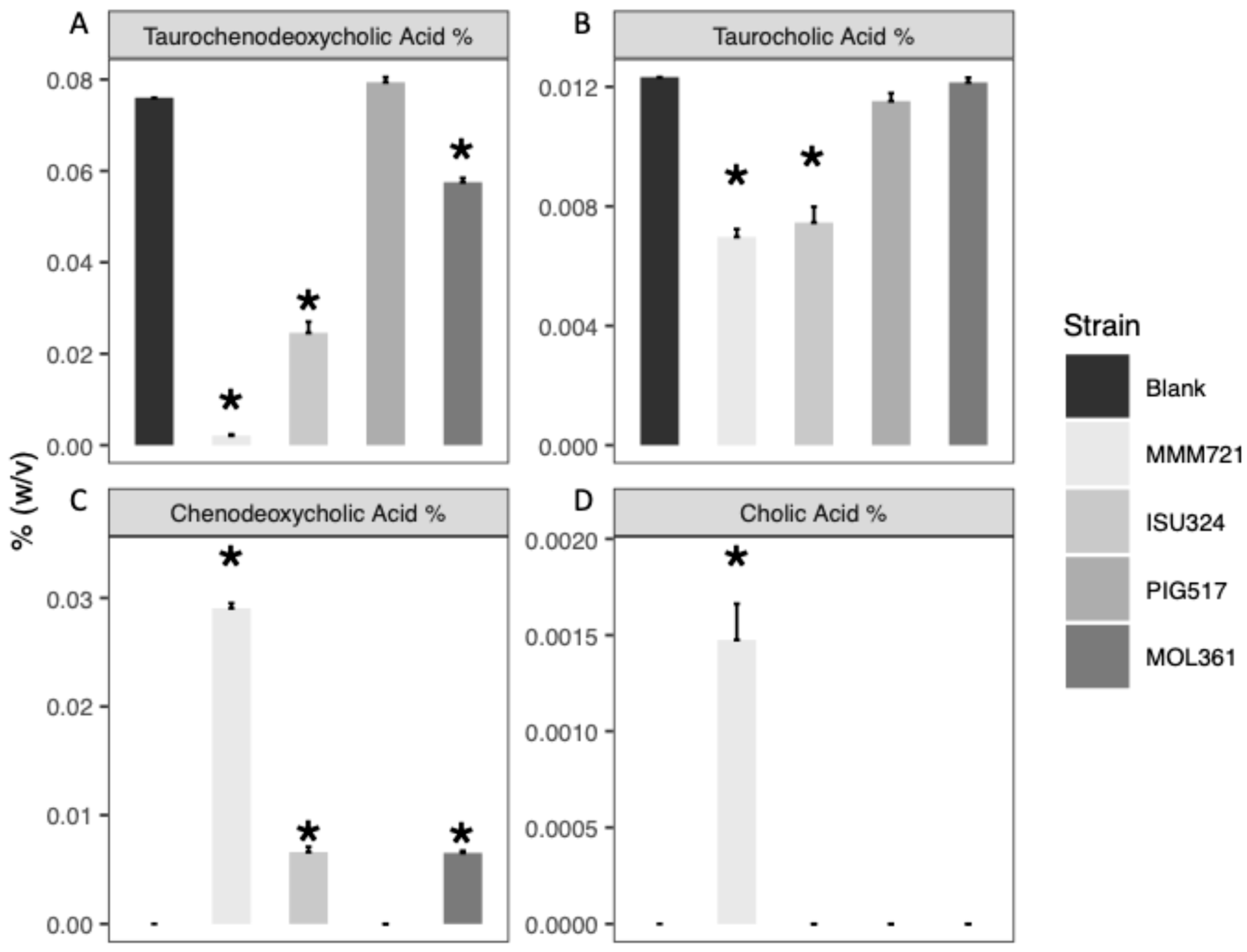
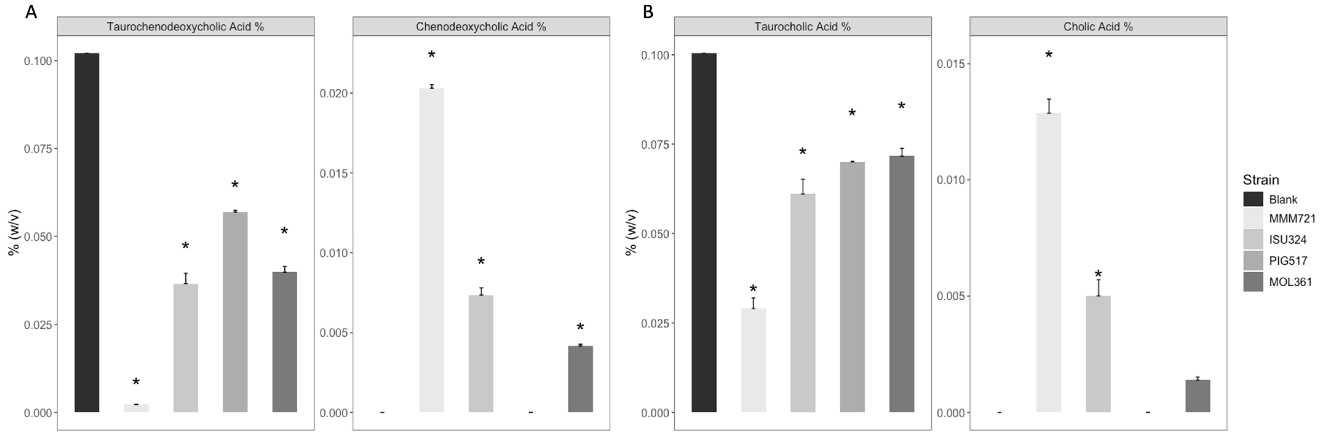
3.1. Both T. Bilis and T. Sanguinis Are Capable of Deconjugating Bile Acids

When Turicibacter isolates were grown in growth media supplemented with 0.1% (v/v) whole chicken bile, the two chicken isolates T. bilis MMM721 and T. bilis ISU324 were capable of significantly decreasing percentages of TCDCA (MMM721 p-value < 0.001; ISU324 p-value < 0.001) and TCA (MMM721 p-value < 0.001; ISU324 p-value < 0.001) in the media (Figure 1A,C). This suggests both chicken T. bilis isolates were capable of deconjugating the major BAs in chicken bile. While both chicken isolates could reduce TCDCA and TCA levels in whole chicken bile-supplemented media, the swine isolate T. bilis PIG517 did not appear capable of significantly decreasing TCA or TCDCA levels in the media (TCDCA p-value = 0.821; TCA p-value = 0.813) (Figure 1A,B). When exposed to whole chicken bile, Turicibacter sanguinis MOL361 significantly reduced TCDCA only (p-value < 0.001) in the media, with no significant decreases to TCA levels (p-value = 0.999) (Figure 1A,B). The reductions in TCDCA corresponded with significant increases to CDCA levels in spent media for T. bilis MMM721, T. bilis ISU324, and T. sanguinis MOL361 (p-value < 0.001) (Figure 1C). While TCA concentrations dropped in cultures from T. bilis MMM721 and T. bilis ISU324 groups, only T. bilis MMM721 saw a significant increase in CA levels in the media (p-value < 0.001) (Figure 1D).
Incubation of strains in media supplemented with 0.1% (w/v) TCDCA or TCA revealed many of the same trends observed with whole chicken bile-supplemented media, though some deviations were observed (Figure 2). Both chicken isolates, Turicibacter bilis MMM721 and T. bilis ISU324, were capable of significantly decreasing TCDCA (MMM721 p-value < 0.001; ISU324 p-value < 0.001) and TCA (MMM721 p-value < 0.001; ISU324 p-value < 0.001) in the media (Figure 2). The decrease of TCDCA and TCA coincided with significant accumulations of CDCA (MMM721 p-value < 0.001; ISU324 p-value < 0.001) and CA (MMM721 p-value < 0.001; ISU324 p-value < 0.003) in the media from T. bilis MMM721 and T. bilis ISU324 cultures (Figure 2). Unlike in whole bile-supplemented ISU324 cultures, TCA-supplemented ISU324 saw the accumulation of CA in the media (Figure 2B). Decreases in TCDCA (p-value <0.001) and TCA (p-value = 0.002) were observed in T. bilis PIG517 cultures supplemented with individual tauro-BAs, but accumulations of CDCA and CA were not observed (Figure 2). Turicibacter sanguinis MOL361 significantly decreased concentrations of both TCA (p-value = 0.004) and TCDCA (p-value < 0.001) in media (Figure 2). Significant CDCA accumulation (p-value = 0.001) was observed in TCDCA-supplemented T. sanguinis MOL361 cultures (Figure 2A). A small accumulation of CA was detected in MOL361 cultures supplemented with TCA, but this increase was not significant (p-value = 0.678) when compared to the control media (Figure 2B).

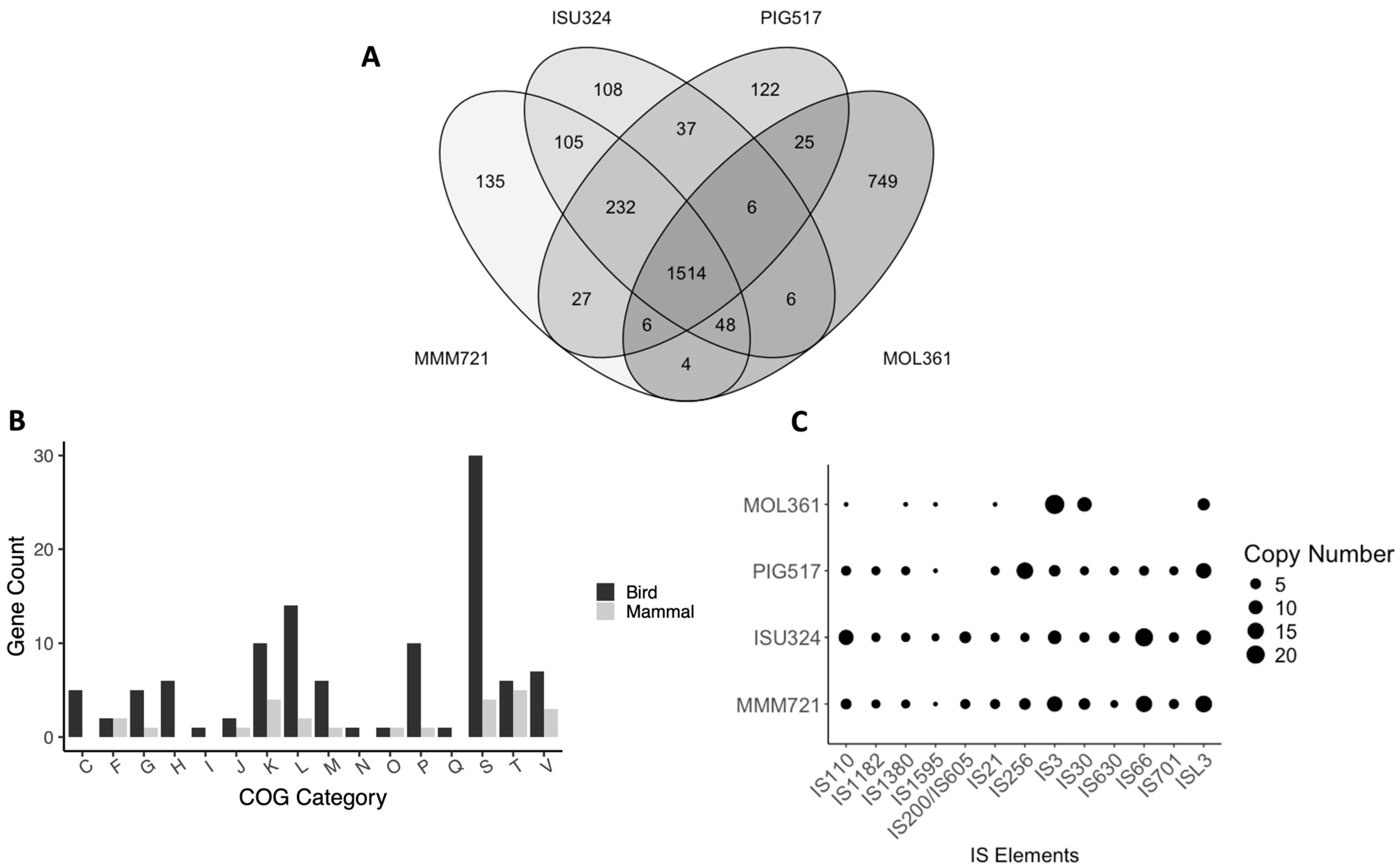
3.2. Comparative Genomics

Turicibacter bilis strains possessed genomes ranging from 2.66–2.79 Mbp in length while the Turicibacter sanguinis MOL361 genome was 3.00 Mbp in length (Supplementary Table S2). The DNA G + C content for the T. bilis genome assemblies, as well as the published T. sanguinis genome, was ~34.5%. The PATRIC annotations identified 2841 coding regions (CDS) for the T. sanguinis MOL361 genome, 2608 CDS for T. bilis MMM721, 2561 CDS for T. bilis ISU324, and 2475 CDS for T. bilis PIG517. All T. bilis strains contained nine rRNA gene operons while the T. sanguinis genome had eight.
Genome comparisons between the three T. bilis strains and T. sanguinis confirmed all Turicibacter bilis strains belonged to the same species (ANIb/ANIm > 95%; dDDH > 70%) which was distinct from T. sanguinis MOL361, confirming the results from our prior short read assemblies [17,61]. T. sanguinis MOL361 was least similar to T. bilis PIG517 (ANIb: 76.59%, ANIm: 86.23%, dDDH: 23.4%), despite both strains being isolated from mammalian samples (Supplementary Tables S3–S5).
Genome annotations identified 1514 CDS shared between the four Turicibacter isolates (Figure 3A). The three T. bilis strains shared an additional 232 CDS that were unique to the T. bilis species while 749 CDS were exclusively found in T. sanguinis MOL361. A total of 105 CDS were exclusively shared between the avian isolates, T. bilis MMM721 and T. bilis ISU324, while only 25 were shared exclusively between strains originating from mammalian hosts, T. bilis PIG517 and T. sanguinis MOL361, suggesting differences based on host. Turicibacter bilis PIG517 possessed 122 unique CDS. Annotated genomes had cluster of orthologous group (COG) categories in similar proportions when comparing the four genomes (Supplemental Figure S1). Nearly 25% of the CDS had COG categorizations that were either not available from the EggNOG-Mapper annotation or their function was unknown. Looking at the COG categories for genes that were shared exclusively between isolates belonging to mammalian- or avian-associated Turicibacter strains, avian strains appeared especially enriched in genes belonging to several COG categories, including L: replication and repair and M: cell wall/membrane/envelope biogenesis. Differences in genes found exclusively within T. bilis or T. sanguinis were also assessed (Supplemental Figure S2). Two of the genes belonging to COG category M that were found exclusively in the avian Turicibacter strains belonged to the CBM50 and GT2 classes of carbohydrate-active enzymes (CAZys), both of which were absent in annotations of the T. bilis PIG517 and T. sanguinis MOL361 genomes. Many the genes from COG category L were transposases belonging to the IS200-like family of transposable elements (TEs), with multiple copies being present in each genome.
To further explore differences in types and counts of transposable elements found in Turicibacter, all four genomes were analyzed with ISEScanner. Turicibacter bilis ISU324 had the most TEs identified (n = 84), T. bilis MMM721 had 82, T. bilis PIG517 had 62, and T. sanguinis MOL361 had 45 TEs identified (Figure 3C). The IS200/IS605 TE was unique to MMM721 and ISU324, which possessed six and four copies, respectively. Several other TEs were unique to T. bilis isolates, including IS1182, IS256, IS630, IS66, and IS701 (Figure 3C).

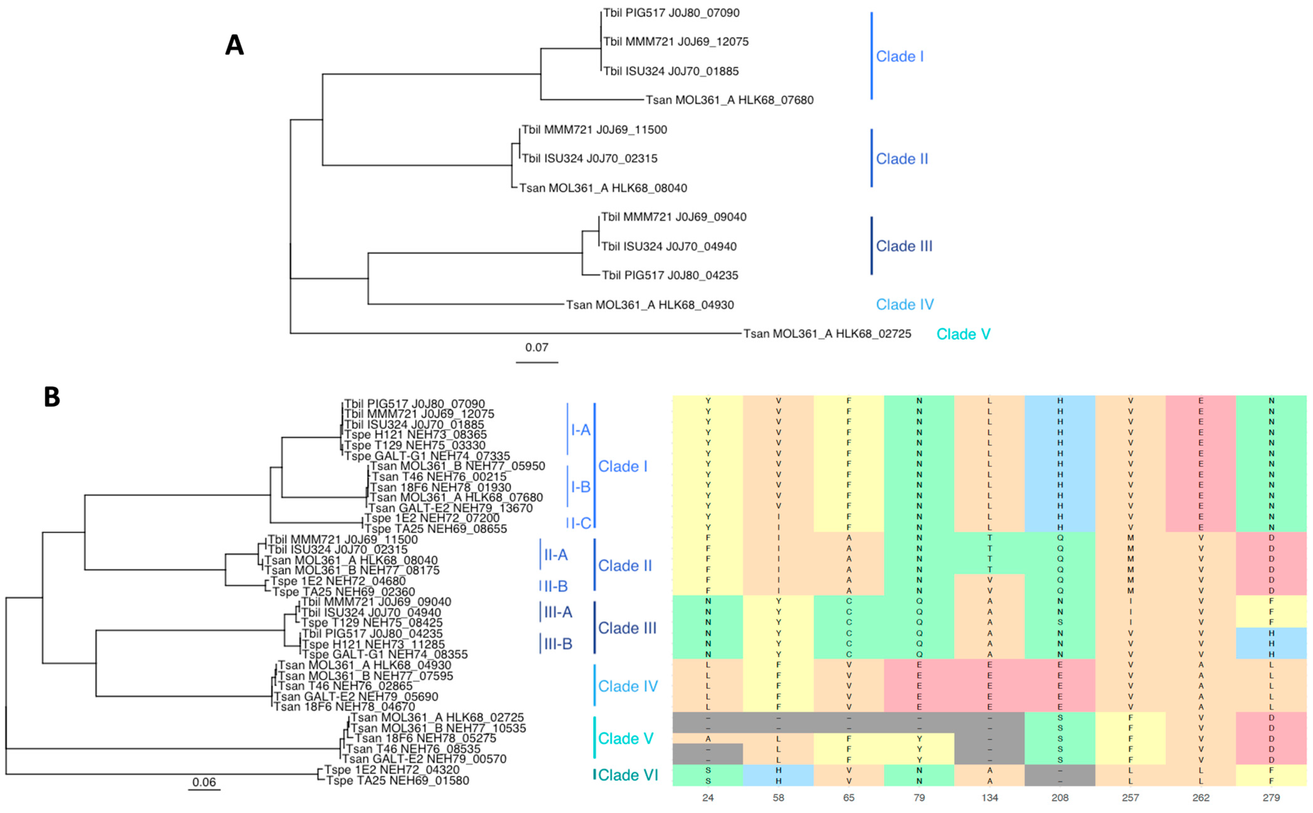
3.3. Bile Salt Hydrolase Genes

Putative BSH genes (Choloylglycine hydrolase, EC 3.5.1.24) were identified from BV-BRC genome annotations and are summarized in Supplementary Table S6. Turicibacter sanguinis MOL361 possessed four BSH genes, though the T. sanguinis MOL361_A HLK68_02725 (741 bp) was ~250 bp shorter than the other three BSH genes encoded in the genome (987 bp), suggesting a potential gene truncation. T. bilis MMM721 and T. bilis ISU324 each possessed 3 BSH genes while T. bilis PIG517 only contained 2 (Supplementary Table S6). A neighbor-joining phylogenetic tree showed bsh partitioned into five distinct clades based on amino acid sequence (Figure 4A). Another phylogeny based on the DNA sequence was largely similar to that of the amino acid tree, with the only difference being the location some of the clade branches vs. Clade V, which served as a “root” for the phylogeny.
Additional BSH genes identified in public sequence data from the genus Turicibacter were incorporated into a new genome alignment and phylogeny to further assess the trends observed with the four Turicibacter strains above (Figure 4B, Supplementary Table S6, Supplementary Figures S3 and S4). These newly incorporated BSH sequences were from a recent analysis of Turicibacter strains isolated from human and mouse specimens in which the strains were assessed for their ability to modify host bile acids, including tauro-conjugated bile acids [41]. The addition of these new BSH genes with known activities vs. tauro-conjugated BAs revealed an additional clade of BSH genes, Clade VI (Figure 4B). The additional resolution provided by further BSH gene incorporation also allowed for the delineation of sub-clades within some of the larger clades.
Previous analyses of BSH genes in other microbial species have identified amino acid residues involved in substrate specificity. These substrate specificity residues are found at positions 24, 58, 65, 79, 134, 208, 257, 262, and 279 of the BSH protein [62,63,64]. To compare the substrate specificity residue profiles between Turicibacter BSH clades and individual strains, the amino acid sequence for all BSH genes from this study were aligned with a well-characterized BSH (LsBSH) belonging to Ligilactobacillus salivarious (AFP87505.1) using Clustal W (Supplementary Figure S5) [58]. The previously described conserved active site residues (based on L. salivarious BSH positioning) were present and conserved across the majority of Turicibacter spp. BSH genes. Cys2, Arg16, Asp18, Asn171, and Arg224 were present in all BSH genes but T. sanguinis (Tsan) MOL361_A HLK68_02725, Tsan MOL361_B NEH77_10535, Tsan GALT-E2 NEH79_00570, and Tsan T46 NEH76_08535 (Supplementary Figure S5) [65]. These BSH all appeared to have truncations at N-terminus and belonged to Clade V of the BSH amino acid phylogeny (Figure 4B; Supplementary Figure S5). Compared to LsBSH, the majority of Turicibacter BSH genes also possessed insertions of amino acid residues at positions 10, 11, 43, 148, and 304 (Supplementary Figure S5). Other notable amino acid insertions and deletions were present in Clades IV, V, and VI (Supplemental Figure S5).
Generally, substrate specific amino acid residue signatures reflected the BSH clades and sub-clades from the amino acid phylogeny (Figure 4B). The one exception was Clade V, which displayed heterogeneity, especially in amino acid residues lost to truncations at the N-terminus. Turicibacter strains that appeared incapable of deconjugating tauro-conjugated BAs, Turicibacter sp. (Tspe) H121, T. bilis (Tbil) PIG517, Tspe T129, and Tsan T46, encoded BSH genes primarily belonging to subclade I-A and Clade III (both III-A and III-B), with Tsan T46 as an outlier (Figure 4B). Interestingly, Tspe GALT-G1, a tauro-deconjugating strain, possessed two BSH genes falling into subclade I-A (Tspe GALT-G1 NEH74_07335) and subclade III-B (Tspe GALT-G1 NEH74_08355), with their respective amino acid sequences being 100% identical to Tspe T129 NEH75_03330, and Tbil PIG517 J0J80_04235 and Tspe H121 NEH73_11285, respectively (Supplementary Figure S5) [41].

4. Discussion

The genus Turicibacter is a common, yet understudied, member of the gastrointestinal microbiota [4,5,7,8,10]. The host range of the Turicibacter genus is broad, suggesting species, and strains, may exhibit adaptations to cope with differences in the intestinal environment between animal hosts, where the relative abundance of Turicibacter appears highest [17,66]. Bile is a major stressors acting upon bacteria of the small intestine, also serving as a substrate which can be utilized by the members of the bacterial community [19,20,36,67,68,69]. Recent work has identified Turicibacter sanguinis as a BA-modifying organism [37,39,40,41]. Here, we investigated genomic features of three strains of T. bilis: MMM721, ISU324, and PIG517 and the type strain of the other species within the genus, T. sanguinis MOL361. Genomic analyses identified inter- and intraspecies similarities and differences in gene content and transposase repertoire which may reflect adaptations within their respective host environments. All Turicibacter isolates encoded multiple BSH genes from strains with different specificities and activity levels for specific BAs.
Like other Turicibacter, the genomes of T. bilis MMM721, T. bilis ISU324, and T. bilis PIG517 consisted of a single chromosome with no plasmids [6,45]. Turicibacter bilis MMM721, T. bilis ISU324, and T. bilis PIG517 were genetically distinct from T. sanguinis [17]. Over 100 coding regions were unique to T. bilis MMM721 and T. bilis ISU324. In conjunction with ANI and dDDH metrics, this overlap in gene content highlights relatedness between the two chicken isolates. The high degree of similarity between these two strains likely reflects adaptations to the chicken gut environment, resulting in a similar gene repertoire [70]. Despite some anatomical and physiological similarities between the human and swine gut, T. bilis PIG517 (swine host) and T. sanguinis MOL361 (human host) were the least similar of the Turicibacter isolates [16,17,71,72,73].
Avian T. bilis genomes were enriched in unique genes associated with clusters of orthologous groups (COG) categories L and M, relating to replication and repair, and cell wall/membrane/envelope biogenesis, respectively. Within COG category L, the avian T. bilis isolates each possessed multiple copies of the IS200/IS605 transposon family, a family of transposable elements (TEs) not identified in the two mammalian-derived isolates. The IS200/IS605 transposon group encompasses a large and broadly distributed family of TEs found in both Gram-positive and Gram-negative bacteria [74,75]. Transposable elements have been hypothesized to take part in “genomic streamlining”, or the trimming of unnecessary genes through transposon insertion and disruption of non-vital genes, allowing for rapid adaptation to the host environment [76,77]. The two avian Turicibacter isolates possessed the same families of TEs in similar abundances throughout the genome, likely indicating a shared ancestral strain of Turicibacter. This similarity in gene and TE content was not observed when comparing the T. bilis PIG517 and T. sanguinis MOL361, despite both isolates originating from mammalian host species. A pangemonic analysis of Turicibacter isolates from a variety of host species would further identify how Turicibacter species can colonize the intestinal tract of such a broad range of animal host species, with particular emphasis on transposon burden to lend insight into the potential role these mobile genetic elements play in the adaptation of Turicibacter to multiple intestinal environments.
COG category M was also enriched in the T. bilis strains originating from chickens. Genes for two carbohydrate-active enzymes (CAZymes), belonging to the CBM50 and GT2 families, were identified exclusively in the two chicken-associated strains of T. bilis. Carbohydrate-binding module 50 (CMB50) is a family of glycosyl hydrolase-binding proteins which aid in the cleavage of peptidoglycan or chitin molecules [78,79]. Glycosyltransferase family 2 (GT2) represents a large group of enzymes implicated in a variety of activities involving the synthesis of glycosidic bonds [80]. In bacteria, GT2 glycosyltransferases are primarily implicated in polysaccharide capsule synthesis which likely aids in bacterial survival in the intestine while also modulating the host immune system [80,81,82]. Interestingly, GT2 was observed in metagenomically assembled Turicibacter genomes from the chicken intestinal tract, suggesting GT2 may play a role in the colonization or persistence of Turicibacter in the poultry intestine [83]. However, more Turicibacter strains isolated from poultry, as well as other species, will be required to fully explore this potential association.
Bile and BAs are common stressors that Turicibacter must cope with when colonizing the intestinal tract of animals [20,36]. One mechanism Turicibacter may utilize to mitigate the detrimental effects of BAs is through the modification of BAs by bile salt hydrolases (BSHs) [84,85]. Turicibacter sanguinis MOL361 and T. bilis strains ISU324 and MMM721 were all capable of deconjugating the taurine from TCDCA and TCA, decreasing the abundance of the two BAs in liquid culture. Chicken bile is primarily (>95%) composed of TCDCA and TCA, so T. bilis MMM721 and T. bilis ISU324 should be adapted to growth in the presence of these two BAs [31,32]. Deconjugation of TCDCA and TCA could provide T. bilis MMM721 and T. bilis ISU324 with an adaptive advantage in the chicken small intestine [86,87].
Turicibacter sanguinis MOL361 was capable of deconjugating TCDCA and, to a lesser extent, TCA, which is consistent with previous reports [39,41]. This decreased activity toward some tauro-conjugated BAs may reflect how the BA profile of humans is primary glyco-conjugated (~75%), but both TCDCA and TCA can still be found in the liver and gastrointestinal tract at ~10% each, thus, T. sanguinis would still need to act upon it [88]. Turicibacter bilis PIG517 displayed little to no deconjugation of TCA and TCDCA. The BA profile of swine bile is primarily glyco-conjugated BAs (>90%), with TCDCA and TCA making up ~1–2% of the BA pool [89,90]. This suggests TCDCA and TCA are less frequently encountered by pig intestinal microbes, and potentially explains the lack of tauro-associated BSH-activity, especially to TCDCA and TCA, from a pig-adapted isolate like T. bilis PIG517 [90,91,92]. The differences in functionality, both in specificity and apparent activity, of encoded BSHs in the different Turicibacter strains studied here may reflect the host BA profile. Further work needs to be conducted in Turicibacter and other BSH-encoding organisms to characterize this potential link between BSH spectrum of activity and the BA profile of its preferred host species as well as the potential inhibitory effects of bile and BAs from different hosts and how the transformation of these BAs impacts Turicibacter growth dynamics and survival. In this study, spend media samples were collected after 8 h of incubation, which correspond to the late-log to early stationary phase of growth for Turicibacter bilis [37]. Follow-up studies should be conducted to further assess the impacts of growth phase and incubation times on the extent of BA deconjugation for these and other Turicibacter strains.
Our initial BSH gene analysis was expanded to include other Turicibacter strains with known tauro-BA deconjugation phenotypes. Strains with the ability to deconjugate tauro-BAs could indicate a strain can colonize and persist within the poultry gut. All Turicibacter strains possessed multiple BSH genes, which could be grouped into seven clades based on amino acid sequence relatedness. In general, clades reflected the differences in amino acid residues present at positions previously identified to be involved in BSH substrate specificity [62,63,64]. Little intra-clade variability was observed at these specificity residues apart from Clade V in which four of the five BSH genes displayed large deletions/truncations at the N-terminus. Bile salt hydrolase genes with truncations at the N-terminus are likely functionally inactive, as they lack the catalytic Cys2 residue, a requirement for deconjugation of the amino acid from the steroid core of the bile acid, though additional functional studies under a variety of conditions are needed to confirm this [84,93,94].
Trends associated with specific BSH clade’s ability to deconjugate tauro-conjugated BAs were observed. Turicibacter strains that appeared incapable of deconjugating tauro-conjugated BAs were Turicibacter sp. H121, T. bilis PIG517, Turicibacter sp. T129, and T. sanguinis T46 [41]. Interestingly, the BSH genes from these strains fell primarily into the subclade I-A and Clade III, including both subclades III-A and III-B. Strains capable of tauro-deconjugation abilities encoded BSH genes falling primarily into subclades I-B/I-C, Clade II, Clade IV, Clade V, and Clade VI. These results align well with the findings of Lynch and colleagues, who recently profiled the BA specificities of BSH genes from the Turicibacter genus [41]. Differences in BSH activity spectra could not be explained solely through clade identity or even analysis of amino acid residues previously associated with BA specificity from the literature [62,63,64]. The outlier strains, T. sanguinis T46 and Turicibacter sp. GALT-G1, possess highly similar, or even identical, BSH genes to other Turicibacter strains but display opposite tauro-BA deconjugation phenotypes, suggesting that other factors may play a role in BSH expression and activity spectra [41]. Further studies into the deconjugative capabilities of bsh knockout mutants would provide greater understanding of the spectrum of activity and rate of deconjugation for Turicibacter BSH from different clades and sub-clades, both individually and in combination.
Overall, possessing multiple, unique BSH genes with specificities for different BAs likely improves Turicibacter survival in response to the diverse BA pools present in the guts of vertebrates, enhancing the ability of Turicibacter strains to specifically colonize the gastrointestinal tract of a preferred host, or host range, based on their BSH repertoire. The activity of these Turicibacter BSH appears not only to be based upon sequence identity, but is likely context dependent as well, with pH, growth phase, and BA concentration potentially playing roles in deconjugation rates [41,95]. Research into individual BSH from Turicibacter and assessing expression levels or activity spectra when exposed to a variety of physiologically relevant conditions should provide further insights into how these BSH aid in the colonization and persistence of Turicibacter in the intestine of poultry and other livestock species.
Human pathogens, like Campylobacter jejuni, Clostridioides difficile, and Escherichia coli, all recognize and utilize specific BAs as chemical signals to induce germination, expression of colonization factors, or enhance virulence mechanisms [43,96,97,98,99]. Focusing research efforts into Turicibacter and other BA-modifying commensals could inform targeted interventions to reduce or even prevent pathogen colonization and inhibit pathogen virulence in the host.

5. Conclusions

Here, we assess the genomic relatedness of three strains of the newly described species Turicibacter bilis and Turicibacter sanguinis MOL361. These four isolates were grown in the presence of chicken bile and its two primary bile acids, taurocholic acid or taurochenodeoxycholic acid, followed by analysis of bile acid biotransformation products. Each Turicibacter strain tested possessed multiple bile salt hydrolase genes, and all but T. bilis PIG517 were capable of bile salt hydrolase activity in vitro, as displayed by significant reductions in taurocholic acid and taurochenodeoxycholic acid and the accumulation of their deconjugation products, cholic acid and chenodeoxycholic acid, respectively, which is likely a reflection of the prevalence of these specific bile acids in the gastrointestinal tract of their respective host species. The greatest activity was observed with T. bilis MMM721, closely followed by T. bilis ISU324, both of which were isolated from the surface of chicken eggshells. Overall, this work highlights the ability of Turicibacter species to modify chicken bile and its primary bile acids, aiding in its persistence within the chicken gut. Further studies should be conducted to assess the activity rates and substrate preferences of individual bile salt hydrolase enzymes within the genus Turicibacter and the implications of this activity on successful host colonization and nutritional absorption should be characterized.

Supplementary Materials

The following supporting information can be downloaded at https://www.mdpi.com/article/10.3390/bacteria4030035/s1, Figure S1. Clusters of orthologous groups (COG) category abundance of CDS identified in Turicibacter strains analyzed in this study: Figure S2. Clusters of orthologous groups (COG) category counts of CDS that were shared between and unique either Turicibacter bilis or Turicibacter sanguinis: Figure S3. DNA-based phylogeny of bsh of the four Turicibacter strains analyzed in this study: Figure S4. DNA-based phylogeny of bsh from select Turicibacter BSH genes with known activity vs. tauro-conjugated bile acids: Figure S5. Amino acid sequence alignment of select Turicibacter BSH genes with known activity vs. tauro-conjugated bile acids with LsBSH (Ligilactobacillus salivarious; AFP87505.1) as a reference: Table S1. Genomes used in this study: Table S2. Genome assembly statistics for T. bilis MMM721, T. bilis ISU324, and T. bilis PIG517: Table S3. Pairwise ANIb results comparing T. bilis MMM721, T. bilis ISU324, and T. bilis PIG517 and T. sanguinis MOL361: Table S4. Pairwise ANIm results comparing T. bilis MMM721, T. bilis ISU324, and T. bilis PIG517 and T. sanguinis MOL361: Table S5. Pairwise dDDH results comparing T. bilis MMM721, T. bilis ISU324, and T. bilis PIG517 and T. sanguinis MOL361: Table S6. List of bile salt hydrolase gene used in this study.

Author Contributions

Conceptualization, J.J.M. and T.L.; methodology, J.J.M. and T.L.; software, J.J.M. and L.S.; validation, J.J.M., L.S. and T.L.; formal analysis, J.J.M., L.S. and T.L.; investigation, J.J.M.; resources, L.S. and T.L.; data curation, J.J.M. and L.S.; writing—original draft preparation, J.J.M.; writing—review and editing, J.J.M., L.S. and T.L.; visualization, J.J.M. and T.L.; supervision, T.L.; project administration, T.L.; funding acquisition, T.L. All authors have read and agreed to the published version of the manuscript.

Funding

This work was supported by the USDA project number 5030-31320-004-00D. Mention of trade names or commercial products in this publication is solely for the purpose of providing specific information and does not imply recommendation or endorsement by the USDA. This research was supported by an appointment to the Agricultural Research Service (ARS) Research Participation Program administered by the Oak Ridge Institute for Science and Education (ORISE) through an interagency agreement between the U.S. Department of Energy (DOE) and the U.S. Department of Agriculture (USDA). ORISE is managed by ORAU under DOE contract number DE- SC0014664. All opinions expressed in this paper are the authors’ and do not necessarily reflect the policies and views of USDA, ARS, DOE, or ORAU/ORISE.

Institutional Review Board Statement

Not applicable.

Informed Consent Statement

Not applicable.

Data Availability Statement

Whole genome sequence data for this study are publicly available through the National Center for Biotechnology Information (NCBI). Specific NCBI accession numbers are listed in Tables S1 and S6.

Acknowledgments

The authors would like to thank Daniel Nielson, Cassidy Klima, and David Alt for their assistance generating long-read sequence data on the Oxford Nanopore MinION.

Conflicts of Interest

The authors declare no conflicts of interest.

References

  1. Ogawa, Y.; Ooka, T.; Shi, F.; Ogura, Y.; Nakayama, K.; Hayashi, T.; Shimoji, Y. The genome of Erysipelothrix rhusiopathiae, the causative agent of swine erysipelas, reveals new insights into the evolution of firmicutes and the organism’s intracellular adaptations. J Bacteriol. 2011, 193, 2959–2971. [Google Scholar] [CrossRef]
  2. Davis, J.J.; Xia, F.; Overbeek, R.A.; Olsen, G.J. Genomes of the class Erysipelotrichia clarify the firmicute origin of the class Mollicutes. Int. J. Syst. Evol. Microbiol. 2013, 63 Pt 7, 2727–2741. [Google Scholar] [CrossRef]
  3. Verbarg, S.; Göker, M.; Scheuner, C.; Schumann, P.; Stackebrandt, E. The Families Erysipelotrichaceae emend., Coprobacillaceae fam. nov., and Turicibacteraceae fam. nov. In The Prokaryotes: Firmicutes and Tenericutes; Rosenberg, E., DeLong, E.F., Lory, S., Stackebrandt, E., Thompson, F., Eds.; Springer: Berlin/Heidelberg, Germany, 2014; pp. 79–105. [Google Scholar]
  4. Köhler, T.; Dietrich, C.; Scheffrahn, R.H.; Brune, A. High-resolution analysis of gut environment and bacterial microbiota reveals functional compartmentation of the gut in wood-feeding higher termites (Nasutitermes spp.). Appl. Env. Environ. Microbiol. 2012, 78, 4691–4701. [Google Scholar] [CrossRef] [PubMed]
  5. Looft, T.; Allen, H.K.; Cantarel, B.L.; Levine, U.Y.; Bayles, D.O.; Alt, D.P.; Henrissat, B.; Stanton, T.B. Bacteria, phages and pigs: The effects of in-feed antibiotics on the microbiome at different gut locations. Isme J. 2014, 8, 1566–1576. [Google Scholar] [CrossRef] [PubMed]
  6. Auchtung, T.A.; Holder, M.E.; Gesell, J.R.; Ajami, N.J.; Duarte, R.T.; Itoh, K.; Caspi, R.R.; Petrosino, J.F.; Horai, R.; Zárate-Bladés, C.R. Complete Genome Sequence of Turicibacter sp. Strain H121, Isolated from the Feces of a Contaminated Germ-Free Mouse. Genome Announc. 2016, 4, e00114-16. [Google Scholar] [CrossRef] [PubMed]
  7. Siegerstetter, S.C.; Schmitz-Esser, S.; Magowan, E.; Wetzels, S.U.; Zebeli, Q.; Lawlor, P.G.; O’Connell, N.E.; Metzler-Zebeli, B.U. Intestinal microbiota profiles associated with low and high residual feed intake in chickens across two geographical locations. PLoS ONE 2017, 12, e0187766. [Google Scholar] [CrossRef]
  8. Johnson, T.A.; Sylte, M.J.; Looft, T. In-feed bacitracin methylene disalicylate modulates the turkey microbiota and metabolome in a dose-dependent manner. Sci. Rep. 2019, 9, 8212. [Google Scholar] [CrossRef]
  9. Wu, M.; Li, J.; An, Y.; Li, P.; Xiong, W.; Li, J.; Yan, D.; Wang, M.; Zhong, G. Chitooligosaccharides Prevents the Development of Colitis-Associated Colorectal Cancer by Modulating the Intestinal Microbiota and Mycobiota. Front. Microbiol. 2019, 10, 2101. [Google Scholar] [CrossRef]
  10. Maki, J.J.; Bobeck, E.A.; Sylte, M.J.; Looft, T. Eggshell and environmental bacteria contribute to the intestinal microbiota of growing chickens. J. Anim. Sci. Biotechnol. 2020, 11, 60. [Google Scholar] [CrossRef]
  11. Zhong, Y.; Nyman, M.; Fåk, F. Modulation of gut microbiota in rats fed high-fat diets by processing whole-grain barley to barley malt. Mol. Nutr. Food Res. 2015, 59, 2066–2076. [Google Scholar] [CrossRef]
  12. Getachew, B.; Aubee, J.I.; Schottenfeld, R.S.; Csoka, A.B.; Thompson, K.M.; Tizabi, Y. Ketamine interactions with gut-microbiota in rats: Relevance to its antidepressant and anti-inflammatory properties. BMC Microbiol. 2018, 18, 222. [Google Scholar] [CrossRef] [PubMed]
  13. Ahn, I.S.; Lang, J.M.; Olson, C.A.; Diamante, G.; Zhang, G.; Ying, Z.; Byun, H.R.; Cely, I.; Ding, J.; Cohn, P.; et al. Host Genetic Background and Gut Microbiota Contribute to Differential Metabolic Responses to Fructose Consumption in Mice. J. Nutr. 2020, 150, 2716–2728. [Google Scholar] [CrossRef] [PubMed]
  14. Fung, T.C.; Vuong, H.E.; Luna, C.D.G.; Pronovost, G.N.; Aleksandrova, A.A.; Riley, N.G.; Vavilina, A.; McGinn, J.; Rendon, T.; Forrest, L.R.; et al. Intestinal serotonin and fluoxetine exposure modulate bacterial colonization in the gut. Nat. Microbiol. 2019, 4, 2064–2073. [Google Scholar] [CrossRef] [PubMed]
  15. Gao, H.; Jiang, Q.; Ji, H.; Ning, J.; Li, C.; Zheng, H. Type 1 diabetes induces cognitive dysfunction in rats associated with alterations of the gut microbiome and metabolomes in serum and hippocampus. Biochim. Biophys. Acta Mol. Basis Dis. 2019, 1865, 165541. [Google Scholar] [CrossRef]
  16. Bosshard, P.P.; Zbinden, R.; Altwegg, M. Turicibacter sanguinis gen. nov., sp. nov., a novel anaerobic, Gram-positive bacterium. Int. J. Syst. Evol. Microbiol. 2002, 52, 1263–1266. [Google Scholar] [CrossRef]
  17. Maki, J.J.; Looft, T. Turicibacter bilis sp. nov., a novel bacterium isolated from the chicken eggshell and swine ileum. Int. J. Syst. Evol. Microbiol. 2022, 72, 005153. [Google Scholar] [CrossRef]
  18. Imamura, Y.; Motooka, D.; Nakajima, Y.; Ito, S.; Kitakaze, M.; Iida, T.; Nakamura, S. Turicibacter faecis sp. nov., isolated from faeces of heart failure mouse model. Int. J. Syst Evol. Microbiol. 2024, 74, 006379. [Google Scholar] [CrossRef]
  19. Taranto, M.P.; Fernandez Murga, M.L.; Lorca, G.; de Valdez, G.F. Bile salts and cholesterol induce changes in the lipid cell membrane of Lactobacillus reuteri. J. Appl. Microbiol. 2003, 95, 86–91. [Google Scholar] [CrossRef]
  20. Begley, M.; Gahan, C.G.M.; Hill, C. The interaction between bacteria and bile. FEMS Microbiol. Rev. 2005, 29, 625–651. [Google Scholar] [CrossRef]
  21. Hatton, G.B.; Yadav, V.; Basit, A.W.; Merchant, H.A. Animal Farm: Considerations in Animal Gastrointestinal Physiology and Relevance to Drug Delivery in Humans. J. Pharm. Sci. 2015, 104, 2747–2776. [Google Scholar] [CrossRef]
  22. Zheng, L.; Kelly, C.J.; Colgan, S.P. Physiologic hypoxia and oxygen homeostasis in the healthy intestine. A Review in the Theme: Cellular Responses to Hypoxia. Am. J. Physiol. Cell Physiol. 2015, 309, C350–C360. [Google Scholar] [CrossRef] [PubMed]
  23. Russell, D.W. The enzymes, regulation, and genetics of bile acid synthesis. Annu. Rev. Biochem. 2003, 72, 137–174. [Google Scholar] [CrossRef] [PubMed]
  24. Macierzanka, A.; Torcello-Gómez, A.; Jungnickel, C.; Maldonado-Valderrama, J. Bile salts in digestion and transport of lipids. Adv. Colloid. Interface Sci. 2019, 274, 102045. [Google Scholar] [CrossRef] [PubMed]
  25. Ridlon, J.M.; Kang, D.J.; Hylemon, P.B. Bile salt biotransformations by human intestinal bacteria. J. Lipid Res. 2006, 47, 241–259. [Google Scholar] [CrossRef]
  26. Vessey, D.A. The biochemical basis for the conjugation of bile acids with either glycine or taurine. Biochem. J. 1978, 174, 621–626. [Google Scholar] [CrossRef]
  27. Arrese, M.; Trauner, M. Molecular aspects of bile formation and cholestasis. Trends Mol. Med. 2003, 9, 558–564. [Google Scholar] [CrossRef]
  28. Hofmann, A.F.; Hagey, L.R.; Krasowski, M.D. Bile salts of vertebrates: Structural variation and possible evolutionary significance. J. Lipid Res. 2010, 51, 226–246. [Google Scholar] [CrossRef]
  29. Hofmann, A.F.; Mysels, K.J. Bile acid solubility and precipitation in vitro and in vivo: The role of conjugation, pH, and Ca2+ ions. J. Lipid Res. 1992, 33, 617–626. [Google Scholar] [CrossRef]
  30. Bremer, J. Species differences in the conjugation of free bile acids with taurine and glycine. Biochem. J. 1956, 63, 507–513. [Google Scholar] [CrossRef]
  31. Hagey, L.R.; Vidal, N.; Hofmann, A.F.; Krasowski, M.D. Complex Evolution of Bile Salts in Birds. Auk 2010, 127, 820–831. [Google Scholar] [CrossRef]
  32. Elkin, R.G.; Wood, K.V.; Hagey, L.R. Biliary bile acid profiles of domestic fowl as determined by high performance liquid chromatography and fast atom bombardment mass spectrometry. Comp. Biochem. Physiol. Part B 1990, 96, 157–161. [Google Scholar] [CrossRef]
  33. Lin, J.; Sahin, O.; Michel, L.O.; Zhang, Q. Critical role of multidrug efflux pump CmeABC in bile resistance and in vivo colonization of Campylobacter jejuni. Infect. Immun. 2003, 71, 4250–4259. [Google Scholar] [CrossRef]
  34. Sun, J.; Deng, Z.; Yan, A. Bacterial multidrug efflux pumps: Mechanisms, physiology and pharmacological exploitations. Biochem. Biophys. Res. Commun. 2014, 453, 254–267. [Google Scholar] [CrossRef]
  35. Suzuki, S.; Kimoto-Nira, H.; Suganuma, H.; Suzuki, C.; Saito, T.; Yajima, N. Cellular fatty acid composition and exopolysaccharide contribute to bile tolerance in Lactobacillus brevis strains isolated from fermented Japanese pickles. Can. J. Microbiol. 2014, 60, 183–191. [Google Scholar] [CrossRef]
  36. Ridlon, J.M.; Harris, S.C.; Bhowmik, S.; Kang, D.J.; Hylemon, P.B. Consequences of bile salt biotransformations by intestinal bacteria. Gut Microbes 2016, 7, 22–39. [Google Scholar] [CrossRef]
  37. Maki, J.J.; Lippolis, J.D.; Looft, T. Proteomic response of Turicibacter bilis MMM721 to chicken bile and its bile acids. BMC Res. Notes 2022, 15, 236. [Google Scholar] [CrossRef]
  38. Cabral, D.J.; Small, D.M.; Lilly, H.S.; Hamilton, J.A. Transbilayer movement of bile acids in model membranes. Biochemistry 1987, 26, 1801–1804. [Google Scholar] [CrossRef] [PubMed]
  39. Kemis, J.H.; Linke, V.; Barrett, K.L.; Boehm, F.J.; Traeger, L.L.; Keller, M.P.; Rabaglia, M.E.; Schueler, K.L.; Stapleton, D.S.; Gatti, D.M.; et al. Genetic determinants of gut microbiota composition and bile acid profiles in mice. PLoS Genet. 2019, 15, e1008073. [Google Scholar] [CrossRef] [PubMed]
  40. Fu, J.; Kuipers, F. Systems genetics approach reveals cross-talk between bile acids and intestinal microbes. PLoS Genet. 2019, 15, e1008307. [Google Scholar] [CrossRef] [PubMed]
  41. Lynch, J.B.; Gonzalez, E.L.; Choy, K.; Faull, K.F.; Jewell, T.; Arellano, A.; Liang, J.; Yu, K.B.; Paramo, J.; Hsiao, E.Y. Gut microbiota Turicibacter strains differentially modify bile acids and host lipids. Nat. Commun. 2023, 14, 3669. [Google Scholar] [CrossRef]
  42. Alenezi, T.; Fu, Y.; Alrubaye, B.; Alanazi, T.; Almansour, A.; Wang, H.; Sun, X. Potent Bile Acid Microbial Metabolites Modulate Clostridium perfringens Virulence. Pathogens 2023, 12, 1202. [Google Scholar] [CrossRef]
  43. Taheri, N.; Mahmud, A.K.M.F.; Sandblad, L.; Fällman, M.; Wai, S.N.; Fahlgren, A. Campylobacter jejuni bile exposure influences outer membrane vesicles protein content and bacterial interaction with epithelial cells. Sci. Rep. 2018, 8, 16996. [Google Scholar] [CrossRef] [PubMed]
  44. Bansal, M.; Alenezi, T.; Fu, Y.; Almansour, A.; Wang, H.; Gupta, A.; Liyanage, R.; Graham, D.B.; Hargis, B.M.; Sun, X. Specific Secondary Bile Acids Control Chicken Necrotic Enteritis. Pathogens 2021, 10, 1041. [Google Scholar] [CrossRef] [PubMed]
  45. Maki, J.J.; Nielsen, D.W.; Looft, T. Complete Genome Sequence and Annotation for Turicibacter sanguinis MOL361(DSM 14220). Microbiol. Resour. Announc. 2020, 9, e00475-20. [Google Scholar] [CrossRef] [PubMed]
  46. Wick, R.R.; Judd, L.M.; Gorrie, C.L.; Holt, K.E. Completing bacterial genome assemblies with multiplex MinION sequencing. Microb. Genom. 2017, 3, e000132. [Google Scholar] [CrossRef]
  47. Wick, R.R.; Judd, L.M.; Holt, K.E. Performance of neural network basecalling tools for Oxford Nanopore sequencing. Genome Biol. 2019, 20, 129. [Google Scholar] [CrossRef]
  48. Wick, R.R.; Judd, L.M.; Gorrie, C.L.; Holt, K.E. Unicycler: Resolving bacterial genome assemblies from short and long sequencing reads. PLoS Comput. Biol. 2017, 13, e1005595. [Google Scholar] [CrossRef]
  49. Wattam, A.R.; Davis, J.J.; Assaf, R.; Boisvert, S.; Brettin, T.; Bun, C.; Conrad, N.; Dietrich, E.M.; Disz, T.; Gabbard, J.L.; et al. Improvements to PATRIC, the all-bacterial Bioinformatics Database and Analysis Resource Center. Nucleic Acids Res. 2017, 45, D535–D542. [Google Scholar] [CrossRef]
  50. Meier-Kolthoff, J.P.; Auch, A.F.; Klenk, H.-P.; Göker, M. Genome sequence-based species delimitation with confidence intervals and improved distance functions. BMC Bioinform. 2013, 14, 60. [Google Scholar] [CrossRef]
  51. Richter, M.; Rosselló-Móra, R.; Oliver Glöckner, F.; Peplies, J. JSpeciesWS: A web server for prokaryotic species circumscription based on pairwise genome comparison. Bioinformatics 2016, 32, 929–931. [Google Scholar] [CrossRef]
  52. Wattam, A.R.; Brettin, T.; Davis, J.J.; Gerdes, S.; Kenyon, R.; Machi, D.; Mao, C.; Olson, R.; Overbeek, R.; Pusch, G.D.; et al. Assembly, Annotation, and Comparative Genomics in PATRIC, the All Bacterial Bioinformatics Resource Center. In Comparative Genomics: Methods and Protocols; Setubal, J.C., Stoye, J., Stadler , P.F., Eds.; Springer: New York, NY, USA, 2018; pp. 79–101. [Google Scholar]
  53. Huerta-Cepas, J.; Forslund, K.; Coelho, L.P.; Szklarczyk, D.; Jensen, L.J.; von Mering, C.; Bork, P. Fast Genome-Wide Functional Annotation through Orthology Assignment by eggNOG-Mapper. Mol. Biol. Evol. 2017, 34, 2115–2122. [Google Scholar] [CrossRef] [PubMed]
  54. Xie, Z.; Tang, H. ISEScan: Automated identification of insertion sequence elements in prokaryotic genomes. Bioinformatics. 2017, 33, 3340–3347. [Google Scholar] [CrossRef] [PubMed]
  55. Giardine, B.; Riemer, C.; Hardison, R.C.; Burhans, R.; Elnitski, L.; Shah, P.; Zhang, Y.; Blankenberg, D.; Albert, I.; Taylor, J. Galaxy: A platform for interactive large-scale genome analysis. Genome Res. 2005, 15, 1451–1455. [Google Scholar] [CrossRef] [PubMed]
  56. Kearse, M.; Moir, R.; Wilson, A.; Stones-Havas, S.; Cheung, M.; Sturrock, S.; Buxton, S.; Cooper, A.; Markowitz, S.; Duran, C.; et al. Geneious Basic: An integrated and extendable desktop software platform for the organization and analysis of sequence data. Bioinformatics 2012, 28, 1647–1649. [Google Scholar] [CrossRef]
  57. Olson, R.D.; Assaf, R.; Brettin, T.; Conrad, N.; Cucinell, C.; Davis, J.J.; Dempsey, D.M.; Dickerman, A.; Dietrich, E.M.; Kenyon, R.W.; et al. Introducing the Bacterial and Viral Bioinformatics Resource Center (BV-BRC): A resource combining PATRIC, IRD and ViPR. Nucleic Acids Res. 2023, 51, D678–D689. [Google Scholar] [CrossRef]
  58. Wang, Z.; Zeng, X.; Mo, Y.; Smith, K.; Guo, Y.; Lin, J. Identification and characterization of a bile salt hydrolase from Lactobacillus salivarius for development of novel alternatives to antibiotic growth promoters. Appl. Environ. Microbiol. 2012, 78, 8795–8802. [Google Scholar] [CrossRef]
  59. Sergi, M.; Montesano, C.; Napoletano, S.; Pizzoni, D.; Manetti, C.; Colistro, F.; Curini, R.; Compagnone, D. Analysis of Bile Acids Profile in Human Serum by Ultrafiltration Clean-up and LC-MS/MS. Chromatographia 2012, 75, 479–489. [Google Scholar] [CrossRef]
  60. Christinat, N.; Valsesia, A.; Masoodi, M. Untargeted Profiling of Bile Acids and Lysophospholipids Identifies the Lipid Signature Associated with Glycemic Outcome in an Obese Non-Diabetic Clinical Cohort. Biomolecules 2020, 10, 1049. [Google Scholar] [CrossRef]
  61. Kim, M.; Oh, H.S.; Park, S.C.; Chun, J. Towards a taxonomic coherence between average nucleotide identity and 16S rRNA gene sequence similarity for species demarcation of prokaryotes. Int. J. Syst. Evol. Microbiol. 2014, 64, 346–351. [Google Scholar] [CrossRef]
  62. Xu, F.; Hu, X.-J.; Singh, W.; Geng, W.; Tikhonova, I.G.; Lin, J. The complex structure of bile salt hydrolase from Lactobacillus salivarius reveals the structural basis of substrate specificity. Sci. Rep. 2019, 9, 12438. [Google Scholar] [CrossRef]
  63. Öztürk, M.; Önal, C. Asparagine 79 is an important amino acid for catalytic activity and substrate specificity of bile salt hydrolase (BSH). Mol Biol Rep. 2019, 46, 4361–4368. [Google Scholar] [CrossRef]
  64. Karlov, D.S.; Long, S.L.; Zeng, X.; Xu, F.; Lal, K.; Cao, L.; Hayoun, K.; Lin, J.; Joyce, S.A.; Tikhonova, I.G. Characterization of the mechanism of bile salt hydrolase substrate specificity by experimental and computational analyses. Structure 2023, 31, 629–638.e5. [Google Scholar] [CrossRef] [PubMed]
  65. Öztürk, M.; Kılıçsaymaz, Z.; Önal, C. Site-Directed Mutagenesis of Bile Salt Hydrolase (BSH) from Lactobacillus plantarum B14 Confirms the Importance of the V58 and Y65 Amino Acids for Activity and Substrate Specificity. Food Biotechnol. 2023, 37, 74–88. [Google Scholar] [CrossRef]
  66. Karasov, W.H.; Douglas, A.E. Comparative digestive physiology. Compr. Physiol. 2013, 3, 741–783. [Google Scholar] [CrossRef] [PubMed]
  67. Dong, Z.; Lee, B.H. Bile salt hydrolases: Structure and function, substrate preference, and inhibitor development. Protein Sci. 2018, 27, 1742–1754. [Google Scholar] [CrossRef]
  68. Vital, M.; Rud, T.; Rath, S.; Pieper, D.H.; Schlüter, D. Diversity of Bacteria Exhibiting Bile Acid-inducible 7α-dehydroxylation Genes in the Human Gut. Comput. Struct. Biotechnol. J. 2019, 17, 1016–1019. [Google Scholar] [CrossRef]
  69. Heinken, A.; Ravcheev, D.A.; Baldini, F.; Heirendt, L.; Fleming, R.M.T.; Thiele, I. Systematic assessment of secondary bile acid metabolism in gut microbes reveals distinct metabolic capabilities in inflammatory bowel disease. Microbiome 2019, 7, 75. [Google Scholar] [CrossRef]
  70. Van Rossum, T.; Ferretti, P.; Maistrenko, O.M.; Bork, P. Diversity within species: Interpreting strains in microbiomes. Nat. Rev. Microbiol. 2020, 18, 491–506. [Google Scholar] [CrossRef]
  71. Zhang, Q.; Widmer, G.; Tzipori, S. A pig model of the human gastrointestinal tract. Gut Microbes 2013, 4, 193–200. [Google Scholar] [CrossRef]
  72. Bendixen, E.; Danielsen, M.; Larsen, K.; Bendixen, C. Advances in porcine genomics and proteomics—A toolbox for developing the pig as a model organism for molecular biomedical research. Brief. Funct. Genom. 2010, 9, 208–219. [Google Scholar] [CrossRef]
  73. Van de Vliet, M.; Joossens, M. The Resemblance between Bacterial Gut Colonization in Pigs and Humans. Microorganisms 2022, 10, 1831. [Google Scholar] [CrossRef]
  74. Beuzón, C.R.; Chessa, D.; Casadesús, J. IS200: An old and still bacterial transposon. Int. Microbiol. 2004, 7, 3–12. [Google Scholar]
  75. Altae-Tran, H.; Kannan, S.; Demircioglu, F.E.; Oshiro, R.; Nety, S.P.; McKay, L.J.; Dlakić, M.; Inskeep, W.P.; Makarova, K.S.; Macrae, R.K.; et al. The widespread IS200/IS605 transposon family encodes diverse programmable RNA-guided endonucleases. Science 2021, 374, 57–65. [Google Scholar] [CrossRef] [PubMed]
  76. Hendry Tory, A.; Freed Lindsay, L.; Fader, D.; Fenolio, D.; Sutton Tracey, T.; Lopez Jose, V. Ongoing Transposon-Mediated Genome Reduction in the Luminous Bacterial Symbionts of Deep-Sea Ceratioid Anglerfishes. mBio 2018, 9. [Google Scholar] [CrossRef] [PubMed]
  77. Siguier, P.; Gourbeyre, E.; Varani, A.; Ton-Hoang, B.; Chandler, M. Everyman’s Guide to Bacterial Insertion Sequences. Microbiol. Spectr. 2015, 3, Mdna3–Mdna0030. [Google Scholar] [CrossRef] [PubMed]
  78. Onaga, S.; Taira, T. A new type of plant chitinase containing LysM domains from a fern (Pteris ryukyuensis): Roles of LysM domains in chitin binding and antifungal activity. Glycobiology 2008, 18, 414–423. [Google Scholar] [CrossRef]
  79. Steen, A.; Buist, G.; Leenhouts, K.J.; El Khattabi, M.; Grijpstra, F.; Zomer, A.L.; Venema, G.; Kuipers, O.P.; Kok, J. Cell wall attachment of a widely distributed peptidoglycan binding domain is hindered by cell wall constituents. J. Biol. Chem. 2003, 278, 23874–23881. [Google Scholar] [CrossRef]
  80. Lombard, V.; Golaconda Ramulu, H.; Drula, E.; Coutinho, P.M.; Henrissat, B. The carbohydrate-active enzymes database (CAZy) in 2013. Nucleic Acids Res. 2014, 42, D490–D495. [Google Scholar] [CrossRef]
  81. Islam, S.T.; Lam, J.S. Synthesis of bacterial polysaccharides via the Wzx/Wzy-dependent pathway. Can. J. Microbiol. 2014, 60, 697–716. [Google Scholar] [CrossRef]
  82. Hsieh, S.A.; Allen, P.M. Immunomodulatory Roles of Polysaccharide Capsules in the Intestine. Front. Immunol. 2020, 11, 690. [Google Scholar] [CrossRef]
  83. Segura-Wang, M.; Grabner, N.; Koestelbauer, A.; Klose, V.; Ghanbari, M. Genome-Resolved Metagenomics of the Chicken Gut Microbiome. Front. Microbiol. 2021, 12, 726923. [Google Scholar] [CrossRef]
  84. Liang, L.; Yi, Y.; Lv, Y.; Qian, J.; Lei, X.; Zhang, G. A Comprehensive Genome Survey Provides Novel Insights into Bile Salt Hydrolase (BSH) in Lactobacillaceae. Molecule. 2018, 23, 1157. [Google Scholar] [CrossRef] [PubMed]
  85. Begley, M.; Hill, C.; Gahan, C.G. Bile salt hydrolase activity in probiotics. Appl. Environ. Microbiol. 2006, 72, 1729–1738. [Google Scholar] [CrossRef] [PubMed]
  86. Dussurget, O.; Cabanes, D.; Dehoux, P.; Lecuit, M.; Buchrieser, C.; Glaser, P.; Cossart, P. Listeria monocytogenes bile salt hydrolase is a PrfA-regulated virulence factor involved in the intestinal and hepatic phases of listeriosis. Mol. Microbiol. 2002, 45, 1095–1106. [Google Scholar] [CrossRef]
  87. Delpino, M.V.; Marchesini, M.I.; Estein, S.M.; Comerci, D.J.; Cassataro, J.; Fossati, C.A.; Baldi, P.C. A bile salt hydrolase of Brucella abortus contributes to the establishment of a successful infection through the oral route in mice. Infect. Immun. 2007, 75, 299–305. [Google Scholar] [CrossRef]
  88. Chiang, J.Y. Recent advances in understanding bile acid homeostasis. F1000Research 2017, 6, 2029. [Google Scholar] [CrossRef]
  89. Zhou, P.; Yan, H.; Zhang, Y.; Qi, R.; Zhang, H.; Liu, J. Growth performance, bile acid profile, fecal microbiome and serum metabolomics of growing-finishing pigs fed diets with bile acids supplementation. J. Anim. Sci. 2023, 101, skad393. [Google Scholar] [CrossRef]
  90. Fang, W.; Wen, X.; Meng, Q.; Wu, W.; Everaert, N.; Xie, J.; Zhang, H. Alteration in bile acids profile in Large White pigs during chronic heat exposure. J. Therm. Biol. 2019, 84, 375–383. [Google Scholar] [CrossRef]
  91. Kuramoto, T.; Miyamoto, J.; Konishi, M.; Hoshita, T.; Masul, T.; Une, M. Bile acids in porcine fetal bile. Biol. Pharm. Bull. 2000, 23, 1143–1146. [Google Scholar] [CrossRef]
  92. Song, M.; Yang, Q.; Zhang, F.; Chen, L.; Su, H.; Yang, X.; He, H.; Liu, F.; Zheng, J.; Ling, M.; et al. Hyodeoxycholic acid (HDCA) suppresses intestinal epithelial cell proliferation through FXR-PI3K/AKT pathway, accompanied by alteration of bile acids metabolism profiles induced by gut bacteria. FASEB J. 2020, 34, 7103–7117. [Google Scholar] [CrossRef]
  93. Lodola, A.; Branduardi, D.; De Vivo, M.; Capoferri, L.; Mor, M.; Piomelli, D.; Cavalli, A. A Catalytic Mechanism for Cysteine N-Terminal Nucleophile Hydrolases, as Revealed by Free Energy Simulations. PLoS ONE 2012, 7, e32397. [Google Scholar] [CrossRef] [PubMed]
  94. Tanaka, H.; Hashiba, H.; Kok, J.; Mierau, I. Bile salt hydrolase of Bifidobacterium longum-biochemical and genetic characterization. Appl. Environ. Microbiol. 2000, 66, 2502–2512. [Google Scholar] [CrossRef] [PubMed]
  95. Foley, M.H.; O’Flaherty, S.; Allen, G.; Rivera, A.J.; Stewart, A.K.; Barrangou, R.; Theriot, C.M. Lactobacillus bile salt hydrolase substrate specificity governs bacterial fitness and host colonization. Proc. Natl. Acad. Sci. USA 2021, 118, e2017709118. [Google Scholar] [CrossRef] [PubMed]
  96. Joffre, E.; Nicklasson, M.; Álvarez-Carretero, S.; Xiao, X.; Sun, L.; Nookaew, I.; Zhu, B.; Sjöling, Å. The bile salt glycocholate induces global changes in gene and protein expression and activates virulence in enterotoxigenic Escherichia coli. Sci. Rep. 2019, 9, 108. [Google Scholar] [CrossRef]
  97. Tam, J.; Icho, S.; Utama, E.; Orrell, K.E.; Gómez-Biagi, R.F.; Theriot, C.M.; Kroh, H.K.; Rutherford, S.A.; Lacy, D.B.; Melnyk, R.A. Intestinal bile acids directly modulate the structure and function of C. difficile TcdB toxin. Proc. Natl. Acad. Sci. USA 2020, 117, 6792–6800. [Google Scholar] [CrossRef]
  98. Winston, J.A.; Theriot, C.M. Impact of microbial derived secondary bile acids on colonization resistance against Clostridium difficile in the gastrointestinal tract. Anaerobe 2016, 41, 44–50. [Google Scholar] [CrossRef]
  99. Doranga, S.; Conway, T. OmpC-Dependent Bile Tolerance Contributes to E. coli Colonization of the Mammalian Intestine. Microbiol. Spectr. 2023, 11, e0524122. [Google Scholar] [CrossRef]
Figure 1. Bile salt hydrolase activity of Turicibacter strains after 8 h of exposure to 0.1% (v/v) whole chicken bile. The concentrations (% w/v) of (A) taurochenodeoxycholic acid, (B) taurocholic acid, (C) chenodeoxycholic acid, and (D) cholic acid were identified from the targeted metabolomics data. Error bars represent the mean ± standard error of the mean. Statistical analyses were performed using an analysis of variance (ANOVA) followed by Tukey’s “Honest Significant Difference” method. An asterisk (*) indicates significant differences (adjusted p-value < 0.05) in bile acids between inoculated tubes and uninoculated controls (blank) after 8 h. Limit of detection (LOD) for taurochenodeoxycholic acid/chenodeoxycholic acid = 0.0001% (w/v). LOD for taurocholic acid/cholic acid = 0.0003% (w/v).
Bacteria 04 00035 g001
Figure 2. Bile salt hydrolase activity of Turicibacter strains after 8 h of exposure to either 0.1% (w/v) taurochenodeoxycholic acid or 0.1% (w/v) taurocholic acid. The concentrations (% w/v) of (A) taurochenodeoxycholic acid and chenodeoxycholic acid, and (B) taurocholic acid and cholic acid were identified from targeted metabolomics data. Error bars represent the mean ± standard error of the mean. Statistical analyses were performed using an analysis of variance (ANOVA) followed by Tukey’s “Honest Significant Difference” method. An asterisk (*) indicates significant differences (adjusted p-value < 0.05) in bile acids between inoculated tubes and uninoculated controls (blank) after 8 h. Limit of detection (LOD) for taurochenodeoxycholic acid/chenodeoxycholic acid = 0.0001% (w/v). LOD for taurocholic acid/cholic acid = 0.0003% (w/v).
Bacteria 04 00035 g002
Figure 3. Genomic analysis of Turicibacter strains included in this study. (A) Venn diagram of shared and unique coding regions (CDS) when comparing the four Turicibacter strains included in this study. (B) Barchart of clusters of orthologous groups (COG) categorization of CDS that were shared between and unique to Turicibacter strains isolated from either a bird or mammalian host. (C) Dotplot of insertion sequences (IS elements) identified in the four Turicibacter strains included in this study.
Bacteria 04 00035 g003
Figure 4. (A) Amino acid alignment-based phylogeny of bile salt hydrolase (BSH) genes from the four Turicibacter strains from this study. (B) Amino acid alignment-based phylogeny of select Turicibacter BSH genes with known activity vs. tauro-conjugated bile acids. Clades and sub-clades are annotated in different shades of blue. Amino acid residues with purported roles in substrate specificity are annotated for each bsh and denoted in the grid to the right. Noted amino acid positions are in relation to their position when aligned to LsBSH (Ligilactobacillus salivarious; AFP87505.1) as a reference. Amino acid residues are colored according to their respective chemical properties (Chemistry color scheme) as identified by the ggmsa (v1.3.4) package in R. Red denotes acidic amino acid residues (D and E). Blue denotes basic amino acid residues (H). Green denotes polar amino acid residues (C, N, Q, S, and T). Yellow denotes aromatic amino acid residues (F and Y). Orange denotes nonpolar amino acid residues (A, I, L, M, and V).
Bacteria 04 00035 g004
Disclaimer/Publisher’s Note: The statements, opinions and data contained in all publications are solely those of the individual author(s) and contributor(s) and not of MDPI and/or the editor(s). MDPI and/or the editor(s) disclaim responsibility for any injury to people or property resulting from any ideas, methods, instructions or products referred to in the content.

