Article

First Culturing of Potential Bacterial Endophytes from the African Sahelian Crop Fonio Grown Under Abiotic Stress Conditions

by
Roshan Pudasaini
1,
Eman M. Khalaf
1,2,
Dylan J. L. Brettingham
1 and
Manish N. Raizada
1,*
1
Department of Plant Agriculture, University of Guelph, Guelph, ON N1G 2W1, Canada
2
Department of Microbiology and Immunology, Faculty of Pharmacy, Damanhour University, Damanhour 22514, Egypt
*
Author to whom correspondence should be addressed.
Bacteria 2025, 4(3), 31; https://doi.org/10.3390/bacteria4030031
Submission received: 29 November 2024 / Revised: 8 May 2025 / Accepted: 26 June 2025 / Published: 30 June 2025

Abstract

In the African Sahel, fonio (Digitaria sp.) is a cereal crop that alleviates mid-season hunger before other main crops are harvested. As fonio is valued for its ability to grow under low nutrient and drought conditions, it was hypothesized that it may contain endophytic bacteria that can tolerate such extreme stress. White fonio seeds were obtained from a dry environment (Mali) and a moderate rainfall environment (Guinea). Plants were grown indoors on field soil mixed with sand to mimic Sahelian soils, grown at 30 °C, and exposed to drought, optimal water, and low nitrogen stress conditions. In total, 73 cultured bacteria were classified using full-length 16S rRNA sequencing followed by searching three 16S reference databases. Selected strains were tested in vitro for tolerance to relevant abiotic stresses. Including nine isolates from seeds, the candidate root/shoot endophytes spanned 27 genera and 18–39 top-match species. Several well-known nitrogen-fixing bacteria were cultured, including Ensifer. Leaves were dominated by Bacilli (spore-formers known to withstand dry conditions). There were five root isolates of Variovorax. Leifsonia was isolated from the leaves and showed 100% sequence identity with seed isolates, suggestive of transmission from seed to shoot. In vitro experiments showed that seed isolates, including Leifsonia, survived diverse abiotic stresses relevant to the Sahel. Combined, these results suggest that white fonio hosts stress-tolerant microbiota, and points to Leifsonia as a candidate seed-to-plant transmitted endophyte, pending confirmation by future whole genome sequencing. This microbial collection serves as a starting point for long-term experiments to understand stress tolerance in this under-studied crop.

1. Introduction

The small grain cereal crop, Fonio (Digitaria spp.), is a staple food source for farmers in the semi-arid regions of around 15 countries in the West African Sahel (Figure 1A). Harvested in 6–8 weeks, fonio is the fastest maturing cereal crop [1,2], and it helps to mitigate mid-season hunger before farmers can harvest primary crops like sorghum, rice, or pearl millet [1,3]. Fonio has been valued by the West African rural communities as human food, and as straw for livestock feed [3]. Fonio is used in many traditional and modern dishes, including couscous, porridge, bread, cookies, and traditional liquor [4,5]. It is a gluten-free whole grain food source that provides several essential micronutrients and minerals, mainly methionine, cysteine, iron, and zinc [6,7], which are low in other cereals like rice, maize, or wheat. Fonio is also considered to be good for diabetic patients and is known as a palatable and digestible baby food [4,6].
For the past 5000 years, small scale farmers in the West African Sahel have been growing fonio (Figure 1A–D) [white grain type: Digitaria exilis (Kippist) Stapf, and black grain type: Digitaria iburua] [1]. Today, the black landraces are limited primarily to Nigeria and to parts of Benin and Togo, whereas the white landraces grow from east to west along the African Sahel (Figure 1A) [8,9].
Even though local communities value fonio for its cultural, nutritional, and climate adaptation benefits, it remains underutilized, mainly because of low yield, problems of lodging/shattering, and associated drudgery. Nevertheless, fonio is known for its ability to perform in dry and marginal/low-nutrient soils [5]. Unfortunately, as this crop has received very little attention from the scientific community, there is limited information on how the crop survives under these abiotic stress conditions.
Microbiomes have been shown to promote plant health under stress [10,11]. As part of the plant microbiome, endophytes are defined as microbes that inhabit plant tissues internally without causing disease symptoms [12,13,14]. Many endophytes have been found to support host plants by promoting growth and tolerance to abiotic/biotic stress in exchange for the carbon needed for their survival [15,16]. Some endophytes (e.g., Bacillus spp., Pseudomonas spp.) produce plant growth regulators, such as auxin and cytokinin, that promote root and/or shoot growth [17,18,19]. Critically, endophytes promote plant survival on nutrient-limiting soils by diverse mechanisms, including phosphate solubilization, nitrogen fixation, and nutrient uptake (nitrogen, phosphorous, magnesium, zinc, etc.) [20,21,22]. With respect to adaptation to dry conditions, endophytes have also been found to produce specialized signals and substrates/hormones, like abscisic acid (ABA) or 1-aminocyclopropane-1-carboxylic acid (ACC) deaminase, that regulate plant stress responses such as stomatal closure to prevent water loss and restrict stress-induced senescence [23,24]. Some endophytes have also been reported to increase drought tolerance in host plants by altering the levels of osmolytes (e.g., sugars, amino acids) [25,26].
In a recent foundational study, Tabassum et al. [27] reported the seed microbiomes of 126 white fonio accessions from across West Africa. They identified nearly 14,000 bacterial ASVs, including a core seed microbiome. Using three fonio accessions, they demonstrated that the majority of the bacterial taxa could be vertically transmitted across two seed generations. Furthermore, they identified the host genetic loci that promote seed microbiome diversity. Combined, these results suggested that there has been long-term selection on the fonio microbiomes.
As a crop adapted to drought and marginal soils, in parallel to Tabassum et al. [27], it was hypothesized that fonio vegetative tissues (Figure 1C,D) might host bacterial endophytes that can survive arid and low nutrient environments, as seen in other crops [16,28,29,30]. As the long-term goal is to conduct functional experiments, the present work was focused on culturing bacteria and used full length 16S rRNA sequencing for taxonomic identification. The specific research objectives were as follows: (i) to culture bacterial endophytes from the roots and leaves of white fonio accessions from contrasting moisture regimes in West Africa; (ii) to culture and to identify bacterial endophytes from these accessions when grown under controlled drought conditions, and to evaluate the changes in bacterial endophytic community composition compared to optimal water conditions; (iii) to culture and to identify bacterial endophytes from fonio plants grown under low nitrogen; and (iv) to undertake functional testing of select strains for abiotic stress tolerance.
As the greatest diversity of white fonio occurs in Guinea and Mali [31,32,33], two white fonio accessions were selected for this study, from Mali (CM05836) and from Guinea (CM07354) (Figure 1A) [33]. The Mali accession was from the Mopti region, associated with the Fulani people, and was selected because its collection site was recorded as having the lowest rainfall in a survey of 166 white fonio accessions available [33]. Furthermore, its genome has been fully sequenced and thus represents the genetic model for this crop [33]. Mali is also known to have nutrient-depleted soils [34,35]. By contrast, the Guinea accession was from the Konsankoro region, associated with the Malinke ethnic group, and was selected because it originates from a relatively wet environment (top quartile, in the same accession survey) [33]; additionally, the majority of white fonio cultivation occurs in Guinea [31,36]. The two accessions were shown to belong to distinct genetic clusters [33].

2. Materials and Methods

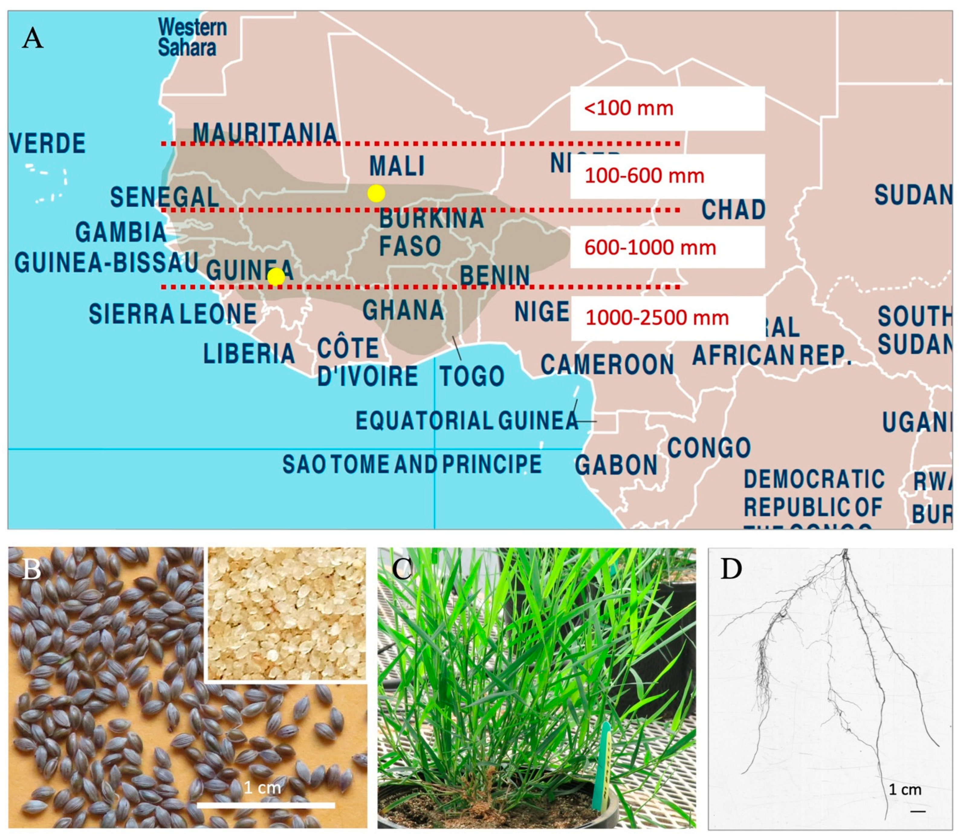
2.1. Plant Genetic Material

Two accessions (landraces) of white fonio (Digitaria exilis) were obtained from The Research Institute for Development (IRD) France. Accession CM05836 represents a dry area (mean precipitation 333 mm, mean temp 27.9 °C) from Mali associated with the Fulani people [Mopti region, latitude 15.05; longitude −3.88; altitude, 263 m above sea level (MASL)], whereas CM07354 represents a high rainfall area (mean precipitation 998 mm, mean temp 24.1 °C) from Guinea associated with the Malinke ethnic group (Konsankoro region, latitude 9.03; longitude −9.00; 537 MASL) [33].

2.2. Plant Growth Conditions and Experimental Design

Fonio seeds (non-sterilized) were sown in 4 L poly pots (~10 seeds sown, then thinned to one seedling per pot) containing a mix (2:1:2), respectively, of sand, perlite, and field soil from the Elora Research Station, University of Guelph, Elora, ON, Canada. Plants were grown under a mixture of fluorescent lamps (LED18ET8/4/850, GE, East Cleveland, OH, USA) and incandescent bulbs (Ultra LED A21-15 W, Sylvania, Markham, ON, Canada) in an indoor growth room (Crop Science Growth Facility, University of Guelph) at an intensity of ~300 μmol/m2/s at canopy level for a 12/12 h day/night photoperiod. The day/night temperature was 30 °C/25 °C, and the RH was 65%.
Drought experiment: Soil moisture levels were measured daily (9–11 am) using a FieldScout TDR 150 Soil Moisture Meter (Spectrum Tech. Inc., Aurora, CO, USA), with the entire probe (12 cm) inserted into the soil. The meter was set to ‘standard soil’ calibration mode for all measurements. Each reading was taken 2–3 times per pot, and averaged. Based on the moisture readings, pots were watered manually to maintain the desired volumetric water content (VWC) [drought: 6% VWC; optimal: 26% VWC) during the period of drought treatment (21–42 days after emergence (DAE)]. However, for the drought treatment, watering was decreased starting at 14 DAE to achieve the desired moisture levels in the pots progressively by 21 DAE. In terms of nutrients, 10 mL/pot of the fertilizer stock solution [50 g/L of NPK 24:10:20 (#10535, Plant Products, Leamington, ON, Canada) + 1.2 g/L MgSO4] was supplied at the time of planting, whereas no fertilizer was supplied after planting until the end of the drought treatment (i.e., until tissue harvest) to avoid adding additional water. In a split plot design, two moisture treatments (6% and 26% VWC) were set as the main plot, and two fonio accessions (CM05836; CM07354) as subplots (randomized), with 3 replicates.
Low nitrogen experiment: The low nitrogen experiment was conducted at a different time than the drought experiment, with constant, optimal watering (26% VWC). Fertilizer lacking nitrogen was formulated [(27 g/L of NPK 0:20:20 consisting of zero nitrogen, 21 g/L KH2PO4, and 6 g/L KCl) to which was added 1.2 g/L MgSO4, and 3 g/L micronutrient mix (#10050, Plant Products, Leamington, ON, Canada)]. The nutrients were supplied continuously in the drip irrigation line (diluted 1:100 in 120 mL/pot of water) three days each week, starting at germination until tissue harvest.

2.3. Tissue Sampling

Leaf and root samples (Figure 1C,D) from three biological replicates (i.e., from 3 pots: 1 plant/pot) of all treatments were collected at 43 DAE. Six leaves per plant (representing older, mid age, and young leaves) and full-length crown roots (~5 g/plant) were collected per replicate. For bacterial sampling, each tissue sample was pooled from the three replicates (plants). From the low nitrogen experiment, stem tissue was also collected (three 5 cm pieces, including base, middle, and top segments). Collected samples were washed once with tap water, then moved to a sterile laminar flow hood, where they were washed with sterile water then surface sterilized. For surface sterilization, the roots were dipped in 70% ethanol for 5 min followed by 2% sodium hypochlorite for 5 min, whereas the leaves and stems were dipped in 70% ethanol for 3 min followed by 2% sodium hypochlorite for 3 min; all tissues were then washed 10 times with double distilled sterile water. The final washes from all the samples were added onto LB plates (see below for recipe) and incubated for 3 days at 30 °C to confirm the absence of any microbial growth.
For seed sampling, a pool of ~200 seeds/accession was used. The seeds were previously multiplied by growing source seed from Mali and Guinea in pots containing commercial potting mix with no added microbes (Sunshine Mix, Sun Gro Horticulture, Agawam, MA, USA) in a growth chamber at the University of Guelph. Seeds were soaked in distilled water for 48 h, then surface sterilized using the protocol used for the roots.

2.4. Bacterial Isolation Media

LB agar was prepared as follows, per L: 10 g tryptone, 5 g yeast extract, 5 g NaCl, 12 g agar, at pH 7. LGI agar (N-free medium) was prepared as follows, per L: 5 g sucrose, 0.01 g FeCl3·6H2O, 0.8 g K3PO4, 0.2 g MgSO4·7H2O, 0.2 g CaCl2, 0.002 g Na2MoO4·2H2O, 12 g agar, at pH 6, followed by autoclaving. The same recipes excluding agar were used to prepare the respective liquid media.

2.5. Bacterial Culturing

Surface sterilized tissues and seeds were ground in an autoclaved mortar and pestle along with 1 mL 50 mM sodium phosphate buffer (14.43 mL of 1 M Na2HPO4, and 10.58 mL of 1 M NaH2PO4, in a final volume of 500 mL of autoclaved ddH2O at pH 7) per gram of fresh tissue weight. After grinding, 50 μL of the mixture was serially diluted three times in 450 μL of sodium phosphate buffer, resulting in 10×, 100×, and 1000× dilutions. Each dilution was spread onto both LB and LGI agar plates separately and incubated at 30 °C for up to 3 days. Unique colonies from each plate were selected based on colony colour and morphology; single colonies were streaked onto new plates for purification.

2.6. Bacterial DNA Isolation and Sequencing

Purified isolates were cultured in their respective liquid media at 30 °C for 1–2 days in a shaker incubator. Bacterial DNA was extracted using the QIAamp DNA mini kit (cat# 51306, Qiagen, Germantown, MD, USA). Thereafter, 2 μL of genomic DNA were quantified using a Qubit v1.2 fluorometer (#Q32857, Qubit Systems Inc., Kingston, ON, Canada) with two standards. Polymerase chain reaction (PCR) amplification of the 16S rRNA gene (V1–V9) was performed using the primer set 27F [5′-AGAGTTTGATCMTGGCTCAG-3′] and 1492R [5′-GGTTACCTTGTTACGACTT-3′] in a PTC200 DNA Thermal Cycler (MJ Scientific, Waltham, MA, USA). The PCR mixtures contained ~100 ng of genomic DNA, 20 μL of GoTaq Green Master Mix (M712C, Promega, Madison, WI, USA), 2 μL of each primer (10 μM), and nuclease-free H2O to a final volume of 40 μL. The PCR conditions were as follows: an initial denaturation at 96 °C for 3 min, followed by 35 amplification cycles at 94 °C for 30 s, 48 °C for 30 s, 72 °C for 90 s, then a final extension at 72 °C for 7 min [37]. An amplicon size of ~1400 bp was verified via using gel electrophoresis and purified from liquid using the Illustra GFX DNA and Gel Band Purification Kit (#28-9034, GE Healthcare, Waukesha, WI, USA). Purified samples were then quantified using a Qubit v1.2 fluorometer (#Q32857, Molecular Probes, Life Technologies, Waltham, MA, USA) with two standards. Purified amplicons were kept at −20 °C.
Purified amplicons were then processed using the BigDye Terminator v3.1 Labeling Reactions (Applied Biosystems, Foster City, CA, USA). Each sample was subjected to two reactions (forward and reverse primers). For each reaction, 28 ng per kb of template DNA (41 ng for primer set 27F/1492R) and 10 pmol of primer were dried in a 96-well plate (using an open thermocycler) at 96 °C for 10 min. A master mix of BigDye/buffer/water 1:2:9 was mixed, then 12 µL was added to each reaction tube. The 96-well plate was subsequently sealed with film, vortexed gently, and briefly spun down before thermocycling using the following conditions: initial denaturation at 96 °C for 2 min, followed by 30 amplification cycles at 96 °C for 30 s, 48 °C for 15 s, 60 °C for 4 min, followed by incubation at 10 °C. Samples were subsequently submitted to the AAC Genomics Facility (University of Guelph, Guelph, ON, Canada) for sequencing in a 3730 DNA analyzer (Applied Biosystems, Foster City, CA, USA) of the 16S V1–V9 amplicons.

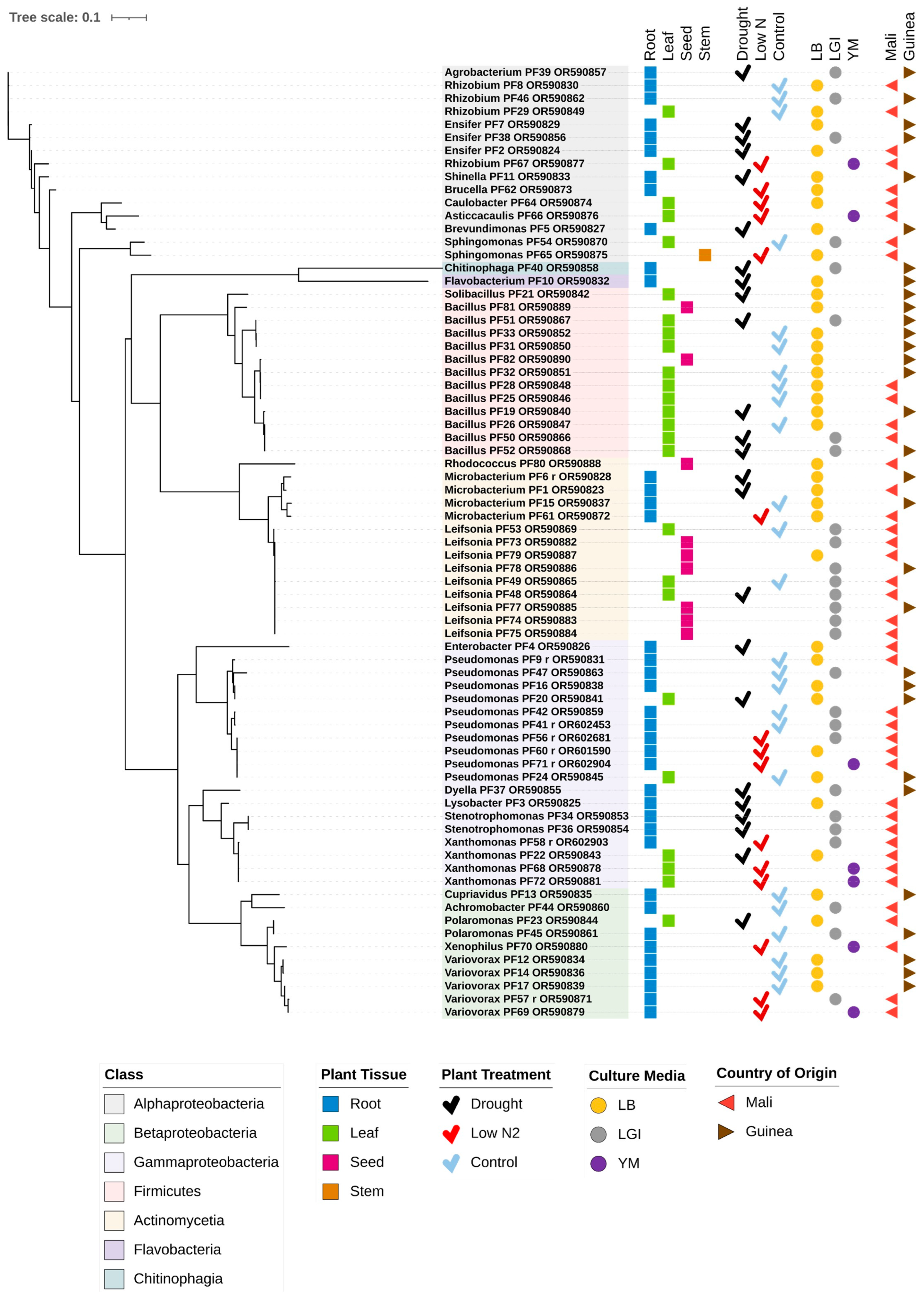
2.7. 16S rRNA Gene Analysis and Phylogenetic Tree Construction

Sanger sequences of the 16S rRNA gene (V1–V9 region) of the cultured fonio isolates were received as ab1 files and converted into the FASTA file format using the seqret tool (European Molecular Biology Open Software Suite, EMBOSS, version 6.6.0.0) in the command line [38]. Sequences were trimmed and edited using UGENE v.48.1 [39], then contigs were generated using CAP3, a sequence assembly program [40] embedded into UGENE software. For identification, sequences were searched against three databases during September 2023, including NCBI using BLASTN (standard database) [41], EzBioCloud through the 16S-based ID service [42], and SpeciesFinder from the Center for Genomic Epidemiology [43]. The sequences from this study were deposited in GenBank, and accession numbers were added to their corresponding strains (Supplementary Materials, Table S1). For all seed and Leifsonia isolates, GenBank (16S rRNA database) and EzBioCloud were re-accessed on 21 February 2025 to capture taxonomic updates (Supplementary Materials, Figures S1 and S2).
Sanger sequences of the 16S rRNA gene (V1–V9 region) of the cultured fonio isolates were used to generate a phylogenetic tree. To generate the tree, first, a multiple sequence alignment was performed using the MUSCLE program [44] in the command line. Subsequently, the GTR+G (general time reversible model of nucleotide substitution) model with default parameters was used using the RAxML-NG tool [45]. The phylogenetic tree was visualized and annotated using iTOL online software [46].

2.8. Bacterial Transmission Bioinformatic Analysis

Potential transmission of the Leifsonia taxa from seed to leaf was tested bioinformatically using multiple sequence alignment (Clustal W, European Bioinformatics Institute and Kyoto University Bioinformatics Center (http://www.bic.kyoto-u.ac.jp/)) [47,48] of full length 16S rRNA sequences (V1–V9). Seed versus leaf isolates were considered to match when they had 100% sequence identities (except for rare ‘N’ sequences) across a minimal length of >1000 bp (after trimming).

2.9. GenBank Accession Numbers

The 16S rRNA sequences were deposited in NCBI GenBank on 27 September 2023. The GenBank accession numbers of the bacterial strains in this study are noted in the Supplementary Materials, Table S1 (Column D).

2.10. Functional Abiotic Stress Tolerance Experiments

A total of 14 isolates were functionally tested for abiotic stress tolerance in vitro. All 9 seed isolates were tested, given their potential to be vertically transmitted, including 6 Leifsonia (PF73, PF74, PF75, PF77, PF78, PF79), 2 Bacilli (PF81, PF82), and 1 Rhodococcus (PF80). The remaining 5 isolates (from leaf and root) were selected as positive controls, based on the reported stress tolerance of their respective taxonomic group in the literature. Specifically, 2 additional Bacilli (PF50, PF 52), 2 Ensifer (PF2, PF38), and 1 Variovorax (PF57 from Mali) strains were selected as potential positive controls for tolerance to drought, low nitrogen, and acidity + aluminum toxicity, respectively.

2.10.1. Growth on Nitrogen-Free Media

As described by Thompson and Raizada [49], a method to test bacteria for their ability to grow on a nitrogen-free media was adapted from the American Type Culture Collection’s “ATCC medium: 1312 Azospirillum amazonense (LGI medium)”. Individual strains were streaked from frozen glycerol stocks onto 20 mL ATCC LGI nitrogen-free agar plates (100 mm × 15 mm). The ATCC LGI media recipe was as follows in ddH2O: 0.2 g/L K2HPO4, 0.6 g/L KH2PO4, 0.02 g/L CaCl2·2H2O, 0.2 g/L MgSO4·7H2O, 0.002 g/L Na2MoO4·2H2O, 0.01 g/L FeCl3, 5 g/L sucrose, and 15 g/L Bacto-agar, followed by autoclaving. The pH was adjusted to 6.0 by using HCl. The plates were incubated at 37 °C in an anaerobic chamber (Anaerobe Systems AS-580, American Laboratory Trading, East Lyme, CT, USA) with 5% H2, 5% CO2, and 90% N2 for 4 days. Isolates which grew after 4 days were restreaked onto fresh LGI plates to deplete any residual nitrogen. A single isolate (PF81), which did not grow in the first round, was re-streaked from the LB plates onto fresh LGI plates to confirm the absence of growth. There were 3 replicates per round. Each round of plates was incubated in the anaerobic chamber at 37 °C for 4 days.

2.10.2. Drought Tolerance

As a proxy for drought tolerance, a PEG-6000 (polyethylene glycol) tolerance protocol was followed, adapted from Hernández-Fernández et al. [50] and Latif et al. [51]. Isolates were grown for 2 days as LB liquid cultures, then aliquots of 10 μL were inoculated into 500 μL LB (pH 7) amended with 0%, 10%, 30%, and 40% (w/v) PEG-6000 in sterile 96 deep well plates. There were 3 replicates per PEG treatment, each on a separate 96-well plate, with the position of each isolate randomized differently on each of the 3 replicates. Negative controls received no bacteria to check for background contamination. Plates were sealed with a breathable membrane and incubated at 30 °C at 200 rpm for 2 days. OD600 readings were taken using a SpectraMax 384 Plus spectrophotometer (Molecular Devices, San Jose, CA, USA) using 110 μL aliquots, with fresh, sterile LB broth as the blank. Optical density measurements were normalized to the respective rank-ordered 0% PEG OD600 reading and multiplied by 100 to obtain a percentage, and then the mean percentage was calculated and categorized as follows: susceptible to PEG (<10%), moderate resistance (10–50%), and high resistance (>50%).

2.10.3. Acid and Aluminum Tolerance

To test for strain tolerance to combined acid and aluminum, an adapted protocol from Huang et al. [52] and Lim et al. [53] was followed as described by Thompson and Raizada [49]. Glucose Medium (GM) was used. The GM broth consisted of 1% glucose, 0.05% peptone, 0.02% yeast extract, and 0.02% MgSO4·7H2O, adjusted to pH 4.5 with HCl, followed by autoclaving. The media was amended with filter-sterilized AlCl3·6H2O (4 mM stock in cooled GM broth) to add aluminum as per the concentrations noted. To ensure that the isolates had the capacity to grow in GM, they were initially grown for 2 days in GM broth at neutral pH (pH 7), then 10 μL of each GM-grown isolate was used to inoculate 500 µL of GM broth containing 0% AlCl3·6H2O, 0.1 mM AlCl3·6H2O or 0.4 mM AlCl3·6H2O at pH 4.5 in 96 deep well plates. There were 3 replicates per treatment, each on a separate 96-well plate, with the position of each isolate randomized differently on each of the 3 replicates. Plates were incubated at 30 °C and 200 rpm for two days. The OD600 readings were taken using 110 μL aliquots of each sample in a SpectraMax 384 Plus spectrophotometer (Molecular Devices, San Jose, CA, USA), with fresh GM broth used as the blank. Optical density measurements were normalized to the respective rank-ordered 0% AlCl3 OD600 reading and multiplied by 100 to obtain a percentage, and then the mean percentage was calculated and categorized as follows: susceptible to AlCl3 (<10%), moderate resistance (10–50%), and high resistance (>50%).

3. Results

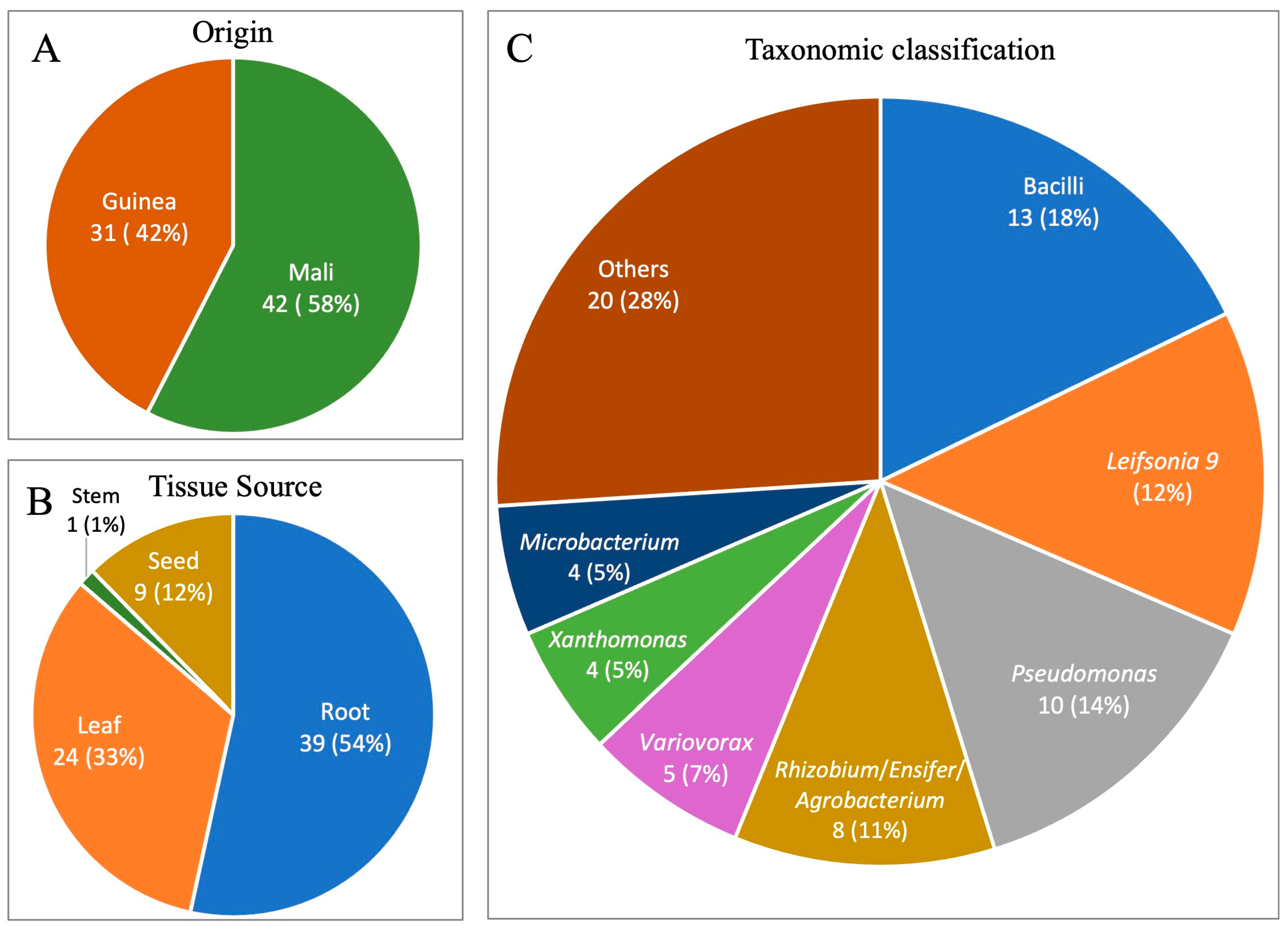
3.1. Overview of Bacterial Isolates from Fonio

From the white fonio accessions from Guinea and Mali (Figure 1A), a total of 73 bacterial isolates were cultured from surface-sterilized tissues and could be classified. Of these, 42 isolates were cultured from the Mali accession, and 31 from the Guinea accession (Figure 2A). In total, 39 were from the root, 24 were from the shoot, 9 were from the seed, and 1 was from the stem (Figure 2B, Figure 3A and Figure 4). The fonio endosphere was revealed to possess the following culturable bacterial diversity: the isolates spanned diverse phyla and classes including Alpha-, Beta-, and Gamma-proteobacteria, Firmicutes, Actinomycetia, Flavobacteria, and Chitinophagia (Figure 4), encompassing 27 genera and 18–39 unique species, depending on the software used (Table 1 and Table 2, Figure 4). However, 73% of the isolates (53/73) belonged to only seven clades at the genus level, dominated by Bacilli (13 isolates), Pseudomonas (10 isolates), and Leifsonia (9 isolates) (Figure 2C and Figure 4). The phylogenetic tree construction showed that the isolates from Mali versus Guinea spanned similar phyla, classes, and genera, and were not distinct (Figure 4).

3.2. Potential Bacterial Endophytes Isolated from White Fonio Leaves and Roots in the Drought Versus the Optimal Water Experiment

Mali vs. Guinea: In the drought/optimal water experiment, in the roots plus the leaves, similar taxonomic diversity was cultured from the Mali accession at the genus level (15 unique genera) compared to the Guinea accession (17 unique genera) (Table 1, Figure 4). Several genera were shared between the two accessions (Rhizobium, Ensifer, Microbacterium, Pseudomonas, Bacillus), while a few genera were unique to an accession (e.g., two isolates of Stenotrophomonas in Mali), but primarily as single isolates (e.g., Enterobacter in Mali, and Chitinophaga in Guinea). Variovorax was unique to Guinea but isolated from the low nitrogen experiment from Mali (Figure 2C, Table 1 and Table 2); similarly, Leifsonia was unique to Mali, but isolated from the Guinea seeds (Figure 2C, Figure 3A and Figure 4, Table 1).
Root vs. Shoot: In the drought/optimal water experiment, 11/20 leaf isolates were Bacilli from both accessions, whereas no Bacilli were identified amongst the 29 root isolates (Table 1, Figure 4). Some genera were only isolated from the roots, including Variovorax (five isolates, including the low nitrogen experiment), Microbacterium (four isolates across both experiments), and Ensifer (three isolates), while Leifsonia was only isolated from the leaves (three isolates). Rhizobium and Pseudomonas were cultured from both the roots and the leaves (Figure 4).
Drought vs. Optimal Water: In the drought/optimal water experiment, from both accessions, 26 isolates were cultured from the vegetative tissues grown under optimal water conditions, while 33 isolates came from the drought-grown plants (Table 1, Figure 4). Five diverse Pseudomonas isolates were cultured from the roots of plants grown under optimal water conditions, but none from the roots of drought-grown plants (Table 1, Figure 4). Amongst the nitrogen-fixing bacteria, the three isolates of Ensifer were solely isolated from the drought-grown plants, while the three isolates of Rhizobium came only from the well-watered plants (Table 1, Figure 4). Other prevalent genera (e.g., Bacillus, Leifsonia) were isolated in the plants grown under both optimal water and drought conditions.

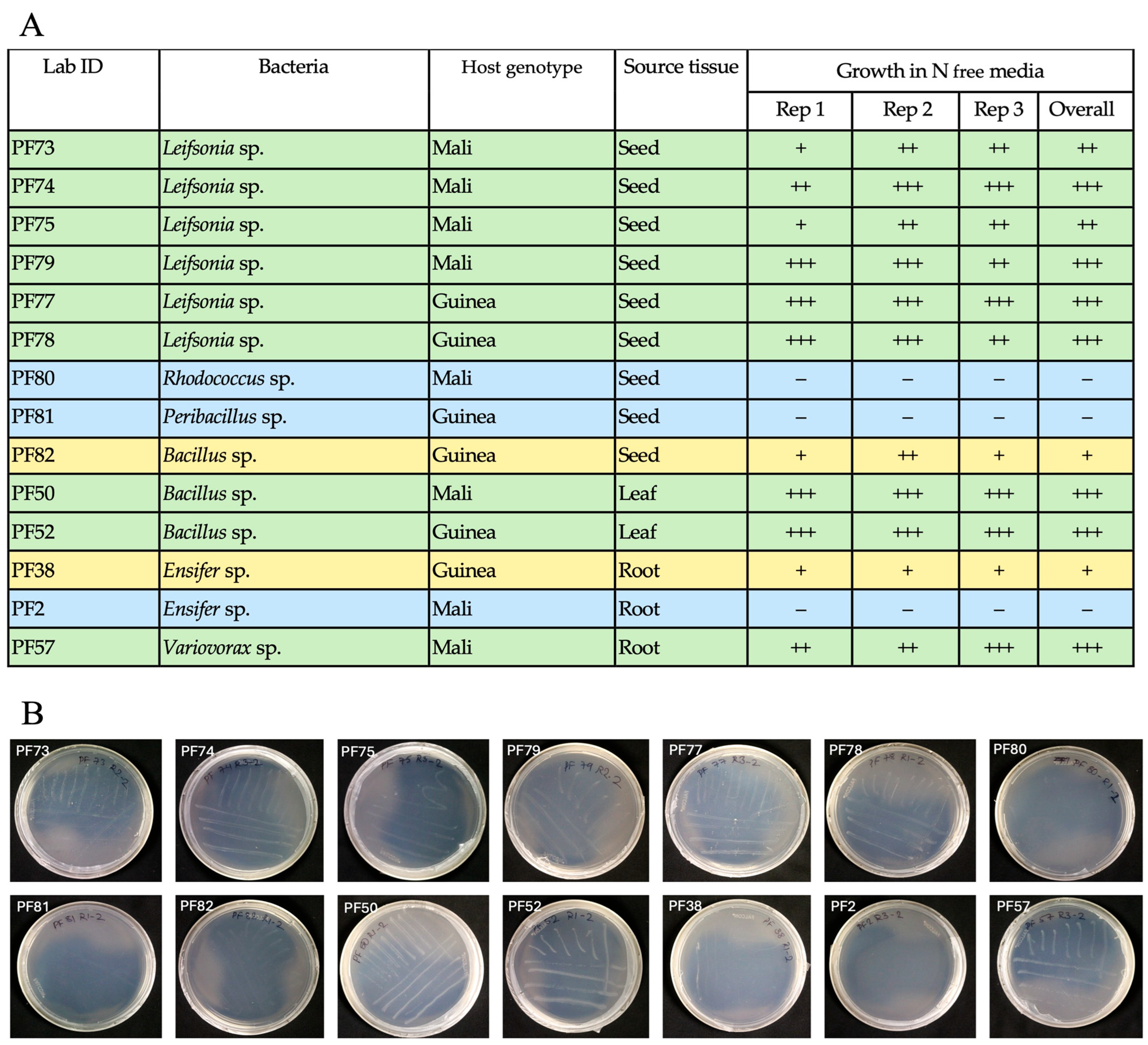
3.3. Potential Bacterial Endophytes from White Fonio Grown Under Low Nitrogen Stress

In a later experiment, 15 isolates spanning 11 genera were cultured from vegetative tissues of the Mali and Guinea accessions grown under low nitrogen with optimal water conditions (Table 2, Figure 4). Some genera were uniquely cultured from the low nitrogen experiment that were absent in the drought-optimal water experiment (e.g., Brucella, Caulobacter, Asticcacaulis, Xenophilus), but these were single isolates. More prevalent genera (Rhizobium, Sphingomonas, Microbacterium, Pseudomonas, Xanthomonas, Variovorax) were shared between both experiments. Bacillus, Leifsonia, and Ensifer were not cultured from low nitrogen-grown plants.

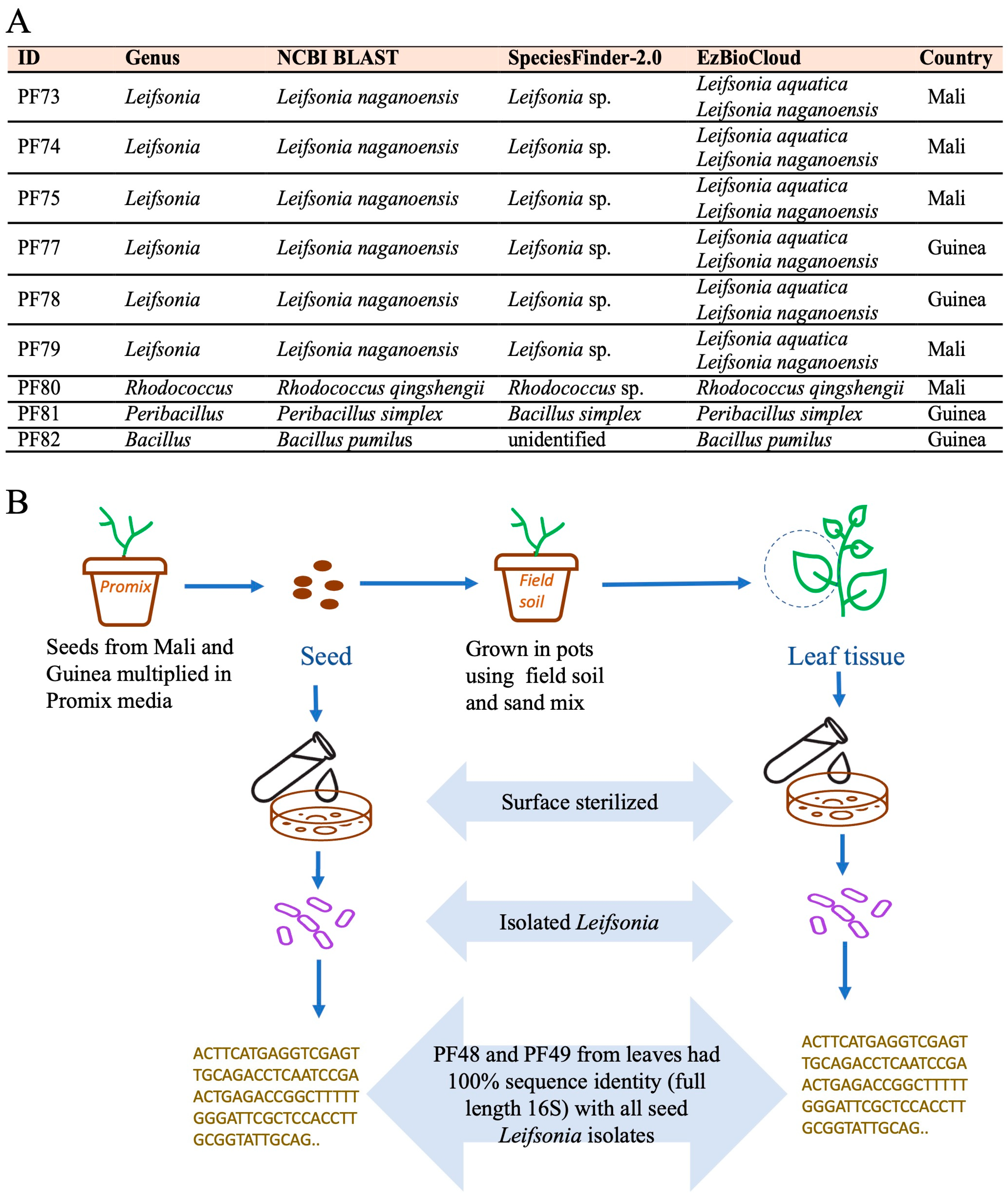
3.4. Potential Bacterial Endophytes from White Fonio Seeds and Possible Transmission Dynamics

Next, to examine the potential transmission from seeds to vegetative tissues, potential endophytic bacteria were also cultured from the surface sterilized Mali and Guinea fonio seeds (Figure 3 and Figure 4). Of the nine isolates, six were Leifsonia, two were Bacilli, and one was Rhodococcus. The Bacilli and Rhodococcus seed isolates did not match the isolates from the vegetative tissues. All six Leifsonia seed isolates had identical full length 16S rRNA gene sequences, despite originating from different countries. Furthermore, the Leifsonia seed isolates (from Mali and Guinea) showed 100% 16S rRNA sequence identity with the leaf isolates PF48 and PF49 (from Mali) (Figure 3, Supplementary Materials, Figure S3), consistent with their possible transmission from seed to shoot after germination.

3.5. Functional Abiotic Stress Tolerance Experiments for Seed Isolates

As seed endophytes have the potential to be inherited, all nine seed isolates, including six Leifsonia, two Bacilli, and one Rhodococcus, were tested in vitro for tolerance to abiotic stresses. Stresses were selected based on their relevance to the edaphic environments from which the seeds originated in the Sahel, specifically water limitation, high aluminum, low pH, and low nitrogen. Five leaf/root isolates were also tested as positive controls (e.g., Ensifer growth under low nitrogen). All nine seed isolates showed tolerance to moderate water limitation, but only a subset (including three Leifsonia and two Bacilli) showed modest growth under severe water limitation (Table 3). All nine seed isolates showed modest to excellent tolerance to low pH and aluminum, including the Leifsonia isolates (Table 3). All six Leifsonia isolates and one Bacillus grew on nitrogen-free media after sequential re-streaking, which was performed to deplete any residual nitrogen (Figure 5).

4. Discussion

4.1. General Discussion

This study identified the potential bacterial endophytes from the leaves and roots of white fonio grown under extreme abiotic stress conditions. It was theorized that generations of fonio farmers may have inadvertently selected for a stress-resistant microbiome in this crop. The fonio accessions in this study are associated with the Dogon, Malinke, and Fulani peoples of West Africa [33], who highly value fonio; for example, Dogon farmers describe this crop as an “image of the original atom whence the universe sprang” [3]. This respect shown is because fonio is a resilient crop, adapted to the arid, low nutrient conditions of the African Sahel [3,54]. The bacterial isolates from this study will permit long-term functional experiments to help understand the contribution of the fonio microbiome to the resiliency of this crop.
The total cultured library from this study spanned 27 genera; however, nearly 75% of the 73 isolates belonged to only 7 genera complexes (summarized in Figure 4). This study revealed that the cultured endosphere from the fonio leaves and roots (as well as a few from seeds) include endophytic bacteria observed in other plants, including other cereals (e.g., Agrobacterium-Rhizobium, Bacillus, Microbacterium, Pseudomonas, Sphingomonas, Stenotrophomonas, Enterobacter, Xanthomonas) [12,14,16,55]. Recently, a study of 126 fonio accessions [27] showed that the seeds were dominated by Bacillus and Pseudomonas, consistent with this study showing their dominance in the leaves and roots, despite differences in the methodology used. Tabassum et al. [27] used short-read V5–V7 16S Next Generation Sequencing of the microbiome (culture-independent), whereas the present study used longer amplicon (V1–V9) 16S Sanger sequencing of cultured isolates.
No major differences were observed in the culturable taxonomic composition of the bacterial endophytes retrieved from drought versus well-watered fonio plants, with some exceptions (Pseudomonas, Ensifer, Rhizobium). This result is consistent with the findings of Tabassum et al. [27], which showed that the fonio seed microbiome correlated with host accession altitude, longitude, and temperature, but not with precipitation. Interestingly, two genera that were not observed in the fonio seeds [27], namely Brucella and Xenophilus, were cultured here from nitrogen-stressed roots. This difference could be due to the divergent 16S rRNA sequencing strategies used or demonstrates that fonio microbiomes differ between the tissues (see below) and/or under nutrient stress conditions. However, these observed similarities or biases, including comparisons of root versus shoot, Mali versus Guinea, or different nitrogen levels, should be viewed as preliminary, and must be validated by future high throughput culture-independent, microbiome sequencing experiments. Nevertheless, the results provide intriguing preliminary evidence that fonio is inhabited by bacteria previously reported to tolerate abiotic stress and to promote plant growth. Furthermore, the study points to Leifsonia as a potential seed-to-leaf transmitted microbe in this crop, pending confirmation by whole genome sequencing. This study also describes the potential of Leifsonia to tolerate specific abiotic stresses relevant to the Sahel, based on the in vitro results.

4.2. The Fonio Endosphere Contains Bacteria Known to Tolerate Abiotic Stress and Promote Plant Growth

A striking feature of the fonio leaf endosphere was that 11/20 isolates were Bacilli; furthermore, of the 9 seed isolates, 2 were also Bacilli (Figure 4). Bacilli are spore-formers which can tolerate hot and dry conditions [56] and, hence, may be adapted to the harsh conditions of the African Sahel [57,58]. Here, three of the four Bacilli tested showed growth tolerance to moderate water limitation, including two seed-derived strains (Table 3). Several studies have shown that Bacillus inoculants can promote drought tolerance in diverse plants, by maintaining the optimal transpiration rate, by improving photosynthetic efficiency, by producing enzymes that scavenge reactive oxygen species (ROS), by decreasing electrolyte leakage, by increasing osmoprotectants, such as proline and sugars, by producing extracellular polymers, by regulating diverse phytohormones, including indole-3-acetic acid, which promotes root growth for improved water and nutrient uptake, and by inducing host plant genes that promote tolerance to drought stress [56,59,60,61,62]. Bacillus inoculants have also been known to act as biofertilizers to promote growth in low nutrient soils [56,63], which are prevalent in the African Sahel [64]. Here, three of the four Bacilli isolates tested could grow in the absence of nitrogen (Figure 5).
In the context of the nutrient-limiting habitat of the Sahel, it was also interesting to culture the genera previously shown to be nitrogen fixers (e.g., Rhizobium, Ensifer, Bacillus, Microbacterium, Stenotrophomonas) [65,66,67]. Of these, the Ensifer isolates from both the Guinea and Mali fonio accessions were somewhat surprising. This is because, in the comprehensive literature search, we could find almost no reports of Ensifer in grasses including cereals, with some exceptions [68,69,70,71], including, interestingly, from the rhizosphere of a salt-tolerant grass from West Africa (Senegal) [68]. Ensifer also exhibits a degree of host-geographic specificity within the legume family [72]. Here, one of the two Ensifer strains tested could grow in the absence of nitrogen (Figure 5).
Also noteworthy was that, of the 39 root cultures, 5 were Variovorax, isolated from both the Guinea and Mali accessions (Figure 4). Only one of these was functionally tested, but it showed tolerance to moderate water limitation, low pH, aluminum, and low nitrogen (Table 3, Figure 5). In a systematic study [73] involving Arabidopsis plants exposed to abiotic stress, Variovorax was shown to be the sole genus capable of reversing the root growth inhibition caused by a 185 member bacterial synthetic community. This activity was associated with an auxin degradation operon [73].
Moving forward, functional in planta studies are needed to understand the potential contributions of Bacilli, Variovorax, nitrogen fixers such as Ensifer, and other isolates to abiotic stress tolerance in fonio.

4.3. Similarities and Differences Between Fonio Seed and Leaf/Root Endophytes

In total, Tabassum et al. [27] identified 191 core bacterial taxa in seeds across West Africa. Of the 27 cultured genera identified here, primarily from the leaves/roots, 23 were also identified as seed endophytes by Tabassum et al. [27]. The four exceptions were Agrobacterium, Solibacillus, Brucella, and Xenophilus. In three fonio accessions, Tabassum et al. [27] further showed that 52–74% of the bacterial amplicon sequence variants (ASVs) were transmitted from parental seed to progeny seed. Combined, these results suggest that many vertically transferred seed endophytes may be founders of the vegetative plant microbiome community in this crop.

4.4. Leifsonia Is a Candidate Seed-to-Leaf Transmitted Endophyte in Fonio

Leifsonia sp. have previously been reported as endophytes of coffee [74], pear [75], rice [76], tea [77], and maize [78]. Some Leifsonia isolates have been reported as plant growth promoters [77,79,80]. Here, Leifsonia were isolated as presumptive endophytes from white fonio seeds from both Mali and Guinea, consistent with the recent findings from Tabassum et al. [27] that reported seven Leifsonia ASVs in the white fonio seed endosphere. In the present study, the majority (six/nine isolates) of the potential endophytes isolated from the seeds belonged to Leifsonia, all of which showed 100% 16S rRNA gene sequence identity with at least two leaf isolates. Combined, these observations suggest that Leifsonia is an important seed endophyte associated with fonio that transmits to the leaves after germination, with the possibility that it is inherited inter-generationally in West Africa. Any transmission conclusions, however, require Leifsonia whole genome sequencing (WGS), and for inheritance, requires culturing and WGS across successive plant generations.
The identification of the Leifsonia isolates, using full-length 16S rRNA sequencing, was not consistent among the reference 16S databases used: an updated NCBI BLAST (BLAST+ 2.16.0) consistently showed all nine isolates to most closely match L. naganoensis (~99.9% identity, Supplementary Materials, Figure S1), while the EzBioCloud best match was equally L. aquatica or L. naganoensis (~99.6–99.9%, Supplementary Materials, Figure S2). The recent microbiome study by Tabassum et al. [27] used shorter-read V5-V7 sequencing and, hence, cannot clarify the Leifsonia taxonomy. Based on whole genome sequencing, L. aquatica and L. naganoensis are close relatives [81], which explains the results from EzBioCloud. Future whole genome sequencing will finalize the taxonomy of the fonio isolates but represents a limitation of this study.
Unfortunately, no functional information about L. aquatica and L. naganoensis could be found in the literature. However, the closest identified species [81] is L. shinshuensis, which has been shown in rice to be a root endophyte (strain A1) that could promote root and shoot growth, confer modest plant resistance to Al3+, and is itself highly tolerant to Al3+ [77]. Aluminum tolerance may be of interest, since a large fraction of the soil in Guinea, the center of white fonio [36] cultivation, suffers from aluminum toxicity [2,82]. It is noteworthy from this study that the majority of Leifsonia isolates could grow in the aluminum, including two strains under high aluminum (Table 3). In pear, L. shinshuensis was isolated as a root endophyte, and showed the ability to solubilize phosphate, secrete IAA (indole compounds), and grow on a nitrogen-free medium [75]. Consistent with this finding, here, all six Leifsonia isolates were able to grow in the absence of nitrogen in vitro (Figure 5).
In the context of drought, Leifsonia sp. C5G2 isolated from the water-stressed coleus rhizosphere was shown to promote photosynthetic health in two ornamental species under severe drought and recovery [80]. Consistent with this prior finding, here, all six Leifsonia seed isolates showed tolerance to moderate water limitation, including three isolates under severe water limitation (Table 3). Furthermore, a comparative analysis of the Leifsonia genomes showed the presence of bacterial osmotic stress genes, though fewer in the L. nagoensis cluster [81]. Similarly, a Leifsonia endophyte genome was also shown to encode oxidative stress tolerance-encoding genes [76].
Given these prior reports of the potential plant benefits conferred by Leifsonia sp., and its potential seed-to-shoot transmission in fonio, it is proposed that this bacterial genus should be prioritized in future experiments concerning the fonio microbiome.

5. Conclusions

Here, the potential endophytic bacteria were cultured for the first time from white fonio, a subsistence crop from the African Sahel. The plants were grown indoors under water limitation or sufficient water, and bacteria were cultured primarily from the roots and shoots. Bacteria that survived the drought conditions included well known stress tolerant genera, including Bacilli. Evidence was found that Leifsonia is a candidate seed-to-shoot transmitted endophyte in this crop, pending further validation. Preliminary in vitro experiments show that fonio seed isolates, including Leifsonia, can survive a diversity of abiotic stress conditions relevant to the Sahel. This strain collection is available to the research community, as a starting point for experiments to understand how this orphan crop survives stress conditions.

Supplementary Materials

The following supporting information can be downloaded at: https://www.mdpi.com/article/10.3390/bacteria4030031/s1, Table S1: Detailed information for the isolates in this study including 16S rRNA sequences, taxonomies, and GenBank accession numbers; Figure S1: Taxonomic analysis of seed isolates based on the NCBI database; Figure S2: Taxonomic analysis of seed isolates based on the EzBioCloud database; Figure S3: Multi-sequence (CLUSTALW) alignments of Leifsonia bacteria isolated in this study to demonstrate potential transmission dynamics from seed to leaf in white fonio.

Author Contributions

Conceptualization, M.N.R.; methodology, R.P., E.M.K. and M.N.R.; investigation, R.P. and D.J.L.B.; data curation, R.P. and E.M.K.; software, E.M.K.; writing—original draft preparation, R.P.; writing—review and editing, M.N.R., E.M.K., D.J.L.B. and R.P.; visualization, R.P. and E.M.K.; supervision, M.N.R.; project administration, M.N.R.; funding acquisition, M.N.R. All authors have read and agreed to the published version of the manuscript.

Funding

This research was funded by the Natural Sciences and Engineering Research Council (NSERC) of Canada, grant number: 4000924.

Data Availability Statement

The 16S rRNA sequences have been deposited in GenBank under the specific accession numbers as noted in Table S1.

Acknowledgments

We thank Adeline Barnaud (IRD, France) for the gift of white fonio seeds, and Sue Couling (University of Guelph) for growth facility assistance. R.P. was the recipient of the Arrell Graduate Scholarship from the Arrell Food Institute, University of Guelph, Canada.

Conflicts of Interest

The authors declare no conflicts of interest.

References

  1. National Research Council. Lost Crops of Africa, Volume I: Grains; The National Academies Press: Washington, DC, USA, 1996; pp. 59–76. [CrossRef]
  2. Blench, R.M. Vernacular names for African millets and other minor cereals and their significance for agricultural history. Archaeol. Anthropol. Sci. 2016, 8, 1–8. [Google Scholar] [CrossRef]
  3. Cruz, J.; Béavogui, F. Fonio, An African Cereal; The French Agricultural Research Centre for International Development CIRAD: Montpellier, France, 2016; pp. 13–41. [Google Scholar]
  4. Jideani, I.A.; Jideani, V.A. Developments on the cereal grains Digitaria exilis (acha) and Digitaria iburua (iburu). J. Food Sci. Technol. 2011, 48, 251–259. [Google Scholar] [CrossRef]
  5. Ballogou, V.Y.; Soumanou, M.M.; Toukourou, F.; Hounhouigan, J.D. Indigenous knowledge on landraces and fonio-based food in Benin. Ecol. Food Nutr. 2014, 53, 390–409. [Google Scholar] [CrossRef] [PubMed]
  6. Zhu, F. Fonio grains: Physicochemical properties, nutritional potential, and food applications. Compr. Rev. Food Sci. Food Saf. 2020, 19, 3365–3389. [Google Scholar] [CrossRef]
  7. Temple, V.J.; Bassa, J.D. Proximate Chemical Composition of Acha (Digitaria exilis) Grain. J. Sci. Food Agric. 1991, 56, 561–563. [Google Scholar] [CrossRef]
  8. Vodouhe, S.R.; Achigan-Dako, G.E.; Dansi, A.; Adoukonou-Sagbadja, H. Fonio: A treasure for West Africa. In Proceedings of the Plant Genetic Resources and Food Security in West and Central Africa, Regional Conference, Ibadan, Nigeria, 26–30 April 2004; Vodouhe, R.S., Atta-Krah, K., Achigan-Dako, G.E., Eyog-Matig, O., Avohou, H., Eds.; Bioversity International: Rome, Italy, 2007; pp. 219–223. [Google Scholar]
  9. Wang, X.; Chen, S.; Ma, X.; Yssel, A.E.J.; Chaluvadi, S.R.; Johnson, M.S.; Gangashetty, P.; Amidou, F.; Sanogo, M.D.; Zwaenepoel, A.; et al. Genome sequence and genetic diversity analysis of an under-domesticated orphan crop, white fonio (Digitaria exilis). GigaScience 2021, 10, giab013. [Google Scholar] [CrossRef]
  10. Leach, J.E.; Triplett, L.R.; Argueso, C.T.; Trivedi, P. Communication in the Phytobiome. Cell 2017, 169, 587–596. [Google Scholar] [CrossRef]
  11. Busby, P.E.; Soman, C.; Wagner, M.R.; Friesen, M.L.; Kremer, J.; Bennett, A.; Morsy, M.; Eisen, J.A.; Leach, J.E.; Dangl, J.L. Research priorities for harnessing plant microbiomes in sustainable agriculture. PLoS Biol. 2017, 15, e2001793. [Google Scholar] [CrossRef] [PubMed]
  12. Hardoim, P.R.; van Overbeek, L.S.; Berg, G.; Pirttilä, A.M.; Compant, S.; Campisano, A.; Döring, M.; Sessitsch, A. The Hidden World within Plants: Ecological and Evolutionary Considerations for Defining Functioning of Microbial Endophytes. Microbiol. Mol. Biol. Rev. 2015, 79, 293–320. [Google Scholar] [CrossRef]
  13. White, J.F.; Kingsley, K.L.; Zhang, Q.; Verma, R.; Obi, N.; Dvinskikh, S.; Elmore, M.T.; Verma, S.K.; Gond, S.K.; Kowalski, K.P. Review: Endophytic microbes and their potential applications in crop management. Pest Manag. Sci. 2019, 75, 2558–2565. [Google Scholar] [CrossRef]
  14. Babalola, O.O.; Adedayo, A.A. Endosphere microbial communities and plant nutrient acquisition toward sustainable agriculture. Emerg. Top. Life Sci. 2023, 7, 207–217. [Google Scholar] [CrossRef]
  15. Card, S.; Johnson, L.; Teasdale, S.; Caradus, J. Deciphering endophyte behaviour: The link between endophyte biology and efficacious biological control agents. FEMS Microbiol. Ecol. 2016, 92, 114. [Google Scholar] [CrossRef]
  16. Mao, W.; Wu, Y.; Li, F.; Tang, W.; Gong, W.; Han, X.; White, J.F.; Ji, X.; Li, H. Seed endophytes and their roles in host plant stress resistance. J. Soil Sci. Plant Nutr. 2023, 23, 2927–2937. [Google Scholar] [CrossRef]
  17. Nadal, M.C.; dos Reis Ferreira, G.M.; Andrade, G.V.S.; Buttrós, V.H.; Rodrigues, F.A.; da Silva, C.M.; Martins, A.D.; Rufato, L.; Luz, J.M.Q.; Dória, J.; et al. Endophytic bacteria can replace the need for synthetic auxin during in vitro rooting of Pyrus communis. Agronomy 2022, 12, 1226. [Google Scholar] [CrossRef]
  18. Abideen, Z.; Cardinale, M.; Zulfiqar, F.; Koyro, H.W.; Rasool, S.G.; Hessini, K.; Darbali, W.; Zhao, F.; Siddique, K.H.M. Seed Endophyte bacteria enhance drought stress tolerance in Hordeum vulgare by regulating, physiological characteristics, antioxidants and minerals uptake. Front. Plant Sci. 2022, 13, 980046. [Google Scholar] [CrossRef]
  19. Orozco-Mosqueda, M.d.C.; Santoyo, G.; Glick, B.R. Recent advances in the bacterial phytohormone modulation of plant growth. Plants 2023, 12, 606. [Google Scholar] [CrossRef]
  20. Sajjad Mirza, M.; Ahmad, W.; Latif, F.; Haurat, J.; Bally, R.; Normand, P.; Malik, K.A. Isolation, partial characterization, and the effect of plant growth-promoting bacteria (PGPB) on micro-propagated sugarcane in vitro. Plant Soil 2001, 237, 47–54. [Google Scholar] [CrossRef]
  21. Johnston-Monje, D.; Raizada, M.N. Plant and endophyte relationships: Nutrient management. In Comprehensive Biotechnology, 2nd ed.; Moo-Young, M., Ed.; Elsevier B.V.: Amsterdam, The Netherlands, 2011; pp. 713–727. [Google Scholar]
  22. Szilagyi-Zecchin, V.J.; Ikeda, A.C.; Hungria, M.; Adamoski, D.; Kava-Cordeiro, V.; Glienke, C.; Galli-Terasawa, L.V. Identification and characterization of endophytic bacteria from corn (Zea mays L.) roots with biotechnological potential in agriculture. AMB Express 2014, 4, 26. [Google Scholar] [CrossRef]
  23. Siddikee, M.A.; Chauhan, P.S.; Anandham, R.; Han, G.H.; Sa, T. Isolation, characterization, and use for plant growth promotion under salt stress, of ACC deaminase-producing halotolerant bacteria derived from coastal soil. J. Microbiol. Biotechnol. 2010, 20, 1577–1584. [Google Scholar] [CrossRef]
  24. Zhu, J.K. Abiotic Stress Signaling and Responses in Plants. Cell 2016, 167, 313–324. [Google Scholar] [CrossRef]
  25. Vardharajula, S.; Ali, S.Z.; Grover, M.; Reddy, G.; Bandi, V. Drought-tolerant plant growth promoting Bacillus spp.: Effect on growth, osmolytes, and antioxidant status of maize under drought stress. J. Plant Interact. 2011, 6, 1–14. [Google Scholar] [CrossRef]
  26. Ali, S.Z.; Sandhya, V.; Rao, L.V. Isolation and characterization of drought-tolerant ACC deaminase and exopolysaccharide-producing fluorescent Pseudomonas sp. Ann. Microbiol. 2014, 64, 493–502. [Google Scholar] [CrossRef]
  27. Tabassum, N.; Ahmed, H.I.; Parween, S.; Sheikh, A.H.; Saad, M.M.; Krattinger, S.G.; Hirt, H. Host genotype, soil composition, and geo-climatic factors shape the fonio seed microbiome. Microbiome 2024, 12, 11. [Google Scholar] [CrossRef]
  28. Xu, L.; Naylor, D.; Dong, Z.; Simmons, T.; Pierroz, G.; Hixson, K.K.; Kim, Y.M.; Zink, E.M.; Engbrecht, K.M.; Wang, Y.; et al. Drought delays development of the sorghum root microbiome and enriches for monoderm bacteria. Proc. Natl. Acad. Sci. USA 2018, 115, E4284–E4293. [Google Scholar] [CrossRef] [PubMed]
  29. Walitang, D.I.; Kim, C.G.; Kim, K.; Kang, Y.; Kim, Y.K.; Sa, T. The influence of host genotype and salt stress on the seed endophytic community of salt-sensitive and salt-tolerant rice cultivars. BMC Plant Biol. 2018, 18, 51. [Google Scholar] [CrossRef]
  30. Dastogeer, K.M.G.; Tumpa, F.H.; Sultana, A.; Akter, M.A.; Chakraborty, A. Plant microbiome–an account of the factors that shape community composition and diversity. Curr. Plant Biol. 2020, 23, 100161. [Google Scholar] [CrossRef]
  31. Adoukonou-Sagbadja, H.; Wagner, C.; Dansi, A.; Ahlemeyer, J.; Daïnou, O.; Akpagana, K.; Ordon, F.; Friedt, W. Genetic diversity and population differentiation of traditional fonio millet (Digitaria spp.) landraces from different agro-ecological zones of West Africa. Theor. Appl. Genet. 2007, 115, 917–931. [Google Scholar] [CrossRef]
  32. Olodo, K.F.; Gueye, M.C.; Calatayud, C.; Diop, B.M.; Kane, N.A.; Ngom, A.; Ntui, V.O.; Barreto, M.M.S.; Uyoh, E.A.; Abraham, S.; et al. EST-SSR development for Digitaria exilis and its relatives D. iburua and D. longiflora from transcriptome sequences. Plant Genet. Resour. Characterisation Util. 2019, 17, 280–284. [Google Scholar] [CrossRef]
  33. Abrouk, M.; Ahmed, H.I.; Cubry, P.; Šimoníková, D.; Cauet, S.; Pailles, Y.; Bettgenhaeuser, J.; Gapa, L.; Scarcelli, N.; Couderc, M.; et al. Fonio millet genome unlocks African orphan crop diversity for agriculture in a changing climate. Nat. Commun. 2020, 11, 4488. [Google Scholar] [CrossRef]
  34. Sidibé, D. Mali Agricultural Pilot Soil Baseline and Background Research. Mali Agricultural Pilot, Research Report. OXFAM America. 2013. Available online: http://www.oxfamamerica.org/static/media/files/Mali_Agricultural_Study_Report_-_Soil_Baseline_and_Background_Research.pdf (accessed on 17 December 2023).
  35. Touré, I.; Larjavaara, M.; Savadogo, P.; Bayala, J.; Yirdaw, E.; Diakite, A. Land degradation along a climatic gradient in Mali: Farmers’ perceptions of causes and impacts. Land Degrad. Dev. 2020, 31, 2804–2818. [Google Scholar] [CrossRef]
  36. FAOSTAT. Crop and Livestock Products. Food and Agriculture Organization of the United Nations. Available online: https://www.fao.org/faostat/en/#data (accessed on 29 November 2023).
  37. Shehata, H.R.; Lyons, E.M.; Jordan, K.S.; Raizada, M.N. Bacterial endophytes from wild and ancient maize are able to suppress the fungal pathogen Sclerotinia homoeocarpa. J. Appl. Microbiol. 2016, 120, 756–769. [Google Scholar] [CrossRef] [PubMed]
  38. Chojnacki, S.; Cowley, A.; Lee, J.; Foix, A.; Lopez, R. Programmatic access to bioinformatics tools from EMBL-EBI update: 2017. Nucleic Acids Res. 2017, 45, 550–553. [Google Scholar] [CrossRef]
  39. Okonechnikov, K.; Golosova, O.; Fursov, M. Unipro UGENE: A unified bioinformatics toolkit. Bioinformatics 2012, 28, 1166–1167. [Google Scholar] [CrossRef] [PubMed]
  40. Huang, X.; Madan, A. CAP3: A DNA sequence assembly program. Genome Res. 1999, 9, 868–877. [Google Scholar] [CrossRef]
  41. Schoch, C.L.; Ciufo, S.; Domrachev, M.; Hotton, C.L.; Kannan, S.; Khovanskaya, R.; Leipe, D.; McBeigh, R.; O’Neill, K.; Robbertse, B.; et al. NCBI Taxonomy: A comprehensive update on curation, resources and tools. Database 2020, 2020, baaa062. [Google Scholar] [CrossRef] [PubMed]
  42. Yoon, S.; Ha, S.; Kwon, S.; Lim, J.; Kim, Y.; Seo, H.; Chun, J. Introducing EzBioCloud: A taxonomically united database of 16S rRNA gene sequences and whole-genome assemblies. Int. J. Syst. Evol. Microbiol. 2017, 67, 1613–1617. [Google Scholar] [CrossRef]
  43. Larsen, M.V.; Cosentino, S.; Lukjancenko, O.; Saputra, D.; Rasmussen, S.; Hasman, H.; Sicheritz-Pontén, T.; Aarestrup, F.M.; Ussery, D.W.; Lund, O. Benchmarking of methods for genomic taxonomy. J. Clin. Microbiol. 2012, 52, 1529–1539. [Google Scholar] [CrossRef]
  44. Edgar, R.C.; Drive, R.M.; Valley, M. MUSCLE: Multiple sequence alignment with high accuracy and high throughput. Nucleic Acids Res. 2004, 32, 1792–1797. [Google Scholar] [CrossRef]
  45. Kozlov, A.M.; Darriba, D.; Flouri, T.; Morel, B.; Stamatakis, A. RAxML-NG: A fast, scalable and user-friendly tool for maximum likelihood phylogenetic inference. Bioinformatics 2019, 35, 4453–4455. [Google Scholar] [CrossRef]
  46. Letunic, I.; Bork, P. Interactive tree of life (iTOL) v5: An online tool for phylogenetic tree display and annotation. Nucleic Acids Res. 2021, 49, W293–W296. [Google Scholar] [CrossRef]
  47. Madeira, F.; Pearce, M.; Tivey, A.R.N.; Basutkar, P.; Lee, J.; Edbali, O.; Madhusoodanan, N.; Kolesnikov, A.; Lopez, R. Search and sequence analysis tools services from EMBL-EBI in 2022. Nucleic Acid. Res. 2022, 50, W276–W279. [Google Scholar] [CrossRef]
  48. Sievers, F.; Wilm, A.; Dineen, D.; Gibson, T.J.; Karplus, K.; Li, W.; Lopez, R.; McWilliam, H.; Remmert, M.; Söding, J.; et al. Fast, scalable generation of high-quality protein multiple sequence alignments using Clustal Omega. Mol. Syst. Biol. 2011, 7, 539. [Google Scholar] [CrossRef] [PubMed]
  49. Thompson, M.E.H.; Raizada, M.N. The microbiome of fertilization-stage maize silks (style) encodes genes and expresses traits that potentially promote survival in pollen/style niches and host reproduction. Microorganisms 2024, 12, 1473. [Google Scholar] [CrossRef]
  50. Hernández-Fernández, G.; Galán, B.; Carmona, M.; Castro, L.; García, J. Transcriptional response of the xerotolerant Arthrobacter sp. Helios strain to PEG-induced drought stress. Front. Microbiol. 2022, 13, 1009068. [Google Scholar] [CrossRef]
  51. Latif, M.; Bukhari, S.A.H.; Alrajhi, A.A.; Alotaibi, F.S.; Ahmad, M.; Shahzad, A.N.; Dewidar, A.Z.; Mattar, M.A. Inducing drought tolerance in wheat through exopolysaccharide-producing rhizobacteria. Agronomy 2022, 12, 1140. [Google Scholar] [CrossRef]
  52. Huang, S.; Wang, X.; Liu, X.; He, G.; Wu, J. Isolation, identification, and characterization of an aluminum-tolerant bacterium Burkholderia sp. SB1 from an acidic red soil. Pedosphere 2018, 28, 905–912. [Google Scholar] [CrossRef]
  53. Lim, J.C.; Goh, K.M.; Shamsir, M.S.; Ibrahim, Z.; Chong, C.S. Characterization of aluminum resistant Anoxybacillus sp. SK 3–4 isolated from a hot spring. J. Basic Microbiol. 2015, 55, 514–519. [Google Scholar] [CrossRef] [PubMed]
  54. Small, E. Teff & Fonio—Africa’s sustainable cereals. Biodiversity 2015, 16, 27–41. [Google Scholar] [CrossRef]
  55. Abdelfattah, A.; Tack, A.J.M.; Lobato, C.; Wassermann, B.; Berg, G. From seed to seed: The role of microbial inheritance in the assembly of the plant microbiome. Trends Microbiol. 2023, 31, 346–355. [Google Scholar] [CrossRef]
  56. Etesami, H.; Jeong, B.R.; Glick, B.R. Potential use of Bacillus spp. as an effective biostimulant against abiotic stresses in crops—A review. Curr. Res. Biotech. 2023, 5, 100128. [Google Scholar] [CrossRef]
  57. Hilhorst, T.; Muchena, E.M. (Eds.) Nutrients on the Move: Soil Fertility Dynamics in African Farming Systems; International Institute for Environment and Development: London, UK, 2000; pp. 83–117. [Google Scholar]
  58. Sissoko, K.; van Keulen, H.; Verhagen, J.; Tekken, V.; Battaglini, A. Agriculture, livelihoods and climate change in the West African Sahel. Reg. Environ. Change 2011, 11, 119–125. [Google Scholar] [CrossRef]
  59. Lopes, R.; Tsui, S.; Gonçalves, P.J.R.O.; de Queiroz, M.V. A look into a multifunctional toolbox: Endophytic Bacillus species provide broad and underexploited benefits for plants. World J. Microbiol. Biotechnol. 2018, 34, 94. [Google Scholar] [CrossRef]
  60. Abdelaal, K.; Alkahtani, M.; Attia, K.; Hafez, Y.; Király, L.; Künstler, A. The role of plant growth-promoting bacteria in alleviating the adverse effects of drought on plants. Biology 2021, 10, 520. [Google Scholar] [CrossRef] [PubMed]
  61. Poveda, J.; González-Andrés, F. Bacillus as a source of phytohormones for use in agriculture. Appl. Microbiol. Biotechnol. 2021, 105, 8629–8645. [Google Scholar] [CrossRef] [PubMed]
  62. Tsotetsi, T.; Nephali, L.; Malebe, M.; Tugizimana, F. Bacillus for plant growth promotion and stress resilience: What have we learned? Plants 2022, 11, 2482. [Google Scholar] [CrossRef] [PubMed]
  63. Mahapatra, S.; Yadav, R.; Ramakrishna, W. Bacillus subtilis impact on plant growth, soil health and environment: Dr. Jekyll and Mr. Hyde. J. Appl. Microbiol. 2022, 132, 3543–3562. [Google Scholar] [CrossRef]
  64. Doso Jnr, S. Land degradation and agriculture in the Sahel of Africa: Causes, impacts and recommendations. J. Agric. Sci. Appl. 2014, 3, 67–73. [Google Scholar] [CrossRef]
  65. Xing, Y.X.; Wei, C.Y.; Mo, Y.; Yang, L.T.; Huang, S.L.; Li, Y.R. Nitrogen-fixing and plant growth-promoting ability of two endophytic bacterial strains isolated from sugarcane stalks. Sugar Tech. 2016, 18, 373–379. [Google Scholar] [CrossRef]
  66. Patel, J.K.; Archana, G. Diverse culturable diazotrophic endophytic bacteria from Poaceae plants show cross-colonization and plant growth promotion in wheat. Plant Soil 2017, 417, 99–116. [Google Scholar] [CrossRef]
  67. Rosenblueth, M.; Ormeño-Orrillo, E.; López-López, A.; Rogel, M.A.; Reyes-Hernández, B.J.; Martínez-Romero, J.C.; Reddy, P.M.; Martínez-Romero, E. Nitrogen fixation in cereals. Front. Microbiol. 2018, 9, 1794. [Google Scholar] [CrossRef]
  68. Fall, F.; le Roux, C.; Bâ, A.M.; Fall, D.; Bakhoum, N.; Faye, M.N.; Kane, A.; Ndoye, I.; Diouf, D. The rhizosphere of the halophytic grass Sporobolus robustus Kunth hosts rhizobium genospecies that are efficient on Prosopis juliflora (Sw.) DC and Vachellia seyal (Del.) P.J.H. Hurter seedlings. Syst. Appl. Microbiol. 2019, 42, 232–239. [Google Scholar] [CrossRef] [PubMed]
  69. Han, H.; Wu, X.; Yao, L.; Chen, Z. Heavy metal-immobilizing bacteria combined with calcium polypeptides reduced the uptake of Cd in wheat and shifted the rhizosphere bacterial communities. Environ. Pollut. 2020, 267, 115432. [Google Scholar] [CrossRef]
  70. Xi, J.; Qian, K.; Shan, L.; Huang, J.; Yan, Y. The potential of mineral weathering of halophilic-endophytic bacteria isolated from Suaeda salsa and Spartina anglica. Arch. Microbiol. 2022, 204, 561. [Google Scholar] [CrossRef] [PubMed]
  71. Bahulikar, R.A. Prevalence of Deltaproteobacterial sequences in nifH gene pools associated with the rhizosphere of native switchgrass from Tall Grass Prairie (Oklahoma, USA). Biotech 2023, 13, 210. [Google Scholar] [CrossRef]
  72. Andrews, M.; Andrews, M.E. Specificity in legume-rhizobia symbioses. Int. J. Mol. Sci. 2017, 18, 705. [Google Scholar] [CrossRef]
  73. Finkel, O.M.; Salas-González, I.; Castrillo, G.; Conway, J.M.; Law, T.F.; Teixeira, P.J.P.L.; Wilson, E.D.; Fitzpatrick, C.R.; Jones, C.D.; Dangl, J.L. A single bacterial genus maintains root growth in a complex microbiome. Nature 2020, 587, 103–108. [Google Scholar] [CrossRef] [PubMed]
  74. Duong, B.; Xuan Nguyen, H.; Viet Phan, H.; Colella, S.; Quang Trinh, P.; Thi Hoang, G.; Thi Nguyen, T.; Marraccini, P.; Lebrun, M.; Duponnois, R. Identification and characterization of Vietnamese coffee bacterial endophytes displaying in vitro antifungal and nematicidal activities. Microbiol. Res. 2021, 242, 126613. [Google Scholar] [CrossRef]
  75. Liaqat, F.; Eltem, R. Identification and characterization of endophytic bacteria isolated from in vitro cultures of peach and pear rootstocks. 3 Biotech 2016, 6, 120. [Google Scholar] [CrossRef]
  76. Battu, L.; Ulaganathan, K. Whole genome sequencing and identification of host-interactive genes in the rice endophytic Leifsonia sp. ku-ls. Funct. Integr. Genom. 2020, 20, 237–243. [Google Scholar] [CrossRef]
  77. Jiang, X.; Li, W.W.; Han, M.; Chen, G.; Wu, J.; Lai, S.; Fu, Z.; Zhang, S.; Deng, W.W.; Gao, L.; et al. Aluminum-tolerant, growth-promoting endophytic bacteria as contributors in promoting tea plant growth and alleviating aluminum stress. Tree Physiol. 2022, 42, 1043–1058. [Google Scholar] [CrossRef]
  78. Verma, A.; Shameem, N.; Jatav, H.S.; Sathyanarayana, E.; Parray, J.A.; Poczai, P.; Sayyed, R.Z. Diversity of bacterial endophytes of maize (Zea mays) and their functional potential for micronutrient biofortification. Curr. Microbiol. 2022, 79, 6. [Google Scholar] [CrossRef] [PubMed]
  79. Kang, S.M.; Khan, A.L.; You, Y.H.; Kim, J.G.; Kamran, M.; Lee, I.J. Gibberellin production by newly isolated strain Leifsonia soli SE134 and its potential to promote plant growth. J. Microbiol. Biotechnol. 2014, 24, 106–112. [Google Scholar] [CrossRef] [PubMed]
  80. Nordstedt, N.P.; Jones, M.L. Isolation of rhizosphere bacteria that improve quality and water stress tolerance in greenhouse ornamentals. Front. Plant Sci. 2020, 11, 826. [Google Scholar] [CrossRef] [PubMed]
  81. Nordstedt, N.P.; Roman-Reyna, V.; Jacobs, J.M.; Jones, M.L. Comparative genomic understanding of gram-positive plant growth-promoting Leifsonia. Phytobiomes J. 2021, 5, 263–274. [Google Scholar] [CrossRef]
  82. Cox, C.M.; Koo, J. Soil fertility. In Atlas of African Agriculture Research and Development: Revealing Agriculture’s Place in Africa; Sebastian, K., Ed.; International Food Policy Research Institute (IFPRI): Washington, DC, USA, 2014; pp. 40–41. [Google Scholar] [CrossRef]
Figure 1. Origin of white fonio accessions and tissue morphology. (A) Sources of white fonio accessions used in this study from West Africa (yellow dots). The shaded area indicates the cultivation region for white fonio along with annual rainfall zones (red dotted lines) (Map adapted from: Maps world, CC BY-SA 4.0). Pictures of white fonio: (B) seeds (inset shows dehulled seeds as white), (C) shoot, and (D) root system.
Bacteria 04 00031 g001
Figure 2. Overview of the 73 potential bacterial endophytes isolated from white fonio in this study by (A) geographic origin, (B) tissue source, and (C) classification at the genus level.
Bacteria 04 00031 g002
Figure 3. Potential bacterial endophytes isolated from white fonio seeds and hypothesized transmission dynamics. (A) List of isolates from surface sterilized white fonio seeds. See Figures S1 and S2 for taxonomic analysis. (B) Preliminary evidence for Leifsonia transmission from seed to leaves based on 100% sequence matches of the full length 16S rRNA gene (see Figure S3 for DNA sequence alignments).
Bacteria 04 00031 g003
Figure 4. Summary of potential endophytic bacterial isolates from white fonio. From left to right, the following are shown: the phylogenetic relationships of the isolates based on the 16S rRNA (V1–V9) gene; the genus name of each isolate; the isolate name (PF number); the GenBank accession number (OR number); the plant source tissue; the stress condition in which the plants were grown; the bacteriological culture media used (LB, LGI, YM, of which LGI had minimal nitrogen); and the country origin of the source plants (the Mali source was from a lower rainfall environment than the Guinea source).
Bacteria 04 00031 g004
Figure 5. Ability of selected bacterial isolates to grow on nitrogen-free media. (A) Growth of isolates on nitrogen-free media (LGI-agar). The strains had been restreaked from LGI agar onto new LGI agar to deplete any residual nitrogen. Rating: +++ good, ++ medium, + low, − no growth. (B) Representative pictures of the second generation LGI plates, reflecting varying levels of growth of the isolates tested.
Bacteria 04 00031 g005
Table 1. Potential endophytic bacterial isolates cultured from the root and shoot of white fonio [Mali (M) and Guinea (G) accessions] grown under drought compared to optimal water conditions.
IDGenusNCBI BLAST SpeciesFinder-2.0EzBioCloud OriginTissue SourceTreatment
PF44AchromobacterAchromobacter spaniusAchromobacter
xylosoxidans
Achromobacter
mucicolens
MRootOptimum water
PF39AgrobacteriumAgrobacterium
tumefaciens
Agrobacterium sp. Agrobacterium
tumefaciens
GRootDrought
PF5BrevundimonasUncultured
bacterium
Brevundimonas vesicularisBrevundimonas
vesicularis
GRootDrought
PF40ChitinophagaChitinophaga sp. Chitinophaga sp. Chitinophaga
ginsengisoli
GRootDrought
PF13CupriavidusCupriavidus sp. Cupriavidus sp.Cupriavidu
campinensis
GRootOptimum water
PF37DyellaDyella
jiangningensis
Dyella
jiangningensis
Dyella jiangningensisGRootDrought
PF2EnsiferEnsifer adhaerensEnsifer adhaerensEnsifer morelensisMRootDrought
PF38EnsiferEnsifer sp. UnknownEnsifer adhaerensGRootDrought
PF7EnsiferEnsifer adhaerensSinorhizobium sp. Ensifer adhaerensGRootDrought
PF4EnterobacterEnterobacter
ludwigii
Enterobacter sp. Enterobacter ludwigiiMRootDrought
PF10FlavobacteriumFlavobacterium
hauense
UnknownFlavobacterium sp.GRootDrought
PF3LysobacterLysobacter sp.Lysobacter soliLysobacter panacisoliMRootDrought
PF1MicrobacteriumMicrobacterium sp. Microbacterium sp. Microbacterium
saccharophilum
MRootDrought
PF62MicrobacteriumMicrobacterium chocolatumMicrobacterium sp.Microbacterium
atlanticum
GRootDrought
PF15MicrobacteriumMicrobacterium
foliorum
Microbacterium sp.Microbacterium
aerolatum
GRootOptimum water
PF45PolaromonasPolaromonas sp. Polaromonas sp. Polaromonas
ginsengisoli
GRootOptimum water
PF16PseudomonasPseudomonas sp. Pseudomonas sp. Pseudomonas solaniGRootOptimum water
PF41PseudomonasPseudomonas mosseliiPseudomonas mosseliiPseudomonas mosseliiMRootOptimum water
PF42PseudomonasPseudomonas mosseliiPseudomonas sp.Pseudomonas mosseliiMRootOptimum water
PF47PseudomonasPseudomonas
nitroreducens
Pseudomonas sp.Pseudomonas
nitritireducens
GRootOptimum water
PF9PseudomonasPseudomonas
paralcaligenes
Pseudomonas
alcaligenes
Pseudomonas
alcaligenes
MRootOptimum water
PF46RhizobiumAgrobacterium
tumefaciens,
Rhizobium sp.
Rhizobium sp.Agrobacterium
radiobacter
GRootOptimum water
PF8RhizobiumAgrobacterium
tumefaciens
Rhizobium sp.Agrobacterium
radiobacter
MRootOptimum water
PF11ShinellaUnknownUnknownShinella curvataGRootDrought
PF34StenotrophomonasStenotrophomonas maltophiliaStenotrophomonas maltophiliaStenotrophomonas sp.MRootDrought
PF36StenotrophomonasStenotrophomonas maltophiliaStenotrophomonas maltophiliaStenotrophomonas sp.MRootDrought
PF12VariovoraxVariovorax
beijingensis
Variovorax sp. Variovorax paradoxusGRootOptimum water
PF14VariovoraxVariovorax
beijingensis
Variovorax sp. Variovorax paradoxusGRootOptimum water
PF17VariovoraxVariovorax
beijingensis
Variovorax sp.Variovorax paradoxusGRootOptimum water
PF19BacillusBacillus subtilisBacillus subtilisBacillus tequilensisGLeafDrought
PF50BacillusBacillus sp.Bacillus subtilisBacillus siamensisMLeafDrought
PF51BacillusBacillus
thuringiensis
Bacillus sp.Bacillus proteolyticusGLeafDrought
PF52BacillusBacillus siamensisBacillus subtilisBacillus siamensisGLeafDrought
PF25BacillusBacillus safensisBacillus safensisBacillus safensisMLeafOptimum water
PF26BacillusBacillus sp. Bacillus sp. Bacillus siamensisMLeafOptimum water
PF28BacillusBacillus sp. unidentifiedBacillus safensisMLeafOptimum water
PF31BacillusBacillus toyonensisBacillus cereusBacillus toyonensisGLeafOptimum water
PF32BacillusBacillus safensisunidentifiedBacillus safensisGLeafOptimum water
PF33BacillusBacillus sp. Bacillus cereusBacillus toyonensisGLeafOptimum water
PF48LeifsoniaLeifsonia
naganoensis
Leifsonia sp. Leifsonia aquatica
Leifsonia naganoensis
MLeafDrought
PF49LeifsoniaLeifsonia
naganoensis
Leifsonia sp.Leifsonia aquatica
Leifsonia naganoensis
MLeafOptimum water
PF53LeifsoniaLeifsonia
naganoensis
Leifsonia sp.Leifsonia aquatica
Leifsonia naganoensis
MLeafOptimum water
PF23PolaromonasPolaromonas sp. Polaromonas sp.Polaromonas
ginsengisoli
MLeafDrought
PF20PseudomonasPseudomonas sp. Pseudomonas sp. Pseudomonas solaniGLeafDrought
PF24PseudomonasPseudomonas
jessenii
Pseudomonas sp. Pseudomonas mooreiGLeafOptimum water
PF29RhizobiumAgrobacterium
tumefaciens
Rhizobium sp.Agrobacterium
radiobacter
MLeafOptimum water
PF21SolibacillusSolibacillus sp. Solibacillus sp.Solibacillus isronensisGLeafDrought
PF5SphingomonasSphingomonas
echinoides
Sphingomonas sp.Sphingomonas
echinoides
MLeafOptimum water
PF22XanthomonasXanthomonas sp. Xanthomonas
oryzae pv.
Oryzae
Xanthomonas
maliensis
MLeafDrought
Table 2. Potential endophytic bacterial isolates cultured from the root and shoot of white fonio (Mali genotype) growing under low nitrogen conditions.
IDGenusNCBI BLASTSpeciesFinder-2.0EzBioCloudTissue Source
PF62BrucellaOchrobactrum sp. Ochrobactrum sp. Brucella triticiRoot
PF61MicrobacteriumMicrobacterium oxydansMicrobacterium oxydansMicrobacterium oxydansRoot
PF56PseudomonasPseudomonas chlororaphisPseudomonas fluorescensPseudomonas mooreiRoot
PF60PseudomonasPseudomonas mooreiPseudomonas sp.Pseudomonas
vancouverensis
Root
PF71PseudomonasPseudomonas mooreiPseudomonas sp.Pseudomonas
vancouverensis
Root
PF57VariovoraxVariovorax paradoxusVariovorax sp.Variovorax sp.Root
PF69VariovoraxVariovorax sp. Variovorax sp.Variovorax sp.Root
PF58XanthomonasXanthomonas maliensisXanthomonas citri pv.
Punicae
Xanthomonas euroxantheaRoot
PF72XanthomonasXanthomonas sp. Xanthomonas sp.Xanthomonas maliensisRoot
PF70XenophilusVariovorax sp. Xenophilus aerolatusXenophilus aerolatusRoot
PF66AsticcacaulisAsticcacaulis sp. Asticcacaulis sp.Asticcacaulis benevestitusLeaf
PF64CaulobacterCaulobacter rhizosphaeraeUncultured bacteriumCaulobacter sp.Leaf
PF67RhizobiumRhizobium sp.Rhizobium mongolenseRhizobium sp.Leaf
PF68XanthomonasXanthomonas sp. Xanthomonas sp. Xanthomonas maliensisLeaf
PF65SphingomonasSphingomonas sp.Sphingomonas mucosissimaSphingomonas oleiStem
Table 3. Tolerance of isolates in vitro to water limitation (PEG6000), acidity and aluminum (AlCl3).
Lab IDBacteriaHost GenotypeSource Tissue1 Tolerance [Percent OD600 Versus Negative Control Without Stress]
10% PEG30% PEG40% PEG0.1 mM AlCl3 at pH 4.50.4 mM AlCl3 at pH 4.5
2 MAXMINAVGMAXMINAVGMAXMINAVGMAXMINAVGMAXMINAVG
PF73Leifsonia sp.MaliSeed9710199252223645115130114282527
PF74Leifsonia sp.Mali Seed111108111116109101072576136823
PF75Leifsonia sp.Mali Seed675761545433121140131446958
PF79Leifsonia sp.Mali Seed1079610410471179158531366944
PF77Leifsonia sp.GuineaSeed9491958566669612710888182108
PF78Leifsonia sp.GuineaSeed10411711126915189137360681199
PF80Rhodococcus sp.MaliSeed90969498912797313612293317
PF81Peribacillus sp.GuineaSeed6767671411145442910041125022
PF82Bacillus sp.GuineaSeed645059241317745236237152322
PF50Bacillus sp.Mali Leaf63606111687564628391158
PF52Bacillus sp.GuineaLeaf899222233474746111
PF38Ensifer sp.GuineaRoot494949333333981571146510976
PF2Ensifer sp.Mali Root473841947878726567677473
PF57Variovorax sp.MaliRoot554650107815266350654217574
1 Rating: BLUE <10% growth compared to negative control (0% PEG or no AlCl3); YELLOW 10–50% growth compared to negative control; GREEN >50% growth compared to negative control. 2 MAX: comparison between maximum values among three replicates for each treatment; MIN: comparison between minimum values among three replicates for each treatment; AVG: comparison between average values of all three replicates for each treatment.
Disclaimer/Publisher’s Note: The statements, opinions and data contained in all publications are solely those of the individual author(s) and contributor(s) and not of MDPI and/or the editor(s). MDPI and/or the editor(s) disclaim responsibility for any injury to people or property resulting from any ideas, methods, instructions or products referred to in the content.

