Article

Antimicrobial Resistance and Prevalence of β-lactamase Genes Among Multidrug-Resistant Acinetobacter baumannii Isolates from Infected Diabetic Foot Ulcers

by
Diwan Mahmood Khan
1,2,3,4,
Venkatakrishna I. Rao
2,
M. S. Moosabba
3,
Davoodbasha MubarakAli
5,6,* and
Muhammed Manzoor
4,*
1
Department of Microbiology, Lord Buddha Koshi Medical College & Hospital, Saharsa 852221, India
2
Department of Microbiology, Yenepoya Medical College & Hospital, Yenepoya Deemed to be University, Mangalore 575018, India
3
Department of General Surgery, Yenepoya Medical College & Hospital, Yenepoya Deemed to be University, Mangalore 575018, India
4
Yenepoya Research Centre, Yenepoya Deemed to be University, Mangalore 575018, India
5
School of Life Sciences, B.S. Abdur Rahman Crescent Institute of Science and Technology, Chennai 600048, India
6
Crescent Global Outreach Mission (CGOM): Research & Development, B.S. Abdur Rahman Crescent Institute of Science and Technology, Chennai 600048, India
*
Authors to whom correspondence should be addressed.
Bacteria 2025, 4(2), 24; https://doi.org/10.3390/bacteria4020024
Submission received: 3 March 2025 / Revised: 22 April 2025 / Accepted: 7 May 2025 / Published: 12 May 2025

Abstract

:
Diabetic foot infections (DFIs) are a severe complication of diabetes and are increasing in prevalence globally. The microbiology of DFIs exhibits significant regional variation, with Acinetobacter baumannii frequently emerging as the predominant pathogen. This study aimed to investigate the microbiological profile of A. baumannii in DFIs of different Wagner grades. Pus and tissue specimens from 480 diabetic patients treated for DFIs between September 2016 and August 2019 were collected, and antimicrobial susceptibility testing was performed. Multiplex PCR was conducted to amplify extended spectrum β-lactamase (ESBL) and metallo-β-lactamase (MBL) genes. A. baumannii had a prevalence of 14.58% in DFIs, with 100% resistance to cephalosporins. Among the 70 A. baumannii isolates, 19 (27.14%) were ESBL producers and 43 (61.43%) were MBL producers. blaTEM was the most prevalent gene (52.94%) in ESBL producers; blaNDM-1 was the most prevalent gene (52.94%) in MBL producers. Our findings highlight the need for regular antimicrobial susceptibility testing, molecular surveillance, and robust antimicrobial stewardship programmes to effectively manage A. baumannii DFIs and mitigate their resistance.

1. Introduction

Diabetic foot infections (DFIs) are among the most severe complications in patients with type 2 diabetes mellitus, often leading to cellulitis, abscesses, necrotizing fasciitis, gangrene, septic arthritis, tendonitis, and osteomyelitis of the lower limbs [1,2]. Approximately 25% of patients with type 2 diabetes mellitus experience foot infection during their lifetime [3,4,5]. Over one-third of individuals with diabetes develop diabetic foot ulcers (DFUs), and half of these ulcers progress to DFIs [6]. The incidence of DFIs is 10 times higher than that of non-DFIs, with male patients being more commonly affected than female patients [7].
In recent decades, there has been a global shift in the microbiological profile of DFIs, with an increasing prevalence of Gram-negative bacteria over Gram-positive bacteria [8,9,10,11,12]. The most frequently isolated Gram-negative pathogens in DFIs include Pseudomonas aeruginosa, Escherichia coli, A. baumannii, Klebsiella species, and Proteus species [12,13,14,15]. Many studies have reported that DFIs can be polymicrobial or monomicrobial, with immune deficiency and decreased healing capacity contributing to the progression of bacterial infections [16,17].
A. baumannii is a highly virulent Gram-negative pathogen and the third most common cause of DFIs after Staphylococcus aureus and P. aeruginosa [18]. The occurrence of A. baumannii infection in DFIs ranges from 11 to 33% [19,20]. Our previous study revealed that biofilm formation, cell surface hydrophobicity, and gelatinase production were significantly higher in A. baumannii isolates from patients with DFIs, highlighting their key roles in infection pathogenesis [21]. The rapid rise of carbapenem-resistant A. baumannii isolates over the past decade has been concerning [22]. In 2018, the WHO ranked carbapenem-resistant A. baumannii (CRAB) as a top priority for antibiotic research and development [23].
Extended-spectrum β-lactamases (ESBLs), AmpC β-lactamases, and metallo-β-lactamases (MBLs) have emerged globally as the key drivers of antimicrobial resistance in Gram-negative bacteria [24,25]. Despite advancements in therapies, the increasing resistance among A. baumannii isolates has rendered many conventional treatments ineffective, highlighting the need for alternative strategies. This study aimed to investigate the microbiological profile, prevalence, and distribution of A. baumannii infections in DFIs, detect ESBL and MBL production, and assess antibiotic susceptibility patterns to inform targeted treatment strategies.

2. Materials and Methods

2.1. Sample Collection

A total of 480 patients with DFIs admitted to the Department of General Surgery at Yenepoya University Hospital, a tertiary care hospital in Mangalore, India, between September 2016 and August 2019 were included in the study. Samples were collected according to the Meggit’s Wagner DFU classification system grade, the International Working Group on DFI/PEDIS (IWGDF), and the University of Texas criteria. Pus exudates and tissue specimens were collected from all patients with DFUs. Patients aged >18 years who were diagnosed with DFU infections caused by A. baumannii were included. These patients were required to have active foot ulcers classified under Meggit’s Wagner diabetic foot ulcer classification system, specifically grades III to V, indicating moderate-to-severe infection and tissue involvement.
The exclusion criteria aimed to minimize sample redundancy and ensure a homogeneous study population regarding diagnosis and infection type. Duplicate samples from the same patient were excluded from this study. Patients with non-DFU infections were not eligible to exclude confounding infections from other conditions. Additionally, Meggit’s Wagner grades 0–II, which represent minor or pre-ulcerative conditions, PEDIS grade 1–2, and University of Texas stage 1–2 were excluded.

2.2. Bacterial Isolation and Antibiogram Profile of A. baumannii

All isolates were identified using standard microbiological techniques and confirmed using the BD Phoenix 100 system (Becton Dickinson, Sparks, MD, USA). Leeds Acinetobacter agar base medium was used to confirm multidrug-resistant A. baumannii. Antibiogram testing was performed using the Kirby–Bauer method on Mueller–Hinton agar, and the results were interpreted according to the CLSI 2016 guidelines [26].
The antibiotics tested included amikacin (30 µg), imipenem (10 µg), meropenem (10 µg), piperacillin (100 µg), piperacillin/tazobactam (110 µg), levofloxacin (5 µg), ciprofloxacin (5 µg), co-trimoxazole (trime-thoprim/sulfamethoxazole) (1.25/23.75 µg), ceftriaxone (30 µg), cefotaxime (30 µg), ceftazidime (30 µg), cefepime (30 µg), gentamicin (10 µg), tobramycin (10 µg), tetracycline (30 µg), cefoperazone/sulbactam (100 µg), and ceftriaxone/sulbactam (45 µg) from Hi-Media Laboratories (Mumbai, India). For quality control, E. coli (ATCC 25922), E. coli (ATCC 35218), and P. aeruginosa (ATCC 27853) were used. Multidrug-resistant isolates were tested for colistin and polymyxin B using E-test strips and the micro-broth dilution method. Breakpoints ≤2 µg/L were considered susceptible, whereas those >4 µg/L and >8 µg/L were regarded as resistant [27].

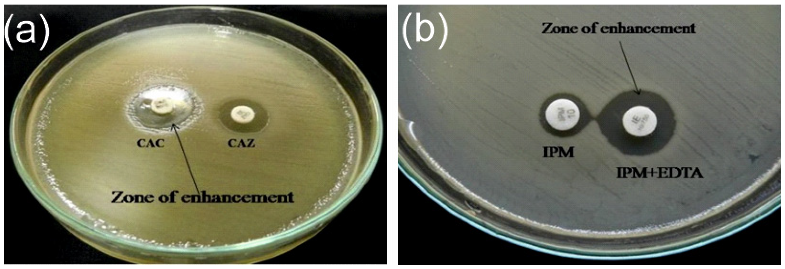
2.3. Extended Spectrum β-lactamase and Metallo-β-lactamase

ESBL and MBL production were detected using standard methods. The ESBL phenotype was identified using the combined disc diffusion method, with E. coli (ATCC 25922) as a negative control and A. baumannii (ATCC 19606) as a positive control. A result was considered positive if the zone of inhibition around ceftazidime/clavulanic acid was ≥5 mm compared to ceftazidime alone. MBL detection was conducted using the imipenem-EDTA combined disc test, with isolates showing ≥7 mm inhibition zone size on the imipenem-EDTA disc compared with the imipenem disc alone classified as MBL producers. P. aeruginosa (ATCC 27853) served as the positive control for MBL detection.

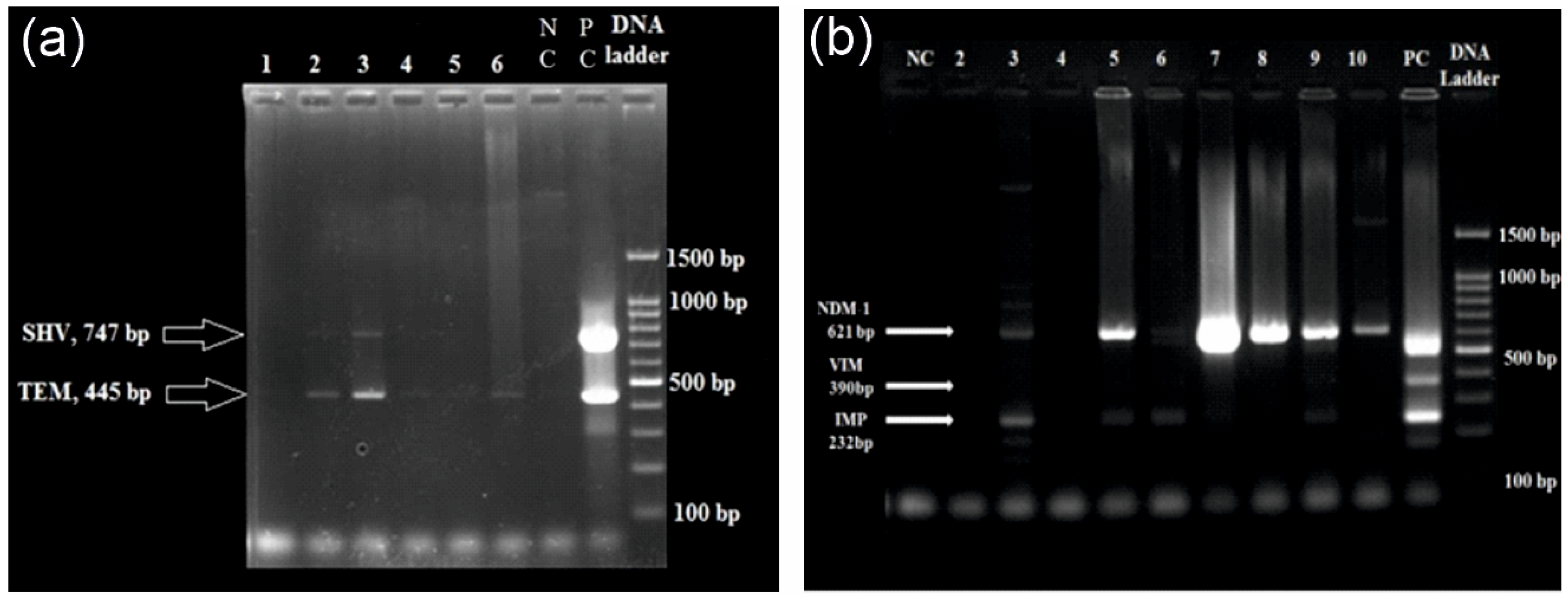
2.4. DNA Extraction and Multiplex qPCR

DNA was extracted from overnight cultures grown in Luria–Bertani (LB) broth using the Easy Tissue and Cell Genomic DNA Purification Kit following the manufacturer’s instructions. Extracted DNA was stored at −20 °C until PCR analysis. Multiplex PCR was performed to amplify ESBL genes (blaCTX-M, blaSHV, and blaTEM) and MBL genes (blaIMP, blaVIM, and blaNDM-1). The selection of isolates for multiplex PCR was based on high virulence factors and coexistence of both ESBL and MBL genes in a single isolate. Primer sequences are listed in Table S1.
PCR was conducted using Ampliqon Taq DNA polymerase 2X master mix RED in a 25 µL reaction volume, including primers at 10 pmol/µL and 2 µL of the DNA template. Amplification was performed in a Veriti™ Thermal Cycler (Applied Biosystems, Foster City, CA, USA) under the following conditions: initial denaturation at 95 °C for 15 min, followed by 30 cycles of 95 °C for 30 s, 60 °C for 30 s, and 72 °C for 2 min for ESBL genes, and 94 °C for 30 s, 52 °C for 40 s, and 72 °C for 50 s for MBL genes. The final extension step was performed at 72 °C for 5 min. The amplified products were separated on 2% agarose gel, stained with ethidium bromide, and visualized using a gel documentation system (Major Science, CA, USA).

2.5. Statistical Analysis

Statistical analysis was performed using SPSS software v. 29.0.2.0 (IBM Corp., Armonk, NY, USA). Data are expressed as the mean ± standard deviation (SD) and frequency (%). χ2 and Fisher’s exact tests were used to assess the associations between antibiotic resistance mechanisms (ESBL and MBL) and clinical outcomes. Statistical significance was set at p < 0.05.

3. Results

3.1. Patient Characteristics

This study included all patients diagnosed with DFIs who presented to the tertiary care hospital during the study period. A total of 480 participants were included in this study; 365 (76%) were male, and the mean (SD) age of all participants was 56.5 (11.4) years. There was no significant difference in age between the two groups (p = 0.116). Among all participants, 20.5% had Wagner Grade 2, 40.3% had Wagner grade 3, 30.7% had Wagner grade 4, and 7.5% had Wagner grade 5, with no significant differences observed between males and females. There were no significant differences in hypertension (p = 0.071), ischemic heart disease (p = 0.321), peripheral vascular disease (p = 0.361), retinopathy (p = 0.443), or nephropathy (p = 0.213) between males and females. However, smoking (p < 0.001) and alcohol consumption (p < 0.001) differed between groups, with a higher prevalence in males (Table 1).
Biochemical markers, such as haemoglobin (p = 0.032), random blood sugar (p < 0.001), fasting blood sugar (p = 0.002), and postprandial blood sugar (p < 0.001), showed significant sex differences. Additionally, urine ketone bodies differed between the groups (p = 0.034, Table 1).
Seventy (14.58%) A. baumannii isolates were obtained from the DFIs. Table S2 presents the source-wise distribution of microbial isolates from the DFIs. Of these, 54 isolates were obtained from pus and exudates, and 16 were obtained from tissue samples. Among the 70 isolates, three were from healed wounds, 25 from unhealed major wounds, and 42 from unhealed minor wounds according to the Meggit’s Wagner and PEDIS grading systems. The PEDIS grading showed a similar distribution with three healed, 25 unhealed major, and 42 unhealed minor isolates.

3.2. Antimicrobial Susceptibility and ESBL/MBL Detection

The antimicrobial susceptibility of A. baumannii isolated from DFIs (n = 70) is shown in Table 2. Most isolates exhibited resistance to β-lactams, with 100% resistance to cephalosporins, including cefepime, ceftazidime, ceftriaxone, and cefotaxime. There was high resistance to carbapenems, such as imipenem and meropenem (resistance rates 91.4% and 88.6%, respectively). There was also high resistance to fluoroquinolones, such as ciprofloxacin and levofloxacin (resistance rates 94.3% and 90.0%, respectively) and to tetracycline and trimethoprim/sulfamethoxazole (94.3%) (Table 2).
Among the 70 isolates from DFIs, 19 (27.14%) were ESBL producers and 43 (61.43%) were MBL producers (Figure 1).

3.3. Multiplex qPCR

Multiplex PCR assays targeted both ESBL and MBL genes. An agarose gel showing PCR amplification of ESBL and MBL genes is presented in Figure 2.
Of the 70 A. baumannii isolates, 17 exhibited both multiple resistance genes (ESBL and MBL) and high virulence potential. Among the 17 ESBL producers, blaTEM (52.94%) was the most commonly detected gene. Combination patterns, such as blaCTX-M + blaSHV + blaTEM (5.88%) and blaSHV + blaTEM (5.88%), were also observed (Table 3). blaNDM-1 was the most prevalent gene in MBL producers (52.94%), followed by blaIMP + blaNDM-1 (17.64%). The total resistance of MBL producers was notably higher (82.35%) than that of non-MBL producers (88.24%) (Table 3).

4. Discussion

In this study, we isolated and characterized 70 A. baumannii isolates from DFIs of patients treated at a tertiary care hospital in India. Our findings revealed a 14.58% prevalence of A. baumannii in DFIs. Most isolates were resistant to β-lactams, with 100% resistance to cephalosporins. More than half of the A. baumannii isolates were MBL producers. These results emphasize the growing threat posed by multidrug-resistant bacteria found in DFIs.
In our study, the incidence of DFIs was higher in males, with a male-to-female ratio of 3.17:1. This is consistent with previous findings from India, such as Jain et al. [28] (male-to-female ratio: 2.1:1) and Sekhar et al. [29] (male-to-female ratio: 2.5:1), and studies from other countries, including Kuwait, where Al Benwan et al. [13] reported a male-to-female ratio of 2.8:1. The male predominance in DFUs may be linked to factors such as increased risk behaviours, including poor glycaemic control, smoking, and alcohol consumption [30,31].
Our study reported a higher prevalence (14.6%) of A. baumannii in DFIs, consistent with previous findings that A. baumannii is a key pathogen in DFIs [32]. Most of our isolates exhibited resistance to β-lactams, including cefepime, ceftazidime, ceftriaxone, and cefotaxime. There was also high resistance to carbapenems, such as imipenem and meropenem (resistance rates 91.4% and 88.6%, respectively). Similar to our findings, previous reports indicated that carbapenem resistance in A. baumannii strains is approximately 90% [33]. An increased prevalence of carbapenem-resistant A. baumannii isolates has been reported in numerous countries across Northern and Eastern Europe and in the Levant countries of the Arab League [34,35]. Carbapenem resistance is often linked to widespread co-resistance across various antibiotic classes [36].
Similar to carbapenems, higher resistance was observed for fluoroquinolones, with ciprofloxacin and levofloxacin resistance rates of 94.3% and 90.0%, respectively. Recent reports have indicated that the resistance of A. baumannii to fluoroquinolones ranges from 50 to 73% in various regions; resistance rates have significantly increased in recent years in developing countries, reaching 75–97.7% [23,37,38]. Resistance to tetracycline and trimethoprim/sulfamethoxazole was also notably high in our study (94.3%), consistent with recent transcriptomic studies that indicated increasing resistance of A. baumannii to tigecycline [39]. In the current study, 27.14% of A. baumannii isolates were ESBL producers. We observed an increased prevalence of ESBL-producing A. baumannii when compared with other studies reported by Safari et al. [40] (7%) and Ranjbar et al. [41] (16%) but a lower prevalence when compared with studies by Farajnia et al. [42] (70%) and Azizi et al. [43] (53.8%). Prolonged hospitalization, ICU admission, invasive procedures, and broad-spectrum antibiotic use have contributed to the spread of A. baumannii-resistant organisms [44]. In the present study, 52.94%, 5.88%, 5.88%, and 5.88% of the isolates carried blaTEM, blaCTX-M, blaSHV + blaTEM, and blaCTX-M + SHV + TEM, respectively. Consistent with our findings, other studies have reported that 20% and 52.1% of A. baumannii isolates carry blaTEM-1 and blaTEM, respectively [41,44]. These findings highlight the critical role of ESBL and MBL enzymes as key resistance mechanisms that drive the spread of multidrug-resistant A. baumannii.
Although our study provides valuable insights into the prevalence, antimicrobial resistance, and molecular characteristics of A. baumannii in patients with DFI, several limitations must be acknowledged. First, this was conducted at a single tertiary care hospital, which may limit the generalizability of the findings to other healthcare settings, geographic regions, and ethnic groups. Second, the absence of genome sequencing of these isolates may have restricted our understanding of the complex genetic mechanisms underlying multidrug resistance profiling in clinical A. baumannii isolates. Therefore, further studies with larger cohorts and multicentre approaches are needed to track the emergence and spread of resistant clinical A. baumannii strains. Prevention of DFIs is crucial in patients requiring palliative care, where the focus is on improving quality of life and further avoiding complications. Our study did not assess A. baumannii colonization at other body sites, which is a limitation. However, further studies are needed to assess the prevalence and impact of such colonization in this patient population. Some clinical isolates, including non-ESBL and non-MBL producers, exhibited ESBL and MBL genes, which may appear inconsistent with their phenotypic resistance profiles. Potential explanations include the coexistence of resistance genes, gene expression regulation, and genetic heterogeneity. These discrepancies highlight the need for more sensitive testing methods to better align genetic and phenotypic resistance profiles.

5. Conclusions

Our study highlights the significant role of A. baumannii in DFIs, driven by its high prevalence and high multidrug resistance, particularly to carbapenems, cephalosporins, and fluoroquinolones. More than half of the isolates were MBL producers, which emphasizes resistance mechanisms such as ESBLs and MBLs. Continuous surveillance, tailored treatment strategies, antimicrobial stewardship, and understanding of genetic factors, such as carbapenemase production, are crucial to improve patient outcomes.

Supplementary Materials

The following supporting information can be downloaded at: https://www.mdpi.com/article/10.3390/bacteria4020024/s1. Table S1: List of primers used in this study. Table S2: Distribution of microbial isolates from DFIs by source.

Author Contributions

Conceptualization, D.M.K., V.I.R., M.S.M., D.M. and M.M.; methodology, D.M.K., V.I.R. and M.S.M.; formal analysis, D.M.K., V.I.R., M.S.M., D.M. and M.M.; investigation, D.M.K. and M.M.; resources, V.I.R. and M.S.M., data curation, D.M.K., V.I.R. and M.S.M., writing—original draft preparation, D.M.K. and M.M.; writing—review and editing, D.M.K., V.I.R., M.S.M., D.M. and M.M.; visualization, D.M.K. and M.M.; supervision, V.I.R., M.S.M. and D.M. All authors have read and agreed to the published version of the manuscript.

Funding

This research received no external funding.

Institutional Review Board Statement

This study adhered to the principles of the Declaration of Helsinki and ethical guidelines for research involving human participants. Ethical approval was obtained from the Institutional Ethics Committee of the Yenepoya Deemed to be University (Reg. No-YU2016/172).

Informed Consent Statement

Written informed consent has been obtained from the patient(s) to publish this paper.

Data Availability Statement

The data that support the findings of this study are available from the corresponding author upon reasonable request.

Conflicts of Interest

The authors declare no conflicts of interest.

References

  1. Cigna, E.; Fino, P.; Onesti, M.G.; Amorosi, V.; Scuderi, N. Diabetic foot infection treatment and care. Int. Wound J. 2016, 13, 238–242. [Google Scholar] [CrossRef] [PubMed]
  2. Lauri, C.; Leone, A.; Cavallini, M.; Signore, A.; Giurato, L.; Uccioli, L. Diabetic foot infections: The diagnostic challenges. J. Clin. Med. 2020, 9, 1779. [Google Scholar] [CrossRef]
  3. Noor, S.; Khan, R.U.; Ahmad, J. Understanding diabetic foot infection and its management. Diabetes Metab. Syndr. 2017, 11, 149–156. [Google Scholar] [CrossRef] [PubMed]
  4. Burgess, J.L.; Wyant, W.A.; Abdo Abujamra, B.; Kirsner, R.S.; Jozic, I. Diabetic wound-healing science. Medicina 2021, 57, 1072. [Google Scholar] [CrossRef]
  5. McDermott, K.; Fang, M.; Boulton, A.J.M.; Selvin, E.; Hicks, C.W. Etiology, epidemiology, and disparities in the burden of diabetic foot ulcers. Diabetes Care 2023, 46, 209–221. [Google Scholar] [CrossRef] [PubMed]
  6. Edmonds, M.; Manu, C.; Vas, P. The current burden of diabetic foot disease. J. Clin. Orthop. Trauma. 2021, 17, 88–93. [Google Scholar] [CrossRef]
  7. Hadi, P.; Rampal, S.; Neela, V.K.; Cheema, M.S.; Sarawan Singh, S.S.; Kee Tan, E.; Sinniah, A. Distribution of causative microorganisms in diabetic foot infections: A ten-year retrospective study in a tertiary care hospital in central Malaysia. Antibiotics 2023, 12, 687. [Google Scholar] [CrossRef] [PubMed]
  8. Turhan, V.; Mutluoglu, M.; Acar, A.; Hatipoglu, M.; Onem, Y.; Uzun, G.; Ay, H.; Oncul, O.; Gorenek, L. Increasing incidence of Gram-negative organisms in bacterial agents isolated from diabetic foot ulcers. J. Infect. Dev. Ctries. 2013, 7, 707–712. [Google Scholar] [CrossRef]
  9. El-Hazmi, M.M. Bacteriological profile of diabetic foot infections in a teaching hospital in Saudi Arabia. J. Pure Appl. Microbiol. 2015, 9, 1933–1943. [Google Scholar]
  10. Jouhar, L.; Jaafar, R.F.; Nasreddine, R.; Itani, O.; Haddad, F.; Rizk, N.; Hoballah, J.J. Microbiological profile and antimicrobial resistance among diabetic foot infections in Lebanon. Int. Wound J. 2020, 17, 1764–1773. [Google Scholar] [CrossRef]
  11. Alhubail, A.; Sewify, M.; Messenger, G.; Masoetsa, R.; Hussain, I.; Nair, S.; Tiss, A. Microbiological profile of diabetic foot ulcers in Kuwait. PLoS ONE 2020, 15, e0244306. [Google Scholar] [CrossRef] [PubMed]
  12. Qu, Y.D.; Ou, S.J.; Zhang, W.; Li, J.X.; Xia, C.L.; Yang, Y.; Liu, J.B.; Ma, Y.F.; Jiang, N.; Wang, Y.Y.; et al. Microbiological profile of diabetic foot infections in China and worldwide: A 20-year systematic review. Front. Endocrinol. 2024, 15, 1368046. [Google Scholar] [CrossRef] [PubMed]
  13. Al Benwan, K.; Al Mulla, A.; Rotimi, V.O. A study of the microbiology of diabetic foot infections in a teaching hospital in Kuwait. J. Infect. Public Health 2012, 5, 1–8. [Google Scholar] [CrossRef]
  14. Son, S.T.; Han, S.K.; Lee, T.Y.; Namgoong, S.; Dhong, E.S. The microbiology of diabetic foot infections in Korea. J. Wound Manag. Res. 2017, 13, 8–12. [Google Scholar] [CrossRef]
  15. Sannathimmappa, M.B.; Nambiar, V.; Aravindakshan, R.; Al Khabori, M.S.; Al-Flaiti, A.H.; Al-Azri, K.N.; Al-Reesi, A.K.; Al Kiyumi, A.R. Diabetic foot infections: Profile and antibiotic susceptibility patterns of bacterial isolates in a tertiary care hospital of Oman. J. Educ. Health Promot. 2021, 10, 254. [Google Scholar] [CrossRef] [PubMed]
  16. Hitam, S.A.; Asma’Hassan, S.; Maning, N.U. The significant association between polymicrobial diabetic foot infection and its severity and outcomes. Malays. J. Med. Sci. 2019, 26, 107. [Google Scholar] [CrossRef]
  17. Sun, H.; Ma, Y.; Heng, H.; Liu, X.; Liang, J.; Geng, H. Microbiological Distribution, Antimicrobial Susceptibility and Risk Factors of Polymicrobial Infections in Diabetic Foot. Clin. Lab. 2024, 70. [Google Scholar] [CrossRef]
  18. Howard, A.; O’Donoghue, M.; Feeney, A.; Sleator, R.D. Acinetobacter baumannii: An emerging opportunistic pathogen. Virulence 2012, 3, 243–250. [Google Scholar] [CrossRef]
  19. Lahiri, K.K.; Mani, N.S.; Purai, S.S. Acinetobacter spp as nosocomial pathogen: Clinical significance and antimicrobial sensitivity. Med. J. Armed Forces India 2004, 60, 7–10. [Google Scholar] [CrossRef]
  20. Bali, N.K.; Fomda, B.A.; Bashir, H.; Zahoor, D.; Lone, S.; Koul, R.A. Emergence of carbapenem-resistant Acinetobacter in a temperate north Indian State. Br. J. Biomed. Sci. 2013, 70, 156–160. [Google Scholar] [CrossRef]
  21. Khan, D.M.; Manzoor, M.A.; Rao, I.V.; Moosabba, M.S. Evaluation of biofilm formation, cell surface hydrophobicity and gelatinase activity in Acinetobacter baumannii strains isolated from patients of diabetic and non-diabetic foot ulcer infections. Biocatal. Agric. Biotechnol. 2019, 18, 101007. [Google Scholar] [CrossRef]
  22. Zarrilli, R.; Pournaras, S.; Giannouli, M.; Tsakris, A. Global evolution of multidrug-resistant Acinetobacter baumannii clonal lineages. Int. J. Antimicrob. Agents 2013, 41, 11–19. [Google Scholar] [CrossRef] [PubMed]
  23. Kyriakidis, I.; Vasileiou, E.; Pana, Z.D.; Tragiannidis, A. Acinetobacter baumannii antibiotic resistance mechanisms. Pathogens 2021, 10, 373. [Google Scholar] [CrossRef] [PubMed]
  24. Gupta, V. An update on newer β-lactamases. Indian J. Med. Res. 2007, 126, 417–427. [Google Scholar]
  25. Goel, V.; Hogade, S.A.; Karadesai, S.G. Prevalence of extended-spectrum beta-lactamases, AmpC beta-lactamase, and metallo-beta-lactamase producing Pseudomonas aeruginosa and Acinetobacter baumannii in an intensive care unit in a tertiary care hospital. J. Sci. Soc. 2013, 40, 28–31. [Google Scholar] [CrossRef]
  26. Humphries, R.M.; Ambler, J.; Mitchell, S.L.; Castanheira, M.; Dingle, T.; Hindler, J.A.; Koeth, L.; Sei, K. CLSI methods development and standardization working group best practices for evaluation of antimicrobial susceptibility tests. J. Clin. Microbiol. 2018, 56, 10–128. [Google Scholar] [CrossRef]
  27. Andrews, J.M. Determination of minimum inhibitory concentrations. J. Antimicrob. Chemother. 2001, 48 (Suppl. S1), 5–16. [Google Scholar] [CrossRef]
  28. Manisha, J.; Mitesh, P.H.; Nidhi, S.K.; Modi, D.J.; Vegad, M.M. Spectrum of Microbial Flora in Diabetic Foot Ulcer and Antibiotic Sensitivity Pattern in Tertiary Care Hospital in Ahmedabad, Gujarat. Nat. J. Med. Res. 2012, 3, 354–357. [Google Scholar]
  29. Sekhar, S.; Unnikrishnan, M.K.; Rodrigues, G.S.; Vyas, N.; Mukhopadhyay, C. Antimicrobial susceptibility pattern of aerobes in diabetic foot ulcers in a South-Indian tertiary care hospital. Foot 2018, 37, 95–100. [Google Scholar] [CrossRef] [PubMed]
  30. Iacopi, E.; Pieruzzi, L.; Riitano, N.; Abbruzzese, L.; Goretti, C.; Piaggesi, A. The weakness of the strong sex: Differences between men and women affected by diabetic foot disease. Int. J. Low. Extrem. Wounds 2023, 22, 19–26. [Google Scholar] [CrossRef]
  31. Sahu, S.S.; Chaudhary, V.; Sharma, N.; Kumari, S.; Pal, B.; Khurana, N. Prevalence and risk factors associated with diabetic foot ulcer in India: A systematic review and meta-analysis. Int. J. Infect. Dev. Ctries. 2024. [Google Scholar] [CrossRef]
  32. Castellanos, N.; Nakanouchi, J.; Yüzen, D.I.; Fung, S.; Fernandez, J.S.; Barberis, C.; Tuchscherr, L.; Ramirez, M.S. A study on Acinetobacter baumannii and Staphylococcus aureus strains recovered from the same infection site of a diabetic patient. Curr. Microbiol. 2019, 76, 842–847. [Google Scholar] [CrossRef] [PubMed]
  33. Isler, B.; Keske, Ş.; Aksoy, M.; Azap, Ö.K.; Yilmaz, M.; Yavuz, S.Ş.; Aygün, G.; Tigen, E.; Akalın, H.; Azap, A.; et al. Antibiotic overconsumption and resistance in Turkey. Clin. Microbiol. Infect. 2019, 25, 651–653. [Google Scholar] [CrossRef] [PubMed]
  34. Moghnieh, R.A.; Kanafani, Z.A.; Tabaja, H.Z.; Sharara, S.L.; Awad, L.S.; Kanj, S.S. Epidemiology of common resistant bacterial pathogens in the countries of the Arab League. Lancet Infect. Dis. 2018, 18, e379–e394. [Google Scholar] [CrossRef]
  35. European Centre for Disease Prevention and Control. Antimicrobial Resistance in the EU/EEA (EARS-Net)—Annual Epidemiological Report 2020. Stockholm: ECDC. 2022. Available online: https://www.ecdc.europa.eu/en/publications-data/surveillance-antimicrobial-resistance-europe-2022 (accessed on 1 March 2025).
  36. Tacconelli, E.; Carrara, E.; Savoldi, A.; Harbarth, S.; Mendelson, M.; Monnet, D.L.; Pulcini, C.; Kahlmeter, G.; Kluytmans, J.; Carmeli, Y.; et al. Discovery, research, and development of new antibiotics: The WHO priority list of antibiotic-resistant bacteria and tuberculosis. Lancet Infect. Dis. 2018, 18, 318–327. [Google Scholar] [CrossRef]
  37. Zaki, M.E.; Abou ElKheir, N.; Mofreh, M. Molecular study of quinolone resistance determining regions of gyrA gene and parC genes in clinical isolates of Acintobacter baumannii resistant to fluoroquinolone. Open Microbiol. J. 2018, 12, 116. [Google Scholar] [CrossRef]
  38. Vázquez-López, R.; Solano-Gálvez, S.G.; Juárez Vignon-Whaley, J.J.; Abello Vaamonde, J.A.; Padró Alonzo, L.A.; Rivera Reséndiz, A.; Muleiro Álvarez, M.; Vega López, E.N.; Franyuti-Kelly, G.; Álvarez-Hernández, D.A.; et al. Acinetobacter baumannii resistance: A real challenge for clinicians. Antibiotics 2020, 9, 205. [Google Scholar] [CrossRef] [PubMed]
  39. Li, L.; Hassan, K.A.; Tetu, S.G.; Naidu, V.; Pokhrel, A.; Cain, A.K.; Paulsen, I.T. The transcriptomic signature of tigecycline in Acinetobacter baumannii. Front. Microbiol. 2020, 11, 565438. [Google Scholar] [CrossRef]
  40. Safari, M.; Nejad, A.S.; Bahador, A.; Jafari, R.; Alikhani, M.Y. Prevalence of ESBL and MBL encoding genes in Acinetobacter baumannii strains isolated from patients of intensive care units (ICU). Saudi J. Biol. Sci. 2015, 22, 424–429. [Google Scholar] [CrossRef]
  41. Ranjbar, R.; Tolon, S.S.; Zayeri, S.; Sami, M. The frequency of antibiotic resistance and ESBLs among clinically Acinetobacter baumannii strains isolated from patients in a major hospital in Tehran, Iran. Open Microbiol. J. 2018, 12, 254. [Google Scholar] [CrossRef]
  42. Farajnia, S.; Azhari, F.; Alikhani, M.Y.; Hosseini, M.K.; Peymani, A.; Sohrabi, N. Prevalence of PER and VEB type extended spectrum betalactamases among multidrug resistant Acinetobacter baumannii isolates in North-West of Iran. Iran. J. Basic Med. Sci. 2013, 16, 751. [Google Scholar] [PubMed]
  43. Azizi, M.; Mortazavi, S.H.; Etemadimajed, M.; Gheini, S.; Vaziri, S.; Alvandi, A.; Kashef, M.; Ahmadi, K. Prevalence of extended-spectrum β-Lactamases and antibiotic resistance patterns in Acinetobacter baumannii isolated from clinical samples in Kermanshah, Iran. Jundishapur J. Microbiol. 2017, 10, e61522. [Google Scholar] [CrossRef]
  44. Zarabadi-Pour, M.; Peymani, A.; Habibollah-Pourzereshki, N.; Sarookhani, M.R.; Karami, A.A.; Javadi, A. Detection of extended-spectrum ß-lactamases among Acinetobacter baumannii isolated from hospitals of Qazvin, Iran. Ethiop. J. Health Sci. 2021, 31, 229–236. [Google Scholar] [CrossRef] [PubMed]
Figure 1. (a) ESBL detection using the double-disc synergy test of A. baumannii. A zone of enhancement >5 mm with a β-lactamase inhibitor indicates ESBL production. CAZ—ceftazidime; CAC—ceftazidime + clavulanic acid combination. (b) MBL detection using the IPM + EDTA disc combination test. MBL producers are resistant to imipenem (IPM) but sensitive to the IPM-EDTA combination. A zone of enhancement >7 mm indicates MBL production.
Bacteria 04 00024 g001
Figure 2. (a) Agarose gel showing PCR amplification of the ESBL gene in A. baumannii. Lanes 1–6: isolates; Lane 7: negative control (NC); Lane 8: positive control (PC); Lane 9: DNA ladder [100 bp DNA ladder]. (b) Agarose gel showing PCR amplification of the MBL gene in A. baumannii. Lane 1: negative control (NC); Lanes 2–10: samples; Lane 11: positive control (PC); Lane 12: DNA ladder [100 bp DNA ladder].
Bacteria 04 00024 g002
Table 1. Characteristics of all participants included in this study.
All MalesFemalesDifference Between Males and Females
480 (100.00%)365 (76%)115 (24%)p a
Age, y56.5 (11.4)56.9 (11.1)55.1 (12.3)0.116
Wagner Grade, n (%)Grade 298 (20.5)70 (19.2)28 (24.4)0.237
Grade 3199 (40.3)154 (42.2)45 (39.1)
Grade 4147 (30.7)117 (35.5)30 (26.1)
Grade 536 (7.5)24 (6.6)12 (10.4)
Diabetes mellitus, n (%)480 (100.00)365 (76)115 (24)-
Hypertension, n (%)249 (51.9)182 (49.9)67 (58.3)0.071
Ischemic heart disease, n (%)80 (16.7)63 (17.3)17 (14.8)0.321
Peripheral vascular disease, n (%)300 (62.5)226 (61.9)74 (64.3)0.361
Retinopathy, n (%)275 (57.7)211 (55.8)64 (55.7)0.443
Nephropathy, n (%)85 (17.7)68 (18.6)17 (14.8)0.213
Smoking, n (%)263 (54.8)260 (71.2)3 (2.6)<0.001
Alcohol use, n (%)218 (45.4)215 (58.9)3 (2.6)<0.001
Haemoglobin, (g/dL)10.9 (4.8)11.2 (5.4)10.1 (1.8)0.032
Random blood sugar, (mg/dL)251.8 (104.5)243.6 (99.6)277.6 (115.3)<0.001
Fasting blood sugar, (mg/dL)141.2 (65.2)135.9 (63.1)115 (68.9)0.002
Postprandial blood sugar, (mg/dL)199.8 (94.2)189.6 (86.8)232.1 (108.7)<0.001
Urine ketone bodies, (mg/dL)70 (14.6)46 (12.6)24 (20.9)0.034
a χ2 (categorical variables) or independent samples t-test (continuous variables). Values are mean (SD) unless otherwise indicated.
Table 2. Antimicrobial susceptibility tests of A. baumannii isolated from DFIs.
AntibioticsSensitiveModerate or Intermediate SensitiveResistant
β-lactams
Aminoglycosides
Amikacin (30 µg)2 (2.9%)0 68 (97%)
Gentamicin (10 µg)3 (4.3%)0 67 (95.7%)
Tobramycin (10 µg)5 (7.1%)0 65 (92.9%)
Carbapenems
Imipenem (10 µg)6 (8.6%)0 64 (91.4%)
Meropenem (10 µg)8 (11.4%)0 62 (88.6%)
Cephalosporins
Cefepime (30 µg)00 70 (100%)
Ceftazidime (30 µg)0 0 70 (100%)
Cefotaxime (30 µg)2 (2.9%)0 68 (97%)
Ceftriaxone (30 µg)0 0 70 (100%)
Fluroquinolones
Ciprofloxacin (5 µg)4 (5.7%)066 (94.3)
Levofloxacin (5 µg)4 (5.7%)3 (4.3%)63 (90%)
Penicillins
Piperacillin (100 µg)1 (1.4%)0 69 (98.6%)
Tetracycline (30 µg)3 (4.3%)1(1.4%)66 (94.3%)
Folate-pathway inhibitors
Trimethoprim/sulfamethoxazole (25 µg) 4 (5.7%)066 (94.3%)
Β-lactam inhibitor combinations
Piperacillin/tazobactam (30 µg)4 (5.7%)3 (4.3%)63 (90%)
Values in parentheses represent the percentage of isolates.
Table 3. Prevalence of ESBL and non-ESBL genes in A. baumannii from DFI patients.
GeneESBL Producer (n = 17)Non-ESBL Producer (n = 17)MBL Producer (n = 17)
Producer (n = 17)
Non-MBL Producer (n = 17)
blaCTX-M1 (5.88%)1 (5.88%)--
blaSHV00--
blaTEM9 (52.94%)5 (29.41%)--
blaCTX-M + SHV + TEM1 (5.88%)1 (5.88%)--
blaSHV + blaTEM1 (5.88%)3 (17.64%)--
blaCTX-M + blaSHV01 (5.88%)--
blaIMP--00
blaVIM--00
blaNDM-1--9 (52.94%)10 (58.82%)
blaIMP+ VIM + NDM-1--1 (5.88%)0
blaVIM + blaNDM-1--01 (5.88%)
blaIMP + blaVIM--1 (5.88%)2 (11.76%)
blaIMP + blaNDM-1--3 (17.64%)2 (11.76%)
Total12 (70.59%)11 (64.70%)14 (82.35%)15 (88.24%)
Disclaimer/Publisher’s Note: The statements, opinions and data contained in all publications are solely those of the individual author(s) and contributor(s) and not of MDPI and/or the editor(s). MDPI and/or the editor(s) disclaim responsibility for any injury to people or property resulting from any ideas, methods, instructions or products referred to in the content.

