Article

Efficient Cryopreservation of Human Midbrain Organoids Using Conventional DMSO Protocols with Ice Recrystallization Inhibitors


1
Department of Chemistry and Biomolecular Sciences, University of Ottawa, Ottawa, ON K1N 6N5, Canada
2
The Neuro’s Early Drug Discovery Unit (EDDU), McGill University, 3801 University, Montreal, QC H3A 2B4, Canada
*
Author to whom correspondence should be addressed.
Organoids 2026, 5(3), 32; https://doi.org/10.3390/organoids5030032 (registering DOI)
Submission received: 27 July 2026 / Revised: 3 September 2026 / Accepted: 16 September 2026 / Published: 19 September 2026

Abstract

Human midbrain organoids (hMOs) are a powerful stem cell-derived model for studying human neurodevelopment and neurological disease in vitro; however, their broad adoption is limited by long culture times, high cost, and inter-batch variability. Cryopreservation offers a strategy to address these limitations by enabling long-term storage and reducing experimental heterogeneity, but its effectiveness depends on robust cryoprotective strategies. Here, we evaluated hMO cryopreservation using a standard 10% dimethyl sulfoxide (DMSO) protocol, alone and in combination with a panel of five ice recrystallization inhibitors (IRIs), including betaine-derived ammonium salts and N-aryl-d-gluconamides. Across all conditions, hMOs were successfully recovered following freeze–thawing, with preservation of overall structural integrity, including maintained morphology and reduced cellular debris. Importantly, post-thaw viability and cell recovery in the 10% DMSO condition were comparable to those of fresh controls, demonstrating that conventional DMSO-based cryopreservation is sufficient to maintain hMO survival and gross structural preservation. While supplementation with select IRIs, particularly N-ethyl betaine ethyl ester iodide (NEBEE-I), yielded modest improvements in viability relative to DMSO alone and a non-significant trend toward enhanced electrophysiological activity in multielectrode array recordings, these effects were secondary to the robust performance of the baseline DMSO protocol. Collectively, these findings establish standard 10% DMSO cryopreservation as an effective and broadly applicable method for hMO preservation, while suggesting that small-molecule IRIs may provide incremental benefits in selected outcome measures.

1. Introduction

Human brain organoids have emerged as powerful stem cell-derived models for studying human neurodevelopment and neurological disease in a physiologically relevant in vitro context [1,2,3]. Generated from human pluripotent stem cells (iPSCs), these three-dimensional (3D) cultures self-organize into multicellular structures that recapitulate key features of human brain tissue, including regional patterning, cytoarchitecture, and cellular heterogeneity [2,3,4]. In contrast to conventional two-dimensional systems, brain organoids provide more physiologically complex human neural models for mechanistic studies and preclinical testing [3,5,6].
Among neural organoid systems, human midbrain organoids (hMOs) are a particularly valuable model due to their structural complexity and translational relevance [7,8,9,10]. hMOs recapitulate key features of the ventral midbrain, including dopaminergic neurons and regionally relevant neural cell populations, making them useful for studying midbrain-associated dysfunction and for broader applications in human neural modelling [7,8]. However, their use remains limited by the prolonged culture and maturation times required to obtain developmentally advanced organoids, which increase experimental and financial burden and can make it difficult to standardize culture duration and experimental timing between preparations [11,12,13,14].
Cryopreservation of organoids could help address these challenges by enabling organoids from a single batch to be banked and experiments to be performed at standardized time points, reducing variability associated with differences in culture and maturation timing. These advantages are particularly important for neural organoid systems, where prolonged differentiation timelines and culture heterogeneity hinder reproducibility and throughput [12,15,16]. Cryopreservation has been successfully applied to several non-neuronal organoid systems, including intestinal [17,18,19], hepatic [20,21], renal [15,22], and tumour-derived organoids [23,24], demonstrating that complex 3D tissues can retain viability after optimized cryopreservation protocols.
Despite these advances, cryopreservation of neuronal organoids remains relatively underdeveloped compared to conventional cell-based systems, with published studies focused primarily on cortical and other forebrain-derived neural organoids [25,26]. To our knowledge, systematic cryopreservation of intact human midbrain organoids (hMOs) has not yet been reported. Given differences in developmental patterning and cellular composition between cortical and midbrain systems [2,9], the transferability of existing cryopreservation protocols to hMOs remains uncertain. Neural organoids also present cryobiological challenges associated with their 3D architecture and heterogeneous cellular organization, which can complicate uniform cryoprotectant exposure and heat transfer during freezing and thawing [7,15,25]. Thus, region-specific cryopreservation strategies for complex neuronal organoids remain to be established.
A major source of cryoinjury during cryopreservation is ice recrystallization, where small ice crystals grow into larger, damaging ice crystals during freezing and thawing, inducing mechanical and osmotic stresses that reduce post-thaw viability, recovery, and functionality [27,28,29]. Conventional cryoprotectants, such as dimethyl sulfoxide (DMSO), mitigate cryoinjury by modulating cellular water and solute fluxes [30], thereby reducing osmotic stress, but do not directly inhibit ice recrystallization at concentrations typically used for cryopreservation [27,28,29]. This limitation is particularly relevant for complex 3D systems, such as organoids, where heterogeneous ice formation and ice propagation can compromise post-thaw viability and function [31].
Ice recrystallization inhibitors (IRIs) provide a complementary strategy by suppressing ice crystal growth during freezing and thawing [27,28,29]. Small-molecule carbohydrate- and ammonium-based IRIs are attractive due to their synthetic accessibility, scalability, and cytocompatibility [28]. Supplementation of cryopreservation formulations with small-molecule IRIs has improved outcomes in several sensitive human cell systems, including induced pluripotent stem cells (iPSCs) and neurons derived from iPSCs (iPSC-Ns) [32], as well as hematopoietic stem progenitor cells (HSPCs) [33]. These studies support the potential of IRIs as adjunct cryoprotectants for complex biological systems.
Herein, we investigate whether human midbrain organoids can be cryopreserved while retaining post-thaw viability, recovery, gross morphology, and functional readouts, and whether supplementation with small-molecule IRIs improves these outcomes. To our knowledge, cryopreservation of human midbrain organoids has not yet been reported. Accordingly, this study establishes a baseline cryopreservation framework for hMOs and evaluates the contribution of IRIs in enhancing preservation outcomes in complex 3D neural tissue systems.

2. Materials and Methods

2.1. Synthesis of Compounds

All small-molecule IRIs were synthesized as previously described. Experimental procedures and characterization data are provided in the Supplementary Materials.

2.2. Assessment of Ice Recrystallization Inhibition Activity

Ice recrystallization inhibition activity of select compounds was measured using a splat cooling assay, as previously described [34]. The compound was dissolved in PBS and serially diluted to desired concentrations. A total of 10 μL of sample was dropped from 2 m onto a pre-cooled aluminum block (at −78 °C via dry ice), then transferred to a cryostage held at −6.4 °C for an annealing time of 5 min. Ice wafers were then imaged using a digital camera (Nikon OEM, Nikon Corporation, Tokyo, Japan) fitted to a microscope (430× magnification). Using these images, the cross-sectional areas of each ice crystal was determined using ImageJ software (version 1.54g, National Institutes of Health). From each image, ice crystal areas were binned in 0.001 mm2 increments, and proportional areas were calculated using Microsoft Excel (version 2608, Microsoft Corporation). Ice crystal growth rates (vnorm) were determined as the proportional area > 0.001 mm2 divided by 5 min. Three wafers per concentration (n = 3) were averaged and normalized to the PBS control. Using GraphPad Prism (version 11, GraphPad Software Inc.)., 4-parameter dose–response curves were generated according to Equation (1), allowing for the extrapolation of a half-maximal inhibitory value (IC50) and facilitating comparison between compounds.
v n o r m = 100 1 + 10 ( H i l l S l o p e ) ( l o g I C 50 log c o m p o u n d ) ,

2.3. HepG2 Cell Culture

HepG2 cells (human hepatocellular carcinoma cells, ATCC, HB-8065, Manassas, VA, USA) were plated at a density of 3 × 10⁶ cells in a Corning® U-shaped T75 flask (CorningTM 430641U, Corning Inc., Tewksbury, MA, USA) and cultured in Eagle’s Minimum Essential Medium (MEM, M4655, Sigma-Aldrich Corporation, Oakville, ON, Canada) supplemented with 10% Fetal Bovine Serum (FBS, heat-inactivated, USA- or Canada-origin, GibcoTM, Thermo Fisher Scientific, Waltham, MA, USA), 1% penicillin–streptomycin (10,000 U/mL, 15140122, GibcoTM, Thermo Fisher Scientific Inc., Waltham, MA, USA), 1% MEM non-essential amino acids (100× solution, 11140050, GibcoTM, Thermo Fisher Scientific Inc., Waltham, MA, USA) and 0.1% 1M sodium pyruvate (P5280, Sigma-Aldrich Corporation, Oakville, ON, Canada). Cells were incubated at 37 °C and 5% CO2 and were washed every 2 days with Dulbecco’s PBS (DPBS, 14190-144, GibcoTM, Thermo Fisher Scientific Inc., Waltham, MA, USA) and the culture medium changed until 80–100% confluency was achieved. Cells were detached using AccutaseTM (STEMCELL Technologies Canada Inc., Vancouver, BC, Canada) and cells counts were obtained using a 1:4 dilution with Trypan Blue dye (K940, VWR International, LLC, Radnor, PA, USA) using a Bright-LineTM hemocytometer (Z359629, Cambridge Instruments Inc., Buffalo, NY, USA).

2.4. Assessment of Cytotoxicity Using a Resazurin Assay

The cultured HepG2 cells were plated in a cell culture-treated 96-well plate (CorningTM CostarTM 3596, Corning Inc., Tewksbury, MA, USA) at a density of 10,000 cells/well. The plate was incubated for 20–24 h at 37 °C in 5% CO2, the cell medium was aspirated, and either fresh medium (positive control), medium supplemented with an IRI compound and/or DMSO at a given concentration, or medium supplemented with 1% Triton X (negative control; T8787, Sigma-Aldrich Corporation, Oakville, ON, Canada) was added to the appropriate wells. Acellular blanks were also plated with fresh medium. The plate was incubated again for 4 h or 24 h at 37 °C in 5% CO2. A 10 uL aliquot of 0.125 mg/mL resazurin sodium salt (R7017, Sigma-Aldrich Corporation, Oakville, ON, Canada) in DPBS was added to each well and the plates were incubated for 4 h at 37 °C and 5% CO2. The relative fluorescence units (RFU, 530/590) were measured using a BioTek Synergy H1 microplate reader (BioTek Instruments, Inc., Winooski, VT, USA), and were converted to percentage viability using the following equation:
% c e l l   v i a b i l i t y =   R F U t r e a t m e n t   R F U n e g R F U p o s R F U n e g ×   100 % ,

2.5. iPSC Cell Line Information

The use of iPSCs and iPSC-derived cells in this research was approved by the McGill University Research Ethics Board (A03-M19-22A/eRAP 22-03-027). Two control cell lines generated by the Neuro’s Early Drug Discovery Unit (EDDU), in partnership with the Neuro’s Biobank (C-BIG), were used for this study: (i) AIW002-02 (CBIGi001-A, also known as IPSC0063, https://hpscreg.eu/cell-line/CBIGi001-A, accessed on 11 March 2025), reprogrammed from peripheral blood mononuclear cells of a 37-year-old Caucasian male who had provided informed consent [35]; (ii) QPN929 (CBIGi004-A, also known as IPSC0034, https://hpscreg.eu/cell-line/CBIGi004-A, accessed on 29 July 2025), reprogrammed from peripheral blood mononuclear cells of a 50-year-old Caucasian female who had provided informed consent.

2.6. Maintenance of iPSC and hMO Differentiation

For generation of hMOs, iPSCs were used only after a minimum of two passages following thawing and were not passaged more than ten times. Details on iPSC passaging are found in our QC workflow for iPSC maintenance [35]. hMOs were generated according to the method published in [12]. Briefly, iPSCs with <5% spontaneous differentiation were dissociated and seeded at 10 × 106 viable cells per EB-Disk948 (#eN-eb948u-002, enuvio, Montreal, QC, Canada) in neuronal induction medium supplemented with the ROCK inhibitor Y-27632 (#1254, Biotechne, Toronto, ON, Canada). Neural induction was performed for 2 days, followed by midbrain patterning for 4 days using SB431542 (S1067, Selleck Chemicals, TX, USA), Noggin (120-10C, Peprotech, CT, USA), CHIR99021 (S2924, Selleck Chemicals, TX, USA), and FGF8 (100-25, Peprotech, CT, USA). On day 7, aggregates were embedded in growth-factor-reduced Matrigel (356230, Corning, NY, USA), and maintained in tissue induction medium containing FGF8 (100-25, Peprotech, CT, USA), SHH (100-45, Peprotech, CT, USA), insulin (I9278, Millipore Sigma, MA, USA) and laminin (L20208, Millipore Sigma, MA, USA) for 24 h. On day 8, hMOs were transferred to spinner-flask bioreactors containing final differentiation medium and maintained at 37 °C with 5% CO2 and 42 rpm agitation. Final differentiation medium (FDM) was supplemented with BDNF (450-02, Peprotech, CT, USA), GDNF (450-10, Peprotech, CT, USA), ascorbic acid (A5960, Millipore Sigma, MA, USA), and dibutyryl-cAMP (D0627, Millipore Sigma, MA, USA) to support neuronal maturation. Bioreactor cultures were maintained with weekly medium changes until the desired age for cryopreservation.

2.7. Cryopreservation of Human Midbrain Organoids (hMBOs)

Midbrain organoids were suspended in 1 mL of the desired cryomedium containing final differentiation medium (FDM) with and without 10% (v/v) DMSO and/or IRI compounds at various concentrations. Seven to ten organoids were cryopreserved per cryovial. Cryovials were placed into a Corning® CoolCellTM LX Cell Freezing Container (Corning Inc., Tewksbury, MA, USA) and placed in a −80 °C freezer for 12–18 h. Once frozen, the cryovials were transferred to liquid nitrogen dewars (−196 °C) for storage for 3 to 4 days. Cryovials were thawed in a 37 °C water bath over a period of 2 min, after which the organoids were moved to fresh final differentiation medium (FDM) and recovered for 3 days prior to analysis.

2.8. Brightfield Microscopy of Post-Thaw hMOs

Following cryopreservation and thawing, hMOs were recovered in culture for 3 days prior to imaging. Individual organoids were transferred to ultra-low-attachment (ULA) 6-well plates (Corning Inc., Tewksbury, MA, USA) containing 4 mL of final differentiation medium (FDM). Midbrain organoids were cultured on an orbital shaker at 70 rpm inside a humidified incubator (37 °C, 5% CO2). Brightfield images were acquired using an EVOSTM XL Core microscope (Invitrogen, Thermo Fisher Scientific Inc., Waltham, MA, USA) equipped with a 4× objective. Images were collected under identical illumination and exposure settings for all experimental conditions.
Using ImageJ software, brightfield images were converted to 8-bit grayscale and subjected to uniform thresholding to delineate organoid boundaries. Individual organoids were semi-automatically segmented using the freehand selection tool, ensuring exclusion of debris and fused structures. For each organoid, the cross-sectional area (µm2) and circularity (4π × area/perimeter2; values ranging from 0 to 1, where 1 represents a perfect circle) were calculated using the built-in particle analysis function. Measurements were performed on organoids from a single independent cryopreservation experiment (N = 1), with n = 3 organoids analyzed per condition. All image processing and analysis parameters were kept constant across experimental groups.

2.9. Flow Cytometry Analysis of Dissociated hMOs

Following cryopreservation and thawing, hMOs were recovered in culture for 3 days prior to dissociation. hMOs were washed with 1X D-PBS (MultiCell Technologies Inc., Woonsocket, RI, USA) and treated with TrypLE Express (Thermo Fisher Scientific Inc., Waltham, MA, USA) three times (2 × 10 min, 1 × 5 min) at 37 °C to create a single-cell suspension. Between each incubation period, hMOs were triturated with 1 mL pipet tips. The TrypLE reaction was stopped by adding 1X D-PBS. The single-cell suspension was next filtered through a 30 μM mesh (Miltenyi Biotec, Bergisch Gladbach, Germany) and cells pelleted by centrifugation at 300× g for 5 min. Pelleted cells were suspended in 1X D-PBS and viability staining with Fixable Live/Dead Aqua (Invitrogen, Thermo Fisher Scientific Inc., Waltham, MA, USA) was performed for 30 min at RT (protected from light). Cells were then washed with 1X D-PBS and centrifuged at 300× g for 5 min. The flow cytometry voltage was set according to optimal PMT sensitivity using the peak 2 (Spherotech Inc., Lake Forest, IL, USA) voltration technique described previously by Maeker and Trotter [36]. Compensation control was performed with Ultracomp beads (Thermo Fisher Scientific Inc., Waltham, MA, USA). Cellular debris was identified based on a forward scatter (FSC) versus side scatter (SSC) plot and expressed as the percentage of total recorded events falling within the debris gate. Singlet cells were identified using SSC-A versus FSC-A, and viable cells were defined as Live/Dead Aqua-negative events. Viability was calculated as the percentage of viable singlet cells relative to the total singlet population. Cell recovery (cell count) was determined as the total number of viable singlet cells obtained from each dissociated hMO and reported as viable cells per organoid. For each condition, between 1 and 3 independent organoids were analyzed per experiment, depending on sample availability. All data were acquired on an Attune NxT (Thermo Fisher Scientific Inc., Waltham, MA, USA). All data generated were analyzed with FlowJo (Version 10.6, Becton-Dickinson Biosciences, Milpitas, CA, USA). Data are presented as mean ± SD where n ≥ 3; for experiments with fewer replicates, individual data points are shown. Statistical significance was assessed using Graphpad Prism (version 11).

2.10. Multielectrode Array (MEA) Recordings

Spontaneous neuronal electrical activity was measured by MEA, as described previously [37]. Midbrain organoids were cultivated on PO/laminin pre-coated MEA plates (CytoView MEA 24; Axion BioSystems, Inc., Atlanta, USA). Recordings of spontaneous activity were performed three days after thawing. Different measurements (number of spikes and mean firing rate) were taken during consecutive 10 min recordings using a Maestro Edge MEA System (Axion Biosystems Inc., Atlanta, USA). During the recordings, the environmental chamber was maintained at 37 °C with 5% CO2. The raw extracellular signals were bandpass-filtered between 200 hz and 33 kHz. Action potentials (spikes) were detected using a threshold set at 6 times the standard deviation of the baseline electrode noise. Measurements were analyzed with AxIS Navigator software (version 1.5.1.12 (Axion BioSystems, Atlanta, GA, USA). Data was then exported to Microsoft Excel software and graphed using GraphPad Prism software (version 11).

3. Results and Discussion

3.1. Selection of Ice Recrystallization Inhibitors and Formulations

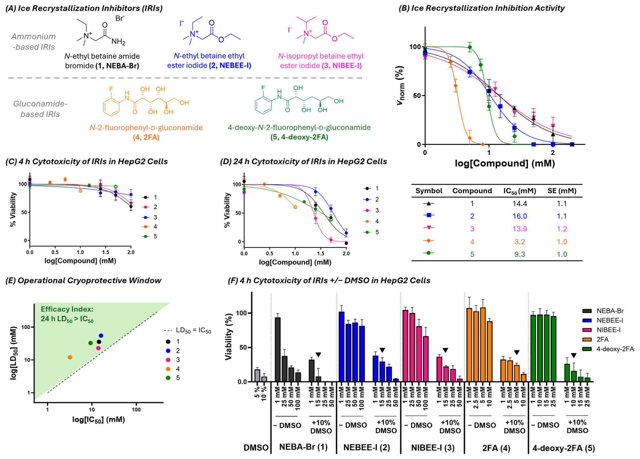
Suitable small-molecule IRIs were first identified based on ice recrystallization inhibition performance and cytocompatibility under cryopreservation-relevant conditions. Five compounds from two structurally distinct families were selected for screening: betaine-derived ammonium salts and N-aryl-d-gluconamides (Figure 1A). Ice recrystallization inhibition dose–response analysis confirmed that all compounds exhibited measurable activity, with IC50 values spanning 3 to 16 mM (Figure 1B), consistent with previously reported effective ranges for small-molecule IRIs [28].
To assess cytocompatibility under cryopreservation-relevant conditions, all compounds were evaluated in HepG2 cells following 4 h exposure, reflecting typical handling times in cryopreservation workflows. HepG2 cells were selected as a well-established model for preliminary cytocompatibility screening. Although neuronal cultures would provide a more directly relevant model for assessing neuronal-specific responses to cryopreservation, HepG2 cells provide a practical and reproducible system for initial evaluation of cytocompatibility. At the 4 h timepoint, all IRIs exhibited minimal toxicity across the concentration ranges tested (Figure 1C), with viability largely maintained up to the aqueous solubility limit or maximum tested concentration (100 mM). To further define the compatibility window, extended 24 h exposure studies were performed to provide a conservative toxicity estimate via cellular LD50 values (Figure 1D). While ammonium-based IRIs 13 produced complete dose–response profiles, gluconamide-based IRIs 45 were limited by solubility constraints, and consequently, LD50 values for 2FA (4) and 4-deoxy-2FA (5) were estimated by nonlinear regression.
Combination of IRI efficacy (IC50) and cytotoxicity (24 h LD50) defined a favourable operational cryoprotective window for each compound (Figure 1E), whereby ice recrystallization inhibition could be achieved at concentrations below cytotoxic levels for all compounds. Ammonium-based IRIs 13 further demonstrated high solubility (>0.5 M), whereas gluconamide derivatives were limited to 10 mM (2FA, 4) and 25 mM (4-deoxy-2FA, 5).
Candidate IRIs were then evaluated in the presence of 10% (v/v) dimethyl sulfoxide (DMSO), the most widely used cryoprotectant. As expected, cytotoxicity was primarily driven by DMSO rather than the IRI compounds (Figure 1F), with all IRIs remaining largely non-toxic at cryopreservation-relevant exposure times of 4 h. IRI concentrations (▼) were selected as final cryopreservation formulations based on two criteria: (i) post-exposure viability that was greater than or equal to that observed with 10% DMSO alone, indicating no additional cytotoxicity, and (ii) demonstrable ice recrystallization inhibition within the active region of the dose–response curve shown in Figure 1B. Concentrations corresponding to the upper plateau of the dose–response curve (e.g., 1 mM) were excluded, since ice crystal growth was effectively uninhibited. For 2FA (4), the selected concentration of 5 mM was additionally supported by previous evidence of cytocompatibility and successful cryopreservation of iPSC-derived neurons (iPSC-Ns) at the same concentration [32]. These selected conditions were carried forward for evaluation in hMO cryopreservation experiments.

3.2. Post-Thaw Morphological Integrity and Recovery of hMBOs

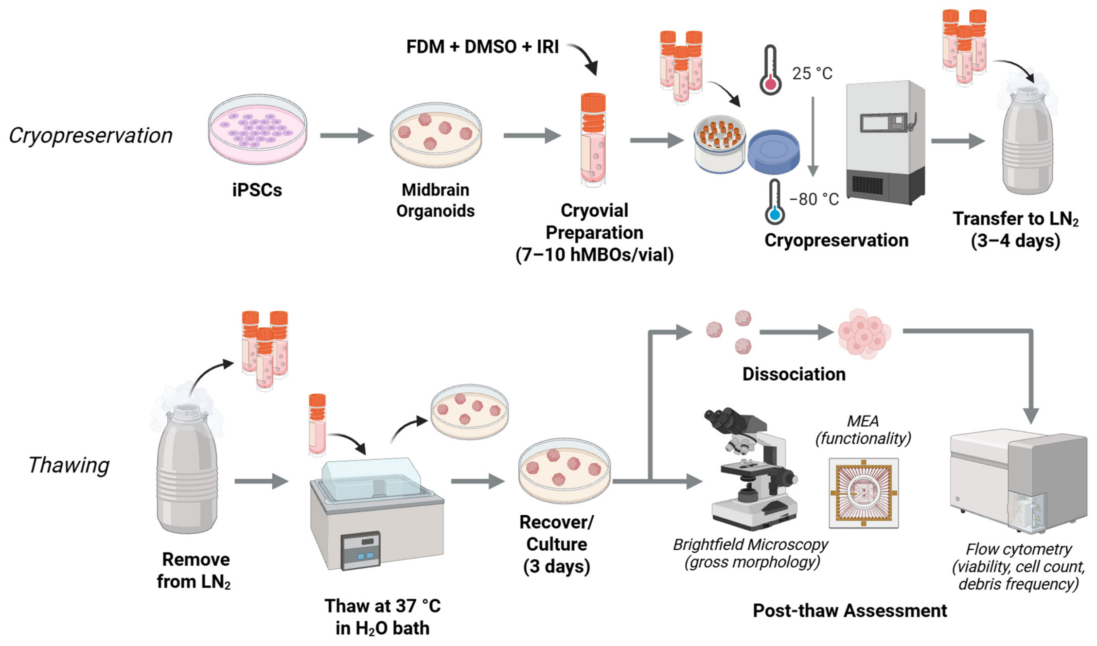
Human midbrain organoids were cryopreserved in final differentiation medium (FDM) supplemented with 10% DMSO, with or without an IRI additive, followed by rapid thawing and a recovery period prior to post-thaw analysis (Figure 2). Organoids were pooled (7–12 per cryovial), cooled to −80 °C using a Corning® CoolCellTM LX Cell Freezing Container, stored in liquid nitrogen, rapidly thawed, and cultured for 3 days prior to assessment.
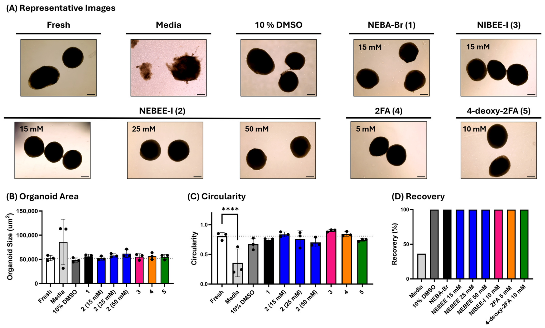
Cryopreservation in all DMSO-containing formulations, with or without IRI supplementation, preserved overall organoid morphology relative to fresh controls (Figure 3A). In all DMSO conditions, organoids retained a spheroidal architecture and well-defined boundaries. In contrast, cryopreservation in medium without cryoprotectant resulted in morphological deterioration, as expected, including irregular size distribution, loss of defined edges, and reduced apparent optical density, consistent with compromised structural integrity.
Quantitatively, mean organoid area did not differ significantly across all cryopreserved conditions relative to age-matched fresh controls (Figure 3B), indicating preservation of gross size post-cryopreservation. However, organoids cryopreserved in medium alone exhibited increased variability in size distribution relative to other conditions.
Circularity, calculated using ImageJ software from the projected area and perimeter of each organoid, is reported on a scale from 0 to 1, where a value of 1 represents a perfect circle and healthy organoid morphology. Circularity was significantly reduced in the FDM-only condition compared to the fresh control (****, p < 0.0001; Figure 3C), which is consistent with the observation of irregular boundaries and visible blebbing by microscopy. In contrast, all DMSO-containing cryopreservation conditions, including DMSO-only and DMSO with IRI supplementation, showed no significant differences in circularity relative to fresh controls and displayed minimal surface blemishing upon visual inspection via microscopy. Circularity values ranged from 0.57 to 0.78 for the 10% DMSO condition and 0.61–0.95 for the 10% DMSO with IRI condition, versus 0.73–0.85 for the fresh control, indicating that preservation of overall organoid shape and boundary integrity is maintained when a cryoprotectant is present.
Recovery of intact organoids following cryopreservation was dependent on cryoprotectant supplementation (Figure 3D). DMSO-containing conditions achieved 100% recovery of intact organoids following thawing, regardless of whether IRIs were present, and regardless of the exact number of organoids pooled within the cryovial (between 7 and 12 hMOs per cryovial were tested). In contrast, the medium-only control yielded approximately 30% recovery, with only 3 of 11 organoids recovered intact and substantial cellular debris observed by microscopy. The poor recovery and extensive structural damage in this condition precluded subsequent analysis by flow cytometry and/or MEA. Collectively, these findings demonstrate that DMSO-based cryopreservation formulations effectively preserve organoid morphology, structural integrity, and post-thaw recovery following cryopreservation, but IRI supplementation does not yield any added benefit for these post-thaw metrics. However, these assessments are limited to gross organoid morphology and recovery; they do not determine whether cryopreservation alters cytoarchitecture or cellular organization within the organoids, as no histological or microscopic analyses were performed beyond macroscopic assessment. Furthermore, these experiments do not reveal whether specific cell populations are differentially affected by cryopreservation, and additional cell type-specific characterization would be required to address this question.

3.3. Post-Thaw Viability and Preliminary Functionality of Cryopreserved hMBOs

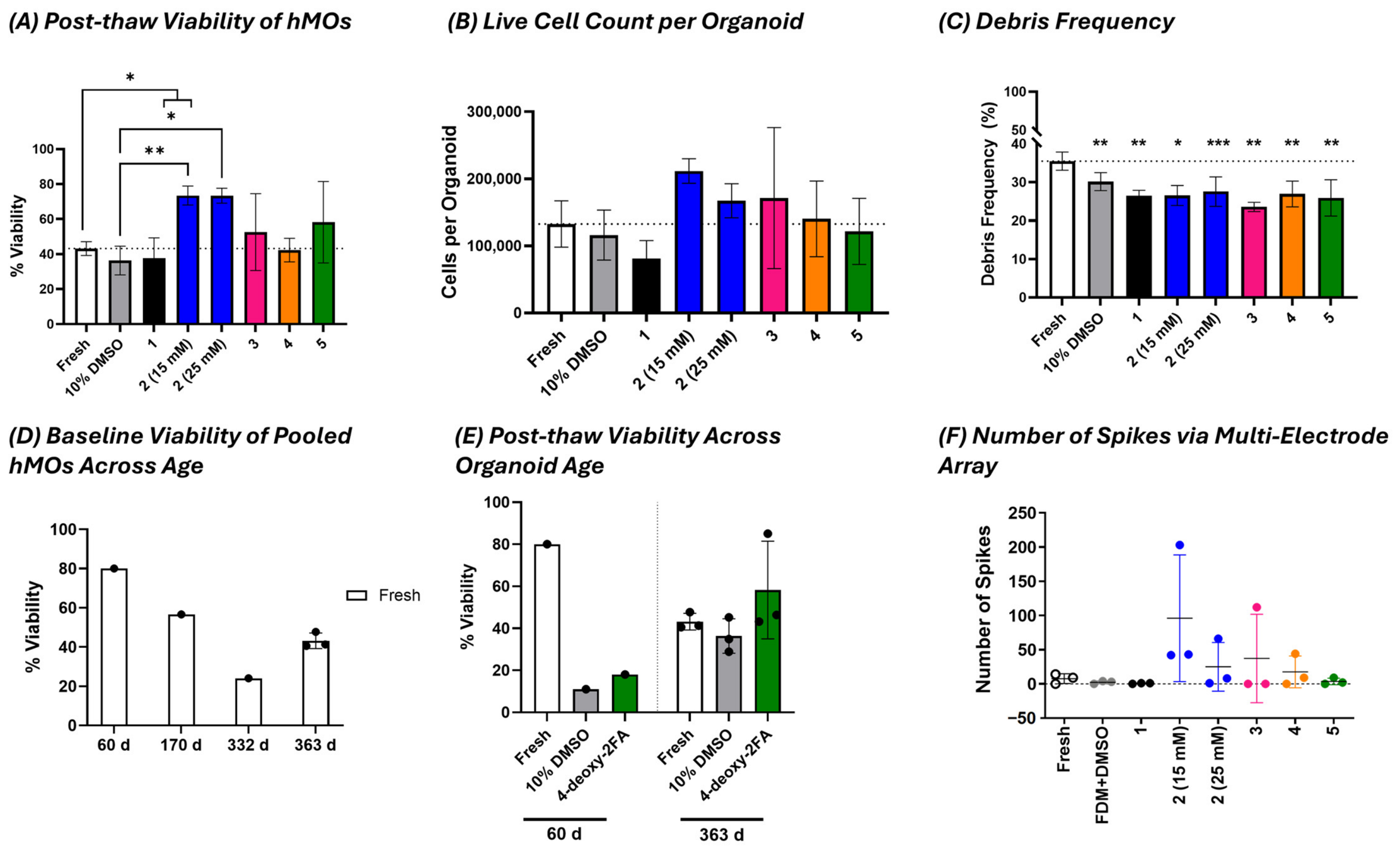
Post-thaw cellular viability was assessed by flow cytometry to evaluate the impact of a panel of seven cryopreservation formulations on hMBO survival (Figure 4A), including DMSO alone and DMSO supplemented with IRIs at concentrations defined in Figure 1F. Each condition consisted of a single cryopreservation experiment (N = 1 cryovial per condition), from which three organoids were independently dissociated and analyzed by flow cytometry (n = 3 technical measurements per condition). Organoids were compared to age-matched fresh controls differentiated from the same batch.
When compared to age-matched fresh controls processed in parallel, NEBEE-I (2) cryopreserved samples exhibited a statistically significant increase in measured viability at both tested concentrations (* p < 0.05 for 15 mM and 25 mM compared to fresh; Figure 4A). To further assess post-thaw outcomes, live cell counts (cell yield) per organoid were also quantified from organoids within the same differentiation, age and cryovial for each of the seven cryopreservation formulations and compared to age-matched controls from the same hMO batch (Figure 4B). No significant differences were observed between IRI-supplemented formulations and the 10% DMSO control, indicating comparable recovery of total viable cells within a given freezing event. Furthermore, no significant difference was found between the cryopreserved condition and the age-matched fresh control, indicating minimal cell loss from the cryopreservation process.
In addition, all cryopreservation conditions, including DMSO-only and IRI-supplemented formulations, showed a significant reduction in cell debris frequency relative to fresh controls (Figure 4C). These differences are most likely attributable to variations in sample processing rather than intrinsic differences in cell health, as fresh organoids typically require more extensive mechanical dissociation, which can increase shear-induced membrane damage and contribute to elevated debris formation and apparent reductions in viability. In contrast, cryopreserved organoids yield more uniform single-cell suspensions following thaw, which may reduce dissociation-associated artefacts in flow cytometry-based readouts. Accordingly, these findings should likely be interpreted in the context of processing-dependent differences in dissociation efficiency rather than as evidence of improved biological viability relative to fresh tissue. However, the present study did not directly assess extracellular matrix (ECM) integrity, and therefore, potential ECM alterations contributing to differences in dissociation efficiency cannot be excluded. Direct assessment of ECM integrity following cryopreservation would be an important direction for future studies.
To assess whether baseline organoid viability varied with maturation stage, fresh hMOs were evaluated across selected time points (60, 170, 332, and 363 days; Figure 4D). Fresh viability showed moderate variability across ages, with higher values observed at earlier stages (60 days) and lower values at intermediate stages (332 days), indicating inherent maturation-associated variability in dissociation-dependent viability. Additional phenotypic characterization would be valuable to determine whether these differences reflect shifts in cellular composition during maturation. For example, more mature organoids may contain fewer progenitor cells and a greater proportion of terminally differentiated cell types, which may differ in their susceptibility to dissociation- and/or cryopreservation-induced stress. Further cell type-specific analyses would be required to test this hypothesis.
To directly compare cryopreservation performance, matched analyses were performed at early (60 days) and late (363 days) stages (Figure 4E), where post-thaw viability of organoids cryopreserved in 10 mM 4-deoxy-2FA (5) supplemented with 10% DMSO was compared against 10% DMSO alone and age-matched fresh controls. At 60 days, both cryopreservation conditions resulted in reduced viability relative to fresh controls, whereas at 363 days, post-thaw viability was comparable to fresh levels. Across both time points, no consistent difference was observed between 4-deoxy-2FA (5) and DMSO-only conditions. Together, these results suggest that while baseline viability varies with organoid maturation stage, cryopreservation outcomes may be dependent on developmental state, with improved cryopreservation outcomes observed in more mature hMOs. Additional phenotypic characterization, such as cell type-specific flow cytometry or RNA sequencing, would help determine whether age-dependent differences in cellular composition underlie these observations, and provide mechanistic insight into the differential cryopreservation responses observed across maturation stages.
Finally, exploratory electrophysiological measurements were performed using multielectrode array (MEA) recordings to evaluate functional recovery following cryopreservation (Figure 4F). For each condition, three individual 363-day-old hMOs were recorded independently on the MEA platform, and spike activity was quantified for each organoid. Baseline spontaneous firing rates in fresh organoids were low and variable, reflecting the inherently heterogeneous nature of MEA recordings in mature organoid systems. Electrophysiological readouts are strongly influenced by factors such as electrode–tissue coupling, local network engagement, and spatial positioning of active regions on the array, all of which contribute to substantial variability between recordings even in age-matched mature cultures. Given the low and inconsistent firing activity observed in fresh controls, and the fact that MEA activity varies at different time points and in different cell types, MEA measurements present challenges as a robust metric of post-thaw functional recovery. This limitation highlights a broader challenge in assessing the functionality of complex neuronal tissues such as hMOs, where sensitive and reproducible functional endpoints remain difficult to establish. Future studies may benefit from complementary electrophysiological techniques, such as calcium imaging or patch-clamp recordings [25,38], which can provide higher-resolution measurements of neuronal activity. However, such approaches typically require tissue sectioning or dissociation, introducing additional challenges and potentially compromising the structural integrity of the organoid.
Despite the challenges associated with obtaining robust MEA measurements, hMOs preserved with NEBEE-I (2) exhibited increased spike frequencies relative to both fresh and 10% DMSO controls, although these differences did not reach statistical significance. Given the low baseline firing rates and substantial variability inherent to organoid MEA recordings, the absence of statistical significance should be interpreted cautiously. Given that MEA analysis was performed three days post-thaw, longer recovery periods could be explored together with increasing the number of organoids per test. Importantly, the MEA dataset serves as an initial functional assessment that complements the structural integrity, recovery, and viability data presented above. Together, these findings suggest that cryopreservation with NEBEE-I (2) does not impair, and may potentially support, the maintenance of neuronal activity following thawing.
Collectively, these results demonstrate that hMBOs can be effectively cryopreserved using a standard 10% DMSO-based protocol, which preserves post-thaw viability, cellular recovery, and overall structural integrity. Supplementation with NEBEE-I (2) showed promise as a cryoprotective additive, significantly enhancing post-thaw viability relative to 10% DMSO alone within the same hMO batch and exhibiting a non-significant trend towards increased electrophysiological activity. Together, these findings establish 10% DMSO as a robust baseline cryopreservation condition for hMBOs, while identifying NEBEE-I (2) as a promising additive for improving post-thaw cell viability and potentially supporting functional preservation.

4. Conclusions

This study provides proof-of-concept evidence that hMBOs can be effectively cryopreserved using a standard 10% DMSO-based protocol while preserving post-thaw viability and overall gross morphology. Within seven different cryopreservation formulations, hMBOs were successfully recovered following cryopreservation using multiple organoid ages, demonstrating the feasibility of conventional DMSO-based cryopreservation for this model. Organoid size and circularity were maintained across all conditions, indicating that gross morphological characteristics were preserved, while post-thaw viability and cell recovery remained comparable to those of age-matched fresh controls.
Although conventional DMSO-based cryopreservation was sufficient to support successful recovery, supplementation with the small-molecule ice recrystallization inhibitor N-ethyl betaine ethyl ester iodide (NEBEE-I, 2) consistently maintained or modestly improved several post-thaw metrics relative to DMSO alone without compromising recovery. Given that IRIs act through a distinct biophysical mechanism by suppressing ice recrystallization, these findings suggest that NEBEE-I may provide complementary protection during freezing and thawing. While these benefits were not reflected in large differences in gross morphology or recovery under the conditions examined, they support continued investigation of IRIs as adjunctive cryoprotectants capable of mitigating subtle forms of cryoinjury that may not be captured by conventional structural or viability assays.
Functional assessment using microelectrode arrays (MEAs) did not provide consistent evidence of spontaneous electrical activity in cryopreserved organoids, irrespective of cryopreservation formulation. However, fresh age-matched organoids likewise failed to exhibit detectable electrophysiological activity under the same assay conditions. Consequently, the absence of MEA activity cannot be attributed to the cryopreservation process or the cryoprotective formulation and instead suggests that the MEA assay, as implemented here, was not sufficiently sensitive or appropriate for evaluating functional capacity in this organoid model. Future studies employing more mature organoids or alternative functional readouts may better resolve whether adjunctive cryoprotectants such as IRIs preserve higher-order cellular function and internal organization beyond the morphological and viability endpoints assessed here. Overall, these finding provide proof-of-concept support for 10% DMSO as a baseline cryopreservation strategy for hMBOs while highlighting the potential of IRIs such as NEBEE-I as mechanistically distinct additives for further optimization of organoid cryopreservation protocols.

Supplementary Materials

The following supporting information can be downloaded at https://www.mdpi.com/article/10.3390/organoids5030032/s1. Figure S1. Representative flow cytometry gating strategy used for viability analysis with Fixable Viability Dye (FVD) Aqua; General Experimental can be found in this section [1,2,3,4,5,39].

Author Contributions

Conceptualization, T.M.D. and R.N.B.; methodology, E.M.W., C.R., P.L. and J.S.; formal analysis E.M.W., C.R., P.L. and J.S.; investigation, E.M.W., C.R., P.L. and J.S.; data curation, E.M.W.; writing—original draft preparation, E.M.W.; writing—review and editing, E.M.W., R.N.B., C.R., P.L., J.S. and T.M.D.; funding acquisition, R.N.B. and T.M.D. All authors have read and agreed to the published version of the manuscript.

Funding

R.N.B. acknowledges the Natural Sciences and Engineering Research Council of Canada (NSERC, RGPIN 16338-2024) for financial support. E.M.W. acknowledges Canadian Blood Services for the Graduate Fellowship Program (GFP) award and financial support. The views herein do not necessarily reflect the views of Canadian Blood Services or the federal, provincial, or territorial governments of Canada. TMD was supported through the TRIDENT preclinical trials initiative funded by the Government of Canada’s New Frontiers in Research Fund Transformation (NFRFT-20022-00051) stream.

Institutional Review Board Statement

The study was conducted in accordance with the Declaration of Helsinki and approved by the Institutional Review Board (Research Ethics Board) of McGill University (protocol code 22-03-027, approved 4 February 2025). Human-derived cell lines were obtained from The Neuro Biobank, which handled patient consent; no direct patient interaction or additional informed consent was required for this study.

Informed Consent Statement

Patient consent was waived since the human-derived cell lines were obtained from The Neuro Biobank, which obtained informed consent from the original participants, and no direct patient interaction or additional informed consent was required for the present study.

Data Availability Statement

The original contributions presented in this study are included in the article/Supplementary Materials. Further inquiries can be directed to the corresponding author.

Acknowledgments

Thanks to Genevieve Dorval for her help in facilitating transfer of materials between sites and to The Neuro Biobank (C-BIG) for providing access to the iPSCs used in this project.

Conflicts of Interest

R.N.B. is co-founder and Chief Scientific Officer of PanTHERA CryoSolutions Inc. (a part of BioLife Solutions Inc.). All other authors declare no conflicts of interest.

References

  1. Kim, J.; Koo, B.-K.; Knoblich, J.A. Human Organoids: Model Systems for Human Biology and Medicine. Nat. Rev. Mol. Cell Biol. 2020, 21, 571–584. [Google Scholar] [CrossRef] [Scilit] [PubMed]
  2. Lancaster, M.A.; Renner, M.; Martin, C.-A.; Wenzel, D.; Bicknell, L.S.; Hurles, M.E.; Homfray, T.; Penninger, J.M.; Jackson, A.P.; Knoblich, J.A. Cerebral Organoids Model Human Brain Development and Microcephaly. Nature 2013, 501, 373–379. [Google Scholar] [CrossRef] [Scilit] [PubMed]
  3. Quadrato, G.; Nguyen, T.; Macosko, E.Z.; Sherwood, J.L.; Min Yang, S.; Berger, D.R.; Maria, N.; Scholvin, J.; Goldman, M.; Kinney, J.P.; et al. Cell Diversity and Network Dynamics in Photosensitive Human Brain Organoids. Nature 2017, 545, 48–53. [Google Scholar] [CrossRef] [Scilit] [PubMed]
  4. Lancaster, M.A.; Knoblich, J.A. Generation of Cerebral Organoids from Human Pluripotent Stem Cells. Nat. Protoc. 2014, 9, 2329–2340. [Google Scholar] [CrossRef] [Scilit] [PubMed]
  5. Toh, H.S.Y.; Choo, X.Y.; Sun, A.X. Midbrain Organoids-Development and Applications in Parkinson’s Disease. Oxf. Open Neurosci. 2023, 2, kvad009. [Google Scholar] [CrossRef] [Scilit] [PubMed]
  6. Fiorenzano, A.; Sozzi, E.; Kastli, R.; Iazzetta, M.R.; Bruzelius, A.; Arlotta, P.; Parmar, M. Advances, Challenges, and Opportunities of Human Midbrain Organoids for Modelling of the Dopaminergic System. EMBO J. 2025, 44, 4181–4195. [Google Scholar] [CrossRef] [Scilit] [PubMed]
  7. Smits, L.M.; Reinhardt, L.; Reinhardt, P.; Glatza, M.; Monzel, A.S.; Stanslowsky, N.; Rosato-Siri, M.D.; Zanon, A.; Antony, P.M.; Bellmann, J.; et al. Modeling Parkinson’s Disease in Midbrain-like Organoids. npj Park. Dis. 2019, 5, 5. [Google Scholar] [CrossRef] [Scilit] [PubMed]
  8. Monzel, A.S.; Smits, L.M.; Hemmer, K.; Hachi, S.; Moreno, E.L.; Van Wuellen, T.; Jarazo, J.; Walter, J.; Brüggemann, I.; Boussaad, I.; et al. Derivation of Human Midbrain-Specific Organoids from Neuroepithelial Stem Cells. Stem Cell Rep. 2017, 8, 1144–1154. [Google Scholar] [CrossRef] [Scilit] [PubMed]
  9. Jo, J.; Xiao, Y.; Sun, A.X.; Cukuroglu, E.; Tran, H.-D.; Göke, J.; Tan, Z.Y.; Saw, T.Y.; Tan, C.-P.; Lokman, H.; et al. Midbrain-like Organoids from Human Pluripotent Stem Cells Contain Functional Dopaminergic and Neuromelanin-Producing Neurons. Cell Stem Cell 2016, 19, 248–257. [Google Scholar] [CrossRef] [Scilit] [PubMed]
  10. Cui, X.; Li, X.; Zheng, H.; Su, Y.; Zhang, S.; Li, M.; Hao, X.; Zhang, S.; Hu, Z.; Xia, Z.; et al. Human Midbrain Organoids: A Powerful Tool for Advanced Parkinson’s Disease Modeling and Therapy Exploration. npj Park. Dis. 2024, 10, 189. [Google Scholar] [CrossRef] [Scilit] [PubMed]
  11. Gordon, A.; Yoon, S.-J.; Tran, S.S.; Makinson, C.D.; Park, J.Y.; Andersen, J.; Valencia, A.M.; Horvath, S.; Xiao, X.; Huguenard, J.R.; et al. Long-Term Maturation of Human Cortical Organoids Matches Key Early Postnatal Transitions. Nat. Neurosci. 2021, 24, 331–342. [Google Scholar] [CrossRef] [Scilit] [PubMed]
  12. Mohamed, N.-V.; Lépine, P.; Lacalle-Aurioles, M.; Sirois, J.; Mathur, M.; Reintsch, W.; Beitel, L.K.; Fon, E.A.; Durcan, T.M. Microfabricated Disk Technology: Rapid Scale up in Midbrain Organoid Generation. Methods 2022, 203, 465–477. [Google Scholar] [CrossRef] [Scilit] [PubMed]
  13. Yao, X.; Kang, J.H.; Kim, K.-P.; Shin, H.; Jin, Z.-L.; Guo, H.; Xu, Y.-N.; Li, Y.-H.; Hali, S.; Kwon, J.; et al. Production of Highly Uniform Midbrain Organoids from Human Pluripotent Stem Cells. Stem Cells Int. 2023, 2023, 3320211. [Google Scholar] [CrossRef] [Scilit] [PubMed]
  14. Kim, D.; Youn, J.; Kim, J.; Lee, J.; Yoon, J.; Kim, D.S. From Organoid Culture to Manufacturing: Technologies for Reproducible and Scalable Organoid Production. npj Biomed. Innov. 2026, 3, 12. [Google Scholar] [CrossRef] [Scilit] [PubMed]
  15. Mashouf, P.; Tabibzadeh, N.; Kuraoka, S.; Oishi, H.; Morizane, R. Cryopreservation of Human Kidney Organoids. Cell. Mol. Life Sci. 2024, 81, 306. [Google Scholar] [CrossRef] [Scilit] [PubMed]
  16. Nickels, S.L.; Modamio, J.; Mendes-Pinheiro, B.; Monzel, A.S.; Betsou, F.; Schwamborn, J.C. Reproducible Generation of Human Midbrain Organoids for in Vitro Modeling of Parkinson’s Disease. Stem Cell Res. 2020, 46, 101870. [Google Scholar] [CrossRef] [Scilit] [PubMed]
  17. Lee, B.E.; Lee, B.J.; Lee, K.J.; Lee, M.; Lim, Y.J.; Choi, J.K.; Keum, B. A Simple and Efficient Cryopreservation Method for Mouse Small Intestinal and Colon Organoids for Regenerative Medicine. Biochem. Biophys. Res. Commun. 2022, 595, 14–21, Correction in Biochem. Biophys. Res. Commun. 2022, 602, 186. [Google Scholar] [CrossRef] [Scilit] [PubMed]
  18. Tsai, Y.-H.; Czerwinski, M.; Wu, A.; Dame, M.K.; Attili, D.; Hill, E.; Colacino, J.A.; Nowacki, L.M.; Shroyer, N.F.; Higgins, P.D.R.; et al. A Method for Cryogenic Preservation of Human Biopsy Specimens and Subsequent Organoid Culture. Cell. Mol. Gastroenterol. Hepatol. 2018, 6, 218–222.e7. [Google Scholar] [CrossRef] [Scilit] [PubMed]
  19. Pleguezuelos-Manzano, C.; Puschhof, J.; van den Brink, S.; Geurts, V.; Beumer, J.; Clevers, H. Establishment and Culture of Human Intestinal Organoids Derived from Adult Stem Cells. Curr. Protoc. Immunol. 2020, 130, e106. [Google Scholar] [CrossRef] [Scilit] [PubMed]
  20. Nagai, W.; Inui, J.; Ueyama-Toba, Y.; Asano, R.; Mizuguchi, H. Cryopreservation of Hepatocyte Organoid-Derived Cells Generated from Human iPS Cells. Biol. Pharm. Bull. 2025, 48, 1391–1398. [Google Scholar] [CrossRef] [Scilit] [PubMed]
  21. Kim, H.; Kim, S.K.; Oelgeschläger, M.; Park, H. Prediction of Acute Hepatotoxicity With Human Pluripotent Stem Cell-Derived Hepatic Organoids. Curr. Protoc. 2024, 4, e1015. [Google Scholar] [CrossRef] [Scilit] [PubMed]
  22. Gulieva, R.E.; Higgins, A.Z. Human Induced Pluripotent Stem Cell Derived Kidney Organoids as a Model System for Studying Cryopreservation. Cryobiology 2021, 103, 153–156. [Google Scholar] [CrossRef] [Scilit] [PubMed]
  23. Larsen, B.M.; Kannan, M.; Langer, L.F.; Leibowitz, B.D.; Bentaieb, A.; Cancino, A.; Dolgalev, I.; Drummond, B.E.; Dry, J.R.; Ho, C.-S.; et al. A Pan-Cancer Organoid Platform for Precision Medicine. Cell Rep. 2021, 36, 109429. [Google Scholar] [CrossRef] [Scilit] [PubMed]
  24. He, A.; Powell, S.; Kyle, M.; Rose, M.; Masmila, E.; Estrada, V.; Sicklick, J.K.; Molinolo, A.; Kaushal, S. Cryopreservation of Viable Human Tissues: Renewable Resource for Viable Tissue, Cell Lines, and Organoid Development. Biopreserv. Biobank. 2020, 18, 222–227. [Google Scholar] [CrossRef] [Scilit] [PubMed]
  25. Xue, W.; Li, H.; Xu, J.; Yu, X.; Liu, L.; Liu, H.; Zhao, R.; Shao, Z. Effective Cryopreservation of Human Brain Tissue and Neural Organoids. Cell Rep. Methods 2024, 4, 100777. [Google Scholar] [CrossRef] [Scilit] [PubMed]
  26. Mojica-Perez, S.; Stokes, K.; Jacobs, S.; Huang, J.; Vaid, S.; Yuan, Y.; Pearson, C.A.; Montes, D.; Tidball, A.; VanHeyningen, D.; et al. Cryopreservation of Human Cortical Organoids Using Vitrification. bioRxiv 2025. [Google Scholar] [CrossRef] [Scilit]
  27. Murray, K.A.; Gibson, M.I. Chemical Approaches to Cryopreservation. Nat. Rev. Chem. 2022, 6, 579–593. [Google Scholar] [CrossRef] [Scilit] [PubMed]
  28. McMunn, L.E.; Walsh, E.M.; Ben, R.N. Targeted Development and Optimization of Small-Molecule Ice Recrystallization Inhibitors (IRIs) for the Cryopreservation of Biological Systems. Cryoletters 2024, 45, 69–87. [Google Scholar] [CrossRef] [Scilit]
  29. Deller, R.C.; Pessin, J.E.; Vatish, M.; Mitchell, D.A.; Gibson, M.I. Enhanced Non-Vitreous Cryopreservation of Immortalized and Primary Cells by Ice-Growth Inhibiting Polymers. Biomater. Sci. 2016, 4, 1079–1084. [Google Scholar] [CrossRef] [Scilit] [PubMed]
  30. Smith, D.J.; Schulte, M.; Bischof, J.C. The Effect of Dimethylsulfoxide on the Water Transport Response of Rat Hepatocytes During Freezing. J. Biomech. Eng. 1998, 120, 549–558. [Google Scholar] [CrossRef] [Scilit] [PubMed]
  31. Murray, K.A.; Gao, Y.; Griffiths, C.A.; Kinney, N.L.H.; Guo, Q.; Gibson, M.I.; Whale, T.F. Chemically Induced Extracellular Ice Nucleation Reduces Intracellular Ice Formation Enabling 2D and 3D Cellular Cryopreservation. JACS Au 2023, 3, 1314–1320. [Google Scholar] [CrossRef] [Scilit] [PubMed]
  32. Alasmar, S.; Huang, J.; Chopra, K.; Baumann, E.; Aylsworth, A.; Hewitt, M.; Sandhu, J.K.; Tauskela, J.S.; Ben, R.N.; Jezierski, A. Improved Cryopreservation of Human Induced Pluripotent Stem Cell (iPSC) and iPSC-Derived Neurons Using Ice-Recrystallization Inhibitors. Stem Cells 2023, 41, 1006–1021. [Google Scholar] [CrossRef] [Scilit] [PubMed]
  33. Briard, J.G.; Jahan, S.; Chandran, P.; Allan, D.; Pineault, N.; Ben, R.N. Small-Molecule Ice Recrystallization Inhibitors Improve the Post-Thaw Function of Hematopoietic Stem and Progenitor Cells. ACS Omega 2016, 1, 1010–1018. [Google Scholar] [CrossRef] [Scilit] [PubMed]
  34. Abraham, S.; Keillor, K.; Capicciotti, C.J.; Perley-Robertson, G.E.; Keillor, J.W.; Ben, R.N. Quantitative Analysis of the Efficacy and Potency of Novel Small Molecule Ice Recrystallization Inhibitors. Cryst. Growth Des. 2015, 15, 5034–5039. [Google Scholar] [CrossRef] [Scilit]
  35. Chen, C.X.-Q.; Abdian, N.; Maussion, G.; Thomas, R.A.; Demirova, I.; Cai, E.; Tabatabaei, M.; Beitel, L.K.; Karamchandani, J.; Fon, E.A.; et al. Standardized Quality Control Workflow to Evaluate the Reproducibility and Differentiation Potential of Human iPSCs into Neurons. SSRN J. 2021. [Google Scholar] [CrossRef] [Scilit]
  36. Maecker, H.T.; Trotter, J. Flow Cytometry Controls, Instrument Setup, and the Determination of Positivity. Cytom. Part A 2006, 69A, 1037–1042. [Google Scholar] [CrossRef] [Scilit] [PubMed]
  37. Maussion, G.; Rocha, C.; Abdian, N.; Yang, D.; Turk, J.; Carrillo Valenzuela, D.; Pimentel, L.; You, Z.; Morquette, B.; Nicouleau, M.; et al. Transcriptional Dysregulation and Impaired Neuronal Activity in FMR1 Knock-Out and Fragile X Patients’ iPSC-Derived Models. Int. J. Mol. Sci. 2023, 24, 14926. [Google Scholar] [CrossRef] [Scilit] [PubMed]
  38. Landry, C.R.; Yip, M.C.; Zhou, Y.; Niu, W.; Wang, Y.; Yang, B.; Wen, Z.; Forest, C.R. Electrophysiological and Morphological Characterization of Single Neurons in Intact Human Brain Organoids. J. Neurosci. Methods 2023, 394, 109898. [Google Scholar] [CrossRef] [Scilit] [PubMed]
  39. Diamante, M. Controlling Ice Growth – From Nucleation to Recrystallization. Ph.D. Thesis, University of Ottawa, Ottawa, ON, Canada, 2026. [Google Scholar]
Figure 1. Selection and preliminary evaluation of ice recrystallization inhibitors (IRIs) for advancement to hMO cryopreservation. (A) Chemical structures and names of five lead compounds. (B) Dose–response curves for ice recrystallization inhibition and corresponding IC50 values. (C) HepG2 cytocompatibility following 4 h exposure measured by resazurin viability staining. (D) HepG2 cytocompatibility following 24 h exposure measured by resazurin viability staining. (E) Operational cryoprotective window comparing IRI efficacy (IC50) and cytotoxicity (24 h LD50). Gluconamide IRIs are limited by solubility, and LD50 values for 2FA and 4-deoxy-2FA were estimated by nonlinear regression (GraphPad Prism, version 11). (F) HepG2 cell viability following 4 h exposure to IRIs alone or in combination with 10% (v/v) DMSO. Concentrations marked with ▼ indicate selected formulation where cell viability and IRI efficacy was balanced. For all panels, error is reported as ±SD.
Organoids 05 00032 g001
Figure 2. Generation, cryopreservation, and post-thaw assessment of human midbrain organoids (hMBOs). Schematic of the experimental workflow used for hMBO cryopreservation and recovery. iPSCs are differentiated into hMBOs, which are pooled (7–12 organoids per cryovial) and suspended in final differentiation medium (FDM) supplemented with cryoprotective agents (CPAs; 10% DMSO with or without an IRI). Cryovials are cooled using an isopropanol freezing container at −80 °C overnight, followed by transfer to liquid nitrogen storage for 3–4 days. Thawing is performed rapidly in a 37 °C water bath (<2 min), followed by transfer to fresh FDM and a 3--day recovery period. Post-thaw outcomes are assessed by brightfield microscopy (gross morphology and structural integrity), flow cytometry (viability, cell counts, debris frequency), and multielectrode array (MEA) recordings (functional activity). For brightfield microscopy and MEA recordings, organoids were assessed individually as independent experimental samples. For flow cytometry, three individual organoids may be pooled following dissociation and analyzed as a single sample (n = 1) or dissociated and analyzed as three independent samples (n = 3); exact experimental details can be found in the Materials and Methods and/or figure captions for each experiment.
Organoids 05 00032 g002
Figure 3. Post-thaw structural and morphological assessment of cryopreserved hMBOs. (A) Representative brightfield images of hMBOs following cryopreservation and thawing in the indicated IRI formulations compared to fresh controls. All IRI conditions were prepared in final differentiation medium (FDM) supplemented with 10% DMSO prior to cryopreservation. Organoids were cryopreserved at 332–365 days of age and imaged after 3 days of post-thaw recovery prior to analysis. Images were acquired at 4× magnification; scale bars = 100 µm. (B) Mean organoid area (µm2) and (C) mean organoid circularity (0–1, 1 indicates perfect circle) quantified by ImageJ from organoids derived from a single independent cryopreservation experiment (N = 1), with n = 3 organoids analyzed per condition. One-way ordinary ANOVA comparing each condition to the fresh control was performed. Error bars represent SD. Asterisks denote statistical significance (**** p < 0.0001). (D) Percentage organoid recovery calculated from a single cryovial (N = 1), where recovery was determined from 7 to 12 organoids per condition as the ratio of intact organoids post-thaw to the total number frozen. For all panels, error is reported as ±SD.
Organoids 05 00032 g003
Figure 4. Post-thaw viability and preliminary functional outcomes after hMBO cryopreservation. For panels (AC), each condition consisted of a single cryopreservation experiment (N = 1 cryovial per condition), from which three 363-day-old organoids were independently dissociated and analyzed by flow cytometry (n = 3 technical replicates per condition). For panels (D,E), each condition consisted of a single cryopreservation experiment (N = 1 cryovial per condition), from which three organoids were dissociated, pooled together, and analyzed by flow cytometry (n = 1 technical replicate per condition from three pooled organoids). Organoids were compared to age-matched fresh controls derived from the same batch. (A) Post-thaw viability of hMBOs assessed by flow cytometry using Live/Dead Fixable Aqua staining. Statistical significance is indicated by asterisks, with * representing p < 0.05 and ** representing p < 0.01. (B) Live cell counts per organoid following dissociation. (C) Cell debris frequency quantified using FSC-SSC gating by flow cytometry. Statistical significance is indicated by asterisks, with * representing p < 0.05, ** representing p < 0.01, and *** representing p < 0.001. (D) Fresh human midbrain organoids (hMOs) were assessed for viability across maturation stages (60, 170, 332, and 363 days) using flow cytometry following dissociation and pooling of three organoids. Data are presented descriptively and reflect variability in baseline viability across organoid ages. (E) Viability of cryopreserved hMOs (10 mM 4-deoxy-2FA with 10% DMSO or 10% DMSO alone) assessed at 60 (young) and 363 (old) days of maturation, with age-matched fresh controls. For 60 days, data represent pooled dissociation of three organoids from a single cryovial (n = 1); for 363 days, three organoids from a single cryovial were analyzed individually by flow cytometry (n = 3). Data are shown descriptively. (F) Electrophysiological activity (number of spikes) measured by multielectrode array (MEA) recordings for three 363-day-old individual organoids per condition. For all panels, error is reported as ±SD.
Organoids 05 00032 g004
Disclaimer/Publisher’s Note: The statements, opinions and data contained in all publications are solely those of the individual author(s) and contributor(s) and not of MDPI and/or the editor(s). MDPI and/or the editor(s) disclaim responsibility for any injury to people or property resulting from any ideas, methods, instructions or products referred to in the content.