Review

Combining Proteomics and Organoid Research to Unravel the Multifunctional Complexity of Kidney Physiology Enhances the Need for Controlled Organoid Maturation

by
Kathrin Groeneveld
1,2,* and
Ralf Mrowka
1,2
1
Experimentelle Nephrologie, KIM III, Universitätsklinikum Jena, Am Klinikum 1 (CeTraMed), 07747 Jena, Germany
2
ThIMEDOP—Thüringer Innovationszentrum für Medizintechnik-Lösungen, Am Klinikum 1 (CeTraMed), 07747 Jena, Germany
*
Author to whom correspondence should be addressed.
Organoids 2025, 4(4), 28; https://doi.org/10.3390/organoids4040028
Submission received: 25 June 2025 / Revised: 28 October 2025 / Accepted: 31 October 2025 / Published: 14 November 2025

Abstract

This review aims to highlight how the study of kidney organoids combined with proteomic analysis can deepen our understanding of renal physiology and disease. Proteomics quantifies proteins in a sample, allowing us to determine which proteins are present, how abundant they are, and how they are modified. These data may reveal the pathways that are active in the kidney organoids and how they change in disease, helping to pinpoint candidate biomarkers. Kidney organoids are three-dimensional structures derived from induced pluripotent stem cells (iPS) that recapitulate many architectural and functional features of the adult organ. Because they can be generated in large numbers under defined conditions, organoids provide a promising platform for testing how genetic mutations, environmental stresses, or drugs affect kidney development and pathology. When proteomic profiles are obtained from mature organoids, researchers can directly link protein-level changes to phenotypic outcomes observed in the model. This integration makes it possible to map disease-related networks at the molecular level and to assess the impact of therapeutic interventions in a system that more closely resembles human kidney tissue than traditional cell lines. A current limitation is that many kidney organoids do not reach the full maturation seen in vivo; they often lack complete segmental differentiation and the functional robustness of adult nephrons. Improving the maturation state of organoids will be essential for accurately modeling chronic kidney diseases and for translating findings into clinically relevant therapies.

1. Methods

During the preparation of this manuscript, we used “consensus.app” [1], “ai-chatbot.tu-ilmenau.de” [2] and “PubMed®” [3] for the purpose of a first literature research. Keywords, such as “renal organoids”, “proteomics”, “renal organoid maturation”, and “kidney proteomics” were used in different combinations, focusing on output from the last ten years. Publications that were finally used within this review were selected based on the interest of the author, the potential impact on future renal organoid research, and relevance to the here revised fields of research.

2. Using Proteomics to Study Organ-Specific Spatial and Dynamic Proteome Patterns in Healthy and Pathological Conditions in Human Kidneys

The human kidney exhibits a complex and diverse proteome that reflects the multifunctional nature of the organ. Proteins related to transport (e.g., aquaporins, ion channels), metabolism (enzymes involved in gluconeogenesis), and structure (collagens, laminins) are highly represented [4,5,6,7,8,9,10,11,12,13].
With the help of proteomics, researchers are able to compile a detailed proteome atlas of the human kidney. In 2021, new ground was broken with a quantitative proteomic map with the work by Al-Majdoub and colleagues highlighting the distribution of enzymes and transporters throughout the human kidney [4]. The study provides insights into the zonal difference in drug metabolism and transport capabilities within the kidney: Cytochrome P450 enzymes (CYPs) are essential in drug metabolism and are found not only in the liver but also in the kidney, aiding in drug detoxification. Uridine 5′-diphospho-glucuronosyltransferases (UGTs) are crucial for phase II drug metabolism, enhancing drug solubility for renal excretion by adding glucuronic acid to drugs. Solute Carrier (SLC) Transporters, a large family of proteins, regulate the movement of substances, including drugs, across cellular membranes in the kidneys. ATP-Binding Cassette (ABC) Transporters, particularly those like P-glycoprotein (ABCB1), are key in drug excretion and resistance by moving drugs out of cells. Peptidases, such as dipeptidyl peptidase-IV (DPP-IV), play a role in peptide drug metabolism and in managing blood pressure and electrolyte balance. Additionally, enzymes involved in the oxidative stress response, like superoxide dismutase (SOD) and glutathione peroxidase (GPX), protect the kidneys from oxidative damage induced by drugs or disease, highlighting the complex interactions within renal biochemical pathways. In 2022, a tissue atlas of the human kidney was meticulously crafted by Hansen, J. et al., setting a new standard for studying the organ [5]. They received nephron segment-specific protein expression profiles. They work on the Kidney Precision Medicine Project (KPMP), which builds a spatially specified human kidney tissue atlas in health and disease with single-cell resolution. This comprehensive map serves as a foundation for dissecting renal pathologies and propels the pursuit of targeted therapies by offering a clear depiction of kidney structure–function–protein relationships. From a molecular standpoint, the exploration into “SLC-omics” by Lewis and colleagues [6] cataloged the nephron’s solute transporters: They generated a database of 431 SLC genes and were able to highlight individual expression features such as sex-specific expression in the proximal tubule and the role of accessory proteins. mRNA expression levels along the renal tube of mice were mapped by Chen et al., utilizing a data set based on RNA Seq experiments [7]. In addition, a map of protein levels along the renal tube was created by Limbutara et al. by applying a LC-MS/MS proteomics data set [8]. This work is fundamental in grasping how kidneys maintain systemic electrolyte and metabolite equilibrium by maintaining site-specific proteins. Together, these detailed location-specific protein function studies provide a robust foundation for pathophysiological studies. These proteomic maps can be used by researchers to evaluate specific protein expression. Specific markers for different kidney cell types (podocytes, tubular cells, and endothelial cells) can be identified, highlighting the cellular complexity of the kidney. Detailed studies of induced changes in protein expression give insight into renal metabolomics and into the development of renal diseases. Specific proteome changes can be used as biomarkers for diagnostics [9,10].
Some researchers focus on changes in the human kidney proteome. Wang and his colleagues added a time factor to their analysis and looked into the spatial dynamics of kidney metabolomics. This way, they were able to show cell fate trajectories in human kidney differentiation [11]. An investigation into the amniotic fluid proteome [12] unveils plastin 3 as a key player in glomerular integrity among fetuses with congenital kidney anomalies. Such findings not only give us more information on developmental kidney diseases but also beckon further research into prenatal interventions.
Proteomic alterations in renal substructure samples due to IgA nephropathy were studied by Yang et al. [13]. Distinct pathway enrichment patterns were revealed, with fatty acid synthesis in the glomerulus and complement and coagulation pathways enriched in the tubules. They also suggest ATP1B1 and COX4I1 in the glomerulus and SLC22A13 in the tubules as promising diagnostic markers for IgA nephropathy and C4A and APEX1 proteins as possible biomarkers for assessing disease progression. Such detailed studies on human kidney organs provide important knowledge when it comes to the introduction of organoid models in kidney disease research. Applications of proteomics and metabolomics to kidney diseases were reviewed by Dubin and Rhee [14]. The systematic review by Davies et al. focuses on proteomics and metabolomics in glomerulonephritis [15].
Other researchers focus on the products of kidney function—such as urine—and draw conclusions about the condition of the organ. The kidney’s special proteomics are very complex, because the organ fulfills highly complex filtration and reabsorption processes. As a result, urine is produced. As urine is easily accessible and can contain much information about previous renal functions, urinary proteomics are being investigated to identify potential new urinary biomarkers. For example, to diagnose and to understand diabetic nephropathy and different types of chronic kidney disease, urinary and histology biomarkers were combined by Lucarelli et al. [16]. They identified urinary proteins differentially expressed in patients who developed end-stage kidney disease. Differential expression was correlated with digital image features for glomeruli and tubules, and both were used as inputs to deep-learning frameworks to predict the kidney disease outcome and stage [16]. Capillary electrophoresis-coupled mass spectrometry was used by Zürbig et al. to profile the low-molecular-weight proteome in urine. They were able to detect subsequent progression to macroalbuminuria via the determination of specific collagen fragments [17]. Siwy et al. combined capillary electrophoresis-coupled mass spectrometry with the extended statistical analysis of 1180 urine samples from patients with different types of chronic kidney disease (CKD). For seven different types of CKD, several potential urinary biomarker peptides were defined and combined into classifiers specific for each CKD [18].
It was hypothesized by Pisitkun, Shen, and Knepper that multiple proteins, which can be used as biomarkers for a diversity of kidney diseases, are delivered to the urine via exosomes. Their study provides insights into a novel mechanism of cellular communication in the kidney but, importantly, gives the implication that exosome isolation can be an efficient first step in the search for urinary biomarkers [9]. These studies demonstrate that urinary proteomics offers robust, non-invasive methods for diagnosing and understanding human nephropathies. It enables early detection and disease differentiation and provides insights into underlying mechanisms.
On the toxicological front, a study on berberrubine-induced nephrotoxicity by Rao et al. describes the delicate protein balance in drug therapies [10]. They found that 103 metabolites were changed after berberrubine application. The researchers identified key proteins from three enriched KEGG pathways, including ERK1/2. They presume that ERK1/2 serves as a binding target involved in berberrubine-induced nephrotoxicity. By delving into metabolomic and proteomic interactions, this research offers invaluable insights into drug safety and efficacy. Finally, the exploration of calcium-binding domains in annexin A2 by Yoodee, S. et al., anticipated in 2025, elucidates a molecular pathway implicated in kidney stone formation [19]. This discovery could hint at new strategies in preventing and treating one of the most prevalent kidney ailments through targeted molecular interventions.
The mature kidney proteome is indicative of fully functional nephrons capable of filtering blood, with proteins related to high metabolic activity and osmoregulation. Proteomic signatures may be used to differentiate between a healthy kidney and various disease states, offering potential biomarkers for diagnosis or treatment targets.

3. Techniques to Generate Complex Renal Organoids

The value of renal organoids for research highly depends on a reproducible protein expression profile that is close to renal physiology. The generation of organoids needs to be controllable and reliably reproducible. Mature organoids should embody the cellular complexity and heterogeneity necessary for nuanced proteomic investigations. Variability in maturation between organoid batches or protocols can confound the interpretation of protein and gene expression patterns, as organoids at different maturation stages show distinct transcriptional signatures [20]. Standardized protocols and large-scale routines are needed to build a stable production line in the lab. Here, we want to give an overview of currently used methods that aim for stable production lines and for higher maturation levels of renal organoids. Table 1 summarizes the methods mentioned here, highlighting their successful improvements (“advantages”) and their limitations.
Przepiorski et al. introduced a simplified method for generating kidney organoids from human pluripotent stem cells, making the process more accessible and efficient for research purposes [21]. A robust cell culture protocol was provided to generate 14-day-old embryoid bodies in large batches. The generation does not ask for special equipment other than culture media and supplements and provides a good base for many researchers to get started on renal organoid generation. Generally, the differentiation of iPSC cells is controlled by growth factors. To ensure the guided development of small renal embryoid bodies into larger and more complex organoids that represent different tissue functions, researchers lean towards refined culture techniques.
The similarities between maturation protocols lie in their focus on replicating the kidney’s microenvironment and developmental cues through advanced bioengineering techniques and the strategic use of biomaterials and growth factors. These methods collectively aim to improve the structural, cellular, and functional biomimicry of kidney organoids to human kidneys in vivo.
Co-culturing: One way to aim for more defined structural patterns with cellular complexity is to introduce different cell types during organoid formation. Mae et al. co-cultured human iPSC-derived ureteric bud cells and ureteric bud tips on a soft hydrogel. The so-generated organoids show epithelial polarity, tubular lumen, and repeated branching potential, although collective duct structures are still immature [22]. The work by Matsumoto et al. evaluated the ability of human iPSC-derived nephron progenitor cells to form chimeric renal organoids using mouse embryonic renal progenitor cells [23]. This interspecies approach underscores the potential for optimizing organoid development through enhanced cell integration and functionality. Through the works of Shi et al., significant advances in integrating collecting systems into kidney organoids were made by fusing the distal nephron to the ureteric bud and recapitulating branching morphogenesis, further advancing the generation of functional and structural complexity within the organoids [24,25]. A method for generating organotypic kidney structures was presented by Tanigawa et al. by integrating pluripotent stem cell-derived renal stroma, emphasizing the importance of the stromal environment in organoid development [26].
Matrices: Across various studies, a consistent theme is the use of matrices that provide spatial cues for organoid maturation. Such matrices can help make organoid growth uniform. Also, depending on the material and structure, they influence the availability of medium ingredients and growth polarity. In a study from Kim and colleagues, a 3D geometrically engineered, permeable membrane-based platform was used to generate consistently grown organoids. They were able to establish polycystic kidney disease and acute kidney injury models [27]. Although permeability and light scattering of the here used nanofiber membrane limit the experimental design of studies, it can still be useful for the large-scale generation of uniform renal organoids of stable maturation. Techniques such as the application of tunable gelatin methacryloyl (GelMA) hydrogels and soft, dynamic hydrogel confinement have been pivotal in improving morphology and reducing epithelial–mesenchymal transition in culture, thus encouraging more precise renal cell-type specification and enhancing the overall organoid structure. The use of hydrogels, as investigated by Clerkin et al. [28] and Geuens et al. [29], offers a supportive matrix crucial for renal cell specification and organoid development by providing a three-dimensional structure for cell growth and differentiation. The incorporation of thiol–ene cross-linked alginate hydrogel encapsulation has been shown to modulate the extracellular matrix of kidney organoids, improving their structural integrity by reducing abnormal collagen deposition. A polyester membrane to culture iPSC-derived kidney organoids was used by Schumacher et al. Additionally, they simulated physiological hypoxia leads and found this approach very effective in enhancing microvasculature formation and patterning [30].
Vascularization has been identified as a critical step towards achieving fully functional kidney organoids. Vascularization is crucial, not just for nutrient and oxygen supply and waste removal. Organoid perfusion can provide paracrine signals for nephron patterning and maturation [31]. Additionally, endothelial–epithelial interactions are implicated in driving the maturation of both nephron and stromal compartments [32]. Methods that include the transplantation of premature organoids to animal tissue are very successful when it comes to functional vascularization. One alternative approach without the requirement of animals uses genetic engineering to help. A successful example would be the genetic inducement of endothelial niches to enable vascularization, promoting multilineage maturation and the emergence of specialized cells like renin-expressing cells [33]. Another alternative approach is to employ organ-on-chip systems.
Chips: Fluidic chips were shown to significantly support and enhance the maturation of renal organoids. By providing controlled fluid flow and a more physiologically relevant environment that includes exposure to sheer stress, these chips can promote fluidic flow, cellular differentiation, and functional maturation beyond what is possible in static cultures [34,35,36]. Microfluidics enables the formation of vascular networks within kidney organoids, including perfusable lumens surrounded by mural cells. This vascularization is critical for supplying nutrients and oxygen, supporting the development of more mature nephron structures and glomerular compartments. Organoids cultured under flow conditions on fluidic chips show increased cellular polarity, adult gene expression, and more mature podocyte and tubular compartments compared to static cultures. Co-culture with human endothelial cells (such as HUVECs) in microfluidic chips leads to the migration and integration of these cells into the organoid tissue, forming interconnected vessel-like structures and further supporting maturation [34].
Mimicking human organs and tissue by controlling the flow of fluidics has formed a class of advanced organoid maturation technologies of its own. There are several recent reviews on it, as the organ-on-chip technologies are constantly advancing [37,38,39].
Collectively, these studies illustrate the rapid development and potential of iPSC-based renal organoid technology in replicating the complex structure and function of the human kidney. By transcending the limitations of traditional 2D cell cultures and opening avenues for modeling kidney diseases, assessing drug effects, and exploring potential regenerative therapies, these advancements mark a significant step forward in biomedical research. Proteomic analysis plays a vital role in understanding the maturation process by identifying key proteins involved in kidney function and development.
Table 1. Overview of organoid maturation methods that represent the diversity of current approaches to enhance complexity and quality of renal organoids. Many protocols are based on Przepiorski’s protocol [21].
RefCell SourceMethodAdvantageLimitationsClinical Model?
[21]hPSCsuspension cell cultuecost effective,
high throughput possible
no flow or vascularization, no CDs,
limited organoid size
[30]hPSCpolyester membraneenhanced sprouting and interconnectivitybatch to batch variability
physiological hypoxiaincreased vessel lengthambient oxygen conc during handling
[27]hPSCpermeable 3D nanofibre membrane (UniMat)enhanced uniformity,
enhanced maturation
not commercially available,
permeability and scattering limits experimental design
PKD,
AKI
[28]hPSCsemi-synthetic hydrogelimproved maturation,
enhanced reproducibility,
controlled environment
DKD
[29]hPSCalginate hydrogelimproved extracellular matrix compositionno structural improvement
[22]hUB, hUB tip soft hydrogel,
co-culture
epithelial polarity,
tubular lumen,
branching,
CD progenitors
immature CDsMCDK
[24,25]hPSC derived UB and NPCsco-culturestructurally integrated CDsefficiency of nephron fusion to CDs and organization needs to be increased
[33]hPSCco-culture,
genetically induced endothelial niche
improved endotheliazation,
improved maturation,
drug-responsive renin expressing cells
[34]hPSC, HUVECsmicrofluidic organ-on-chip,
co-culture
increased endothelial maturation,
vascularization
no high throughput,
transfer of organoids from transwell to chip necessary
[31]hPSCmicrobioreactor arrayconstant perfusion no strong maturation,
high throughput possible
[23]mRPCs,
hPSC derived NPCs
human-mouse chimera,
transplantation,
magnetic sorting of NPCs
increased NPC purity and increased hNPC:mRPC ratio,
increased chimera formation
mouse model,
animals needed,
elaborate protocols
transplantation research
[26]mPSCin vitro induction protocol for SPscomplex structuresmouse model,
animals needed,
elaborate protocols
co-culture with NP and UB
transplantation
[32]hPSCtransplantationimproved vascularization,
improved maturation and morphogenesis
animals needed (chicken embryos),
elaborate protocols
 major protocol categories coded by color:
chip
matrix
co-culture
transplantation
combining co-culture and transplantation
combining co-culture and chip
combining co-culture and matrix
Abbreviations: AKI—acute kidney injury; CD—collective duct; DKD—diabetic kidney disease; hPSC—human pluripotent stem cells; HUVEC—human umbilical vein endothelial cell; MCDK—multi cystic dysplastic kidney; mPSC—mouse pluripotent stem cells; NPC—nephron progenitor cells; PKD—polycystic kidney disease; RPC—renal progenitor cells; SP—stromal progenitor; UB—ureteric bud.

4. Proteomics to Ensure the Complex and Differentiated Maturation of Renal Organoids That Mirrors the Kidney
Renal organoids also express a broad range of kidney-specific proteins. Early-stage organoids might express higher levels of developmental proteins, reflecting their origin from stem cells undergoing differentiation [40]. The presence of extracellular matrix proteins, transporters, and metabolic enzymes has been confirmed in organoids, showing successful maturation to some extent. Organoids express key proteins involved in kidney development and disease, including those linked to podocytopathies and cystic kidney diseases, and their proteomic profiles are more representative of human kidney tissue than traditional cell culture models [41,42]. Within a multidisciplinary cooperation, Rinschen and his colleagues were able to provide the protein expression profiles of renal organoids over cultivation time and during cytokine stress [43]. When exposed to disease-relevant stressors like TNFα, organoids activate inflammatory and complement pathways similar to those seen in human kidney disease, further supporting their relevance for modeling disease mechanisms and biomarker discovery. Renal organoid proteomics has revealed disease-specific pathways, such as fatty acid synthesis in glomeruli and complement activation in tubules in IgA nephropathy [13], and identified key proteins involved in disease progression [14,15]. This integration of organoid omics illuminates the path towards replicating and studying complex kidney diseases in a controlled environment.
Reviews of recent advances confirm that global proteomics and single-cell analyses of kidney organoids provide valuable insights into cell populations and regulatory mechanisms, making them authentic and practical in vitro models for studying kidney development, disease mechanisms, and drug responses, despite some limitations in maturity and complexity compared to adult kidneys [42,44].
Applying the technology of renal organoids, it is important to ensure that the differentiation protocols yield tissue that resembles the gene expression characteristics of the kidney. It is clear from previous studies that the normal kidney tissue has distinct gene signatures that may be altered specifically during disease, such as cancer [45]. Representative examples of kidney-specific genes are given in Table 2.
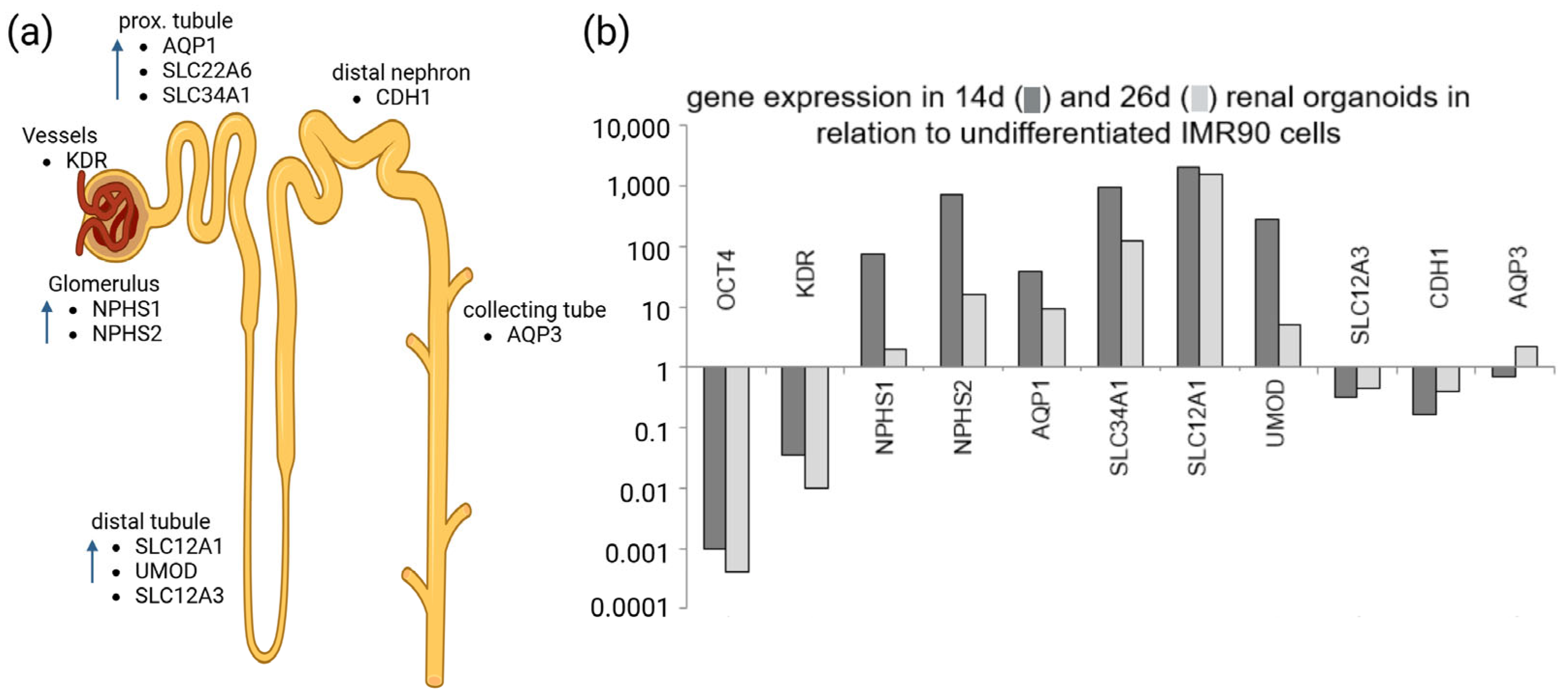
As organoids mature in the culture, they tend to increase the extracellular matrix deposition while decreasing the expression of glomerular proteins, which diverges from the protein composition of fully developed human kidneys [40,41]. The gene expression patterns shown in Figure 1b support this.
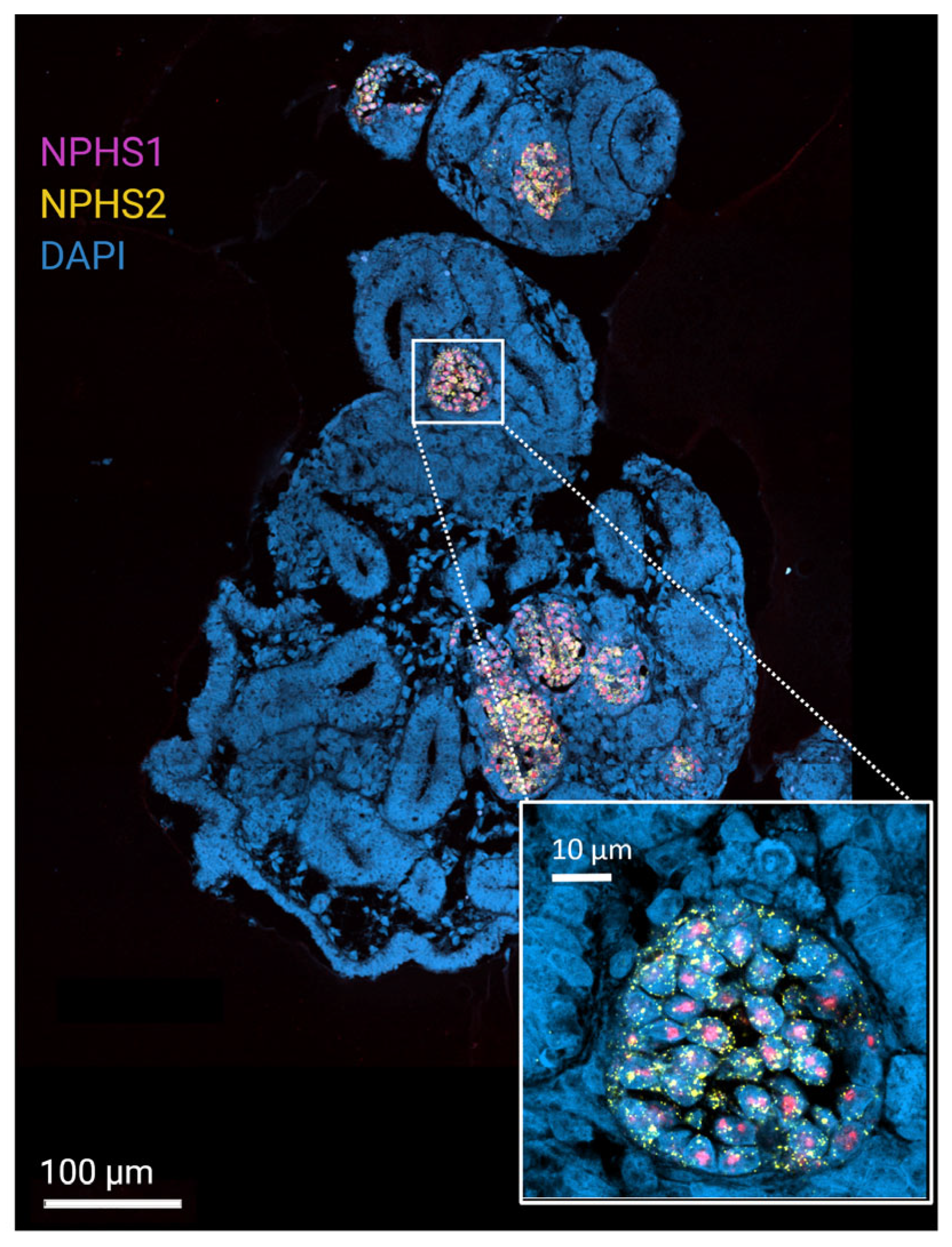
As part of a work by L.L.M. Rutten, targeted increased expression for marker genes of the glomerulus, proximal, and distal tubule, including parts of the Henle loop, as well as the collecting duct, was demonstrated in renal organoids matured for 14 days and for 26 days (Figure 1) using qPCR. The organoids that were used for this gene expression study were generated without fluidic support or any vascularization techniques. The gene expression pattern for the older organoids shows less renal specificity. NPHS1 and NPHS2 were among these marker genes. They code for nephrin and podocin, respectively, both are expressed in renal glomeruli. In another proof-of-concept study, antibody staining methods were used to demonstrate the presence of NPHS1 and NPHS2 mRNA in renal organoids [50] (Figure 2). Both studies support the renal-specific maturation of organoids.
Figure 1. qPCR targeting genes of renal site-specific marker proteins. (a) Expected renal marker genes. Arrows indicate expected increased gene expression in mature renal tissue based on a BioGPS search. (b) qPCR results targeting renal marker genes as displayed in (a). Gene expression based on RNA profiles of 14 d and 26 d old renal organoids in relation to non-differentiated iPS cells that were harvested before organoid initiation. Organoids were generated from iPS(IMR90)-2 cells, acquired from WiCell. Modified figure from Rutten, L.L.M., 2023 [51]. Created in BioRender. Reuter, S. (2025) [52].
Organoids 04 00028 g001
Figure 2. RNAScope staining targeting renal site-specific marker probes for RNA. Confocal laser scanning microscopy image of detection of NPHS1 and NPHS2 mRNA with RNAScope in a 14-day-old renal organoid. Focus-like distribution of hybridization signals for NPHS1 and NPHS2. Organoids were generated from iPS(IMR90)-2 cells, acquired from WiCell. Modified figure from Dilz, J. et al., 2023 [50]. Created in BioRender. Reuter, S. (2025) [53].
Organoids 04 00028 g002
In conclusion, exploring the proteome of renal organoids may offer insights into the complex interplay of proteins in kidney health and disease. By focusing on enzymes, transporters, structural proteins, and disease markers, researchers are uncovering the intricate mechanisms that underpin renal function and pathology. However, it also presents the researchers with the challenge of applying appropriate methods for extended organoid maturation (Section 3) as well as for their downstream analysis (Section 5). The field of proteomics provides a wide range of possible applications.

5. Proteomics to Study Healthy and Pathological Patterns in Matured Renal Organoids

Renal organoid research profits strongly from the interdisciplinary combination of the methodological fields reviewed here. Refined maturation techniques and protocols provide complex renal organoids that mirror the cellular and functional diversity of the human kidney. Proteomic analysis of such organoids can provide insight into (dynamic) protein composition, structure, and function within renal tissues under healthy and pathological conditions. Combining proteomic data with genomic, transcriptomic, and metabolomic data can provide a more comprehensive understanding of kidney function and disease. The following will give you a few examples of recently conducted renal organoid proteomics studies.
Understanding Kidney Development and Maturation: Proteomics can reveal the temporal changes in protein expression throughout organoid development and maturation. A study by Wang G and colleagues looks into the metabolic pathways that underpin cell differentiation within the human kidney [11]. Combining special dynamic metabolomics with single-cell transcriptomics, they found that the tissues’ metabolism changes during differentiation from a renal vesicle toward an S-shaped body and proximal tubules. They also found that their hiPSC-derived renal organoids, which were generated without any of above-described stimuli for complex maturation, show a metabolically immature phenotype, whereas supplementation of the butyrate enhances maturation in these renal organoids. Their study provides a deeper understanding of the metabolic undercurrents that fuel kidney development and underlines the need for such knowledge to support future renal organoid research. Additionally, the critical role that exosomes play in kidney organogenesis was examined by Krause M and associates [54]. They combined proteomics with qPCR and fluorescent labeling methods to analyze the content of ureteric bud cell exosomes as well as the cell-to-cell transfer of exosomes and exosomal RNA. Their findings underscore the complexity of cell-to-cell communication in guiding the developmental pathways of the kidney, which implies its role in organoid maturation as well.
Kidney Disease Mechanisms, Pathophysiology, and Nephrotoxicity: Several studies shed light on the mechanisms and pathophysiology of kidney disease, as well as the toxic responses they can provoke. Exposing renal organoids to various drugs or toxic substances provides invaluable data on how protein expression and signaling pathways are altered in response to nephrotoxic agents. For example, it was investigated by Juliar BA et al. how interferon-γ induces pyroptotic angiopathy and influences APOL1 expression, linking immune responses to the pathology of kidney diseases [55]. To do so, the authors applied microscopy and multi-omics methods to a cell culture, renal organoids, and to biopsy material from patients with kidney disease. In another study, Li L and their team explored, in an interdisciplinary study, which included proteomics, the orphan nuclear receptor COUP-TFII. The receptor exacerbates kidney fibrosis by promoting myofibroblast glycolysis, hinting at the metabolic pathways involved in fibrosis [56]. In another study, Su J and colleagues uncover the mechanisms through which TGF-β, via the RAS effector RREB1, manages fibrogenic and developmental epithelial–mesenchymal transitions (EMTs), offering insights pertinent to both fibrosis and cancer studies [57]. The application of human kidney organoids in studying cisplatin-induced renal injury was explored by Digby JLM, Vanichapol T et al., validating this approach as a highly relevant model for simulating the complexity of kidney responses to toxic insults [58]. This approach is vital for assessing the safety profiles of new medicinal compounds. Yet again, findings underscore the importance of standardization and reliability in the use of advanced models for drug transport and toxicity studies [59].
Comparative proteomic analyses between healthy and diseased renal organoids have begun to pinpoint specific protein biomarkers that correlate with kidney diseases [60], while iPSC-derived kidney organoids have been successfully used to model Alport syndrome, recapitulating disease-specific glomerular basement membrane (GBM) abnormalities [61].
A study that demonstrates how effective interdisciplinary teams of proteomics and organoid researchers can combine their expertise and their methods delivers protein expression profiles for renal organoids at different maturation stages, as well as after cytokine stressor application [40]. A total of 322 differentially expressed proteins were revealed after TNFα treatment. Transcript expression of these 322 proteins was significantly higher in individuals with poorer clinical outcomes in proteinuric kidney disease. They also demonstrated the relevance of renal organoid models for research regarding human kidney diseases.
Engineering renal organoids to carry specific genetic mutations and employing proteomics to study these models can unveil the pathological protein changes associated with genetic kidney diseases [62]. Polycystic kidney disease (PKD), characterized by the formation of numerous cysts in the kidneys, has been a primary focus here. To this end, it was discovered by Scarlat A, Trionfini P et al. how PKD1 mutations disrupt tubular epithelial organoid morphogenesis [63]. Further, Kuraoka S, Tanigawa S et al. highlighted the significance of PKD1 by demonstrating cyst formation in ureteric bud/collecting duct organoids [64]. In another study, genetic drivers of cystogenesis in base-edited human organoids were identified by Vishy CE, Thomas C et al., offering potential therapeutic targets for PKD [65].

6. Proteomics Methods That Are Employed for Kidney and Renal Organoid Research

Many studies of renal organoid proteomics rely on a diverse array of sophisticated techniques. In the following, we provide insight into the possibilities that current proteomics methods can offer:
Quantitative proteomics stands at the forefront of this exploration of methods. The technology predominantly relies on mass spectrometry (MS)-based proteomics to survey the proteome’s landscape efficiently. A key element in quantitative proteomics is the ability to measure the protein levels across various samples, allowing for a comparative analysis of proteomic profiles under different physiological or pathological conditions. In studies such as the one conducted by Al-Majdoub et al. (more details in Section 2), this approach is crucial for charting the abundance and fluctuations in enzymes and transporters [4]. The precision and depth of data generated through MS-based proteomics provide an invaluable resource for constructing detailed proteome maps and possible pathophysiological changes. A study by Hejazi et al. discriminates healthy from diabetic kidneys based on metabolite patterns [66]. Early ischemic injury by MSI was identified by Smaalen et al., demonstrating a possible clinical application of MS-based proteomics [67].
Matrix-Assisted Laser Desorption/Ionization Mass Spectrometry Imaging (MALDI-MSI) offers an exceptional view into the spatial dynamics of proteins within organoid tissues. Organoid samples are coated with a matrix, which ensures the desorption and ionization of sample points without significant fragmentation. For each point, a mass spectrum is generated. This technique allows for the direct analysis of biomolecules from tissue sections without the need for extraction, maintaining spatial integrity. MALDI-MSI is particularly powerful for correlating molecular profiles with histological features. Bindi and her colleagues were able to show proteome variabilities during tumor infiltration in kidney tissues [68]. Disturbances in metabolic distribution patterns in diabetic rats’ kidneys were studied by Wang et al. [69]. As organoid maturation methods evolve rapidly, renal organoids will include more defined renal structures, including location-specific function. We propose that spatial analysis of the respective proteomes will contribute strongly to future organoid-based disease models.
The study of solute transporters benefits immensely from targeted proteomics and functional assays, collectively referred to as “SLC-omics” in the work by Lewis et al. [6]. This approach aims to dissect the expression and activity patterns of solute carriers, crucial for maintaining renal function, across the nephron. It encompasses the combination of proteomic and transcriptomic data, offering a comprehensive view of how these transporters are regulated and function within kidney health and disease contexts. This work provides the first nephron-wide transcriptomic and proteomic atlas of SLC transporters. It is of potential clinical relevance since many SLC transporters are drug targets, such as SGLT2 inhibitors in diabetes, or linked to hereditary renal disorders, such as cystinuria, Fanconi–Bickel syndrome (a glycogen storage disease), and distal renal tubular acidosis. Further aspects in the work by Lewis et al. relate to sex differences in the expression of SLC transporters in proximal tubule S2 vs. S1 segments. Those sex-specific differences might have implications for future sex-specific treatment of renal diseases. In a similar work by Limbutara et al., the authors mapped protein profiles along the nephron path [8]. This was achieved by manually dissecting the nephron in those 14 segments first, including the proximal tubule subsegments, loop of Henle, distal convoluted tubule, connecting tubule, and collecting ducts. The significance of this work is that the authors provide a publicly available atlas of where researchers may access the segment-specific proteomic profiles. An additional paper related to that mapping effort is the paper by Ransick et al., where the authors used single-cell RNA sequencing (scRNA-seq) to profile the cellular landscape of the adult mouse kidney [70]. By sequencing thousands of cells, a high-resolution atlas of renal cell types, gene expression patterns, and their regulation was created by the authors. With this, Ransick et al. provide a comprehensive single-cell atlas of the kidney, which might be used as a foundational reference for dissecting kidney biology at a single-cell resolution. This work might have disease relevance since the single-cell maps may hint to where and how disease processes (for example, fibrosis, diabetic nephropathy, and hypertension) alter specific renal cell populations. Table 3 gives an overview of the protein mapping studies.
Wang et al.’s application of Spatial Dynamic Metabolomics introduces a temporal and spatial dimension to metabolite analysis in kidney differentiation [11]. Leveraging time-resolved mass spectrometry, this technique maps metabolic alterations across the developmental stages of kidney organoids. Such insights into metabolic pathways are invaluable for understanding the processes driving organoid development and the emergence of differentiation patterns. The manipulation of the extracellular matrix (ECM) through methods like the hydrogel encapsulation technique, which was detailed in Geuens et al.’s research, exemplifies the integration of biomaterial science into organoid research [29]. By adjusting the ECM’s composition, such as through thiol–ene crosslinking in alginate hydrogels, researchers can influence organoid growth, differentiation, and structural integrity. This manipulation of ECM components provides a versatile tool for probing the impacts of the microenvironment on organoid physiology.

7. Conclusions

Strongly connected research between the fields of proteomics, organoid research, and data analysis gives valuable input into current research on organ physiology. Focusing on renal organoids, this review demonstrates how such interdisciplinary teams work on modeling complex kidney diseases using organoids and then analyzing these models with proteomic techniques to identify disease-specific proteins. These proteins could serve as biomarkers for the early detection of kidney diseases or may serve as potential new targets for novel therapeutic interventions. Additionally, this approach can be used to test the efficacy and safety of new drugs on kidney organoids, speeding up the process of drug development with a better prediction of drug response in humans.
One of the most challenging aspects of kidney organoids is the standardization of protocols for the robust and reliable production of kidney organoids. Renal organoids derived from iPSCs have been shown to show filtration and reabsorption after implantation in mice kidneys [71]. However, the implementation of strategies to obtain fully functional in vitro filtrating renal organoids without transplantation is a tough task that researchers attack with different approaches.
Merging proteomics with organoid research is a rational and efficient way to advance our understanding of renal biology and pathology. This integrated approach is likely to accelerate the discovery of novel diagnostic and therapeutic strategies for kidney diseases, making it a promising avenue for future renal research and clinical applications.

Author Contributions

The text was drafted and written by K.G., and approval for publication was given by her. Accountability for all aspects of the text was taken by her, and it was assured that questions related to the accuracy or integrity were appropriately investigated and resolved. Conceptualization, R.M. and K.G.; writing—original draft preparation, K.G.; writing—review and editing, R.M. and K.G.; visualization, R.M. and K.G.; supervision, R.M.; project administration, R.M.; funding acquisition, R.M. All authors have read and agreed to the published version of the manuscript.

Funding

A part of the research on which this text is based was funded by the Free State of Thuringia under the grant number 2018 IZN 0002 (Thimedop) and co-financed by funds from the European Union within the framework of the European Regional Development Fund (EFRE). The work was also supported by grant number 01EK1612D (micro-iPSC-profiler) to Ralf Mrowka from the German Ministry of Research and by a Promotion scholarship of the Interdisciplinary Center for Clinical Research (IZKF) to L.M.M. Rutten.

Institutional Review Board Statement

Not applicable.

Informed Consent Statement

Not applicable.

Data Availability Statement

No new data were created or analyzed in this study. Data sharing is not applicable to this article.

Acknowledgments

The authors contributed to the team that published the referenced figures (Figure 1 and Figure 2). Special acknowledgment was made to the primary authors, Lena Lyanne Marcella Rutten and Jasmin Dilz, for granting permission to reuse and modify the figures. Additional gratitude was expressed to Kristina Herold and Nadine Reiher for their support throughout the conceptualization and review stages. The studies that are cited in Figure 1 and Figure 2 utilized iPS(IMR90)-2 cells, acquired from WiCell (Depositor: University of Wisconsin–Laboratory of James Thomson, LotNumber: iPS(IMR90)-2-MCB-01).

Conflicts of Interest

The authors declare no conflicts of interest. The writing of the manuscript and the decision to publish were not influenced by the funders.

References

  1. Consensus: AI-Powered Academic Search Engine. 2025. Available online: https://consensus.app/ (accessed on 23 June 2025).
  2. Ilmenau, T.U. Ai-Chatbot.tu-Ilmenau.de; Technische Universität Ilmenau: Ilmenau, Germany, 2018. [Google Scholar]
  3. USA. National Library of Medicine, National Center for Biotechnology Information. 2025. Available online: https://pubmed.ncbi.nlm.nih.gov/ (accessed on 23 June 2025).
  4. Al-Majdoub, Z.M.; Scotcher, D.; Achour, B.; Barber, J.; Galetin, A.; Rostami-Hodjegan, A. Quantitative Proteomic Map of Enzymes and Transporters in the Human Kidney: Stepping Closer to Mechanistic Kidney Models to Define Local Kinetics. Clin. Pharmacol. Ther. 2021, 110, 1389–1400. [Google Scholar] [CrossRef]
  5. Hansen, J.; Sealfon, R.; Menon, R.; Eadon, M.T.; Lake, B.B.; Steck, B.; Anjani, K.; Parikh, S.; Sigdel, T.K.; Zhang, G.; et al. A reference tissue atlas for the human kidney. Sci. Adv. 2022, 8, eabn4965. [Google Scholar] [CrossRef]
  6. Lewis, S.; Chen, L.; Raghuram, V.; Khundmiri, S.J.; Chou, C.L.; Yang, C.R.; Knepper, M.A. “SLC-omics” of the kidney: Solute transporters along the nephron. Am. J. Physiol. Cell Physiol. 2021, 321, C507–C518. [Google Scholar] [CrossRef]
  7. Chen, L.; Chou, C.L.; Knepper, M.A. A Comprehensive Map of mRNAs and Their Isoforms across All 14 Renal Tubule Segments of Mouse. J. Am. Soc. Nephrol. 2021, 32, 897–912. [Google Scholar] [CrossRef]
  8. Limbutara, K.; Chou, C.L.; Knepper, M.A. Quantitative Proteomics of All 14 Renal Tubule Segments in Rat. J. Am. Soc. Nephrol. 2020, 31, 1255–1266. [Google Scholar] [CrossRef]
  9. Pisitkun, T.; Shen, R.F.; Knepper, M.A. Identification and proteomic profiling of exosomes in human urine. Proc. Natl. Acad. Sci. USA 2004, 101, 13368–13373. [Google Scholar] [CrossRef] [PubMed]
  10. Rao, J.; Wang, T.; Wang, K.; Qiu, F. Integrative analysis of metabolomics and proteomics reveals mechanism of berberrubine-induced nephrotoxicity. Toxicol. Appl. Pharmacol. 2024, 488, 116992. [Google Scholar] [CrossRef] [PubMed]
  11. Wang, G.; Heijs, B.; Kostidis, S.; Rietjens, R.G.J.; Koning, M.; Yuan, L.; Tiemeier, G.L.; Mahfouz, A.; Dumas, S.J.; Giera, M.; et al. Spatial dynamic metabolomics identifies metabolic cell fate trajectories in human kidney differentiation. Cell Stem Cell 2022, 29, 1580–1593.e1587. [Google Scholar] [CrossRef] [PubMed]
  12. Fédou, C.; Camus, M.; Lescat, O.; Feuillet, G.; Mueller, I.; Ross, B.; Buléon, M.; Neau, E.; Alves, M.; Goudounéche, D.; et al. Mapping of the amniotic fluid proteome of fetuses with congenital anomalies of the kidney and urinary tract identifies plastin 3 as a protein involved in glomerular integrity. J. Pathol. 2021, 254, 575–588. [Google Scholar] [CrossRef]
  13. Yang, H.; Liu, F.; Huang, J.; Zheng, F.; Qiu, J.; Zhu, H.; Tang, D.; Yan, Q.; Li, S.-S.; Luo, Z.; et al. Proteomic Insights into IgA Nephropathy: A Comprehensive Analysis of Differentially Expressed Proteins in the Kidney. ACS Omega 2025, 10, 17208–17220. [Google Scholar] [CrossRef]
  14. Dubin, R.; Rhee, E. Proteomics and Metabolomics in Kidney Disease, including Insights into Etiology, Treatment, and Prevention. Clin. J. Am. Soc. Nephrol. CJASN 2019, 15, 404–411. [Google Scholar] [CrossRef]
  15. Davies, E.; McDowell, G.; Oni, L.; Rao, A.; Chetwynd, A. The current use of proteomics and metabolomics in glomerulonephritis: A systematic literature review. J. Nephrol. 2024, 37, 1209–1225. [Google Scholar] [CrossRef] [PubMed]
  16. Lucarelli, N.; Yun, D.; Han, D.; Ginley, B.; Moon, K.C.; Rosenberg, A.Z.; Tomaszewski, J.E.; Zee, J.; Jen, K.Y.; Han, S.S.; et al. Discovery of Novel Digital Biomarkers for Type 2 Diabetic Nephropathy Classification via Integration of Urinary Proteomics and Pathology. medRxiv 2023. [Google Scholar] [CrossRef]
  17. Zürbig, P.; Jerums, G.; Hovind, P.; MacIsaac, R.J.; Mischak, H.; Nielsen, S.E.; Panagiotopoulos, S.; Persson, F.; Rossing, P. Urinary Proteomics for Early Diagnosis in Diabetic Nephropathy. Diabetes 2012, 61, 3304–3313. [Google Scholar] [CrossRef] [PubMed]
  18. Siwy, J.; Zürbig, P.; Argiles, A.; Beige, J.; Haubitz, M.; Jankowski, J.; Julian, B.A.; Linde, P.G.; Marx, D.; Mischak, H.; et al. Noninvasive diagnosis of chronic kidney diseases using urinary proteome analysis. Nephrol. Dial. Transplant. 2016, 32, 2079–2089. [Google Scholar] [CrossRef]
  19. Yoodee, S.; Malaitad, T.; Plumworasawat, S.; Thongboonkerd, V. E53, E96, D162, E247 and D322 in Ca2+-binding domains of annexin A2 are essential for regulating intracellular [Ca2+] and crystal adhesion to renal cells via ERK1/2 and JNK signaling pathways. Arch. Biochem. Biophys. 2025, 769, 110410. [Google Scholar] [CrossRef]
  20. Phipson, B.; Er, P.X.; Combes, A.; Forbes, T.; Howden, S.; Zappia, L.; Yen, H.-J.; Lawlor, K.; Hale, L.; Sun, J.; et al. Evaluation of variability in human kidney organoids. Nat. Methods 2018, 16, 79–87. [Google Scholar] [CrossRef]
  21. Przepiorski, A.; Crunk, A.E.; Holm, T.M.; Sander, V.; Davidson, A.J.; Hukriede, N.A. A Simplified Method for Generating Kidney Organoids from Human Pluripotent Stem Cells. J. Vis. Exp. 2021, 170, e62452. [Google Scholar] [CrossRef]
  22. Mae, S.I.; Ryosaka, M.; Sakamoto, S.; Matsuse, K.; Nozaki, A.; Igami, M.; Kabai, R.; Watanabe, A.; Osafune, K. Expansion of Human iPSC-Derived Ureteric Bud Organoids with Repeated Branching Potential. Cell Rep. 2020, 32, 107963. [Google Scholar] [CrossRef]
  23. Matsumoto, N.; Yamanaka, S.; Morimoto, K.; Matsui, K.; Nishimura, S.; Kinoshita, Y.; Inage, Y.; Fujimori, K.; Kuroda, T.; Saito, Y.; et al. Evaluation of the ability of human induced nephron progenitor cells to form chimeric renal organoids using mouse embryonic renal progenitor cells. Biochem. Biophys. Res. Commun. 2023, 662, 18–25. [Google Scholar] [CrossRef] [PubMed]
  24. Shi, M.; Crouse, B.; Sundaram, N.; Pode Shakked, N.; Thorner, K.; King, N.M.; Dutta, P.; Ester, L.; Zhang, W.; Govindarajah, V.; et al. Integrating collecting systems in human kidney organoids through fusion of distal nephron to ureteric bud. Cell Stem Cell 2025, 32, 1055–1070.e8. [Google Scholar] [CrossRef]
  25. Shi, M.; McCracken, K.W.; Patel, A.B.; Zhang, W.; Ester, L.; Valerius, M.T.; Bonventre, J.V. Human ureteric bud organoids recapitulate branching morphogenesis and differentiate into functional collecting duct cell types. Nat. Biotechnol. 2023, 41, 252–261. [Google Scholar] [CrossRef] [PubMed]
  26. Tanigawa, S.; Tanaka, E.; Miike, K.; Ohmori, T.; Inoue, D.; Cai, C.L.; Taguchi, A.; Kobayashi, A.; Nishinakamura, R. Generation of the organotypic kidney structure by integrating pluripotent stem cell-derived renal stroma. Nat. Commun. 2022, 13, 611. [Google Scholar] [CrossRef]
  27. Kim, D.; Lim, H.; Youn, J.; Park, T.-E.; Kim, D.S. Scalable production of uniform and mature organoids in a 3D geometrically-engineered permeable membrane. Nat. Commun. 2024, 15, 9420. [Google Scholar] [CrossRef]
  28. Clerkin, S.; Singh, K.; Davis, J.L.; Treacy, N.J.; Krupa, I.; Reynaud, E.G.; Lees, R.M.; Needham, S.R.; MacWhite-Begg, D.; Wychowaniec, J.K.; et al. Tuneable gelatin methacryloyl (GelMA) hydrogels for the directed specification of renal cell types for hiPSC-derived kidney organoid maturation. Biomaterials 2025, 322, 123349. [Google Scholar] [CrossRef]
  29. Geuens, T.; Ruiter, F.A.A.; Schumacher, A.; Morgan, F.L.C.; Rademakers, T.; Wiersma, L.E.; van den Berg, C.W.; Rabelink, T.J.; Baker, M.B.; LaPointe, V.L.S. Thiol-ene cross-linked alginate hydrogel encapsulation modulates the extracellular matrix of kidney organoids by reducing abnormal type 1a1 collagen deposition. Biomaterials 2021, 275, 120976. [Google Scholar] [CrossRef]
  30. Schumacher, A.; Roumans, N.; Rademakers, T.; Joris, V.; Eischen-Loges, M.J.; van Griensven, M.; LaPointe, V.L.S. Enhanced Microvasculature Formation and Patterning in iPSC-Derived Kidney Organoids Cultured in Physiological Hypoxia. Front. Bioeng. Biotechnol. 2022, 10, 860138. [Google Scholar] [CrossRef]
  31. Glass, N.R.; Takasako, M.; Er, P.X.; Titmarsh, D.M.; Hidalgo, A.; Wolvetang, E.J.; Little, M.H.; Cooper-White, J.J. Multivariate patterning of human pluripotent cells under perfusion reveals critical roles of induced paracrine factors in kidney organoid development. Sci. Adv. 2020, 6, eaaw2746. [Google Scholar] [CrossRef] [PubMed]
  32. Koning, M.; Dumas, S.; Avramut, M.; Koning, R.; Meta, E.; Lievers, E.; Wiersma, L.; Borri, M.; Liang, X.; Xie, L.; et al. Vasculogenesis in kidney organoids upon transplantation. NPJ Regen. Med. 2022, 7, 40. [Google Scholar] [CrossRef] [PubMed]
  33. Maggiore, J.C.; LeGraw, R.; Przepiorski, A.; Velazquez, J.; Chaney, C.; Vanichapol, T.; Streeter, E.; Almuallim, Z.; Oda, A.; Chiba, T.; et al. A genetically inducible endothelial niche enables vascularization of human kidney organoids with multilineage maturation and emergence of renin expressing cells. Kidney Int. 2024, 106, 1086–1100. [Google Scholar] [CrossRef]
  34. Bas-Cristóbal Menéndez, A.; Du, Z.; van den Bosch, T.P.P.; Othman, A.; Gaio, N.; Silvestri, C.; Quirós, W.; Lin, H.; Korevaar, S.; Merino, A.; et al. Creating a kidney organoid-vasculature interaction model using a novel organ-on-chip system. Sci. Rep. 2022, 12, 20699. [Google Scholar] [CrossRef]
  35. Carrisoza-Gaytan, R.; Kroll, K.T.; Hiratsuka, K.; Gupta, N.R.; Morizane, R.; Lewis, J.A.; Satlin, L.M. Functional maturation of kidney organoid tubules: PIEZO1-mediated Ca2+ signaling. Am. J. Physiol. Cell Physiol. 2023, 324, C757–C768. [Google Scholar] [CrossRef]
  36. Goux Corredera, I.; Amato, G.; Moya-Rull, D.; Garreta, E.; Montserrat, N. Unlocking the full potential of human pluripotent stem cell-derived kidney organoids through bioengineering. Kidney Int. 2025, 108, 38–47. [Google Scholar] [CrossRef] [PubMed]
  37. Nishimura, Y. Revolutionizing renal research: The future of kidney-on-a-chip in biotechnology. Regen. Ther. 2024, 26, 275–280. [Google Scholar] [CrossRef] [PubMed]
  38. Tabibzadeh, N.; Morizane, R. Advancements in therapeutic development: Kidney organoids and organs on a chip. Kidney Int. 2024, 105, 702–708. [Google Scholar] [CrossRef] [PubMed]
  39. Nguyen, V.V.T.; Gkouzioti, V.; Maass, C.; Verhaar, M.C.; Vernooij, R.W.M.; van Balkom, B.W.M. A systematic review of kidney-on-a-chip-based models to study human renal (patho-)physiology. Dis. Model. Mech. 2023, 16, dmm050113. [Google Scholar] [CrossRef]
  40. Lassé, M.; El Saghir, J.; Berthier, C.C.; Eddy, S.; Fischer, M.; Laufer, S.D.; Kylies, D.; Hutzfeldt, A.; Bonin, L.L.; Dumoulin, B.; et al. An integrated organoid omics map extends modeling potential of kidney disease. Nat. Commun. 2023, 14, 4903. [Google Scholar] [CrossRef]
  41. Hutzfeldt, A.; Kretzler, M.; Lindenmeyer, M.; Lassé, M.; Demir, F.; Saghir, E.; Schlüter, H.; Bonin, L.; Harder, J.; Beck, B.; et al. MO059: Trajectory Analysis of the Kidney Organoid Proteome Extends its Modelling Potential of Disease. Nephrol. Dial. Transplant. 2022, 37, gfac063.011. [Google Scholar] [CrossRef]
  42. Tian, P.; Lennon, R. The myriad possibility of kidney organoids. Curr. Opin. Nephrol. Hypertens. 2019, 28, 211–218. [Google Scholar] [CrossRef]
  43. Rinschen, M.M.; Harder, J.L.; Carter-Timofte, M.E.; Zanon Rodriguez, L.; Mirabelli, C.; Demir, F.; Kurmasheva, N.; Ramakrishnan, S.K.; Kunke, M.; Tan, Y.; et al. VPS34-dependent control of apical membrane function of proximal tubule cells and nutrient recovery by the kidney. Sci. Signal. 2022, 15, eabo7940. [Google Scholar] [CrossRef]
  44. Nakazono, Y.; Takahashi, E.; Mae, S.-I.; Kitagawa, F.; Tamai, I.; Morinaga, G.; Kadoguchi, M.; Arakawa, H.; Kudo, T.; Higuchi, D.; et al. Improvement of Protein Expression Profile in Three-Dimensional Renal Proximal Tubular Epithelial Cell Spheroids Selected Based on OAT1 Gene Expression: A Potential In Vitro Tool for Evaluating Human Renal Proximal Tubular Toxicity and Drug Disposition. Drug Metab. Dispos. 2023, 51, 1177–1187. [Google Scholar] [CrossRef]
  45. Lindgren, D.; Eriksson, P.; Krawczyk, K.; Nilsson, H.; Hansson, J.; Veerla, S.; Sjölund, J.; Höglund, M.; Johansson, M.E.; Axelson, H. Cell-Type-Specific Gene Programs of the Normal Human Nephron Define Kidney Cancer Subtypes. Cell Rep. 2017, 20, 1476–1489. [Google Scholar] [CrossRef]
  46. Duan, A.; Wang, H.; Zhu, Y.; Wang, Q.; Zhang, J.; Hou, Q.; Xing, Y.; Shi, J.; Hou, J.; Qin, Z.; et al. Chromatin architecture reveals cell type-specific target genes for kidney disease risk variants. BMC Biol. 2021, 19, 38. [Google Scholar] [CrossRef] [PubMed]
  47. Park, J.; Shrestha, R.; Qiu, C.; Kondo, A.; Huang, S.; Werth, M.; Li, M.; Barasch, J.; Suszták, K. Single-cell transcriptomics of the mouse kidney reveals potential cellular targets of kidney disease. Science 2018, 360, 758–763. [Google Scholar] [CrossRef]
  48. Wuttke, M.; Li, Y.; Li, M.; Sieber, K.B.; Feitosa, M.F.; Gorski, M.; Tin, A.; Wang, L.; Chu, A.Y.; Hoppmann, A.; et al. A catalog of genetic loci associated with kidney function from analyses of a million individuals. Nat. Genet. 2019, 51, 957–972. [Google Scholar] [CrossRef] [PubMed]
  49. Qiu, C.; Huang, S.; Park, J.; Park, Y.; Ko, Y.A.; Seasock, M.J.; Bryer, J.S.; Xu, X.X.; Song, W.C.; Palmer, M.; et al. Renal compartment-specific genetic variation analyses identify new pathways in chronic kidney disease. Nat. Med. 2018, 24, 1721–1731. [Google Scholar] [CrossRef] [PubMed]
  50. Dilz, J.; Auge, I.; Groeneveld, K.; Reuter, S.; Mrowka, R. A proof-of-concept assay for quantitative and optical assessment of drug-induced toxicity in renal organoids. Sci. Rep. 2023, 13, 6167. [Google Scholar] [CrossRef]
  51. Rutten, L.L.M. Morphologische Charakterisierung renaler Organoide aus humanen induzierten pluripotenten Stammzellen. In Medizinischen Fakultät; Friedrich-Schiller-Universität Jena: Jena, Germany, 2023. [Google Scholar]
  52. Reuter, S. qPCR Targeting Genes of Renal Site-Specific Marker Proteins. BioRender. 2025. Available online: https://BioRender.com/h36juj7 (accessed on 11 May 2025).
  53. Reuter, S. RNAScope Staining Targeting Renal Site-Specific Marker Probes for RNA. BioRender. 2025. Available online: https://BioRender.com/24x9svc (accessed on 24 June 2025).
  54. Krause, M.; Rak-Raszewska, A.; Naillat, F.; Saarela, U.; Schmidt, C.; Ronkainen, V.P.; Bart, G.; Ylä-Herttuala, S.; Vainio, S.J. Exosomes as secondary inductive signals involved in kidney organogenesis. J. Extracell. Vesicles 2018, 7, 1422675. [Google Scholar] [CrossRef]
  55. Juliar, B.A.; Stanaway, I.B.; Sano, F.; Fu, H.; Smith, K.D.; Akilesh, S.; Scales, S.J.; El Saghir, J.; Bhatraju, P.K.; Liu, E.; et al. Interferon-γ induces combined pyroptotic angiopathy and APOL1 expression in human kidney disease. Cell Rep. 2024, 43, 114310. [Google Scholar] [CrossRef]
  56. Li, L.; Galichon, P.; Xiao, X.; Figueroa-Ramirez, A.C.; Tamayo, D.; Lee, J.J.; Kalocsay, M.; Gonzalez-Sanchez, D.; Chancay, M.S.; McCracken, K.W.; et al. Orphan nuclear receptor COUP-TFII enhances myofibroblast glycolysis leading to kidney fibrosis. EMBO Rep. 2021, 22, e51169. [Google Scholar] [CrossRef]
  57. Su, J.; Morgani, S.M.; David, C.J.; Wang, Q.; Er, E.E.; Huang, Y.H.; Basnet, H.; Zou, Y.; Shu, W.; Soni, R.K.; et al. TGF-β orchestrates fibrogenic and developmental EMTs via the RAS effector RREB1. Nature 2020, 577, 566–571. [Google Scholar] [CrossRef]
  58. Digby, J.L.M.; Vanichapol, T.; Przepiorski, A.; Davidson, A.J.; Sander, V. Evaluation of cisplatin-induced injury in human kidney organoids. Am. J. Physiol. Renal Physiol. 2020, 318, F971–F978. [Google Scholar] [CrossRef]
  59. Sakolish, C.; Moyer, H.L.; Tsai, H.D.; Ford, L.C.; Dickey, A.N.; Wright, F.A.; Han, G.; Bajaj, P.; Baltazar, M.T.; Carmichael, P.L.; et al. Analysis of reproducibility and robustness of a renal proximal tubule microphysiological system OrganoPlate 3-lane 40 for in vitro studies of drug transport and toxicity. Toxicol. Sci. 2023, 196, 52–70. [Google Scholar] [CrossRef]
  60. Zhang, T.; Widdop, R.E.; Ricardo, S.D. Transition from acute kidney injury to chronic kidney disease: Mechanisms, models, and biomarkers. Am. J. Physiol. Renal Physiol. 2024, 327, F788–F805. [Google Scholar] [CrossRef]
  61. Hirayama, R.; Toyohara, K.; Watanabe, K.; Otsuki, T.; Araoka, T.; Mae, S.-I.; Horinouchi, T.; Yamamura, T.; Okita, K.; Hotta, A.; et al. iPSC-derived type IV collagen α5-expressing kidney organoids model Alport syndrome. Commun. Biol. 2023, 6, 854. [Google Scholar] [CrossRef] [PubMed]
  62. Freedman, B.S.; Brooks, C.R.; Lam, A.Q.; Fu, H.; Morizane, R.; Agrawal, V.; Saad, A.F.; Li, M.K.; Hughes, M.R.; Werff, R.V.; et al. Modelling kidney disease with CRISPR-mutant kidney organoids derived from human pluripotent epiblast spheroids. Nat. Commun. 2015, 6, 8715. [Google Scholar] [CrossRef]
  63. Scarlat, A.; Trionfini, P.; Rizzo, P.; Conti, S.; Longaretti, L.; Breno, M.; Longhi, L.; Xinaris, C.; Remuzzi, G.; Benigni, A.; et al. PKD1 mutation perturbs morphogenesis in tubular epithelial organoids derived from human pluripotent stem cells. Sci. Rep. 2025, 15, 10375. [Google Scholar] [CrossRef] [PubMed]
  64. Kuraoka, S.; Tanigawa, S.; Taguchi, A.; Hotta, A.; Nakazato, H.; Osafune, K.; Kobayashi, A.; Nishinakamura, R. PKD1-Dependent Renal Cystogenesis in Human Induced Pluripotent Stem Cell-Derived Ureteric Bud/Collecting Duct Organoids. J. Am. Soc. Nephrol. 2020, 31, 2355–2371. [Google Scholar] [CrossRef] [PubMed]
  65. Vishy, C.E.; Thomas, C.; Vincent, T.; Crawford, D.K.; Goddeeris, M.M.; Freedman, B.S. Genetics of cystogenesis in base-edited human organoids reveal therapeutic strategies for polycystic kidney disease. Cell Stem Cell 2024, 31, 537–553.e5. [Google Scholar] [CrossRef]
  66. Hejazi, L.; Sharma, S.; Ruiz, A.; Zhang, G.; Tucci, F.C.; Sharma, K. 400-P: Spatial Metabolomics Analysis by MSI-DeepPath Identifies Key Pathways in ZDF Diabetic Kidney Disease Model. Diabetes 2023, 72, 400-P. [Google Scholar] [CrossRef]
  67. van Smaalen, T.C.; Ellis, S.R.; Mascini, N.E.; Siegel, T.P.; Cillero-Pastor, B.; Hillen, L.M.; van Heurn, L.W.E.; Peutz-Kootstra, C.J.; Heeren, R.M.A. Rapid Identification of Ischemic Injury in Renal Tissue by Mass-Spectrometry Imaging. Anal. Chem. 2019, 91, 3575–3581. [Google Scholar] [CrossRef] [PubMed]
  68. Bindi, G.; Monza, N.; de Oliveira, G.S.; Denti, V.; Fatahian, F.; Seyed-Golestan, S.J.; L’Imperio, V.; Pagni, F.; Smith, A. Sequential MALDI-HiPLEX-IHC and Untargeted Spatial Proteomics Mass Spectrometry Imaging to Detect Proteomic Alterations Associated with Tumour Infiltrating Lymphocytes. J. Proteome Res. 2025, 24, 871–880. [Google Scholar] [CrossRef] [PubMed]
  69. Wang, Z.; Fu, W.; Huo, M.; He, B.; Liu, Y.; Tian, L.; Li, W.; Zhou, Z.; Wang, B.; Xia, J.; et al. Spatial-resolved metabolomics reveals tissue-specific metabolic reprogramming in diabetic nephropathy by using mass spectrometry imaging. Acta Pharm. Sin. B 2021, 11, 3665–3677. [Google Scholar] [CrossRef] [PubMed]
  70. Ransick, A.; Lindström, N.O.; Liu, J.; Zhu, Q.; Guo, J.-J.; Alvarado, G.F.; Kim, A.D.; Black, H.G.; Kim, J.; McMahon, A.P. Single-Cell Profiling Reveals Sex, Lineage, and Regional Diversity in the Mouse Kidney. Dev. Cell 2019, 51, 399–413.e7. [Google Scholar] [CrossRef]
  71. van den Berg, C.W.; Koudijs, A.; Ritsma, L.; Rabelink, T.J. In Vivo Assessment of Size-Selective Glomerular Sieving in Transplanted Human Induced Pluripotent Stem Cell-Derived Kidney Organoids. J. Am. Soc. Nephrol. 2020, 31, 921–929. [Google Scholar] [CrossRef]
Table 2. Representative examples of kidney-specific genes and their cell types.
GeneCell Type(s)Function/RoleCitations
SLC34A1Proximal tubulePhosphate transport[46,47]
NPHS1PodocyteFiltration barrier
UMODThick ascending limbSalt handling,
disease risk
[48,49]
SHROOM3Multiple nephron cellsMorphogenesis,
disease association
[49]
Table 3. Overview for three protein mapping studies on kidneys that demonstrate the impact that proteomics-based studies can have on medical research.
Lewis et al., 2021 [6]Limbutara et al., 2020 [8]Ransick et al., 2019 [70]
ApproachSLC-omics of the kidney;
Transcriptomics + Proteomics of SLC transporters across nephron segments
Quantitative Proteomics of all 14 renal tubule segments in rat;
high-resolution proteomics using mass spectrometry + “proteomic ruler”
Single-Cell Profiling of mouse kidney;
Single-Cell RNA sequencing (scRNA-seq)
Data set/
Scale
431 SLC genes curated;
mapped expression in 14 nephron segments (RNA-Seq + LC-MS/MS)
~4234 proteins per segment quantified; 99% proteome coverageThousands of cells sequenced from adult mouse kidney
Key
Findings
Segment-selective transporter expression
Sex differences in proximal tubule
Importance of accessory subunits
Integrated open-access database
Absolute protein counts per cell type;
Consistent patterns with prior physiology;
Created kidney tubule expression atlas (KTEA)
High-resolution kidney cell atlas;
Public interactive atlas;
Sex differences in gene expression;
Regional/stromal diversity;
Lineage insights (e.g., collecting duct cell types)
Clinical/
Research Significance
Informs drug targeting (e.g., SGLT2 inhibitors)Protein-level reference for nephron biologyBasis for precision medicine (sex-specific drug responses)
Explains hereditary transporter disorders (e.g., cystinuria, dRTA)Enables biomarker discoveryMaps cell-specific disease mechanisms
Enables precision medicine approaches to renal transportFacilitates therapeutic development targeting transporters, enzymes, and channelsFramework for cross-species comparison (mouse versus human)
Disclaimer/Publisher’s Note: The statements, opinions and data contained in all publications are solely those of the individual author(s) and contributor(s) and not of MDPI and/or the editor(s). MDPI and/or the editor(s) disclaim responsibility for any injury to people or property resulting from any ideas, methods, instructions or products referred to in the content.