Article

Poultry Mites Contributing to Human Dermatitis: A Retrospective Study in Italy (2010–2024)

by
Iolanda Moretta
1,*,
Simona Principato
2,
Leonardo Brustenga
1 and
Mario Antonello Principato
1,2
1
Department of Veterinary Medicine, University of Perugia, Via San Costanzo 4, 06126 Perugia, Italy
2
Urania Research Centre, Via del Mattatoio Vecchio 7, 06063 Magione, Italy
*
Author to whom correspondence should be addressed.
Poultry 2025, 4(2), 21; https://doi.org/10.3390/poultry4020021
Submission received: 20 February 2025 / Revised: 26 March 2025 / Accepted: 15 April 2025 / Published: 2 May 2025
(This article belongs to the Topic Advances in Infectious and Parasitic Diseases of Animals)

Abstract

:
Dermanyssus gallinae, Ornithonyssus sylviarum, and Ornithonyssus bursa are ectoparasitic bird mites that affect both avian hosts and humans, causing economic losses in poultry farming and dermatological issues in humans. Due to their small size and transient feeding behaviour, mite-induced dermatitis is often misdiagnosed. In this study, over 4900 cases of entomodermatoses, recorded at the Urania Research Centre and the Department of Veterinary Medicine of Perugia—Parasitology Section between 2010–2024, were analysed, selecting cases linked to bird mites. Data on lesion onset, timing, infestation location, environmental factors, lesion type, and symptoms were examined. Avian Dermanyssoidea mites were responsible for about 4% of dermatological cases, with D. gallinae and O. sylviarum being the most prevalent, while O. bursa was less frequent. Species identification was also based on the morphology of the chelicerae and palps, with cases peaking in spring and summer. These mites were widely distributed across Italy, and Ornithonyssus species caused more severe skin reactions than Dermanyssus. Infestations often involved both genera. This study provides the first large-scale analysis of bird mite infestations in Italy, offering new insights into their distribution, infestation dynamics, and clinical symptoms. The findings contribute to improving diagnostic accuracy, pest control strategies, and public health interventions.

1. Introduction

Among the many parasitic bird mites, certain Dermanyssoidea species stand out as notable haematophagous ectoparasites and environmental pests, posing significant health risks [1]. Three species are particularly widespread in the poultry sector: Dermanyssus gallinae (Acarina: Dermanyssidae), Ornithonyssus sylviarum, and Ornithonyssus bursa (Acarina: Macronyssidae) [2]. All these species exhibit remarkable resilience, possessing high reproductive potential and the ability to thrive in diverse environments, even enduring long periods without feeding [3]. Their size is similar (1–2 mm in length), with long, highly mobile legs that enable them to move quickly both on the skin of birds and in the environment [4]. Unlike O. sylviarum, which primarily resides on its host and completes its entire life cycle there, D. gallinae and O. bursa are temporary bird parasites, which are widely dispersed in the environment and exhibit nocturnal feeding behaviour [4].
These three species are parasites of both domestic birds (affecting rural settings and industrial farms) and wild or synanthropic birds. Specifically, D. gallinae is considered the most important ectoparasite in laying henfarms in Europe, where it is responsible for significant economic losses in 80% of farms [1,5]. In North America, O. sylviarum is the most prevalent and harmful parasite in the poultry industry [6,7]. In contrast, in Europe, it is the most commonly detected mite in wild birds and is only occasionally reported in farmed animals [8]. O. bursa has been found in both domestic and wild birds across South America, Africa, Australia, and Asia, while in Europe, it has been reported only sporadically, with avian hosts including swallows, pigeons, parakeets, and chickens [2].
Severe infestations and economic damage occur primarily in egg-laying hens or breeders (chickens or turkeys), which are raised for longer periods than other types of poultry (such as meat birds). This allows large mite populations to develop and be maintained [7]. The rapid multiplication of these Dermanyssoidea mites in farms can cause direct damage to poultry, such as anaemia, malnutrition, and intense pruritus, as well as decreased economic output due to a decline in egg quality and quantity. In severe cases, it may even result in the death of the host [9]. Ornithonyssus sylviarum tends to congregate around vents, where their feeding causes skin inflammation, irritation, scabbing, and anaemia. Sometimes, there is also feather discolouration around vents caused by concentrations of living and dead mites, mite eggs, and mite faeces [10]. However, their impact goes beyond affecting birds; humans are also affected. These mites can infest homes and repeatedly bite people, causing zoonoses characterised by itching and dermatitis, called gamasoidosis [11]. Human cases typically occur in individuals who live in close contact with farmed birds (e.g., farmers, veterinarians, farm staff, visitors) or in homes near nests of wild or synanthropic birds (e.g., pigeon nests in historical city centres) infested by these parasites [5,12,13]. Mites seeking food can infiltrate residential or work environments through ventilation ducts, cracks, and crevices near windows, ceilings, and walls [2]. The health significance of these mites is related to their ability to be transported into confined environments, where they lose their parasitic specificity once trapped inside. After a period of fasting, they begin to repeatedly bite humans [4]. These mites can easily adapt to a variety of environmental conditions, including those in domestic settings, where their usual hosts are absent and humans become substitute hosts [14].
Due to their small size and behaviour of leaving human skin after feeding, infestations by these avian mites often go unnoticed, and the dermatitis they cause may be mistakenly confused with other skin conditions [15]. It is therefore essential to correctly identify the arthropod responsible for cases of human entomodermatosis in order to implement effective environmental pest control measures tailored to the biological cycle and behaviour of the identified species, thereby properly addressing the issue [16,17]. For this purpose, each year, hundreds of environmental dust samples from individuals suspected of entomodermatosis are analysed at the Urania Research Centre and the Department of Veterinary Medicine of Perugia—Parasitology Section (Italy). The aim of this study was to analyse all cases in which poultry mites were identified as responsible for episodes of human dermatitis over the last 15 years (2010–2024) in order to evaluate the origin of the infestations, the distribution of the mites across the national territory, the main periods when these episodes occurred, and the characteristics of the lesions caused by these three Dermanyssoidea mites.

2. Materials and Methods

For this study, we selected and analysed cases from the archives of the laboratories of the Urania Research Centre and the Department of Veterinary Medicine of Perugia—Parasitology Section, covering a period of the past 15 years (2010–2024). These cases involved D. gallinae, O. sylviarum, and O. bursa, which were identified as the causative agents of the skin condition. Environmental dust samples were sent from all over Italy to the aforementioned laboratories in the following ways: (i) directly by private individuals experiencing arthropod bite problems; (ii) by pest control operators during inspections conducted in private homes where individuals were suffering from arthropod bites; and (iii) by family doctors or dermatologists who identified the parasites directly on the skin or clothing of patients presenting lesions associated with entomodermatosis.
The analyses were conducted using the Indoor Dust Direct Examination (E.D.P.A.®) method [18,19].

2.1. Sample Processing

Dust samples were manually collected by the patients in their homes using a broom and a dustpan, sweeping the entire surface of the floor in each room and storing the dust samples in separate plastic jars for each one. The collection followed laboratory instructions, which recommended carrying out the dust collection after a period of two or three days, during which no cleaning had occurred, to obtain more significant and reliable dust samples from the various house environments.
In the laboratories, the dust was sifted through two sieves with 5 mm and 1 mm mesh sizes. Then, two phases followed: (i) dry examination, which involved direct observation of the material in a Petri dish under a stereo microscope to identify traces of arthropods and to study acarological and entomological fragments; and (ii) liquid examination, which involved flotation or sedimentation of the sieved material, followed by observation under a stereo microscope and then under a light microscope. The mites isolated from the dust samples were clarified in lactic acid and mounted on glass slides with Berlese’s solution. Mites were identified under a light microscope at 10× and 40× magnifications by analysing their morphological characteristics and idiosomal chaetotaxy, following the keys currently available, as summarised in the work by Castelli et al. [20].

2.2. Data Collection

The dust samples from each patient were always accompanied by a medical history form that provided information about the onset of the lesions, the geographical origin of the samples, environmental details (e.g., type of dwelling, presence of pets, proximity of poultry farms, or synanthropic bird species at the home), the characteristics of the lesions, and symptoms reported by the patient, often accompanied by significant photos of the lesions. This form, published on the Urania Research Centre website (www.edpa.it accessed on 10 February 2025), includes a declaration of informed consent for the use of personal data for research purposes, which the patient must sign to access the laboratory services.
We obtained information recorded in the databases regarding the period of onset of the lesions and the place of origin. Furthermore, we retrieved the paper records from the archive to identify additional useful information for our study, such as the presence of synanthropic animals or small rural farms.
Finally, distribution maps of the three mite species were created using QGIS (QGIS Development Team, 2024).

3. Results

Between 2010 and 2024, house dust samples of 4909 cases of suspected entomodermatosis were analysed. The E.D.P.A.® analyses revealed that 181 (3.69%) were positive for bird mites, including D. gallinae, O. sylviarum, and O. bursa. D. gallinae was the most commonly diagnosed (83/181 cases, 45.86%), followed by O. sylviarum (81/181 cases, 44.75%) and O. bursa (17/181 cases, 9.39%). There were 22 cases of co-infestation: 18 cases had mixed infestations of D. gallinae and O. sylviarum, 4 cases had D. gallinae and O. bursa, and no case of co-infestation was recorded involving only the two Ornithonyssus species.

3.1. Morphological Characteristics

The morphological characteristics and idiosomal chaetotaxy, following the keys as summarised by Castelli et al. [20], were used for the identification under a light microscope of the three mite species. The microscopic examination of the mites isolated from the environmental samples allowed us also to highlight specific features for each species, some of which are not present in the current identification keys available. For this reason, we have summarised these morphological traits in Figure 1, photographing in detail those features that, in our opinion, consistently allow for the differentiation of these three avian-related Dermanyssoidea mite species: the chelicerae, the sternal shield, the pedipals, and the anal shield.
The chelicerae of D. gallinae are styliform, while in mites belonging to the genus Ornithonyssus, they are cheliform. O. sylviarum almost always differs slightly from O. bursa in the shape and length of the movable digit of the chelicerae, which appears slightly longer (indicated by the arrow). The sternal shield of D. gallinae is narrow in height and very wide, unlike in O. sylviarum, where it appears rectangular, with the lower angles extending and narrowing distally. In contrast, in O. bursa, all four corners of the sternal shield are highly pronounced. The pedipalps exhibit slight differences in the length of the tibial setae, which only reach the apex of the tarsus in D. gallinae. Finally, the anal shield of both Ornithonyssus species is pear-shaped, whereas in D. gallinae, it appears very wide and triangular.

3.2. Timing of Mite Infestation (by Season)

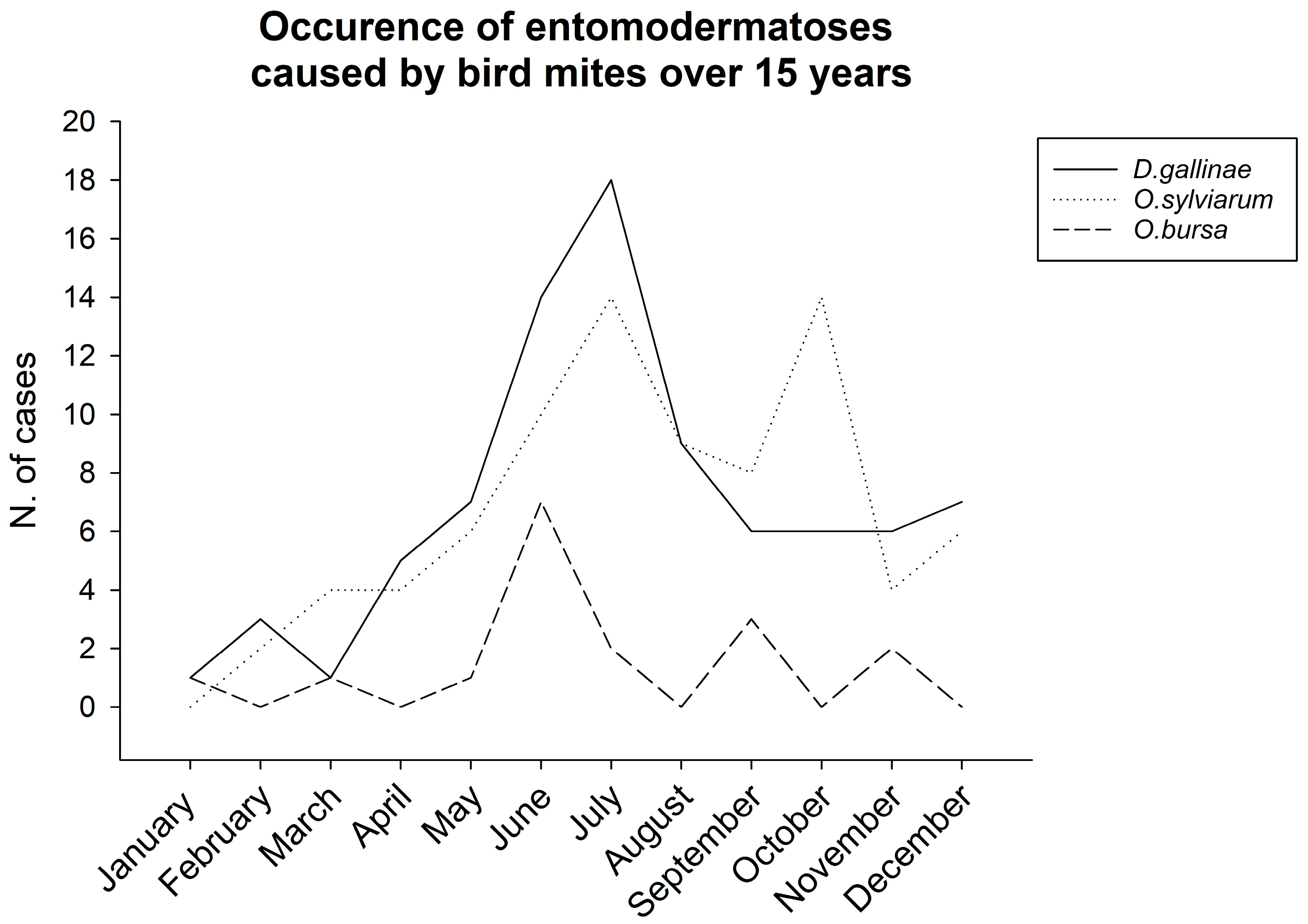
By analysing the period in which the request for environmental dust examination arrived at our laboratories, we reconstructed the distribution curves of infestations caused by the three species of poultry mites in Italy over the last 15 years (Figure 2). The graph shows how D. gallinae had a higher frequency of outbreaks during the summer (particularly between May and August) compared to the two species of Ornithonyssus, which appeared to have more annual peaks, with higher frequencies from late spring to late autumn.

3.3. Geographical Distribution of the Mites

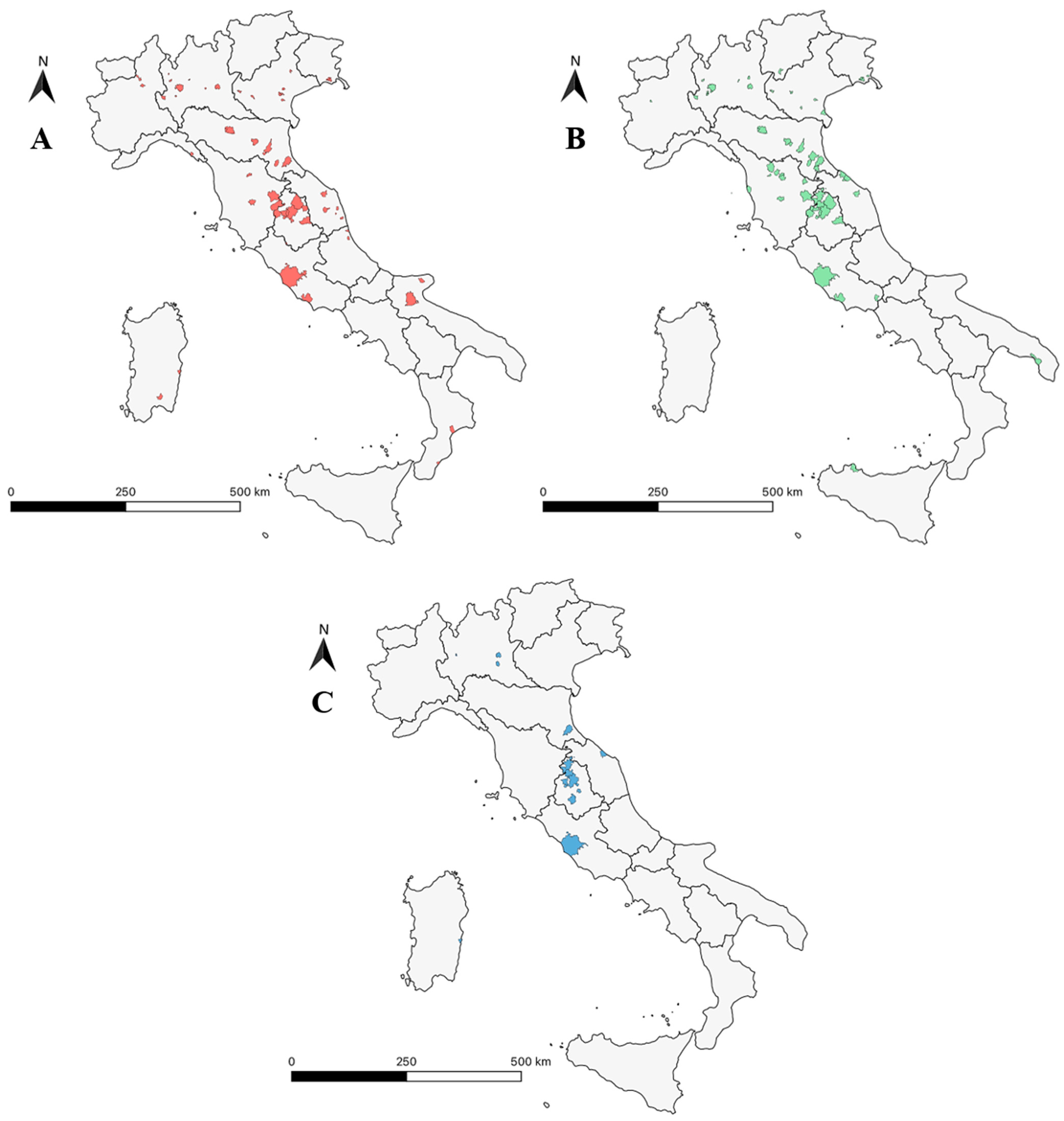
Maps were created in QGIS, showing the distribution of all three species isolated over the years from samples collected from homes located in different regions of Italy, including the islands (Figure 3). In particular, the presence of O. bursa in cases from six different Italian regions is important, which indicates that the species is spreading throughout the national territory.
Almost all positive samples for Dermanyssidae bird mites came from small towns in the inland areas. Analysis of the medical history forms revealed that, in most cases, small family-run poultry farms were the primary source of infestations. However, some cases occurred in strictly urban areas, where patients reported the presence of numerous synanthropic species, particularly pigeons or swallows.
The higher frequency of cases in central Italy was likely due to the higher volume of samples from these areas, owing to their proximity to the laboratories.

3.4. Characteristics of the Skin Lesions

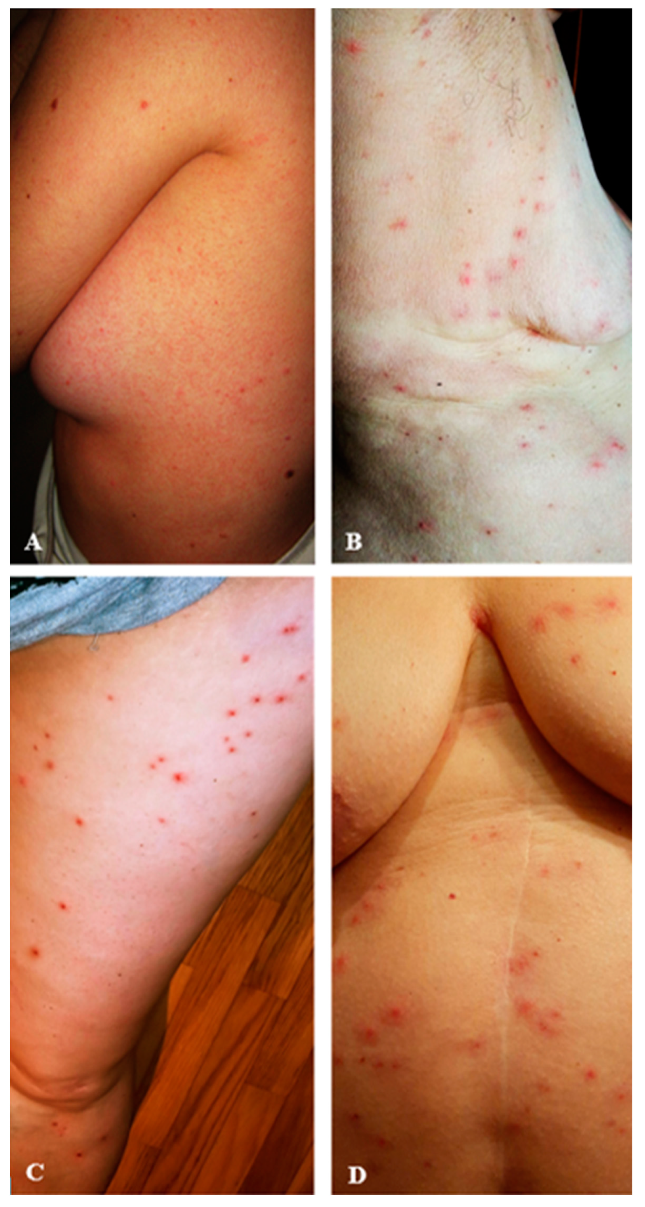
The analysis of the anamnestic forms and of the many photos accompanying the various dust samples allowed us to highlight some differing characteristics in the lesions caused by the three species of mites (Figure 4). Patients with infestation by D. gallinae reported to have generally only a few bites, with only mild redness and itching (Figure 4A). Those with infestation by O. sylviarum mainly had typical micro-papulo-pustular lesions (Figure 4B), and those with O. bursa infestation almost always showed very marked lesions (Figure 4C). Patients with mixed infestation by D. gallinae and O. sylviarum usually had many lesions on their body, which were strongly irritated and itchy (Figure 4D). Therefore, the most severe skin lesions were observed in cases of mixed infestation, when both D. gallinae and Ornithonyssus mites were present simultaneously. Compared to cases with only D. gallinae, the micro-papulo-pustular lesions in mixed infestations were more numerous, often infected (with pus), and the itching was more intense. In general, Ornithonyssus species appeared to be more aggressive towards humans, as they consistently caused visible skin lesions, while D. gallinae, in many of the cases we analysed, caused smaller lesions or just itching and mild skin redness due to a persistent and bothersome crawling sensation on the skin.

4. Discussion

Over the past fifteen years, dust samples from over 4900 suspected cases of entomodermatosis, originating from every Italian region, were analysed by the Urania Research Centre and the Department of Veterinary Medicine of Perugia—Parasitology Section.
In this study, we report that about 4% of these dermatological cases were found to be caused by avian Dermanyssoidea, with the most common species being D. gallinae and O. sylviarum (often associated) and, more rarely, due to O. bursa. This is an important finding, as it represents the first large-scale epidemiological study in the literature. Numerous individual case reports have indeed been described, particularly in recent years [2,4,9,20,21,22,23,24,25,26,27,28,29,30], or there have been literature reviews analysing cases of entomodermatosis caused by poultry mites [16,17,26], but no studies have systematically analysed episodes of human dermatitis caused by Dermanyssoidea. Considering that the analysed samples came from private homes and that, in indoor environments, most human infestations are caused by arthropods transported by pets (mainly fleas, ticks, Sarcoptes scabiei, and Cheyletiella spp.) or by those living inside homes (Pyemotes vetricosus, Glycyphagus domesticus, Cimex lectularius, and Bethylidae wasps), this percentage attributable to poultry mites is not negligible, highlighting the ability of these avian parasites to readily adapt to domestic environments and utilise humans as substitute hosts to sustain their development cycle [4]. The increasing number of reports of attacks on non-avian hosts, including humans and companion animals, may indicate the host expansion of the mites; there may be many factors contributing to this expansion of poultry mites: climate change (especially global warming), the growing population of laying hens, the widespread presence of synanthropic animals, globalisation with increased travel and trade, and the rise in human populations, farms, and small animal densities [14,17,31].
Regarding the distribution of parasites across the national territory, the graphical representation using QGIS allowed us to visualise how the three species are present throughout Italy. Notably, the presence of O. bursa in cases from six different Italian regions indicates that the species is now widespread across the country and no longer confined to the warm island regions of Sicily and Sardinia, where it was previously reported [4,20,32]. Its ubiquitous distribution suggests a significant adaptation of this species to colder temperatures. In effect, while D. gallinae has a cosmopolitan distribution and O. sylviarum is primarily associated with temperate regions, O. bursa is predominantly found in tropical and subtropical areas, although its presence was occasionally recorded in Southern Europe [2,4,20,32]. The majority of the positive samples for Dermanyssidae bird mites came from small towns, mainly located in inland areas. The analysis of the sample origins and environmental data for each case, reported in the medical history forms, revealed the correlation of infestations both with urban and rural contexts, with particular emphasis on the relationship between the presence of domestic and wild birds and the onset of dermatitis: indoor infestations were often linked to small family poultry farms (primarily laying hens), a practice still widespread in small rural villages in Italy. This finding, involving individuals who have daily direct contact with birds, is consistent with the definition of occupational dermatitis as applied to gamasoidosis by several authors [9,13,29,33]. Instead, in urban areas, outbreaks have shown that the presence of synanthropic species, especially pigeons, or abandoned nests (such as those of pigeons or swallows) are the most common causes of infestations inside homes.
The analysis of the distribution over the course of the year of human entomodematosis episodes caused by avian Dermanyssoidea suggests that their occurrence is likely linked to the increased ease of mite entry into homes during spring and summer. This is due to the frequent opening of doors and windows, as well as the presence of pigeons and other birds on roofs, balconies, and windowsills. Sometimes, the infestation of certain households can be linked to the return of swallows from their winter migration and their transportation of O. sylviarum, which is a permanent parasite on the host. Although all three mites have a continuous cycle throughout the year in intensive farms, it is certainly faster during the summer period [5]. These could be the reasons for the peaks of infestation that we observed from late spring to late summer. Autumn outbreaks for O. sylviarum may be due to swallows leaving their nests for seasonal migration, forcing mites left in the nests to seek new hosts for a blood meal.
Regarding the species involved in human gamasoidosis episodes, many authors argue that the species most often responsible is D. gallinae [18,28]. Our data do not support this observation, as the number of episodes in which D. gallinae and O. sylviarum were isolated were practically equal (83 vs. 81), and many cases involved mixed infestations. This was likely due to the difficulty in differentiating D. gallinae from mites of the genus Ornithonyssus. In fact, despite the well-known morphological differences between these mites, their characteristics are often difficult to detect, even when carefully examining them under a microscope. The dorsal idiosomal shields are well known and important for species identification. These differ markedly in their distal part, which is pointed in O. bursa, distinctly narrowed in O. sylviarum, and truncated in D. gallinae [34]. However, visualising the dorsal shield or the anal and genital plates in engorged mites is very challenging due to the presence of blood in the gut. Furthermore, often, only immature stages of the parasite (larvae or nymphs) are isolated, where it is even harder to identify distinctive morphological features. For these reasons, we believed that it was very useful to suggest new identifying features, particularly at the level of chelicerae and pedipalps, which are more easily detectable, even in blood-engorged specimens, and this could help in the correct identification of species during human infestations. In fact, to this day, the microscopic identification of the mite is largely considered the only currently available confirmatory tool for identifying the causative agent, as the clinical history and physical examination of the patient are evidently inadequate for providing a firm diagnosis of gamasoidosis [16], since the lesions caused by poultry mites are non-specific and polymorphic [26]. Symptoms range from simple itching, accompanied by a bothersome crawling sensation, to troublesome dermatitis, often with micro-papulo-pustular lesions, sometimes excoriated due to scratching [11,26]. Mite attacks in residential areas usually occur at night, while occupational cases in poultry farms primarily occur during the day [33]. As a result, more severe skin manifestations are observed in urban settings, where mites find favourable conditions for development (low light, hiding places, high humidity) and can feed for an adequate period (e.g., at night while their host is sleeping). In contrast, when infestations occur in poultry farms and humans are affected, skin lesions occur almost exclusively on exposed areas of the body, and since mites are disturbed by the host’s continuous movement, they are able to bite for only very short periods, failing to engorge; this is especially true with D. gallinae, which often superficially bites the skin without causing visible lesions, resulting only in itchiness and redness [4]. The differential diagnosis includes bites from other biting arthropods (e.g., insects such as mosquitoes, bedbugs, or fleas), scabies, pediculosis, or non-parasitic dermatitis (such as cholinergic urticaria); in some cases, doctors may even refer the patient for psychiatric treatment, mistakenly thinking that these crawling and itching sensations are merely a phobia (e.g., diagnosing it as Ekbom syndrome or delusional ectoparasitosis) [15,17,24,29,30].
A correct diagnosis in cases of gamasoidosis is also essential in order to identify the source of the infestation (often small family poultry farms or nests of pigeons or other birds near windows/balconies/terraces) and implement proper environmental decontamination measures or treatment of infested animals, resolving both the dermatological issue and the infestation in the animals [11,35]. As pure examples, we can report two particular recently diagnosed cases, which highlight how the resolution of the infestation was only possible thanks to the microscopic identification of the poultry mite species involved: a domestic outbreak following the introduction of hunted pheasants infested with O. sylviarum into the home, and a case in an arcade, where, after our diagnosis of a D. gallinae infestation, some pigeon nests were found outside. Occasional diagnosis in humans can also be helpful in the early identification of the presence of gamasides within farms, especially those with a low number of chickens in which the presence of the parasite could be overlooked, allowing for treatment before the infestation reaches very high levels.
As for the lesions, although they are non-specific and the detection of the mite in the domestic environment is essential for diagnostic purposes, in our opinion, their severity reflects the species of Dermanyssoidea responsible; according to our experience, the most severe dermatitis cases were caused by infestations of mites from the Ornithonyssus genus, even if the number of specimens found in the analysed samples was always lower than that of D. gallinae. This demonstrates the greater aggressiveness of species like O. sylviarum and O. bursa compared to D. gallinae, which, when present as the sole species, is responsible only for very mild forms of dermatitis. In our opinion, this may also be due to the different shape of the chelicerae, structures used by mites for feeding. The chelicerae of D. gallinae are long and flexible, resembling needles, whereas those of O. sylviarum and O. bursa are more robust and adapted to tearing the skin. This explains the differences in the observed lesions. The low number of Ornithonyssus specimens found, even in mixed infestations with D. gallinae, is probably the reason why Macronyssidae are often underdiagnosed. In the literature, there are reports of severe dermatitis caused by Dermanyssidae mites [21,23], comparable to the dermatitis that we observed with Ornithonyssus species. This may be due to a misdiagnosis of Macronyssidae mites or to the simultaneous presence of D. gallinae and specimens of the genus Ornithonyssus that are not identified.
Furthermore, in many cases, mites of the genus Dermanyssus were collected directly by the dermatologist on the patient’s skin and sent to the laboratory for identification, without collecting additional samples of environmental dust from the home. This highlights, on the one hand, the high invasiveness of these mites, as they can infiltrate through clothing and quickly reach the skin, but on the other hand, this may have underestimated possible cases of mixed infestations with mites of the genus Ornithonyssus.
This work, with its innovative methodology and large-scale data collection, not only improves the understanding of bird mite infestation dynamics but also provides useful tools for control and prevention, contributing to public health protection and the improvement of environmental sanitation operations. This is particularly important considering that gamasoidosis is an emerging zoodermatosis that can occur in any environment, urban or rural [30]; for this reason, healthcare teams worldwide must be aware of its increasing incidence and include such entomodematoses among the differential diagnoses in cases of acute pruritus or dermatitis with no apparent cause and that are unresponsive to standard treatments [11,15,25,30]. Finally, it should not be overlooked that the potential medical significance of poultry mites is exacerbated by the fact that they can carry and transmit zoonotic diseases of bacterial, viral, and parasitic origin [9,17,36]. So far, the vector role of Dermanyssoidea has been widely demonstrated in avian hosts, but Banović et al. [9] have already reported this possibility in humans, having found Bartonella spp. in an engorged specimen of D. gallinae isolated during an episode of entomodematosis and subsequently confirming serological positivity for Bartonella spp. in one of the individuals with the lesions. Our results, therefore, highlight the importance of conducting further epidemiological research and increasing awareness among both veterinary and medical professionals in order to ensure timely diagnosis, treatment, and preventive measures in the course of gamasoidosis.

5. Conclusions

In conclusion, this first large-scale epidemiological study provides a significant contribution to the analysis of Dermanyssoidea mite infestations in birds, with a particular focus on cases of human dermatitis caused by D. gallinae, O. sylviarum, and O. bursa. Thanks to the rigorous methodology adopted, particularly the microscopic analysis of environmental dust samples, it was possible to observe in detail the morphological characteristics of the species involved, leading to the discovery of new details regarding their chelicerae and palps, which are useful for a more accurate identification. We were able to determine that, in Italy, spring and summer are the peak seasons for most of these dermatitis cases; additionally, the creation of distribution maps highlighted a widespread presence of these species across the country, suggesting their adaptation to environmental conditions different from those traditionally associated with them. Finally, we determined that lesions caused by species belonging to the genus Ornithonyssus were always more severe compared to those due to Dermanyssus and that, in the dust samples examined, both genera were often present.

Author Contributions

Conceptualisation, I.M. and M.A.P.; methodology, M.A.P.; software, S.P. and L.B.; investigation, I.M., S.P. and M.A.P.; data curation, I.M., L.B. and M.A.P.; writing—original draft preparation, I.M.; writing—review and editing, I.M., S.P. and L.B.; supervision, M.A.P. All authors have read and agreed to the published version of the manuscript.

Funding

This research received no external funding.

Institutional Review Board Statement

Not applicable.

Informed Consent Statement

Informed consent was obtained from all subjects involved in this study.

Data Availability Statement

The original contributions presented in this study are included in the article. Further inquiries can be directed to the corresponding author.

Acknowledgments

The authors thank Maria Pia Franciosini from Department of Veterinary Medicine (Perugia, Italy) for her support and valuable insights and Gregory Brandt from EcoSafe Labs and Eco-Safe Pest Control (Dallas, TX, USA) for his support in translating this article into English.

Conflicts of Interest

The authors declare no conflicts of interest.

References

  1. Sparagano, O.A.E.; George, D.R.; Finn, R.D.; Giangaspero, A.; Bartley, K.; Ho, J. Dermanyssus gallinae and chicken egg production: Impact, management, and a predicted compatibility matrix for integrated approaches. Exp. Appl. Acarol. 2020, 82, 441–453. [Google Scholar] [CrossRef]
  2. Waap, H.; Aguin-Pombo, D.; Maia, M. Case Report: Human Dermatitis Linked to Ornithonyssus bursa (Dermanyssoidea: Macronyssidae) Infestation in Portugal. Front. Vet. Sci. 2020, 7, 567902. [Google Scholar] [CrossRef] [PubMed]
  3. Kilpinen, O. How to obtain a bloodmeal without being eaten by a host: The case of poultry red mite, Dermanyssus gallinae. Physiol. Entomol. 2005, 30, 232–240. [Google Scholar] [CrossRef]
  4. Cafiero, M.A.; Viviano, E.; Lomuto, M.; Raele, D.A.; Galante, D.; Castelli, E. Dermatitis due to Mesostigmatic mites (Dermanyssus gallinae, Ornithonyssus [O.] bacoti, O. bursa, O. sylviarum) in residential settings. J. Dtsch. Dermatol. Ges. 2018, 16, 904–906. [Google Scholar] [CrossRef]
  5. Sárkány, P.; Bagi, Z.; Süli, Á.; Kusza, S. Challenges of Dermanyssus gallinae in Poultry: Biological Insights, Economic Impact and Management Strategies. Insects 2025, 16, 89. [Google Scholar] [CrossRef] [PubMed]
  6. Mullens, B.A.; Owen, J.P.; Kuney, D.R.; Szijj, C.E.; Klingler, K.A. Temporal changes in distribution, prevalence and intensity of northern fowl mite (Ornithonyssus sylviarum) parasitism in commercial caged laying hens, with a comprehensive economic analysis of parasite impact. Vet. Parasitol. 2009, 160, 116–133. [Google Scholar] [CrossRef]
  7. Murillo, A.C.; Mullens, B.A. A review of the biology, ecology, and control of the northern fowl mite, Ornithonyssus sylviarum (Acari: Macronyssidae). Vet. Parasitol. 2017, 246, 30–37. [Google Scholar] [CrossRef]
  8. Roy, L.; Chauve, C.M.; Buronfosse, T. Contrasted ecological repartition of the Northern Fowl mite Ornithonyssus sylviarum (Mesostigmata: Macronyssidae) and the chicken red mite Dermanyssus gallinae (Mesostigmata: Dermanyssidae). Acarologia 2010, 50, 207–219. [Google Scholar] [CrossRef]
  9. Banović, P.; Foucault-Simonin, A.; Papić, L.; Savić, S.; Potkonjak, A.; Jurišić, A.; Radenković, M.; Mijatović, D.; Simin, V.; Bogdan, I.; et al. One health approach to study human health risks associated with Dermanyssus gallinae mites. Heliyon 2024, 10, e30539. [Google Scholar] [CrossRef]
  10. Swayne, D.E. Diseases of Poultry, 14th ed.; Wiley-Blackwell: Hoboken, NJ, USA, 2020; pp. 1138–1140. [Google Scholar]
  11. Orton, D.I.; Warren, L.J.; Wilkinson, J.D. Avian mite dermatitis. Clin. Exp. Dermatol. 2000, 25, 129–131. [Google Scholar] [CrossRef]
  12. Mullen, G.R.; Durden, L.A. Medical and Veterinary Entomology, 3rd ed.; Academic Press: Cambridge, MA, USA; Academic Press, Elsevier: London, UK, 2018; pp. 533–595. [Google Scholar]
  13. Moroni, B.; Barlaam, A.; Misia, A.L.; Peano, A.; Rossi, L.; Giangaspero, A. Dermanyssus gallinae in non-avian hosts: A case report in a dog and review of the literature. Parasitol. Int. 2021, 84, 102378. [Google Scholar] [CrossRef]
  14. Cafiero, M.A.; Barlaam, A.; Camarda, A.; Radeski, M.; Mul, M.; Sparagano, O.; Giangaspero, A. Dermanysuss gallinae attacks humans. Mind the gap! Avian Pathol. 2019, 48, S22–S34. [Google Scholar] [CrossRef] [PubMed]
  15. Collgros, H.; Iglesias-Sancho, M.; Aldunce, M.J.; Expósito-Serrano, V.; Fischer, C.; Lamas, N.; Umbert-Millet, P. Dermanyssus gallinae (chicken mite): An underdiagnosed environmental infestation. Clin. Exp. Dermatol. 2013, 38, 374–377. [Google Scholar] [CrossRef]
  16. Kavallari, A.; Küster, T.; Papadopoulos, E.; Hondema, L.S.; Øines, Ø.; Skov, J.; Sparagano, O.; Tiligada, E. Avian mite dermatitis: Diagnostic challenges and unmet needs. Parasite Immunol. 2018, 40, e12539. [Google Scholar] [CrossRef]
  17. George, D.R.; Finn, R.D.; Graham, K.M.; Mul, M.F.; Maurer, V.; Moro, C.V.; Sparagano, O.A. Should the poultry red mite Dermanyssus gallinae be of wider concern for veterinary and medical science? Parasit. Vectors 2015, 8, 178. [Google Scholar] [CrossRef] [PubMed]
  18. Stingeni, L.; Bianchi, L.; Hansel, K.; Neve, D.; Foti, C.; Corazza, M.; Bini, V.; Moretta, I.; Principato, M. Dermatitis caused by arthropods in domestic environment: An Italian multicentre study. J. Eur. Acad. Dermatol. Venereol. 2017, 31, 1526–1533. [Google Scholar] [CrossRef] [PubMed]
  19. Principato, S.; Principato, M.; Stingeni, L.; Mannucci, G.; Moretta, I. Indoor Dust Direct Examination (E.D.P.A.®) and biotic pollution in confined environments. ProScience 2018, 5, 83–89. [Google Scholar]
  20. Castelli, E.; Viviano, E.; Torina, A.; Caputo, V.; Bongiorno, M.R. Avian mite dermatitis: An Italian case indicating the establishment and spread of Ornithonyssus bursa (Acari: Gamasida: Macronyssidae) (Berlese, 1888) in Europe. Int. J. Dermatol. 2015, 54, 795–799. [Google Scholar] [CrossRef]
  21. Cafiero, M.A.; Camarda, A.; Circella, E.; Santagada, G.; Schino, G.; Lomuto, M. Pseudoscabies caused by Dermanyssus gallinae in Italian city dwellers: A new setting for an old dermatitis. J. Eur. Acad. Dermatol. Venereol. 2008, 22, 1382–1383. [Google Scholar] [CrossRef]
  22. Abdigoudarzi, M.; Mirafzali, M.S.; Belgheiszadeh, H. Human Infestation with Dermanyssus gallinae (Acari: Dermanyssidae) in a Family Referred with Pruritus and Skin Lesions. J. Arthropod Borne Dis. 2013, 8, 119–123. [Google Scholar]
  23. de Sousa, M.D.G.; Filho, F.B. Gamasoidosis (bird mite dermatitis): Dermanyssus gallinae in a young boy. Pan Afr. Med. J. 2018, 30, 144. [Google Scholar] [CrossRef] [PubMed]
  24. Navarrete-Dechent, C.; Uribe, P. A case of gamasoidosis caused by Dermanyssus gallinae, misdiagnosed as delusional parasitosis. Clin. Exp. Dermatol. 2018, 43, 950–952. [Google Scholar] [CrossRef]
  25. de Oliveira Alves, A.; Bernardes Filho, F. Gamasoidosis (bird mite dermatitis): A case series in a family. Pediatr. Neonatol. 2018, 59, 102–103. [Google Scholar] [CrossRef] [PubMed]
  26. Cheikhrouhou, S.; Trabelsi, S.; Aloui, D.; Bouchekoua, M.; Khaled, S. Avian mite bites acquired from pigeons: Report of three cases and review of the literature. Tunis. Med. 2020, 98, 241–245. [Google Scholar] [PubMed]
  27. Prouteau, C.; Ameline, M.; Roy, L.; Delaunay, P.; Gangneux, J.P.; Dupuy, A. Prurigo à Dermanyssus gallinae [Gamasoidosis caused by Dermanyssus gallinae]. Ann. Dermatol. Venereol. 2020, 147, 119–122. [Google Scholar] [CrossRef] [PubMed]
  28. Sioutas, G.; Minoudi, S.; Tiligada, K.; Chliva, C.; Triantafyllidis, A.; Papadopoulos, E. Case of Human Infestation with Dermanyssus gallinae (Poultry Red Mite) from Swallows (Hirundinidae). Pathogens 2021, 10, 299. [Google Scholar] [CrossRef]
  29. Sonday, Z.; Todd, G.; Jeebhay, M.F. Occupational bird mite dermatitis (gamasoidosis) among workers in a seed house. Curr. Allergy Clin. Immunol. J. 2023, 36, 178–183. [Google Scholar]
  30. Minagawa, F.H.; Almeida, E.R.M.; Souza, R.C.; Santos, D.C.D.; Haddad Junior, V.; Miot, H.A. Gamasoidosis (avian mite dermatitis) outbreak in a student dormitory. Rev. Soc. Bras. Med. Trop. 2024, 57, e011012024. [Google Scholar] [CrossRef]
  31. Budria, A.; Candolin, U. How does human-induced environmental change influence host-parasite interactions? Parasitology 2014, 141, 462–474. [Google Scholar] [CrossRef]
  32. Masini, P.; Zampetti, S.; Rossetti, M.V.; Biancolini, F.; Miñón Llera, G.; Hansel, K.; Stingeni, L. Gamasoidosis from the tropical fowl mite Ornithonyssus bursa (Acari: Gamasida: Macronyssidae) (Berlese, 1888): Successful eradication of the domestic infestation with dry saturated steam. Int. J. Dermatol. 2022, 61, e230–e232. [Google Scholar] [CrossRef]
  33. Cafiero, M.A.; Galante, D.; Camarda, A.; Giangaspero, A.; Sparagano, O. Why dermanyssosis should be listed as an occupational hazard. Occup. Environ. Med. 2011, 68, 628. [Google Scholar] [CrossRef] [PubMed]
  34. Krantz, G.W.; Walter, D.E. A Manual of Acarology, 3rd ed.; Krantz, G.W., Walter, D.E., Eds.; Texas Tech University Press: Lubbock, TX, USA, 2009. [Google Scholar]
  35. Sparagano, O.A.; George, D.R.; Harrington, D.W.; Giangaspero, A. Significance and control of the poultry red mite, Dermanyssus gallinae. Annu. Rev. Entomol. 2014, 59, 447–466. [Google Scholar] [CrossRef] [PubMed]
  36. Valiente Moro, C.; Chauve, C.; Zenner, L. Vectorial role of some dermanyssoid mites (Acari, Mesostigmata, Dermanyssoidea). Parasite 2005, 12, 99–109. [Google Scholar] [CrossRef] [PubMed]
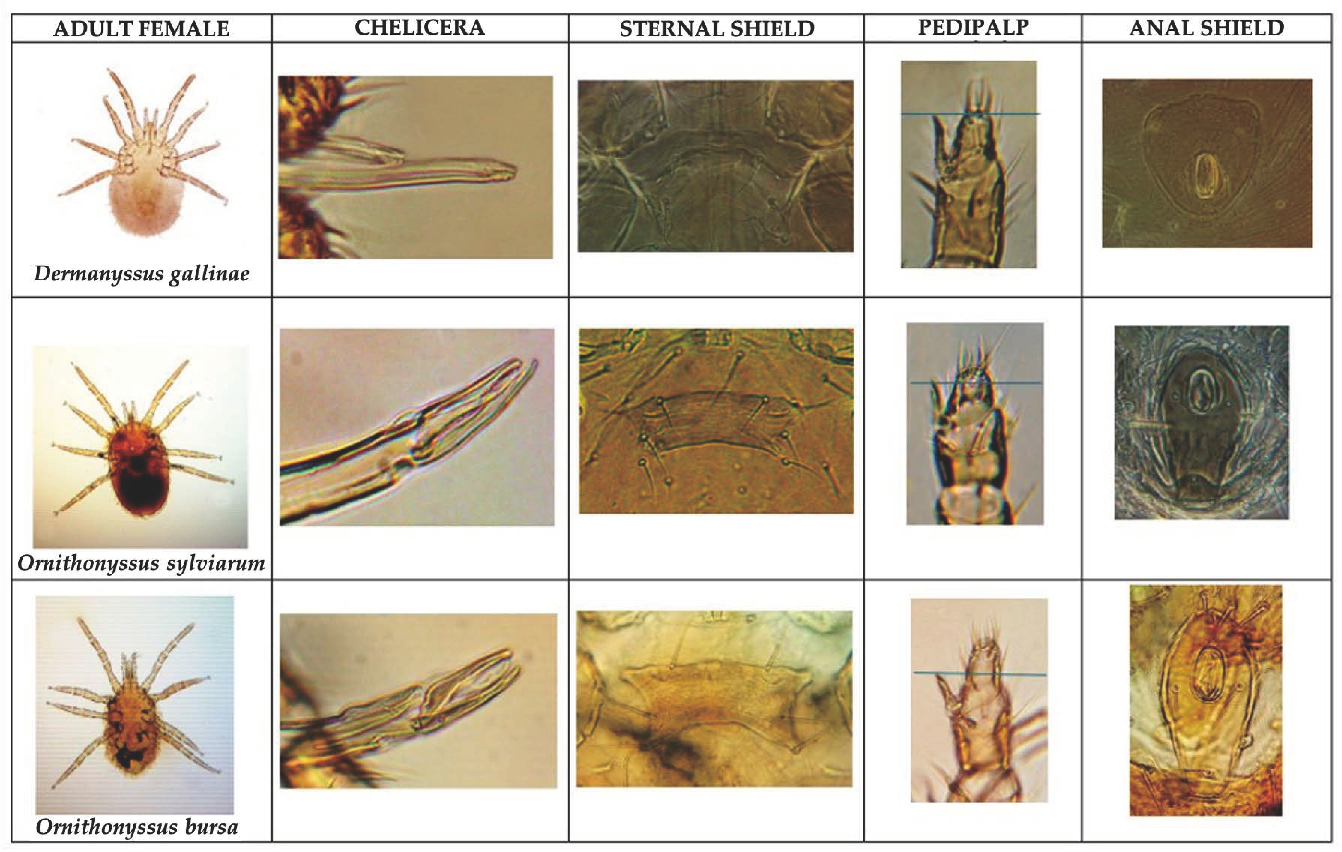
Figure 1. Comparative table showing some morphological characteristics of adult females of the examined Dermanyssoidea mites.
Poultry 04 00021 g001
Figure 2. Graphical representation of the monthly occurrence of bird mite entomodermatosis cases diagnosed in Italy over the past 15 years.
Poultry 04 00021 g002
Figure 3. Geographical distribution of entomodermatosis cases caused by Dermanyssus gallinae (A), Ornithonyssus sylviarum (B), and Ornithonyssus bursa (C) across Italy diagnosed over the past 15 years.
Poultry 04 00021 g003
Figure 4. Lesions caused by Dermanyssoidea bird mites. Patients with infestation by Dermanyssus gallinae (A); Ornithonyssus sylviarum (B); Ornithonyssus bursa (C); Dermanyssus gallinae and Ornithonyssus sylviarum (mixed infestation (D)).
Poultry 04 00021 g004
Disclaimer/Publisher’s Note: The statements, opinions and data contained in all publications are solely those of the individual author(s) and contributor(s) and not of MDPI and/or the editor(s). MDPI and/or the editor(s) disclaim responsibility for any injury to people or property resulting from any ideas, methods, instructions or products referred to in the content.