Article

The Influence of Blue and Red Light on Seed Development and Dormancy in Nicotiana tabacum L.

1
Laboratory of Plant Physiology and Photobiology, Department of Life and Environmental Sciences, Università degli Studi di Cagliari, V.le S. Ignazio da Laconi 13, 09123 Cagliari, Italy
2
Laboratory of Economic and Pharmaceutical Botany, Department of Life and Environmental Sciences, Università degli Studi di Cagliari, V.le S. Ignazio da Laconi 13, 09123 Cagliari, Italy
3
Department of Chemistry, Umeå University, Linnaeus väg 6, 90736 Umeå, Sweden
4
Department of Plant Physiology, Warsaw University of Life Sciences—SGGW, Nowoursynowska Str. 159, 02-776 Warsaw, Poland
*
Authors to whom correspondence should be addressed.
Seeds 2022, 1(3), 152-163; https://doi.org/10.3390/seeds1030014
Submission received: 13 April 2022 / Revised: 12 June 2022 / Accepted: 28 June 2022 / Published: 4 July 2022

Abstract

:
The correct development of seeds is a pivotal requirement for species preservation. This process depends on the balance between sensing the environmental stimuli/stressors and hormone-mediated transduction, which results in physiological responses. The red and blue regions of the electromagnetic spectrum are known to influence seed dormancy and germination. Here, we report on the effects induced by the blue (peak at 430 nm) and red (peak at 650 nm) regions of the electromagnetic spectrum on seeds from photo- and skotomorphogenetic capsules developed under white, blue, or red light. Regardless of exposure, seeds from skotomorphogenetic capsules showed an almost absent dormancy in association with altered germination kinetics. Conversely, in seeds from photomorphogenetic capsules, the exposure to the blue region induced skotomorphogenetic-like effects, while the exposure to the whole visible range (350–750 nm), as well as to only the red region, showed a dose-related trend. The observed differences appeared to be dependent on the wavelengths in the red and to be based on transduction mechanisms taking place in fruits. While the molecular bases of this differential effect need to be clarified, the results hint at the role played by different light wavelengths and intensities in seed development and germination. These findings may be relevant for applications in crop production and species safeguarding.

1. Introduction

Light is one of the most critical factors affecting plant growth, seed development, and germination. The plethora of functions influenced by light is reflected in the large variety of plant photoreceptors [1]. In this respect, the effects on plants span from photosynthesis and primary production to the direct/indirect involvement of light in almost all phases of the life cycle [2,3,4]. In order to interact with light either for signaling (photoreceptors) or for photosynthesis (photosystems and their external antennas), the evolution process shapes dedicated plant chromophores and their apoproteins, exploiting almost the whole range of the electromagnetic spectrum reaching the surface of our planet [5,6]. In signaling, the light quality and quantity are perceived by photoreceptors and influence several processes, including fruit ripening and seed development [7,8], eventually affecting seed germination by modulating both seed dormancy and vitality [9,10]. For all these reasons, plants exploit light not only as a source of energy but, notably, also for acquiring environmental information [11]. In this respect, the influence of red and/or blue photons on dormancy [12] and germination [13] is well known. Its relevancy, however, remains controversial [14,15]. Among others, the blue and red regions of the electromagnetic spectrum affect plant development by a dose-dependent effect that can be cumulative [16,17]. In fact, the dose of photons may trigger either time-dependent effects, dependent on the fluency [18], or time-independent effects, related to the irradiance [17,19]. To acquire more detail, the effect of red light at 666 nm and 730 nm was extensively studied in model organisms, such as Lactuca ssp. and Arabisopsis thaliana [20,21,22], as well as in many other species, and was found to guide seed germination or dormancy through phytochrome activation/inactivation; however, this effect was not always obvious [23].
In more general terms, the whole process of reproduction, including seed development and maturation, from anthesis to pollination first and from fruit ripening to embryogenesis after, is regulated by electromagnetic signals leading to new individuals capable of preserving the species [11,24,25,26,27]. It was previously observed that etiolated fruits of Nicotiana tabacum L. plants produce larger and heavier seeds with altered levels of hormones which are responsible for its absence of dormancy [28]. These observations were connected to a change in the balance between the sink role of fruits, gaining photosynthates from leaves, and their “self-source” ability, by which fruits produce photosynthates through their photosynthetic parenchyma [28]. In the present study, we focused our investigation by narrowing the visible band to the red (peak at 650 nm) and blue (peak at 430 nm) regions of the electromagnetic spectrum. This allowed us to discriminate between the actual photoactive regions in the visible spectrum and the rest (non-photoactive with respect to the observed effects), with the purpose of linking the effect to a specific type of photoreceptor. The readout of this assessment was performed by analyzing the seed quality in terms of the germination kinetics of seeds collected from both photomorphogenetic capsules (PC) and skotomorphogenetic capsules (SC). While SC seeds had an almost absent dormancy with no significant differences between the three conditions tested, consistent with previous studies performed under white light in N. tabacum L. [28], seeds developed in PC presented different behaviors depending on the type of light under which the seed maturation occurred. In particular, PC seeds from capsules developed under white and red light showed a clear dormancy phase, while, unexpectedly, in PC seeds from capsules developed under blue light, the dormancy was absent, showing a skotomorphogenetic-like behavior. The germination profiles suggest a direct photosynthetically active radiation (PAR) effect of the red region in promoting dormancy in seeds under development and maturation with a dose-related trend. The sensing processes and transduction mechanisms built on the base of these observations seem to occur in fruits rather than in other parts of the plant, and photo-perception is confirmed to be a main determinant in seed development and germination. Results are discussed and contextualized in terms of the roles played by the red light in phytochrome activation. The influence of phytochromes on seed dormancy, affecting hormone biosynthesis, levels, and balance, is also taken into account. Considering previous observations [28], the reported data may have relevant applications in crop production or plant preservation for species where, as in tobacco, etiolation of fruits induces bigger and heavier seeds with reduced dormancy.

2. Materials and Methods

2.1. Growth and Cultivation of Tobacco Plants

Seed collection and germination tests on Nicotiana tabacum plants (cv. Petit Havana) were performed in Cagliari (Sardinia, Italy, latitude: 39°22′30″ N; longitude: 9°11′06″ E; altitude: 60 m above the sea level). After harvest, capsules were dried in the dark at 25 °C and 50% relative humidity. Upon drying, the capsules were collected and stored at 18 °C and 50% relative humidity in the dark for 2–3 weeks prior to the start of the experiments. Before germination tests, seeds were collected and imbibed in sterilized tap water for 24 h at room temperature in the dark. After imbibition, seeds were sowed, and plants were grown for 15–20 weeks at 25 °C with 50% relative humidity and under a light regime of 12 h/day. In all the tested conditions, the light source was a LED lamp with a light intensity of 150–200 μmol photons/(s·m2) at 50 cm from the emission source; in all cases, the light source was positioned at a distance from the plants that allowed it to reach an average of ~3000–4000 lux. Plants were first grown under white LED light for 6–8 weeks, and, two weeks before anthesis, they were divided into three experimental groups and kept either under white, blue (peak at 430), or red (peak at 650 nm) LED light. Experiments were performed on at least three test plants for each experimental condition. Finally, seeds resulting from the 6 conditions (white, blue, and red from both etiolated and non-etiolated capsules) were subjected to germination tests performed under white light as described in the Section Germination Tests (see below).

2.2. Membranes Isolation and Solubilization

Plastidial membranes were isolated at 4 °C in the dark [8,29]. After removing the sepals and the seeds, 10 g of capsules were washed in distilled water, dried in paper towels, mixed in a ratio 1:10 (g/mL) with grinding buffer (GB-50 mM MES pH 6.5; 10 mM MgCl2·6H2O; 10 mM CaCl2·2H2O), and blended for 15″. The suspension was subsequently centrifuged (5000× g, 10′, 4 °C) and the pellet resuspended in GB to reach a concentration of 1 mg/mL (final suspension—FS).

2.3. Denaturing Polyacrylamide Gel Electrophoresis (SDS-PAGE)

Sodium dodecyl sulphate-PAGEs with a 10% (w/v) separating gel and a 4% (w/v) stacking gel was used to characterize the solubilized membranes [30]. Briefly, samples were denatured with Rotiload (Roth), boiled for 5′, and centrifuged for 5′, and 5 µL of samples was loaded into the gel. After the electrophoretic separation, the gels were stained with Coomassie brilliant blue G-250.

2.4. Absorption Spectroscopy

For the absorption spectroscopy, 100 μL of membrane suspension was homogenized on ice and solubilized for 20′ under slow shaking at 4 °C in 20 mM n-dodecyl-β-D-maltoside (β-DDM). After a subsequent centrifugation step (17,000× g, 10′, 4 °C), the pellet was discharged, and the supernatant was diluted to 1 mL in GB and used for the measurements. Measurements were performed on a Pharmacia Biotech Ultrospec 4000 spectrophotometer at 4 °C in the range of 370–750 nm with an optical path length of 1 cm (quartz cell, Hellma Analytics).
For the determination of Chl a and Chl b concentrations, the analysis was performed using 80% (v/v) acetone in a ratio 1/100 on 10 μL of FS. Single wavelength measurements were performed at 664 nm and 647 nm and calculated according to [31] using three independent measurements. Amounts of Chl a and b of the controls were used as reference and expressed in relative units as equal to 1. The relative values for the other samples were assigned accordingly.

2.5. Photosynthetic Activity

The photosynthetic parameters of all plants were recorded by using a Mini PPM 100 fluorimeter (EARS, Wageningen, The Netherlands) with a saturating pulse width modulation (PWM) of 7.2 kHz at 455 nm carrying about 2500–3000 μmol PAR [32]. The fluorescence was recorded with an infrared optical bandpass filter in the range of 700–780 nm. The photosynthetic activity of each tested group of plants was evaluated by comparing the maximum fluorescence (Fm), the fluorescence in ambient light (F0), and the photosynthetic yield (Y). Measurements at the end of the experiment on capsules (Section 3.1) and daily measurements on leaves (Section 3.5) were performed in three repetitions. Each measurement was performed on different capsules or leaves from three different plants for each tested group (growing under different light conditions). Measurements are shown as mean ± SD for each experimental group.

2.6. Induction of Etiolation

Etiolation was induced as previously described in [28]. Briefly, in plants on the 2nd day post anthesis (DPA), the senescent corolla was removed, and a triple-layered pocket made of black crepe paper was applied over the green ovary, which was still at the pre-swelling stage. This setup maintained an efficient gas exchange with the environment while the ovary remained shielded from light. The pocket was maintained in place for the entire period of swelling until seed maturation (2–3 weeks). The etiolation was induced in at least three capsules for each test plant.

2.7. Germination Tests

Germination tests were performed according to [28] to investigate whether the light quality during seed maturation affected the seed germination rate. Tests were conducted in triplicate on 30 DPA seeds from different individuals using 25 seeds for each experimental condition.
Seeds were imbibed in tap water for 24 h at room temperature in the dark. Germination tests were performed in sterile Petri dishes. Seeds were placed on a sterile, black layer of porous paper opportunely perforated in order to create a grid of holes to accommodate each seed. This setup prevented the seed from moving while allowing its hydration by an underlying layer of sterile, wet cotton. This setup allowed us to clearly see the early evasion of the primary root as well the overture of the emitted cotyledon leaves.
Seeds were grown for 15 days at 25 °C with 50% relative humidity and under white, cold light with a photoperiod of 12 h/day and a light intensity of 150–200 μmol photons/(s·m2) at a distance from the light source that allowed it to receive an average of ~2500–3000 lux. Depending on the stage of the germination test, data were collected once or twice a day at intervals of 8–12 h. These conditions were selected according to the finding reported previously [28]. Seed viability was tested indirectly by checking the germination rate for all six experimental conditions 6–12 months after harvest. For all groups, a vitality rate of 95–98%, comparable to that of the controls, was observed.

2.8. Spectral Analysis of Light

The spectral analysis of the light sources was performed using a Thunder Optics Mini USB Spectrometer. Measurements in the 350–750 nm range were performed by placing the spectrometer 10 cm from the light source. The intensities of light at 666 nm and 730 nm were recorded and compared between the different light sources. The relative intensities were finally plotted versus seed germinability for each given condition of light.

2.9. Data Analysis

The germination was monitored for 15 days, starting from the sowing day. As in [14], two basic indexes were recorded: the total number of germinated seeds (nt) expressed in % (Nt); and the partial number of germinated seeds (np) from the last check expressed in % (Np). The percentages of the two indexes refer to the total number of seeds sowed (25 per each condition). Thus, the two parameters were defined as: Nt = (nt/25) × 100; Np = [(nt − nt − 1)/25] × 100, where nt is the number of germinated seeds at a given time, and nt − 1 is the number of germinated seeds at the last-but-one measurement. The data were then adapted to a logistic curve using the software mycurvefit (www.mycurvefit.com, accessed on 1 April 2022), and the obtained equation was graphically represented using the software PLOT 2. Germination tests were performed in three repetitions, each one on seeds from a different plant. All the values resulting from each dataset are given as mean ± SD.

3. Results

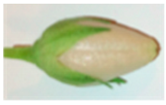
3.1. In the Dark, Ovaries but Not Sepals Undergo Etiolation

As previously observed, when the fruit set of Nicotiana tabacum L. takes place in the dark, the ovary undergoes etiolation. In these conditions, the sepals remain photomorphogenetic (Figure 1a), and the seeds of these fruits carry peculiar germination features [28]. As a further characterization of these samples, the plastidial membranes of the capsules (non-etiolated—NE and etiolated—E) were isolated and analyzed. These experiments showed a clear variation in protein pattern (Figure 1b) and a decrease in the photosynthetic cofactors (Figure 1c) of etiolated tissues (mainly etioplast membranes) when compared with the non-etiolated controls (chloroplast membranes).
A quantification of chlorophylls a and b in the sample indicated a concentration about two orders lower than in the controls (Table 1).
Interestingly, the chlorophyll a/b ratio remained around physiological conditions (normally between 1.8 and 2.2) for both capsules, suggesting the presence of a certain degree of active thylakoids/chloroplasts also in the etiolated tissues and in agreement with a persisting residual photosynthetic activity (Table 1). These results allowed the qualification of the degree of etiolation, indicating not only a deep skotomorphogenetic state for the ovary matured in absence of light but also the retention of a constant background of photosynthetic potential ready to perform.
Differently from what was observed in the dark for the ovary, the sepals did not undergo etiolation. Interestingly, the chlorophyll content in this part of the capsule remained comparable to the one of the non-etiolated tissues (Table 1), suggesting a proxied, regulatory dependence from other sites of the plant, such as the leaves.

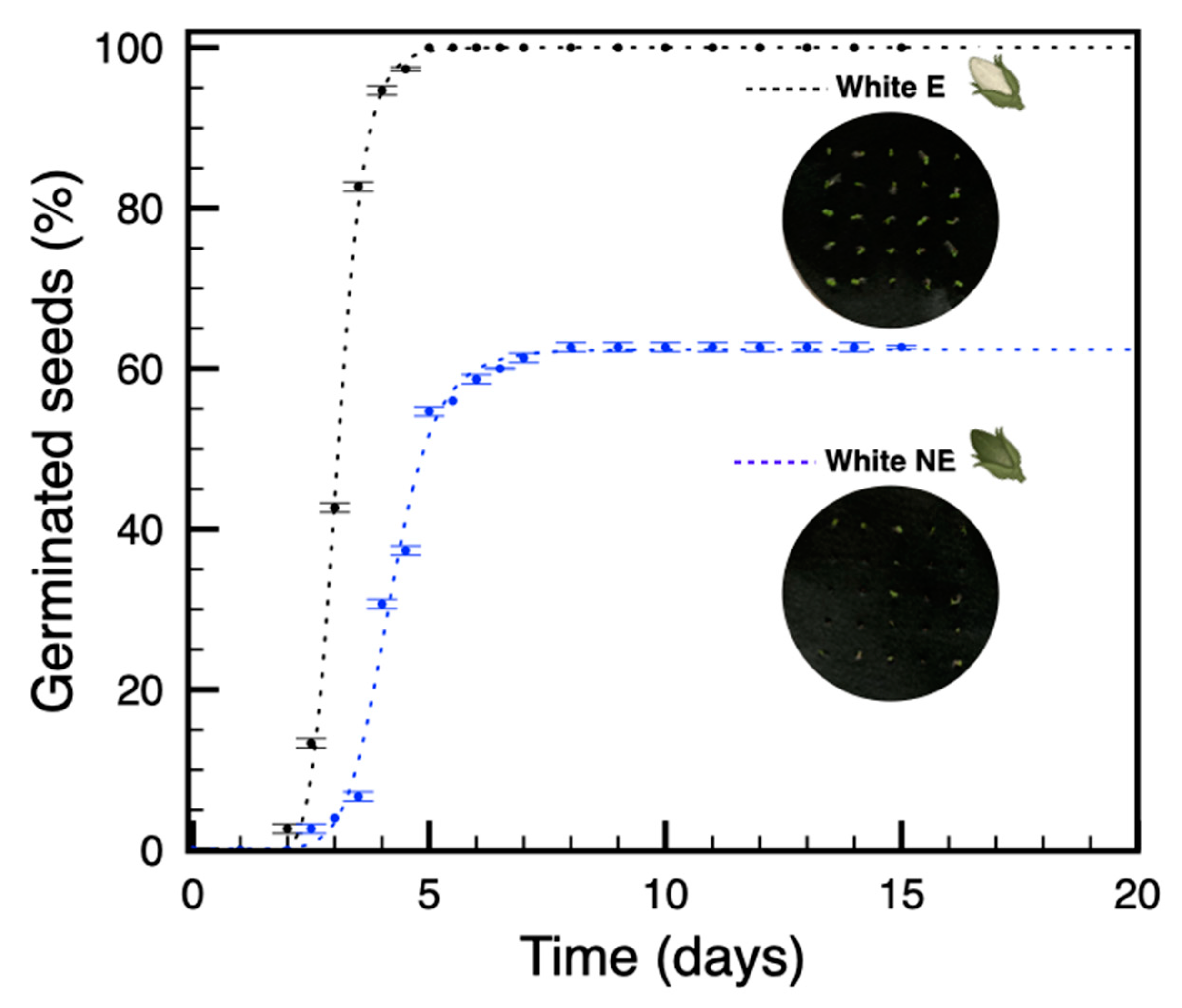
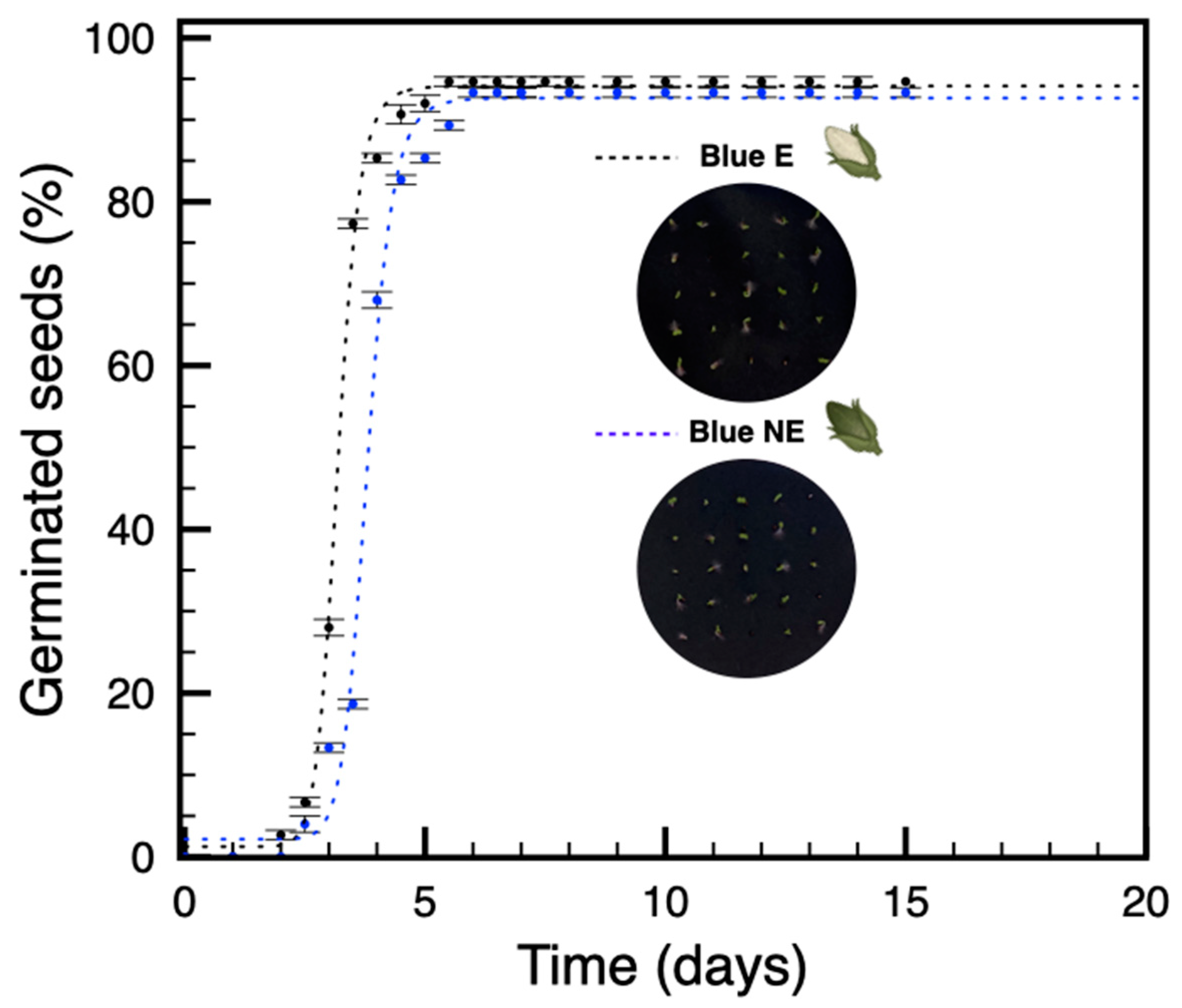
3.2. Seeds from Plants Grown under Blue Light Are Not Dormant

It was previously shown how etiolated capsules from plants grown under white light produce seeds that are characterized by reduced dormancy [28]. In order to expand our understanding of these observations, we investigated whether this effect on dormancy was associated with specific bands in the PAR. A preliminary germination experiment using white light on seeds from the etiolated (CWE) and non-etiolated capsules (CWNE) of plants grown in the presence of white light confirmed the previous findings [28] and allowed us to set a specific control for the tests here reported (Figure 2).
Next, we performed the same experiment, but this time on seeds from the etiolated and non-etiolated capsules of plants grown in the presence of blue light, hereinafter referred to as CBE and CBNE, respectively. Surprisingly, these experiments revealed the absence of dormancy not only in CBE seeds but also in the CBNE ones (Figure 3).
These results suggest that the missing spectral components (red or/and green), which are present in the white light experiments, might be responsible for the observed effect. Consequently, the absence of red/green photons is virtually equivalent to the etiolated condition where the whole light is shielded and none of the photon sources can reach the specific receptors. Minimal differences between CWE and CBE seeds, as well as between CWNE and CBNE seeds, were observed with respect to the time when a given stage of germination was reached; on the contrary, the % of germination changed significantly between the two groups (Table S1).

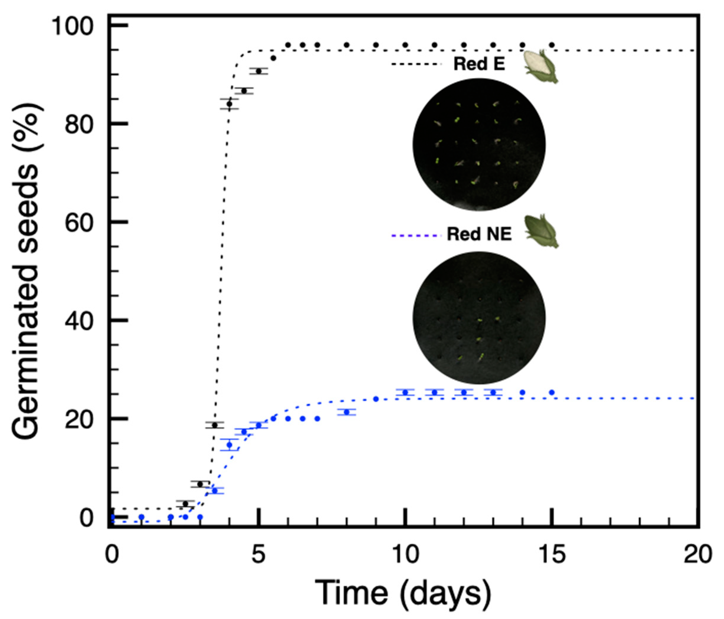
3.3. In Plants Grown under Red Light, Only Seeds from Etiolated Capsules Are Not Dormant

As a further step toward understanding the source of this light-related phenomenon, the same experiments were performed for plants grown under red light. Germination tests were performed on seeds from etiolated and non-etiolated capsules, hereinafter referred to as CRE and CRNE, respectively. Unlike seeds from plants grown under blue light, in these experiments, the red light appeared to promote dormancy in the CRNE seeds, while CRE seeds were not dormant and showed a behavior similar to the CBE condition (Figure 4).
Taking into account the effect associated with the blue light, these differences suggest that the red light might be the trigger responsible for dormancy activation, explaining why the absence of this light quality (experiments in blue light) determined the absence of dormancy in CBE and CBNE seeds (Figure 3). In this case, the differences between CRE and CWE or CBE were found to be more significant with respect to both the time course for each germination stage and the associated % of germination (Table S1).

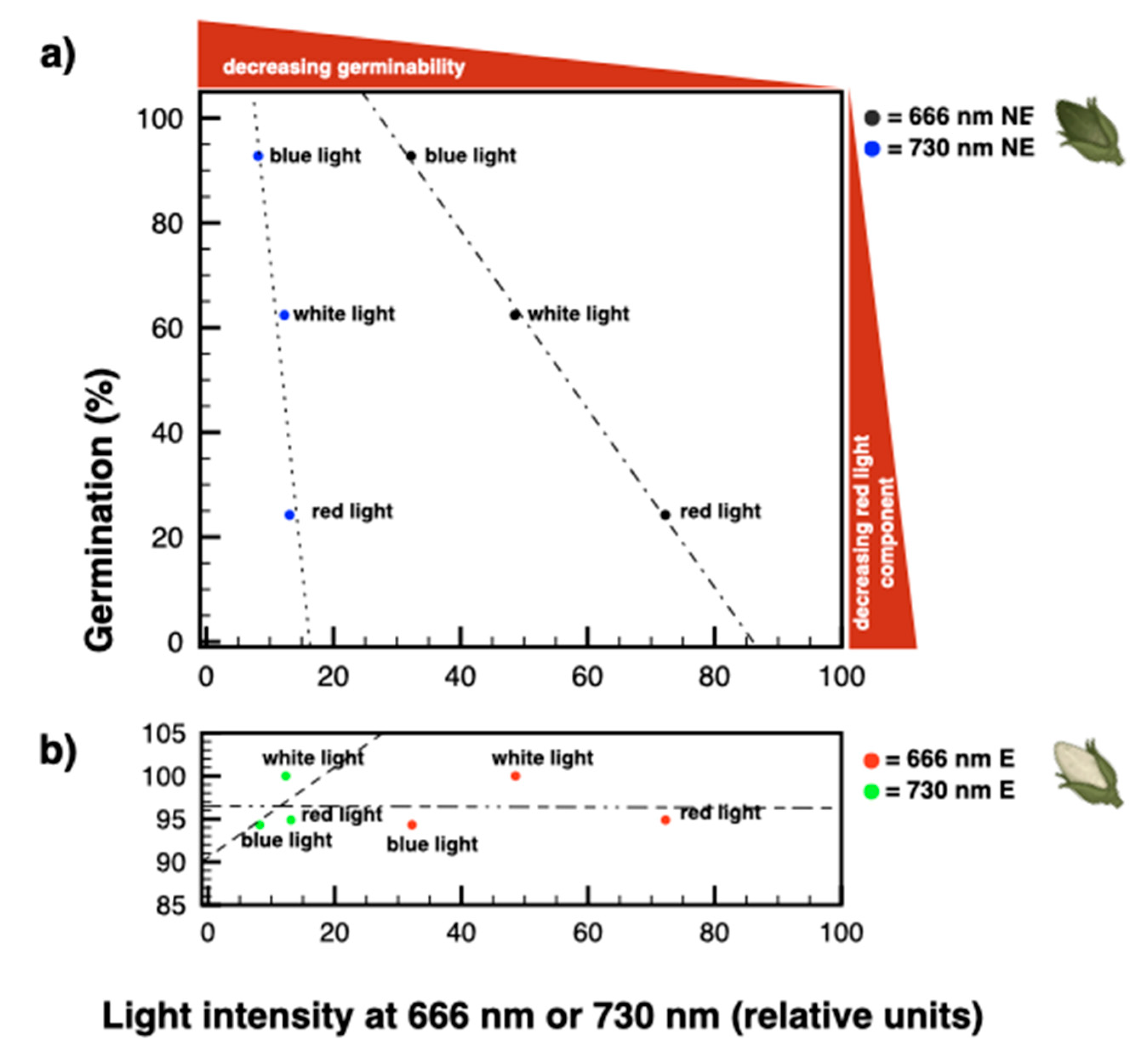
3.4. The Amount and Quality of Red Light Induce a Proportional Increase of Dormancy in Seeds from Non-Etiolated Capsules

To clarify the role of the PAR’s red component, we analyzed the amount of red (666 nm) and far-red (730 nm) light in the LED sources used. Interestingly, the three groups of experiments showed quantitative differences in the non-etiolated conditions with a clear correlation between dormancy and the 666 nm red light necessary for phytochrome activation and an independency between the 730 nm and far-red light (Figure 5a).
In particular, at 666 nm, CRNE seeds had a more pronounced dormancy compared to the CWNE ones, for which the red component of light was less represented (Figure 5a). In the same line, the minimal contribution of red light in the CBNE condition was not sufficient to induce dormancy in the related seeds (Figure 5a). In comparison, there was no evident association with the radiation at 730 nm, a fact that can be explained only partially by the smaller contribution of this band to the light intensity. On the contrary, independent from the level of red radiation considered, the CWE, CBE, and CRE conditions were very similar to each other, confirming, as expected, the absence of induced effects (Figure 5b).

3.5. Plant Growth under Different Lights Has Comparable Photosynthetic Rates

In order to ascertain the general physiological state of the plants subjected to specific light conditions, the photosynthetic performances were assessed daily during the entire experimental time. Background fluorescence (F0) and maximum fluorescence (Fm) levels were used to calculate the photosynthetic yield (Y) and the F0/Fm ratio for leaves of plants from all the experimental groups. These measurements showed only minimal differences; therefore, no stress effect attributable to the light conditions used was detected. Values of photosynthetic efficiency were comparable during the entire experiment, allowing the exclusion of the light conditions from the causes responsible for the observed differences in seed dormancy.
A typical measurement performed five days before the end of the experiment is reported in Table 2, showing only minimal differences compared to the control (white light).

4. Discussions

Seed maturation is a multi-factor process strongly shaped by the environment. The environmental conditions during reproduction, and particularly the conditions of light, affect germination behaviors, determine the degree of dormancy. Light is one of the most active environmental factors known, able to shape the physiology and morphology of organisms and organs, including seeds. Here, we estimated the indirect influence of light on seed differentiation through its direct influence on the maternal plants. Studies were performed by evaluating the dependence of germinability/dormancy on the light conditions under which seeds differentiated. These experiments were performed on plants grown under white light or under polychromatic lights with a dominance of either blue or red components. The type of light influenced seed maturation differentially, evidencing a direct, dose-related effect of the red light on seed dormancy. In agreement with previous studies [28], the complete absence of light induced a deep skotomorphogenetic state of the ovary that retained a residual photosynthetic potential that was ready to perform (Figure 1 and Table 1). This physiological state is associated with the development of seeds with an unlocked dormancy. On the contrary, the absence of only one type of light (red or blue) allowed us to observe the differential effects on the seed dormancy that increased when the red component increased. Consequently, the altered dormancy in seeds developed under dark conditions (etiolation) or unbalanced light can be directly associated with the missing red component of photosynthetically active radiation (Figure 5). Considering that dormancy is also absent in seeds developed under blue light, i.e., under a negligible red component, in this species, seed development and maturation seem to take place by sensing the environment through the presence/absence of red light. These properties are very informative and provide direct evidence for a perception of light strictly localized in the photosynthetic parenchyma of the capsules rather than in the leaves. This is not surprising when considering that, ontogenetically, the flower’s carpels directly derive from leaves and are expected to be similarly influenced by light. N. tabacum is a positive photoblastic species with small seeds [33]. In natural environments, seeds are released from the capsule into the soil, penetrating easily into it. In these conditions, red light, unlike other regions of visible light, can still reach the seed, explaining the observed responsiveness to the red light and its perception through phytochromes.
Systemic processes related to red-light perception are known to take place in leaves and to depend on the phytochrome [34]. Consistently, the ovarium’s carpels are typically characterized by a fully functional photosynthetic parenchyma, and they, like leaves, are expected to depend on stimuli/stressors from red light [35], at least with respect to seed maturation. Further data analyses presented here also evidence the direct influence of the red light at 666 nm rather than of the other phytochrome-sensitive component in the far-red region at around 730 nm (Figure 5). This observation and the related findings are strictly dependent on phytochrome activation rather than on its inactivation or both. Consequently, during tobacco seed maturation, the multi-factor processes creating seed dormancy do not occur according to the reciprocity law. Moreover, seed maturation in tobacco depends on the dose of red light on the ovarium’s carpels and is responsive only to phytochrome activation, indicating that it operates under dose-effect regimes typical for high-irradiance responses.
Being synthesized during seed development, phytochromes mediate between environmental stimuli/stressors and germination, acting indirectly through the levels of hormones and directly by gene regulation mechanisms. According to this last point, it has been known for a long time that phytochromes mediate germination responses on the basis of the red/far red ratio occurring during seed development [20,36]. This is possibly due to the phytochromes’ properties, sensitivity to environmental stimuli/stressors, and their direct/indirect ability to influence germination [37]. Accordingly, depending on the light quality received by the fruit during seed maturation [38], phytochromes can be found in their inactive red-absorbing form (Pr) or in their active far-red-absorbing form (Pfr). In this respect, these biliproteins are well known for directly regulating the germination responses to light [39], but particularly for acting indirectly by up-regulating the conversion of inactive gibberellins into bioactive ones, hence, stimulating germination [40,41]. Phytochromes are also associated with the catabolism of abscisic acid, hence, they also promote germination and reduce dormancy [42]. The levels of these two hormones were found to change in N. tabacum seeds developed in the absence of light [28], and, in the present work, similar hormone-related differences were expected between similar germination profiles and the light quality. Importantly, not only did the germinative behavior in etiolated seeds confirm previous findings, but it was restricted to increasing doses of red light that caused progressive phytochrome activation.
In conclusion, the mechanism of light in phytochromes suggests the specific involvement of these photoreceptors in mediating light signals in the fruit parenchyma, most likely by means of hormone variations, finally resulting in a direct influence on seed maturation. Accordingly, the present study provides direct evidence for similar, primary photo-regulatory mechanisms in the ovarium’s carpel and leaves and suggests a light-mediated environmental influence on seed maturation in tobacco plants.

Supplementary Materials

The following supporting information can be downloaded at: https://www.mdpi.com/article/10.3390/seeds1030014/s1, Table S1: Time course values when a given germination stage is reached.

Author Contributions

Conceptualization: D.P. and E.C. Data curation: D.P., D.F., and E.C. Formal analysis: D.P., D.F. and E.C. Funding acquisition: D.F. and W.P.S. Investigation: A.M., D.P., D.F. and E.C. Methodology: D.P. Project administration: D.P. and E.C. Resources: D.P., A.M., and W.P.S. Supervision: D.P., A.M., and W.P.S. Validation: D.P., D.F. and P.H. Visualization: D.P. and D.F. Writing—original draft: D.P., D.F., E.C., P.H., W.P.S. and A.M. All authors have read and agreed to the published version of the manuscript.

Funding

This research received no external funding.

Institutional Review Board Statement

Not applicable.

Informed Consent Statement

Not applicable.

Data Availability Statement

Not applicable.

Acknowledgments

D.F. and W.P.S. acknowledge The Carl Tryggers Foundation for Scientific Research (CTS 19:324). The authors thank Maria Bonaria Farci for kindly providing the drawings of the N. tabacum capsules and helping with graphics.

Conflicts of Interest

The authors declare no conflict of interest.

References

  1. Möglich, A.; Yang, X.; Ayers, R.A.; Moffat, K. Structure and Function of Plant Photoreceptors. Annu. Rev. Plant Biol. 2010, 61, 21–47. [Google Scholar] [CrossRef] [PubMed] [Green Version]
  2. Petroutsos, D.; Tokutsu, R.; Maruyama, S.; Flori, S.; Greiner, A.; Magneschi, L.; Cusant, L.; Kottke, T.; Mittag, M.; Hegemann, P.; et al. A blue-light photoreceptor mediates the feedback regulation of photosynthesis. Nature 2016, 537, 563–566. [Google Scholar] [CrossRef] [PubMed]
  3. Kami, C.; Lorrain, S.; Hornitschek, P.; Fankhauser, C. Light-Regulated Plant Growth and Development. Curr. Top. Dev. Biol. 2010, 91, 29–66. [Google Scholar] [PubMed] [Green Version]
  4. Collu, G.; Farci, D.; Esposito, F.; Pintus, F.; Kirkpatrick, J.; Piano, D. New insights into the operative network of FaEO, an enone oxidoreductase from Fragaria x ananassa Duch. Plant Mol. Biol. 2017, 94, 125–136. [Google Scholar] [CrossRef] [PubMed]
  5. Li, L.; Ljung, K.; Breton, G.; Schmitz, R.J.; Pruneda-Paz, J.; Cowing-Zitron, C.; Cole, B.J.; Ivans, L.J.; Pedmale, U.V.; Jung, H.-S.; et al. Linking photoreceptor excitation to changes in plant architecture. Genes Dev. 2012, 26, 785–790. [Google Scholar] [CrossRef] [Green Version]
  6. Paik, I.; Huq, E. Plant photoreceptors: Multi-functional sensory proteins and their signaling networks. Semin. Cell Dev. Biol. 2019, 92, 114–121. [Google Scholar] [CrossRef]
  7. Ilić, Z.S.; Fallik, E. Light quality manipulation improves vegetable quality at harvest and postharvest: A review. Environ. Exp. Bot. 2017, 139, 79–90. [Google Scholar] [CrossRef]
  8. Piano, D.; Cocco, E.; Guadalupi, G.; Kalaji, H.M.; Kirkpatrick, J.; Farci, D. Characterization under quasi-native conditions of the capsanthin/capsorubin synthase from Capsicum annuum L. Plant Physiol. Biochem. 2019, 143, 165–175. [Google Scholar] [CrossRef]
  9. Contreras, S.; Bennett, M.A.; Metzger, J.D.; Tay, D. Maternal Light Environment During Seed Development Affects Lettuce Seed Weight, Germinability, and Storability. HortScience 2008, 43, 845–852. [Google Scholar] [CrossRef] [Green Version]
  10. Yang, L.-W.; Liu, S.; Lin, R. The role of light in regulating seed dormancy and germination. J. Integr. Plant Biol. 2020, 62, 1310–1326. [Google Scholar] [CrossRef]
  11. Galvão, V.C.; Fankhauser, C. Sensing the light environment in plants: Photoreceptors and early signaling steps. Curr. Opin. Neurobiol. 2015, 34, 46–53. [Google Scholar] [CrossRef] [PubMed] [Green Version]
  12. Roso, R.; Nunes, U.R.; Müller, C.A.; Paranhos, J.T.; Lopes, S.J.; Dornelles, S.H.B.; Bertagnolli, C.M.; Huth, C.; Forte, C.T.; Menegaes, J.F. Light quality and dormancy overcoming in seed germination of Echium plantagineum L. (Boraginaceae). Braz. J. Biol. 2021, 81, 650–656. [Google Scholar] [CrossRef]
  13. Hernández-Adasme, C.; Silva, H.; Escalona, V. Germination of green and red lettuce cultivars is influenced by LED light spectrum. Acta Hortic. 2022, 1337, 361–366. [Google Scholar] [CrossRef]
  14. Nemahunguni, N.K.; Gupta, S.; Kulkarni, M.G.; Finnie, J.F.; Van Staden, J. The effect of biostimulants and light wavelengths on the physiology of Cleome gynandra seeds. Plant Growth Regul. 2019, 90, 467–474. [Google Scholar] [CrossRef]
  15. Chen, D.; Yuan, Z.; Wei, Z.; Hu, X. Effect of maternal environment on seed germination and seed yield components of Thlaspi arvense. Ind. Crop. Prod. 2022, 181, 114790. [Google Scholar] [CrossRef]
  16. Ptushenko, O.S.; Ptushenko, V.V.; Solovchenko, A.E. Spectrum of Light as a Determinant of Plant Functioning: A Historical Perspective. Life 2020, 10, 25. [Google Scholar] [CrossRef] [Green Version]
  17. Zhong, M.; Zeng, B.; Tang, D.; Yang, J.; Qu, L.; Yan, J.; Wang, X.; Li, X.; Liu, X.; Zhao, X. The blue light receptor CRY1 interacts with GID1 and DELLA proteins to repress GA signaling during photomorphogenesis in Arabidopsis. Mol. Plant 2021, 14, 1328–1342. [Google Scholar] [CrossRef]
  18. Paradiso, R.; Proietti, S. Light-Quality Manipulation to Control Plant Growth and Photomorphogenesis in Greenhouse Horticulture: The State of the Art and the Opportunities of Modern LED Systems. J. Plant Growth Regul. 2021, 41, 742–780. [Google Scholar] [CrossRef]
  19. Landi, M.; Zivcak, M.; Sytar, O.; Brestic, M.; Allakhverdiev, S.I. Plasticity of photosynthetic processes and the accumulation of secondary metabolites in plants in response to monochromatic light environments: A review. Biochim. Biophys. Acta 2019, 1861, 148131. [Google Scholar] [CrossRef]
  20. Hayes, R.G.; Klein, W.H. Spectral quality influence of light during development of Arabidopsis thaliana plants in regulating seed germination. Plant Cell Physiol. 1974, 15, 643–653. [Google Scholar] [CrossRef]
  21. Cammarisano, L.; Donnison, I.S.; Robson, P.R.H. The Effect of Red & Blue Rich LEDs vs Fluorescent Light on Lollo Rosso Lettuce Morphology and Physiology. Front. Plant Sci. 2021, 12, 603411. [Google Scholar] [CrossRef] [PubMed]
  22. Samuolienė, G.; Viršilė, A.; Miliauskienė, J.; Haimi, P.J.; Laužikė, K.; Brazaitytė, A.; Duchovskis, P. The Physiological Response of Lettuce to Red and Blue Light Dynamics Over Different Photoperiods. Front. Plant Sci. 2021, 11, 610174. [Google Scholar] [CrossRef] [PubMed]
  23. Milberg, P.; Andersson, L.; Thompson, K. Large-seeded spices are less dependent on light for germination than small-seeded ones. Seed Sci. Res. 2000, 10, 99–104. [Google Scholar] [CrossRef] [Green Version]
  24. Pinna, M.S.; Mattana, E.; Cañadas, E.M.; Bacchetta, G. Effects of pre-treatments and temperature on seed viability and germination of Juniperus macrocarpa Sm. Comptes Rendus. Biol. 2014, 337, 338–344. [Google Scholar] [CrossRef]
  25. Cuena-Lombraña, A.; Porceddu, M.; Dettori, C.A.; Bacchetta, G. Predicting the consequences of global warming on Gentiana lutea germination at the edge of its distributional and ecological range. PeerJ 2020, 8, e8894. [Google Scholar] [CrossRef]
  26. Handa, A.K.; Tiznado-Hernández, M.E.; Mattoo, A.K. Fruit development and ripening: A molecular perspective. In Plant Biotechnology and Agriculture; Academic Press: Cambridge, MA, USA, 2012; pp. 405–424. [Google Scholar]
  27. El-Keblawy, A.; Soliman, S.; Al-Khoury, R.; Ghauri, A.; Al Rammah, H.; Hussain, S.E.; Rashid, S.; Manzoor, Z. Effect of maturation conditions on light and temperature requirements during seed germination of Citrullus colocynthis from the Arabian Desert. Plant Biol. 2018, 21, 292–299. [Google Scholar] [CrossRef]
  28. Farci, D.; Haniewicz, P.; Cocco, E.; De Agostini, A.; Cortis, P.; Kusaka, M.; Loi, M.C.; Piano, D. The Impact of Fruit Etiolation on Quality of Seeds in Tobacco. Front. Plant Sci. 2020, 11, 563971. [Google Scholar] [CrossRef]
  29. Haniewicz, P.; Floris, D.; Farci, D.; Kirkpatrick, J.; Loi, M.C.; Büchel, C.; Bochtler, M.; Piano, D. Isolation of Plant Photosystem II Complexes by Fractional Solubilization. Front. Plant Sci. 2015, 6, 1100. [Google Scholar] [CrossRef] [Green Version]
  30. Farci, D.; Collu, G.; Kirkpatrick, J.; Esposito, F.; Piano, D. RhVI1 is a membrane-anchored vacuolar invertase highly expressed in Rosa hybrida L. petals. J. Exp. Bot. 2016, 67, 3303–3312. [Google Scholar] [CrossRef] [Green Version]
  31. Porra, R.J.; Thompson, W.A.; Kriedemann, P.E. Determination of accurate extinction coefficients and simultaneous equations for assaying chlorophylls a and b extracted with four different solvents: Verification of the concentration of chlorophyll standards by atomic absorption spectroscopy. Biochim. Biophys. Acta BBA Bioenerg. 1989, 975, 384–394. [Google Scholar] [CrossRef]
  32. De Agostini, A.; Caltagirone, C.; Caredda, A.; Cicatelli, A.; Cogoni, A.; Farci, D.; Guarino, F.; Garau, A.; Labra, M.; Lussu, M.; et al. Heavy metal tolerance of orchid populations growing on abandoned mine tailings: A case study in Sardinia Island (Italy). Ecotoxicol. Environ. Saf. 2019, 189, 110018. [Google Scholar] [CrossRef] [PubMed]
  33. Hashimoto, T.; Yamaki, T. Interaction of gibberellin a3, and inorganic phosphate in tobacco seed germination. Plant Cell Physiol. 1962, 3, 175–187. [Google Scholar] [CrossRef]
  34. Ballaré, C.L. Phytochrome Responses: Think Globally, Act Locally. Trends Plant Sci. 2017, 22, 909–911. [Google Scholar] [CrossRef] [PubMed]
  35. Brazel, A.J.; Ó’Maoiléidigh, D.S. Photosynthetic activity of reproductive organs. J. Exp. Bot. 2019, 70, 1737–1754. [Google Scholar] [CrossRef]
  36. McCullough, J.M.W.; Shropshire, W., Jr. Physiological predetermination of germination responses in Arabidopsis thaliana (1.) HEYNH. Plant Cell Physiol. 1970, 11, 139–148. [Google Scholar] [CrossRef]
  37. Donohue, K.; Heschel, M.S.; Butler, C.M.; Barua, D.; Sharrock, R.A.; Whitelam, G.C.; Chiang, G.C.K. Diversification of phytochrome contributions to germination as a function of seed-maturation environment. New Phytol. 2007, 177, 367–379. [Google Scholar] [CrossRef]
  38. Casal, J.J.; Sánchez, R.A. Phytochromes and seed germination. Seed Sci. Res. 1998, 8, 317–329. [Google Scholar] [CrossRef]
  39. Koornneef, M.; Bentsink, L.; Hilhorst, H. Seed dormancy and germination. Curr. Opin. Plant Biol. 2002, 5, 33–36. [Google Scholar] [CrossRef] [Green Version]
  40. Toyomasu, T.; Kawaide, H.; Mitsuhashi, W.; Inoue, Y.; Kamiya, Y. Phytochrome Regulates Gibberellin Biosynthesis during Germination of Photoblastic Lettuce Seeds. Plant Physiol. 1998, 118, 1517–1523. [Google Scholar] [CrossRef] [Green Version]
  41. Ogawa, M.; Hanada, A.; Yamauchi, Y.; Kuwahara, A.; Kamiya, Y.; Yamaguchi, S. Gibberellin Biosynthesis and Response during Arabidopsis Seed Germination. Plant Cell 2003, 15, 1591–1604, Erratum in Plant Cell 2004, 16, 783. [Google Scholar] [CrossRef] [Green Version]
  42. Seo, M.; Hanada, A.; Kuwahara, A.; Endo, A.; Okamoto, M.; Yamauchi, Y.; North, H.; Marion-Poll, A.; Sun, T.-P.; Koshiba, T.; et al. Regulation of hormone metabolism in Arabidopsis seeds: Phytochrome regulation of abscisic acid metabolism and abscisic acid regulation of gibberellin metabolism. Plant J. 2006, 48, 354–366. [Google Scholar] [CrossRef] [PubMed]
Figure 1. Comparison between non-etiolated and etiolated capsules (a) by assessing the differences in the protein pattern of their plastidial membranes (b). The same samples also showed a different level of photosynthetic pigments, as observed in their absorption spectra after membrane solubilization (c).
Seeds 01 00014 g001
Figure 2. Germination profiles for seeds from etiolated (E—black, dashed line and dots) and non-etiolated capsules (NE—blue, dashed line and dots) from plants grown under white light. The two insets represent typical Petri dishes on the 5th day of the germination test for both types of seed (E and NE).
Seeds 01 00014 g002
Figure 3. Germination profiles for seeds from etiolated (E—black, dashed line and dots) and non-etiolated capsules (NE—blue, dashed line and dots) from plants grown under blue light. The two insets represent typical Petri dishes on the 5/6th days of the germination test for both types of seed (E and NE).
Seeds 01 00014 g003
Figure 4. Germination profiles for seeds from etiolated (E—black, dashed line and dots) and non-etiolated capsules (NE—blue, dashed line and dots) from plants grown under red light. The two insets represent typical Petri dishes on the 5/6th days of the germination test for both types of seed (E and NE).
Seeds 01 00014 g004
Figure 5. Dependence of germination on the dose of red light. Each of the three lamps used (blue, red, and white) had a red component that was measured at 666 nm and 730 nm. (a) In seeds from non-etiolated capsules (NE), the red component at 666 nm increased from blue to white to red light (black dots), and this relative increment is associated with an increment in dormancy. The 730 nm contribution was much less (blue dots), and it was evidently not correlated with dormancy; (b) in seeds from etiolated capsules (E), the red components at 666 nm (red dots) and 730 nm (green dots) are both not correlated with dormancy.
Seeds 01 00014 g005
Table 1. Photosynthetic parameters and chlorophyll content for ovary and sepals in etiolated and non-etiolated samples.
Capsule TypeCapsule PartF0/FmY (%) *Chl a **
(Relative)
Chl b **
(Relative)
Chl a/b
(Ratio)
Seeds 01 00014 i001 Seeds 01 00014 i0020.35 ± 0.0165.3 ± 0.33111.82
Seeds 01 00014 i003n.a. ***n.a. ***0.960.941.87
Seeds 01 00014 i004 Seeds 01 00014 i0050.57 ± 0.0643.3 ± 0.210.0130.0192.20
Seeds 01 00014 i006n.a. ***n.a. ***0.720.781.99
* Y= (Fm − F0)/ Fm = 1 − (F0/Fm); ** = chlorophyll content expressed relatively to the control amount (for details, see Materials and Methods); *** = not applicable.
Table 2. Measures of the main photosynthetic parameters from leaves from plants grown under white, blue, or red LED light.
White Light (Control)Blue LightRed Light
F0/Fm0.286 ± 0.0040.290 ± 0.0030.271 ± 0.002
Y (%) *71.367 ± 0.41670.933 ± 0.32172.767 ± 0.231
* Y= (Fm − F0)/ Fm = 1 − (F0/Fm).
Publisher’s Note: MDPI stays neutral with regard to jurisdictional claims in published maps and institutional affiliations.

