Case Report

Rare Case of Hemoglobin Lepore Trait in a Bangladeshi Patient with Polyarthritis and Fever: Case Description and Brief Literature Review

by
Nira Ferdous
1,2,
Md. Nazrul Islam
2,3,
Abu Talha Mustakim
1 and
Johannes J. Rasker
4,*
1
Department of Medicine, MH Samorita Hospital and Medical College, Tajgaon Dhaka 1208, Bangladesh
2
Modern One Stop Arthritis Care and Research Center® (MOAC&RC®), Dhaka 1209, Bangladesh
3
Department of Rheumatology, Bangladesh Medical University (BMU), Shahbag Dhaka 1000, Bangladesh
4
Faculty of Behavioural, Management and Social sciences, Department Psychology, Health and Technology, University of Twente, Drienerloolaan 5, 7522 Enschede, NB, The Netherlands
*
Author to whom correspondence should be addressed.
Rheumato 2025, 5(4), 16; https://doi.org/10.3390/rheumato5040016
Submission received: 8 August 2025 / Revised: 30 October 2025 / Accepted: 4 November 2025 / Published: 10 November 2025

Abstract

Introduction: In hemoglobinopathies, the amount of globin synthesis in hemoglobin (Hb) or its structure is altered. Clinical features are related to the rate and kind of structural aberrations. The heterozygous form of the Lepore syndrome resembles minor thalassemia both clinically and hematologically. On electrophoresis, abnormal Hb Lepore fractions are found at a rate of 5–15%, with a mildly higher percentage of HbF and lower HbA. In general, Hb Lepore heterozygotes are asymptomatic. Case presentation: A 32-year-old male was admitted to our hospital with complaints of pain and swelling in multiple large joints and high-grade fever for 11 days. His past history was unremarkable; one of his sisters had the β-thalassemia trait. On physical examination, he was moderately anemic, with mild hepatomegaly and normal spleen; both knees and ankles were tender and swollen. Laboratory showed mild microcytic hypochromic anemia with variables similar to the thalassemia trait and signs of inflammation with very high CRP, serum ferritin, and leukocytosis. Blood sugars were increased. Hb electrophoresis showed an abnormal pattern with mild elevation in HbS, normal Hb F, mild reduction in HbA, and high HbA2, compatible with heterozygosity for the Hb Lepore beta chain variant. He was initially diagnosed with diabetes (treated with insulin) and sepsis from unknown origin, but fever and joint pains did not respond to NSAIDs or antibiotics. He had very good response on high-dose methylprednisolone. Undifferentiated arthritis was diagnosed in the patient with Hb Lepore, and he was treated with oral prednisolone and sulfasalazine (SSZ). At follow up, the patient was doing well. He refused further investigations and did not allow testing on his family members. In summary: Hb Lepore is a rare hemoglobinopathy linked to thalassemia, which may manifest with musculoskeletal problems. Our patient with the Hb Lepore trait presented with undifferentiated polyarthritis and fever, but in our case, a causal relationship remains unclear. This is one of the first adult cases of Hb Lepore in Bangladesh and the first with arthritis of unknown origin. The prevalence of Hb Lepore in Bangladesh is unknown.

1. Introduction

The hemoglobin (Hb) Lepore trait is a rare hemoglobinopathy linked to thalassemia, which may manifest with musculoskeletal problems [1,2,3]. We describe a 32-year-old male with the Hb Lepore trait, presenting with undifferentiated polyarthritis and fever. Hb Lepore is a structurally abnormal Hb variant with worldwide distribution. It has an abnormal globin chain, resulting from an unequal crossover of the δ and β genes due to a disarrangement of homologous chromosome during meiosis [1]; it was first described in 1958 in the American Lepore family, with Italian roots [2]. To date, three Hb Lepore variants are known—Hb Lepore Washington Boston (δ87/β116), Hb Lepore Baltimore (δ50/β86), and Hb Lepore Hollandia (δ22/β50)—all characterized by different gene deletion breakpoints and with varying clinical manifestations [3]. Hb Lepore Washington Boston (δ87/β116) is the most common of the three and occurs worldwide [1]. In the homozygous form, there is no HbA and HbA2, and Hb contains only HbF and Lepore, with mean Hb Lepore levels of 15% (8–30%). In heterozygous cases, the Hb contains HbA, Hb Lepore, HbA2, and HbF in varying amounts, with a mean Hb Lepore level of 10% (5–15%) [3]. The Lepore trait’s F fraction is slightly higher than in the beta thalassemia trait [3]. People with homozygous Hb Lepore, who have no normal HbA, present with a clinical picture ranging from thalassemia intermedia to major. In all variants of Hb Lepore, the δβ hybrid chain is synthesized significantly less than the β-chain, consequently with an overall reduction in non-α-globin chains [3]. The clinico-hematological profile of people with Hb Lepore depends on the Hb Lepore mutation and the amount of abnormal Hb genes. Generally, people with the Hb Lepore trait have no symptoms or only mild fatigue and anemia [3]. On the other hand, people with homozygous Hb Lepore are more severely affected with moderate to severe anemia, splenomegaly, and jaundice [4]. In compound heterozygotes for hemoglobins (Hbs) S, C, and E with Hb Lepore, the clinical picture may vary a lot, but, in general, it resembles those of Hbs S, C, or E/β-thalassemia compound states [5,6,7,8].
We aimed to present the clinical case of a 32-year-old male with the Hb Lepore trait, manifesting with polyarthritis and high-grade fever, and to highlight that this is, to the best of our current knowledge, one of the first adult cases of the Hb Lepore trait reported in Bangladesh.

2. Case Presentation

Clinical history and presentation: Our patient is a 32-year-old male who was admitted to our hospital with complaints of pain and swelling in multiple large joints and high-grade fever that persisted for 11 days. He also reported a history of burning micturition with increased frequency and urgency, as well as severe generalized weakness. There was no history of inflammatory low back pain, rash, sexual exposure, or urethral discharge, and no family history of arthritis or psoriasis and gout. His past history was unremarkable, without allergies or repeated or discontinued medications. Family history revealed that one of his sisters had the β-thalassemia trait; however, due to financial constraints, the sister and other family members refused to undergo Hb electrophoresis.
Physical examination: We found the patient moderately anemic with mild hepatomegaly; the spleen was not palpable, and both knees and ankles were tender and swollen.
Complementary investigations: Laboratory: CRP, 312 mg/L; CBC, Hb 7.9 g/dL; total RBC, 4.33 m/μL; HCT, 25.2%; MCV, 58.1 fL; MCH, 18.1 pg; and MCHC, 31.2 g/dL (Table 1). Peripheral blood film (PBF) showed microcytic hypochromic cells with anisopoikilocytosis-like pencil cells, elliptical cells, pear cells, and a few target cells. Total white blood cell count: 24,160 (88% neutrophils) (Table 1); S. Creatinine: 3 mg/dL; uric acid: 8.6 mg/dL. Liver function tests: ALT: 30 U/L; S. Bilirubin: 2.3 mg/dL. Urine: microscopic examination: trace sugar; pus cell: 1–3/HPF; RBC: 2–4/HPF. Immunochromatographic test for Malaria and Kala azar: negative; procalcitonin: 0.21 ng/mL. Fasting blood sugar: 12.9 mmol/L, and 75gm glucose 20 mmol/L 2 h after. Coombs: negative; S. ferritin: 1198.85 ng/mL; anti-CCP: negative; HLA B 27: negative. Mantoux test: 2 mm (non-significant). Hb electrophoresis showed an abnormal pattern with mildly elevated HbS, normal Hb F, mildly reduced HbA, and high HbA2, suggesting a heterozygous Hb Lepore beta chain variant (Figure 1).
Urine culture was negative; blood culture was negative. As there was no history of sexual exposure or urethral discharge, gonorrhea and chlamydia were not tested. As the patient was not able to pay, no synovial analyses or cultures were performed.
X-rays: Lumbosacral spine: no abnormalities (n.a.). Sacro-Iliac joints: n.a. Chest: n.a.
Ultrasound abdomen: mild hepatomegaly; spleen not enlarged.
Echocardiography: minimal pericardial effusion; otherwise, normal.

Hospital Treatment and Evolution

The patient with the Hb Lepore trait was initially diagnosed with diabetes (treated with short-acting regular human insulin) and sepsis from unknown origin. His fever and swollen painful joints did not respond to sulindac, tramadol, or nalbuphine or to antibiotics, consecutively administered as follows: inj. ceftriaxone 1 gm every 12 h (started on the 2nd day of fever before admission to our hospital, and continued for 2 days in hospital); then, for 14 days, combination of inj. meropenem 1 gm every 12 h and inj. moxifloxacin 400 mg once daily for 10 days, followed by 5 days with flucloxacillin 500 mg capsules every 6 h; afterwards, when there was no effect on his symptoms, we started a combination of inj. piperacillin and tazobactam 4.5 gm every 6 h and Tab. linezolid 600 mg every 12 h for 10 days. Clinically, he did not respond to these antibiotics, and CRP 110.8 and serum ferritin 747.9 remained high with normal procalcitonin; for this reason, we administered, on the 5th day of this antibiotic combination, methylprednisolone 1 g daily for 3 days with dramatic improvement on the 2nd day.
Undifferentiated arthritis was diagnosed in the patient with the Hb Lepore trait and diabetes, and we continued prednisolone 20 mg/day for 3 days, and then 10 mg combined with sulfasalazine (SSZ). Two units of red cell concentrate were administered to maintain Hb 10 g/dL. In this regimen, his 104–106 °F temp and joint pain improved, CRP and ferritin normalized, and Creatinine levels were 1.3 mg/dL.
Discharge: The patient was discharged after 22 days with prednisolone prescribed at a tapering dose starting from 10 mg and with SSZ at a dose escalating to 2 gm, calcium + vit D, esomeprazole 20 mg if needed, folic acid 5 mg, and short-acting regular human insulin.
Follow up: After three months, the patient had no joint pains. His regular medications were SSZ 2 gm/day, Gliclazide MR 30 mg twice a day, Empagliflozin + Linagliptin (10/5 mg) once a day, calcium + vit D, and folic acid 5 mg. On physical examination, no joint abnormalities were observed; the liver and spleen were not palpable. Laboratory: CRP 2.9 mg/L; Hb, 11.5 g/dL; total RBC, 6 × 10¹²/L; HCT, 37.1%; MCV, 61.9 fL; MCH, 19.2 pg; and MCHC, 31.6 g/dL. Total white blood cell count: 5.79 × 10⁹/L (57% neutrophils); S. Creatinine: 0.8 mg/dL; uric acid: 8.7 mg/dL. Liver function tests: ALT, 22 U/L; S. ferritin, 443.23 ng/ml. Urine: sediment, n.a. Fasting blood sugar, 6.4 mmol/L, and 2 h after breakfast, glucose, 8.2 mmol/L.HbA1C 5.6%. The patient refused further investigations and did not allow testing on his family members.

3. Discussion

The Hb Lepore trait may manifest with musculoskeletal problems due to the underlying issue of abnormal hemoglobin affecting red blood cell production and function [3]. We describe a 32-year-old male with the Hb Lepore trait, presenting with undifferentiated polyarthritis and fever. Despite the fact that bone and joint involvements are often seen in thalassemia, in our case, a causal relationship remains unclear. Although most Hb Lepore heterozygotes are asymptomatic; some Italian, Greek, Yugoslavian, and Palestinian heterozygotes have demonstrated mild splenomegaly [9,10]. In addition to being healthy, heterozygotes usually exhibit observable microcytosis and hypochromia, as well as normal Hb levels [7]. In our patient, two units of red cell concentrate were administered to maintain Hb around 10g/dL. Clinical signs in homozygotes might vary from thalassemia intermedia to a transfusion-dependent course similar to that for thalassemia major [7]. In our case, Hb electrophoresis techniques identified the Hb Lepore variant as HbD. Family history, clinical correlation, CBC result, iron profile, and high-performance liquid chromatography (HPLC) findings should be interpreted together to ascertain an appropriate diagnosis. Some HPLC techniques show patterns and pictures reliably enough to make a diagnosis [10]. Hb Lepore has been described in many countries, including China [11].
In a UK population screening program, Hb Lepore heterozygotes revealed four out of the five known Lepore variants [12].
In Bangladesh, only two publications were found. Waye et al. [13]. described three children in a Bangladeshi family who had compound heterozygotes for Hb E (alpha 2 beta 2, beta 26Glu Lys) and Hb Lepore Hollandia. All three affected siblings with this unusual combination of mutations had a relatively mild clinical picture, with moderately severe microcytic anemia and no need for transfusions. Aziz et al. described a severely anemic 2-year-old Bangladeshi child suffering from homozygous Hb Lepore. Both parents revealed, on HPLC, a high A2 level and a hump in the download slope of the Hb A2 peak and were diagnosed with Hb Lepore [14].
Strengths: This is one of the first adult cases of Hb Lepore in Bangladesh; it is the first with arthritis of unknown origin.
Limitations: Being in a developing country, we depend significantly on clinical features, biochemical analysis, and capillary electrophoresis to determine the hemoglobinopathy variant. Molecular testing and globin chain analysis are needed to confirm the diagnosis of a specific variant of Hb Lepore [10]; unfortunately, the molecular testing of globin chains and further family testing were not conducted due to financial constraints.

4. Conclusions

We describe a 32-year-old male with the Hb Lepore trait, presenting with undifferentiated polyarthritis and high fever. Heterozygous Hb Lepore is clinically similar to mild thalassemia; thus, in pertinent clinical scenarios, as well as in the case of polyarthritis, it is crucial to remember the uncommon hemoglobinopathy that is Hb Lepore.

Author Contributions

Conceptualization: N.F., M.N.I. and A.T.M.; methodology: all authors; formal analysis, all authors; writing: original draft preparation, N.F., M.N.I. and A.T.M.; writing: review and editing, all authors; supervision, N.F., M.N.I. and J.J.R. All authors contributed to the design of the work, the analysis and interpretation of the data, the drafting of the manuscript, and the approval of the final version. All authors have read and agreed to the published version of the manuscript.

Funding

This research received no external funding.

Institutional Review Board Statement

The institutional ethical review board of MHSMC gave ethical clearance for this project. Ref: MHSHMC/IERB/05-25.

Informed Consent Statement

Written informed consent was obtained from the patient for the publication of this case report.

Data Availability Statement

Research data can be obtained from Nira Ferdous and Md Nazrul Islam.

Acknowledgments

We thank the patient for agreeing with the publication of his disease history.

Conflicts of Interest

The authors declare no conflicts of interest.

References

  1. Bhusal, A.; Bhandari, S.; Seth, T.; Sah, R.P. Homozygous Lepore Syndrome: A Case Report. Ann. Med. Surg. 2022, 80, 104168. [Google Scholar] [CrossRef] [PubMed]
  2. Gerald, P.S.; Diamond, L.K. A new hereditary hemoglobinopathy (the Lepore trait) and its interaction with thalassemia trait. Blood 1958, 13, 835–844. [Google Scholar] [CrossRef] [PubMed]
  3. Behera, S.K.; Mohanty, S.; Acharya, S.; Das, S.; Behera, L. Clinicohematological Profile of Hemoglobin Lepore. Int. J. Pharm. Clin. Res. 2023, 15, 1152–1157. [Google Scholar]
  4. Bozkurt, G.; Baysal, E.; Gu, L.-H.; Huisman, T.H.J. Thalassemia intermedia in two patients with Hb Lepore-beta zero-thalassemia (Frameshift codon 8, -AA). Hemoglobin 1994, 18, 247–250. [Google Scholar] [CrossRef] [PubMed]
  5. Mukhopadhyay, S. Haemoglobin Lepore and beta thalassaemia traits—Prenatal testing by both sequence analysis and MLPA for HBB gene: A case report. Indian J. Obstet. Gynecol. Res. 2024, 11, 112–115. [Google Scholar] [CrossRef]
  6. Huisman, T.H. Compound heterozygosity for Hb S and the hybrid HbS Lepore, P-Nilotic, and Kenya; comparison of hematological and hemoglobin composition data. Hemoglobin 1997, 21, 249–257. [Google Scholar] [CrossRef] [PubMed]
  7. Viprakasit, V.; Pung-Amritt, P.; Suwanthon, L.; Clark, K.; Tanphaichtr, V.S. Complex interactions of deltabeta hybrid haemoglobin (Hb Lepore-Hollandia) Hb E (beta(26G-->A)) and alpha+ thalassaemia in a Thai family. Eur. J. Haematol. 2002, 68, 107–111. [Google Scholar] [CrossRef] [PubMed]
  8. Pasangna, J.; George, E.; Nagaratnam, M. Haemoglobin Lepore in a Malay family: A case report. Malays. J. Pathol. 2005, 27, 33–37. [Google Scholar] [PubMed]
  9. Duma, H.; Efremov, G.; Sadikario, A.; Teodosijev, D.; Mladenovski, B.; Vlaski, R.; Andreeva, M. Study of nine families with haemoglobin-Lepore. Br. J. Haematol. 1968, 15, 161–172. [Google Scholar] [CrossRef] [PubMed]
  10. Asees, M.Y.; Shrateh, O.N.; Jobran, A.W.M.; Assi, A.S. Rare occurrence of hemoglobin Lepore variant in a Palestinian patient: A case report and brief literature review. Ann. Med. Surg. 2023, 85, 5219–5222. [Google Scholar] [CrossRef] [PubMed]
  11. Jiang, F.; Zuo, L.; Li, D.; Li, J.; Tang, X.; Chen, G.; Zhou, J.; Lu, H.; Liao, C. Molecular epidemiology and hematologic characterization of δβ-thalassemia and hereditary persistence of fetal hemoglobin in 125,661 families of greater Guangzhou area, the metropolis of southern China. BMC Med. Genet. 2020, 21, 43. [Google Scholar] [CrossRef] [PubMed]
  12. Guo, L.; Kausar, A.; Old, J.M.; Henderson, S.J.; Gallienne, A.E. Characterization of Hb Lepore variants in the UK population. Hemoglobin 2015, 39, 58–61. [Google Scholar] [CrossRef] [PubMed]
  13. Waye, J.S.; Eng, B.; Patterson, M.; Chui, D.H.; Chang, L.S.; Cogionis, B.; Poon, A.O.; Olivieri, N.F. Hb E/Hb LeporeHollandia in a family from Bangladesh. Am. J. Hematol. 1994, 47, 262–265. [Google Scholar] [CrossRef] [PubMed]
  14. Aziz, M.A.; Khan, W.A.; Das, S.A.; Banu, B. Homozygous Hemoglobin Lepore: A Rare Condition Seen in a Bangladeshi Family. Mymensingh Med. J. 2021, 30, 1172–1176. [Google Scholar] [PubMed]
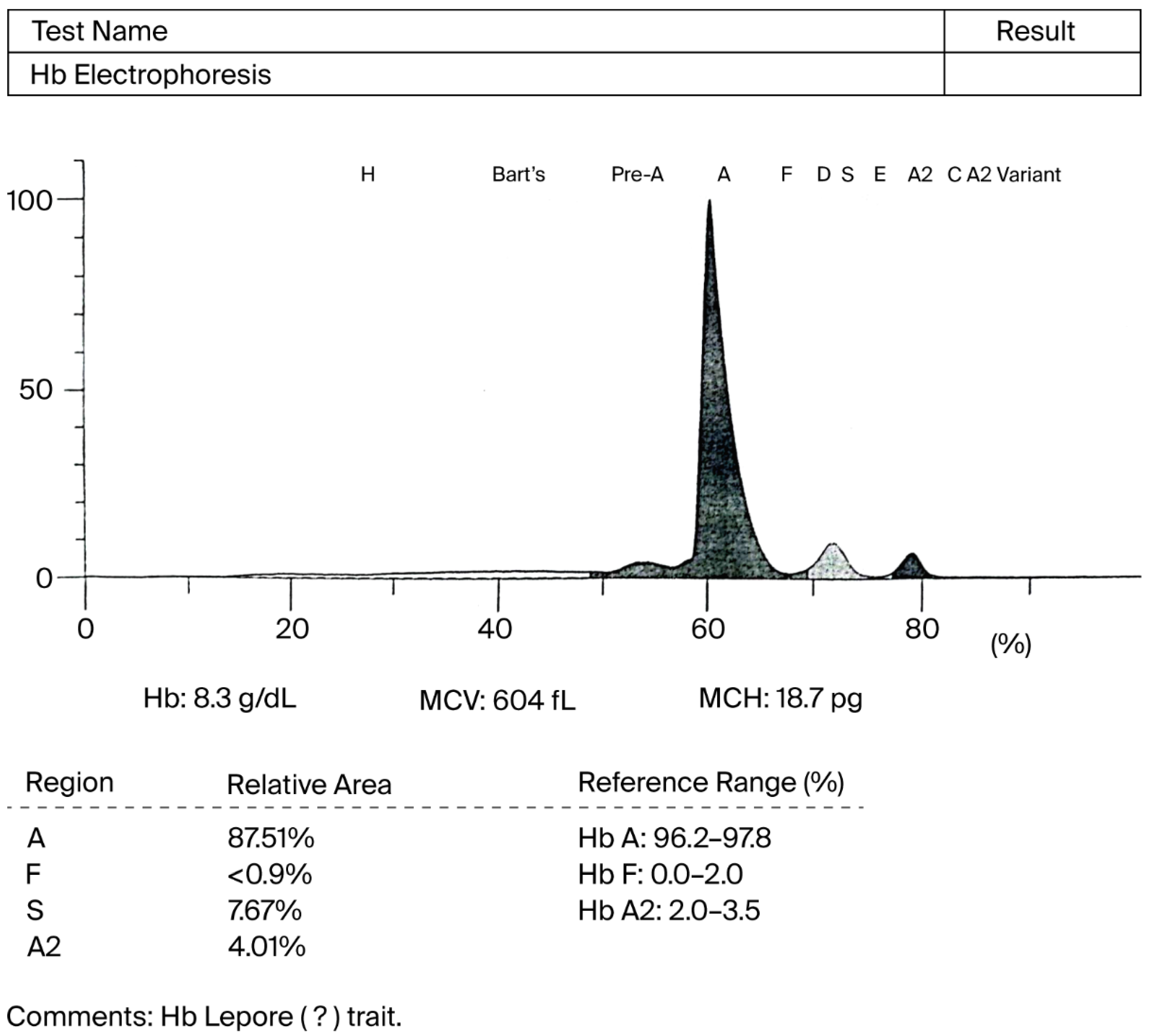
Figure 1. Capillary Hb electrophoresis showing an abnormal pattern with mild elevation in HbS, normal Hb F, mildly reduced HbA, and high HbA2, suggesting a heterozygous beta chain variant of Hb Lepore.
Rheumato 05 00016 g001
Table 1. Complete blood count (CBC), showing low hemoglobin (Hb), low mean corpuscular volume (MCV), low mean corpuscular Hb (MCH), and normal mean corpuscular Hb concentration (MCHC), red blood cell count (RBC), Red Cell Distribution Width-Coefficient of Variation (RDW-CV), Red Cell Distribution Width-Standard Deviation (RDW-SD), Packed Cell Volume (PCV), Platelet Distribution Width (PDW), Mean Platelet Volume (MPV), Plateletcrit (PCT), platelet-large cell ratio (P-LCR), platelet-large cell count (P-LCC), Erythrocyte sedimentation rate (ESR).
Test NameResultUnitReference Range (Adult Male)
Hemoglobin7.9gm/dL12.5–17.0
ESR (Westergren)106mm/1st hr0–10
Total WBC24,160/cumm4000–11,000
Neutrophils86%40–70
Lymphocytes08%20–40
Monocytes04%02–08
Eosinophils02%01–06
Basophils00%00–01
Total Eosinophils483/cumm50–450
Total RBC4.33m/μL4.5–6.5
HCT/PCV25.2%40–54
MCV58.1fL76–94
MCH18.1pg27–32
MCHC31.2g/dL29–34
RDW-CV14.7%11.0–16.0
RDW-SD34.4fl35–56
PDW20.4fL35–56
Total Platelet Count369,000/cumm150,000–450,000
MPV9.8fL7.0–11.0
PCT0.36%0.1–0.2
P-LCR39.0%3.0–43.0
P-LCC144103/uL44.0–140.0
Disclaimer/Publisher’s Note: The statements, opinions and data contained in all publications are solely those of the individual author(s) and contributor(s) and not of MDPI and/or the editor(s). MDPI and/or the editor(s) disclaim responsibility for any injury to people or property resulting from any ideas, methods, instructions or products referred to in the content.