Review

Unlocking the Secrets of Knee Joint Unloading: A Systematic Review and Biomechanical Study of the Invasive and Non-Invasive Methods and Their Influence on Knee Joint Loading

1
CMEMS-R&D Center for Microelectromechanical Systems, School of Engineering, University of Minho, 4710-091 Braga, Portugal
2
Department of Orthopaedic Surgery, William Beaumont Hospital, Royal Oak, MI 48067, USA
*
Author to whom correspondence should be addressed.
Rheumato 2025, 5(3), 8; https://doi.org/10.3390/rheumato5030008
Submission received: 26 March 2025 / Revised: 2 June 2025 / Accepted: 18 June 2025 / Published: 25 June 2025

Abstract

Background/Objectives: This review analyzes the effects of invasive and non-invasive methods of knee joint unloading on knee loading, employing a biomechanical model to evaluate their impact. Methods: PubMed, Web of Science, Cochrane, and Scopus were searched up to 15 May 2024 to identify eligible clinical studies evaluating Joint Space Width, Cartilage Thickness, the Western Ontario and McMaster Universities Osteoarthritis Index, the Knee Injury and Osteoarthritis Outcome Score system, Gait velocity, Peak Knee Adduction Moment, time to return to sports and to work, ground reaction force, and the visual analogue scale pain score. A second search was conducted to select a biomechanical model that could be parametrized, including the modifications that each treatment would impose on the knee joint and was capable of estimate joint loading to compare the effectiveness of each method. Results: Analyzing 28 studies (1652 participants), including 16 randomized clinical trials, revealed significant improvements mainly when performing knee joint distraction surgery, increasing Joint Space Width even after removal, and high tibial osteotomy, which realigns the knee but does not reduce loading. Implantable shock absorbers are also an attractive option as they partially unload the knee but require further investigation. Non-invasive methods improve biomechanical indicators of knee joint loading; however, they lack quantitative analysis of cartilage volume or Joint Space Width. Conclusions: Current evidence indicates a clear advantage in knee joint unloading methods, emphasizing the importance of adapted therapy. However, more extensive research, particularly using non-invasive approaches, is required to further understand the underlying knee joint loading mechanisms and advance the state of the art.

1. Introduction

The human knee joint, a remarkable and intricate structure essential for mobility and weight support [1], is susceptible to knee osteoarthritis (OA), a prevalent degenerative joint disease impacting knee cartilage [2]. This pathology affects around 32.5 million adults in the United States [3], with an estimated annual cost of USD 486.4 billion [4]. Knee OA, resulting from cartilage wear, causes joint pain and severely impacts daily activities, limiting millions of individuals autonomy worldwide [3,4,5,6,7]. Factors like genetics, obesity, deformities, and exercise contribute to its development [5,8]. While there is no cure, treatments aim for symptomatic relief and, in severe cases, total knee arthroplasty (TKA) is the final surgical recourse [2,5,9]. Although TKA is recognized as an alluring solution, its utilization in younger patients (under 65 years) carries an elevated risk of potential future revisions due to the prostheses’ limited lifespan [10].
Several studies have explored the impact of various treatments for knee OA, including systematic reviews on surgeries and comparisons of orthosis efficacy. Takahashi et al. reviews the viability of Knee Joint Distraction (KJD) surgery as a treatment for knee OA, comparing it with other treatments like high tibial osteotomy and total knee arthroplasty. KJD surgery, which uses an external fixator to temporarily unload the joint, has shown improvements in functional outcomes, pain scores, and Joint Space Width at 1-year post-treatment. However, a high risk of pin site infection is noted. The review suggests that KJD could be a potential alternative for younger patients with knee OA, but emphasizes the need for further trials to establish its long-term efficacy and safety compared to contemporary treatments [11]. Bin Abd Razak et al. conducted a parallel systematic review about KJD procedure in younger patients, demonstrating promising results, with improvements in functional outcomes and pain scores. However, once again, it exhibits a high risk of pin site infection. The review emphasizes the need for further research to validate these findings, optimize the procedure, and explore ways to minimize complications. Despite the challenges, KJD is seen as a potential game-changer in managing knee osteoarthritis in younger patients, aligning their findings with Takahashi et al.’s observations [12]. Liu et al. compared High Tibial Osteotomy (HTO) techniques, discussing its biomechanics, and implications for treating medial compartment osteoarthritis with varus deformity. It highlights the importance of understanding HTO’s biomechanics to improve postoperative satisfaction and long-term survival. The article covers various aspects, such as alignment principles, surgical techniques, fixation plates, and the impact of HTO on postoperative gait and knee joint mechanics. It also emphasizes the need for comprehensive studies combining musculoskeletal dynamics modeling and finite element analysis to better understand patient-specific biomechanics after HTO, concluding by stressing the significance of biomechanical environment in addressing complications and enhancing surgical accuracy in HTO [13]. Stiebel et al. discuss the challenges of treating post-traumatic knee OA in young patients. The study highlights the dilemma physicians face due to the lack of therapies that are both safe and effective for long-term joint pain relief and patient acceptance. The document reviews current treatments, including biologics, disease-modifying drugs, partial joint resurfacings, and minimally invasive joint-unloading implants, which are being explored to address this gap. It emphasizes the need for more research to find optimal treatments for young patients with post-traumatic knee OA considering their longer life expectancy and desire to maintain an active lifestyle. The document also explores the pathophysiology of post-traumatic knee OA, detailing how traumatic knee injuries contribute to the development of OA and the subsequent biomechanical changes that exacerbate the condition [14]. Mistry et al. provides a comprehensive literature review on the effectiveness of unloading knee braces in treating unicompartmental knee OA over the past decade. Various studies are discussed that support the use of knee braces to reduce pain, improve function, and enhance quality of life, potentially delaying the need for surgery. It also notes minor complications like soft tissue irritation due to poor fitting, which can be managed with regular follow-ups. The authors conclude that unloader braces are a cost-effective and beneficial treatment for unicompartmental knee osteoarthritis, advocating for a multidisciplinary approach and conservative management before considering surgery [15]. Nevertheless, there is a gap in explanations for mechanical changes in the knee joint, as well as detailed studies comparing various surgical and non-invasive therapies. While these discoveries primarily concern the medical domain, it is critical for a biomedical engineer to understand the reasoning and mechanics underlying the aforementioned approach. As a result, there is a need to conduct biomechanical studies of current solutions, including both invasive and non-invasive procedures, and connect them to the insights offered in the literature.
This systematic review aims to provide a thorough examination of the invasive and non-invasive methods employed for joint distraction, and how they change the biomechanics of the knee. Cartilage lacks a vascular supply, causing it to hinder the delivery of essential nutrients and oxygen for its repair, limiting the human body’s response [5]. Thus, the act of mechanically unloading the knee joint is an approach that seeks to mitigate the degenerative effects of knee conditions by temporarily altering the mechanical forces experienced by the joint by exerting forces to separate the bones of the knee, opposing knee loading and attempting to create space. This process effectively redistributes loading patterns within the knee, potentially promoting cartilage preservation and regeneration. By systematically reviewing the latest research findings, clinical outcomes, and technological advancements, this review aims to offer a comprehensive understanding of the current state of the art of knee joint unloading methods, their effectiveness, and their implications and understanding for knee joint loading. Ultimately, by gaining a deeper insight into these methods and their impact on knee joint loading, we aspire to pave the way for more targeted and efficacious interventions, offering renewed hope to individuals seeking relief from the burdens of knee joint degeneration.

2. Materials and Methods

2.1. Research Strategy

This review adhered to the guidelines outlined in the Preferred Reporting Items for Systematic Reviews and Meta-Analyses (PRISMA) [16] when applicable.

2.1.1. Search Strategy

This review utilized four databases, PubMed, Cochrane, Web of Science, and Scopus, with document retrieval conducted on 15 May 2024. The search strategy involved the terms (“knee” or “lower limb” or “gonarthrosis” or “tibiofemoral” or “patellofemoral”) and (“distraction” or “unloading” or “relief” or “offloading”). Initially, terms related to “cartilage” and “regeneration” were included but yielded limited and irrelevant information, making it challenging to align with predetermined search strategies like PICO [17]. Consequently, a more comprehensive search was conducted with fewer terms to ensure articles with pertinent information were captured. The search encompassed titles, abstracts, and keywords in each database, and where applicable, the articles were limited to trials or clinical trials.

2.1.2. Study Selection

Every article retrieved from the initial search was analyzed to see how relevant it was for this review. This process was performed with the aid of Zotero software version 7.0.15. Duplicate articles were removed using the “find duplicates” option. The remaining files were also manually verified to eliminate any duplicate that could not be found by the algorithm. Articles were excluded if they met the following criteria: (1) reviews, letters to the editor, conference papers and abstracts, meetings, proceedings, and commentaries; (2) the article was not written in English; (3) measured units were not translatable to the international system of units; (4) none of the chosen parameters were evaluated. The articles were then screened based on their abstract, and after it, according to their full text.

2.1.3. Procedure Classification

This article explores a total of six procedures. Three invasive interventions were analyzed: KJD, HTO, and the Implantable Shock Absorber (ISA). Notably, TKA is excluded from consideration as it involves knee joint replacement rather than the distraction mechanism examined in this study. The selected non-invasive interventions comprised orthotic devices, namely Knee Unloading Braces (KUBs), utilizing a three-point pressure system, Knee Distraction Braces (KDBs), employing a mechanism to counteract knee loading, and Physical Therapy (PT) designed to provide knee distraction. The selection of these procedures allows for a comprehensive analysis of various approaches to address knee-related issues, each offering distinct characteristics and applications.

2.1.4. Data Collection and Extraction

The primary outcomes selected for analysis included Radiographic Joint Space Width in mm (JSW), Cartilage Thickness in mm (CT), both total and partial Western Ontario and McMaster Universities Osteoarthritis Index (WOMAC) [18,19], partial and total Knee injury and Osteoarthritis Outcome Score system (KOOS) [20], Gait velocity in m/s (GV), Peak Knee Adduction Moment KNm/Kg (PKAM), time to return to sports (RTS), time to return to work (RTW), ground reaction force in N (GRF) and visual analogue scale in mm (VAS) pain score, all of which were examined. Articles that did not conform to the correct scale or had units incompatible with conversion to the SI system were excluded, even if they satisfied other specified criteria.

2.1.5. Risk of Bias Assessment

The evaluation of bias risk in the incorporated studies utilized the Revised Cochrane risk-of-bias tool for randomized trials (ROB 2) [21] for Randomized Clinical Trials and ROBINS-I [22] for non-Randomized Clinical Trials. The criteria for each domain were tailored to the context of our systematic review, allowing for a specific analysis of bias risk associated with each considered procedure.

2.2. Biomechanical Model

In the selection of an appropriate model, it was considered essential that the model could assess the knee joint’s condition in the coronal plane and that its parameters could be readily obtained and adjusted for this study. Various electronic databases, including OpenSim, PubMed, Embase, and Web of Science, were systematically searched to identify a suitable model for this review that aligns with the specified conditions.
Models such as the one developed by Erdemir use the finite element analysis (FEA) method to describe the geometry and materials of the knee joint [23]. His model was further developed by Chokhandre et al. with the goal of providing free access to three-dimensional finite element representations of the knee joint [24]. However, the utilization of an FEA model, with its multifaceted complexities, surpasses the intended scope of this review, which aims to offer a concise examination of knee joint distraction methodologies without delving into the intricate intricacies of FEA model implementation.
Several models utilize musculoskeletal systems with parameters aiming to comprehend muscle contributions and dynamic interactions. For instance, Marieswaran et al. [25] extended Xu et al.’s original model, initially designed for gait analysis with knee joints and ligaments to assess the strains in connective tissues [26]. Similarly, Arnold et al. developed a musculoskeletal model capable of calculating muscle–tendon lengths and moment arms across various body positions, facilitating detailed examinations of force and moment generation capacities around the ankle, knee, and hip. Despite their importance, these models, falling under musculoskeletal models, face limitations when muscle conditions cannot be parametrized, as observed in this case [27].
For example, in a recent study, a computational multibody knee models to assess the impact of anterior cruciate ligament (ACL) injury on medial meniscus forces. The models predict increased forces, especially in the posterior horn, highlighting vulnerability to injury, particularly with ACL loss. The study also illustrates the role of the deep medial collateral ligament in constraining posterior medial meniscus motion. Multibody models are valuable for analyzing dynamic forces and motion in interconnected structures, offering insights into complex systems [28,29]. However, this study does not focus on studying dynamic knee movement, making multibody models less ideal for this specific application.
Minns’ developed model demonstrates a high degree of accuracy and effectiveness in predicting knee loading [30]. However, its practical implementation encountered challenges stemming from the unavailability of full-leg radiographs and baseline parameter values. This implies that 16 necessary parameters for the model are unavailable. The absence of comprehensive radiographic data for the selected methods, combined with the complexity of numerous parameters, significantly hindered the translation of these methods into the model and hindered comparative analyses.
Kettelkamp’s model was initially designed to assess force distribution on the tibial plateau, primarily in the context of proximal tibial osteotomy [31]. Importantly, Kettelkamp emphasized that its utility extended beyond this specific application. This model is based on a two-leg stance simulation utilizing AP long-leg radiographs, yielding outputs for both medial and lateral knee joint forces and their orientations. However, due to the unavailability of long leg radiographs, we were unable to describe six parameters and thus did not implement this model in our study.
The model developed by Maquet et al. was selected for its robust accuracy, well-defined baseline parameters, and its adaptability to the chosen knee joint distraction methods [32]. While this model also relies on long-leg radiographs, the author’s definition of validation parameters facilitated its implementation in our study. A recent study implemented these three models to assess two-dimensional biomechanical models for predicting knee joint forces in total knee arthroplasty preoperative planning. While the models show potential, their accuracy needs improvement, requiring further optimization. In it, the authors verified that Kettelkamp’s model tends to underestimate knee joint forces (−17% to +128% Body Weight), while Minns’ and Maquet’s models align closely with reference forces (−55% to 80% body weight and 80 to +50% body weight, respectively). Thus, Minns’ and Maquet’s models are considered favorable choices [33].
This model is centered on the tibiofemoral joint, predicting the resultant knee joint force, its orientation, and a lateral muscle force. Within this joint, the tibia experiences the influence of three distinct forces: the partial weight force (P), the muscular force (L), and the resultant force I, as represented in Figure 1.
The muscular force L functions as a tension band that exerts its effect on the tibia, with its line of action crossing paths with that of the partial weight force at point C. It is important to note that the resultant force R also needs to pass through this designated point, C. This alignment of forces is further reinforced by the fact that, in a normally functioning knee, R must inherently traverse through the geometric center of the tibiofemoral contact area, denoted as point G. Consequently, this arrangement yields a statically determinable problem, as depicted in the triangle of forces illustrated in Figure 2A.
As such the resulting Force R, can be expressed through Equation (1).
R = P sin ψ sin β
In order to apply this model to a patient, it needs a one-leg stance anteroposterior long-leg radiograph, including the pelvis, as input. During our investigation, we adhered to the parameters stipulated by Maquet et al.’s model, where the patellar force (P) was deemed to represent 93% of the body weight. Furthermore, the angles, denoted as ψ and β, were determined to be 10.78° and 4.62°, respectively, in accordance with this established model [32]. It is worth noting that due to the unavailability of extensive long leg radiographs for knee modeling, we were constrained from making alterations to these angles, and hence, we retained the original values. All the computational models were implemented using the Python 3.10.0 programming language, ensuring the precision and consistency of our analyses.
In light of these parameters, the resultant force calculated amounted to 215.95% of an individual’s body weight, signifying the substantial biomechanical implications of the imposed conditions.
This model is adaptable to examine valgus and varus knee alignments. Maquet et al. modified the model relations, as illustrated in Figure 2B, to accommodate these variations.
Thus, Equation (1) undergoes modification to become Equation (2). It is noteworthy that when x = 0, both equations converge into the same equation.
R = P 2 + L 2 + 2 P L cos ( β ± x )
To assess stress levels within the knee joint across the range of motion, we employed the conventional stress formula as presented in Equation (3), where R represents the calculated resulting force and S is the area of the contact surface.
σ = R S
No significant data was found that quantified how much this contact area changed with genu varus or valgum. It is widely acknowledged that increasing misalignment generally leads to a decrease in the tibiofemoral contact area, where the lateral contact area decreases with genu varum, and the medial contact area decreases with genu valgum.
In our study, we represented S as a function of the knee deformation angle x through a quadratic relationship, as shown in Equation (4).
S = S m e d i a l + S l a t e r a l ,   i f   x = 0 S = S m e d i a l + S l a t e r a l x 30 ° 2 × 0.75 S m e d i a l ,   i f   x < 0 S = S m e d i a l + S l a t e r a l x 30 ° 2 × 0.75 S l a t e r a l ,   i f   x > 0
Throughout the course of this review, the methodologies employed will systematically adjust the parameters within the model, culminating in a set of outcomes that will serve as the foundation for a biomechanical analysis of knee movement.
In this study, we assumed x [ 30,30 ] ° , where negative values denote genu valgum misalignment and positive values indicate genu varum misalignment. These values were derived from those employed by Maquet et al. [32]. The model was implemented in Python, and the angle step was iteratively adjusted until convergence was achieved.
Schmidt et al. reported a mean tibiofemoral contact area of 452 mm2 for the medial compartment and 314 mm2 for the lateral compartment, totaling 766 mm2. It was assumed that at an extreme angle (30°), only 25% of the corresponding knee compartment would be engaged [34].

3. Results

3.1. Search Results

Identified from the initial 655 records, 29 studies that met the eligibility criteria were subsequently included in this systematic review. The PRISMA flowchart, illustrating the search process, is depicted in Figure A1 and was built using a tool made for this end [35]. The extracted data from the articles is present in Table 1.

3.2. Risk of Bias

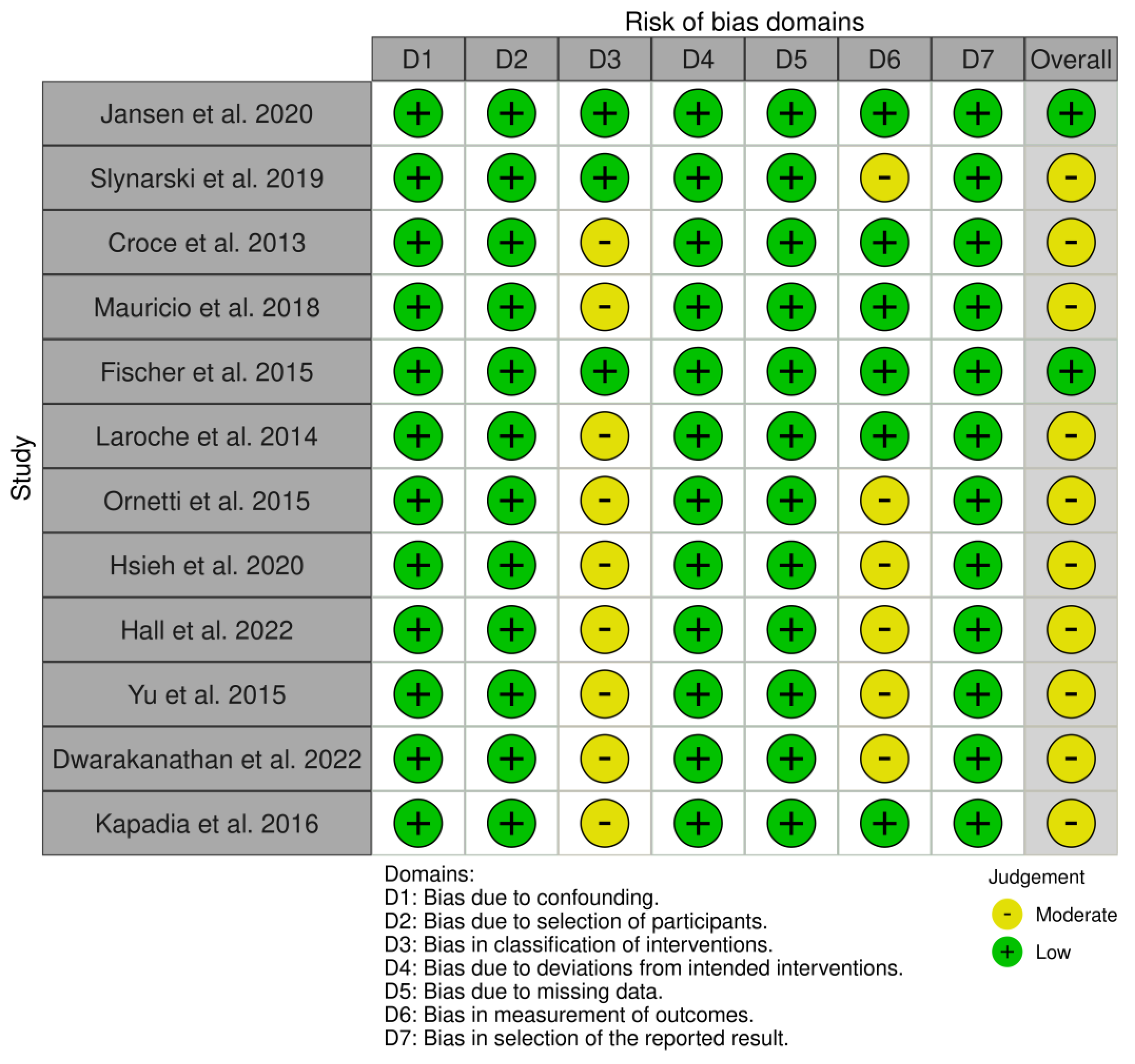
The figures illustrating bias levels, commonly referred to as “traffic lights,” were generated using the RoBVis tool [64]. Figure A2 depicts the risk of bias in randomized trials (RoB 2), indicating that 4 articles were assigned a low risk of bias, while 12 articles elicited some concern. The classification for the latter articles is attributed to parameter D4, specifically addressing bias in the measurement of the outcome. This classification stems from the fact that these outcomes were obtained through questionnaires, and due to their subjective nature, the possibility of bias originating from this method cannot be entirely eliminated.
In Figure A3, the depiction of the Risk Of Bias In Non-randomized Studies of Interventions (ROBINS-I) reveals that 2 articles were classified as having a low risk of bias, while 10 articles were considered to have a moderate level. The moderate bias classification in these articles was attributed to parameter D3, which pertains to the classification of interventions, and D6, which concerns bias in the measurement of outcomes. Regarding parameter D3, numerous articles referred to the orthosis as either a Knee Unloading Brace or a Distraction Rotation Knee Brace. Although such terminology is common, there is ambiguity in defining each term precisely, lacking clear distinctions. As regards parameter D6, once again, it is linked to the reliance on parameters derived from questionnaires. The inherent subjectivity in data collection through questionnaires introduces variability in responses, potentially compromising the accuracy and reliability of the study findings.

3.3. Invasive Methods

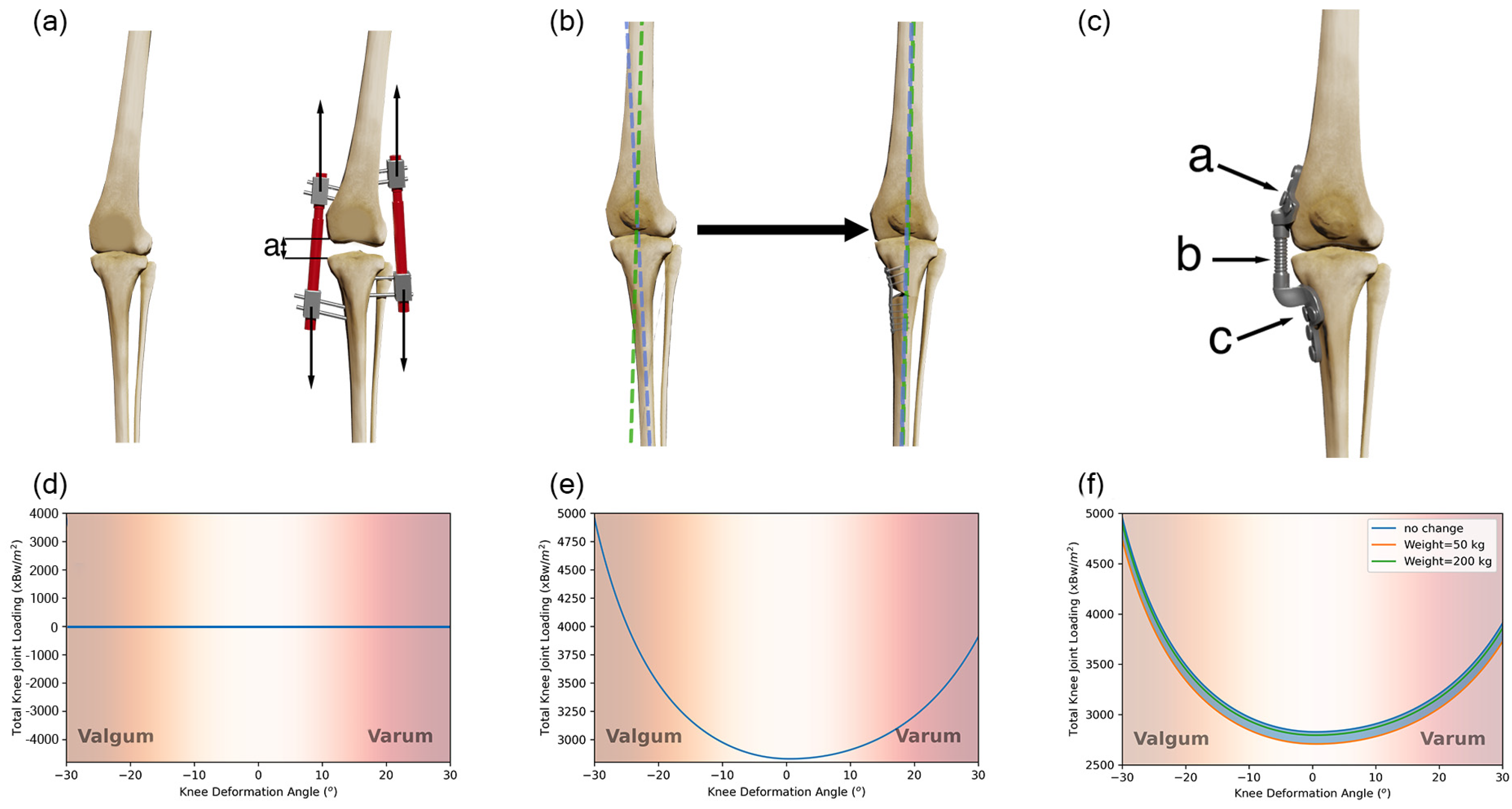
Figure 3 presents an illustration of the invasive methods studied and the results from the biomechanical numerical analysis.

3.3.1. Knee Joint Distraction

In KJD surgery, the surgeon uses external fixation devices to slowly separate the femur and tibia, creating a 2 mm gap initially, which can be adjusted up to 5 mm in the following days (Figure 3a). Full weight bearing is encouraged with crutches. After 6 weeks, the device is removed in an outpatient procedure, and patients can resume full weight bearing. Patients typically experience improved pain/function within a few weeks [65].
This separation interrupts the connection between the femur and tibial condyles, rendering the value of Equation (1) as 0 (Figure 3d). Since condyle contact is no longer present, it results in a null force transmission in this region, considering the resulting stress as 0.
This approach was developed as an option for patients unable to undergo knee replacement surgery (TKA) immediately, aiming to postpone the necessity for revision [65]. Research indicates that this procedure can defer TKA for 5–10 years, depending on individual patient factors [36,41,43,66]. The objective of KJD surgery is to alleviate pain and slow the advancement of knee osteoarthritis by modifying how forces are distributed within the knee joint, promoting improved joint healing and function [65].
Following the completion of the procedure, all patients with knee OA observed positive outcomes and reported an enhancement in their overall condition. Generally, there was a significant rise in JSW in all articles, extending up to two years post-procedure. The observed increase ranged from 0.22 to 1.9 mm, which is deemed substantial in the context of knee OA [39,40,41,67]. Numerous patients noted an escalation in their overall WOMAC score, persisting from 6 weeks to at least 2 years. This score varied between 22 and 38.9, indicating a noteworthy improvement according to patient reports [39,40,41,42]. After 2 years, the patients also noted a reduction in pain levels, registering a decrease of 2.14 to 3.6 mm on a 10 mm VAS [39,41]. At both the one-year and two-year marks, the patients noted a rise in their KOOS scores, suggesting an improvement in their condition. The reported increase ranged from 17 to 28.7 [39,40,42]. Ultimately, one-year post-procedure, a significant portion of patients expressed the ability to resume their previous activities. Specifically, 94% were able to return to work, and 79% resumed participation in sports [43].
Numerous reports emphasize potential challenges and adverse events linked to KJD surgery. Pin tract infections emerged as a prevalent concern, affecting a significant proportion of treated patients. The majority of these infections were successfully managed with oral antibiotics, although certain instances necessitated hospital admission [36,40,41]. Reported adverse events after KJD include post-operative foot drop (managed with an ankle–foot orthosis), cases of osteomyelitis (treated with surgical cleaning and a combination of intravenous and oral antibiotics), pin loosening or breaking (addressed through tightening, refixation, or pressure bandages), and complications such as deep venous thrombosis and pulmonary embolisms (requiring extra anticoagulation). Other issues encompassed suspected compartment syndrome (resulting in frame removal and fasciotomy), pneumonia cases (treated with intravenous antibiotics), post-distraction infections after frame removal (managed with antibiotics), cases of flexion limitation (addressed with manipulation under anesthesia or arthroscopic arthrolysis), and the arthroscopic removal of a loose piece of cartilage/bone in one instance [42].

3.3.2. High Tibial Osteotomy

High tibial osteotomy (HTO) is a surgical procedure aimed at addressing medial knee OA in active, young, or middle-aged patients. The procedure involves wedging open the upper portion of the tibia, leading to a reconfiguration of the knee joint (Figure 3b). This realignment procedure redirects weightbearing from damaged or worn tissue to healthier areas. During the period of bone union, the patients are subject to restricted motion and load-bearing limitations [68].
HTO surgery does not modify the resultant force value; it alters the alignment of this force, consequently changing the contact area. Referring to Figure 2B), it essentially changes the value of x . The representation of the variation in stress in function of the knee deformation angle can be observed in Figure 3e. In it, the σ [ 2829.00 ,   4957.98 ] xBW/m2. The alteration in surface contact area results in a decrease in knee loading of up to 175.25% of its initial value in its most extreme position. This demonstrates the significant impact of knee misalignment on knee loading.
Overall, all patients experienced benefits from the surgery. Every article observed a rise in JSW within the two years following the procedure. This range, varying from 0.2 to 0.88 mm, is deemed substantial in the context of knee osteoarthritis [39,40,67]. Patients noted a rise in their WOMAC scores, ranging from an initial 29 to an eventual 33 within a span of up to two years [37,39,40]. When describing pain, certain patients reported a decrease over a two-year period, ranging from a reduction in VAS scores from 0 mm to 3.85 mm [38,39]. The patients also conveyed an enhancement in their KOOS scores within two years post-surgery, showing an improvement in their condition within a range of 19 to 30 [39,40]. Ultimately, one year post-procedure, a substantial portion of patients indicated their ability to resume previous activities, with 97% successfully returning to work and 80% resuming participation in sports [43].
Patients undergoing this treatment may encounter adverse events. Several occurrences in patients have been documented, such as a broken screw, bleeding, postoperative wound infections, nerve injury, cases of severe pain, and scar formation. A majority of individuals who underwent the procedure required implant removals due to pain/bone consolidation [36,37,38,40].

3.3.3. Implantable Shock Absorber

An Implantable Shock Absorber (Figure 3c) is a device that is implanted in the extra-capsular space along the medial side of the knee joint. It is designed to reduce knee joint load by up to 13 kg and is actively unloading the knee during the stance phase of gait [69]. Recovery of this surgery involves three phases of post-implantation rehabilitation. Phase I focuses on wound healing with activities like wound control, pain management, and elevation. The hospital stay typically ranges from 1 to 5 days. Phase II, spanning 3 to 6 weeks, concentrates on restoring range of motion and resuming daily activities, gradually eliminating crutch use. Common exercises include cycling, single-leg balancing, walking, and quadriceps’ strengthening. Sedentary workers may return to work in 2 to 4 weeks. Phase III, beyond 6 weeks, emphasizes strength-building, return to manual occupations, and potential resumption of sports, with specific timelines provided as general guidelines [70,71].
The average weight of the human body is approximately 62 kg [72], and the offset of 13 kg introduced by this system corresponds to approximately 20.6% of the total body weight. If we change this in Equation (1), the obtained value for R force is 171.47%.
Nevertheless, controlling the patient’s weight is not feasible, and consequently Equation (1) is modified to Equation (5), wherein W is represented as the patient’s body weight.
R = ( 1 13 W ) P sin ψ sin β
To investigate the impact on the overall knee joint loading, we used the correlation between the patient’s body weight (ranging from 50 kg to 200 kg). The maximum and minimum forces were subsequently employed to compute the maximum total knee joint loading, as depicted in Figure 3f.
The ISA System seeks to supplant TKA or HTO treatments with its less invasive, reversible procedure. Preserving tissues and simplifying potential revisions, it presents an alternative approach that addresses patient aversions and offers potential advantages [70].
Clinical trials demonstrate that ISA treatment leads to an enhanced quality of life for patients. Two years post-treatment, the patients noted an enhancement in the Total WOMAC score by 46.2, with specific improvements in the pain and function partial scores of 38.5 and 29.5, respectively [37,44]. No assessment of quantitative parameters was conducted.
ISA studies also documented several adverse events. The primary observed adverse events included pain and infection. A smaller proportion experienced discomfort, and scar formation was also identified. A minority underwent implant removals, some of which due to infection [37,44].

3.4. Non-Invasive Methods

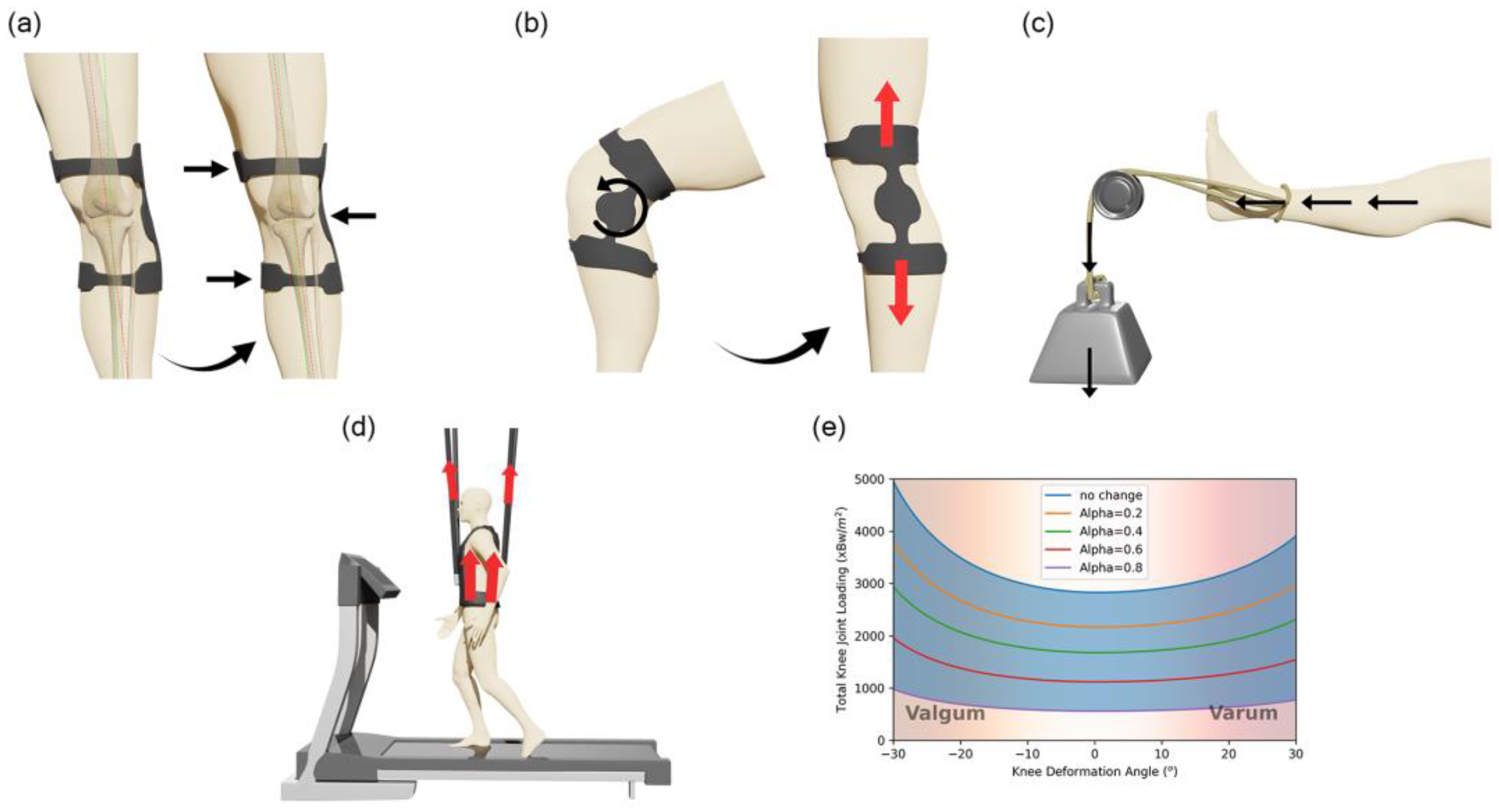
Figure 4 provides an overview of the non-invasive methods examined, along with the outcomes from the biomechanical numerical analysis.

3.4.1. Knee Unloader Brace

A KUB (Figure 4a) is an external orthotic device employing a three-point pressure system to realign the patient’s knee. Despite its name, the brace does not exert a force countering knee load; instead, it redirects force to decrease pressure in the damaged knee area. Because of the three-point pulling principle, this orthosis does not require a rigid structure and is frequently designed as a flexible device. Patients avoid the need for a rehabilitation period and typically experience an immediate change, making it an appealing option [73].
This system bears similarities to high tibial osteotomy (HTO) but is externally applied. Like HTO, in the chosen biomechanical model, it alters the angle, albeit to a lesser extent compared to HTO as it cannot modify the patient’s bone structure. Therefore, it does not fundamentally alter the presented biomechanical model in any manner.
Patients with knee OA displayed a reduction in PKAM ranging from 0.02 to 0.82 KNm/Kg immediately upon wearing the orthosis. Over a span of six months, they reported a sustained decrease of up to 1% in PKAM while utilizing the orthosis. In contrast, healthy patients exhibited an increase of 0.015 KNm/kg in PKAM under similar conditions [45,46,51,52]. Following the immediate application of the orthosis, there was no discernible change in GRF among the patients. However, with continuous use over a period of six months, a notable reduction of 2% in GRF was observed [51]. Upon initial application of the orthosis, the patients experienced an immediate increase in GV by 0.02 m/s. This positive trend persisted, with a further increase of 0.05 at 8 weeks, 0.093 at 3 months, and 0.06 at the 6-month mark. These findings suggest a progressive improvement in GV over the course of orthosis usage [52,53,57,58]. Following two years of usage, MRI scans revealed an average growth in Cartilage Thickness by 0.4 mm [56]. Over a 12-week period, the patients noted an improvement in their well-being, as reflected in the total WOMAC score, which increased within the range of 10.5 to 16.3 [59]. The patients observed a 2.3 reduction in VAS pain within four weeks, and over the course of one year, witnessed a decrease ranging from 0.1 to 2.3 [38,50,54]. Within three months, the patients experienced a notable surge in KOOS total score, ranging from 7.2 to 13, which endured for up to a year with a score of 8.92, and even extended to two years with a substantial increase to 17.8. Additionally, there was an improvement in function over a six-month period, marked by a significant increase of 10.6 [53,56,57,61,62,63].
A small percentage of patients experienced skin irritation, rash, bruises, and discomfort [53,54,61]. Pneumatic KUB devices had limitations, with some patients unable to use the inflated bladder fully due to associated pain and discomfort [45].

3.4.2. Distraction Rotation Knee Brace

DRKB is a specialized orthopedic device designed for the application of knee joint distraction. The primary goal of this orthosis is to induce controlled separation and rotation of the knee joint surfaces, promoting joint unloading. This controlled distraction is particularly beneficial for patients with knee OA (Figure 4b). For the proper transmission of the distraction movement across the leg, this device requires rigidity. Some devices combine the capabilities of both distraction and unloading through a three-point pressure system. This versatile design allows for the simultaneous application of knee joint distraction and targeted unloading [74].
This treatment appears to share a functional principle with the ISA option. Nonetheless, gauging the extent of movement transferred from the skin to the bone proves challenging due to individual variations in parameters like skin elasticity, adipose tissue quantity, and muscle volume among patients. These variations impact the required displacement for movement to transfer from the skin to the bone. Consequently, it is anticipated that the treatment will yield a similar effect [7], though quantifying the added parameter remains elusive.
Five weeks after initiation, the patients observed a decrease in PKAM of 0.018 Nm/Kg [48]. During this period, there was also an increase in GV by 0.1 m/s [48,49]. Additionally, within the same 5-week timeframe, the patients reported a notable rise in the total WOMAC score by 20.7 [48]. Furthermore, VAS pain exhibited an initial decrease of 3.3 after 5 weeks, with this reduction persisting and reaching 2.3 after one year [48,49]. On average, the patients reported a KOOS total score increase of 13.62 at the one-year mark [49]. Certain patients encountered challenges wearing the knee brace due to its rigidity [49].

3.4.3. Physical Therapy

Physical therapy plays a pivotal role in inducing knee joint unloading, employing specialized techniques such as the use of a pulley system or body harness. In the pulley system approach (Figure 4c), a controlled force is applied to pull the femur apart from the tibia, creating a distraction effect on the knee joint. Alternatively, a harness on the body reduces weight-bearing, alleviating pressure on the knee and promoting therapeutic unloading (Figure 4d). These targeted interventions are designed to enhance joint mobility, alleviate pain, and contribute to overall rehabilitation and functional improvement [75,76].
The implementation of a pulley system will not alter the knee joint loading condition during walking since it is applied in a sitting position. Conversely, when utilizing a harness during locomotion, it will decrease the patient’s weight by an amount denoted as α, as depicted in Equation (6).
R = ( 1 α ) P sin ψ sin β
Implementing this modification allows us to observe, in Figure 4e, how it can influence the knee loading condition. As the value of alpha is adjustable, there is a potential to significantly alleviate the knee joint loading on a temporary basis, although this is only achievable during the therapy session.
The immediate reduction in load resulted in a notable increase in GV of 0.03 m/s among healthy patients [47]. Following four weeks of consistent PT, the patients reported a rise of 6.17 in the total WOMAC score [60]. The patients undergoing recurrent PT exhibited a reduction in reported VAS pain, experiencing a decrease of 2.3 mm after four weeks, which persisted for up to a year [50,60]. Following consistent PT, the patients witnessed a substantial improvement in the total KOOS score, with an increase of 19.92 after one month [55].
No significant adverse events were documented during the patients’ participation in PT.

4. Discussion

4.1. Efficiency of Invasive Methods

Our study emphasizes that invasive methods, notably KJD and HTO, were identified as highly effective in distracting the knee joint, despite their higher associated risks, followed by ISA. Non-invasive approaches have shown promising results; however, they lack quantitative parameters related to cartilage or joint distraction, hindering the ability to measure their absolute effectiveness and make a comprehensive comparison with invasive methods.
KJD and the ISA currently stand as the exclusive invasive methods specifically designed for the direct application of joint distraction. Notably, KJD exhibits the ability to increase JSW even after the removal of the device, effectively unloading the knee during its application. This observation finds corroboration in other reviews exclusively addressing the role of KJD, highlighting its efficacy, particularly when applied to younger patients, positioning it as a promising alternative to TKA. However, the necessity for extensive and prolonged RCTs remains crucial for comprehensive validation [77,78]. HTO emerges as a viable approach, but our findings indicate that it does not significantly alter the magnitude of force exerted on the knee. Instead, it induces a shift in knee position, thereby increasing the contact area and indirectly mitigating knee loading conditions. However, the benefits of HTO are limited to patients with misaligned knees, as those with osteoarthritis may not experience substantial advantages from this surgical intervention. While it is acknowledged that knee misalignment poses a risk factor that may eventually lead to osteoarthritis [79], not all end-stage patients develop it [80], making HTO a less universal solution. This perspective aligns with other reviews suggesting that HTO could be a viable treatment option for valgus knee deformities [81,82]. Sport and work participation outcomes were comparable between KJD and HTO, indicating that both interventions are viable choices with similar effectiveness. The ISA presents an intriguing solution by directly applying a load to counteract weight. However, its efficacy is impeded by the patient’s weight, diminishing its effectiveness as weight increases, as demonstrated by our results. Notably, the severity of infections appears to be significantly lower when compared to HTO and KJD, making ISA a more attractive option from the patient’s standpoint. Nonetheless, further trials and implementation are imperative to ascertain its viability given its relatively limited current use [70,83,84]. Another systematic review also underscores the high promise of this method, positioning it as comparable to both invasive and non-invasive alternatives [85].
In addressing the clinical impact of these treatments, specifically patient-centered outcomes, rehabilitation integration, and treatment personalization, while most studies consistently report improvements in JSW, WOMAC, and KOOS scores, there remains a gap in systematically evaluating how these enhancements translate into daily functional gains and individualized recovery pathways. For instance, the reported rates of return to work and sport after knee joint distraction and high tibial osteotomy suggest promising functional recovery, but these metrics alone do not fully capture the nuanced experiences of patients navigating rehabilitation or adapting to personalized regimens. Similarly, while the implantable shock absorber trials report significant improvements in WOMAC scores, the absence of quantitative gait or activity data limits our understanding of long-term patient-specific benefits. Future research should focus on integrating structured rehabilitation protocols, patient-reported satisfaction, and functional mobility assessments to enrich the clinical relevance of these interventions. Such a comprehensive approach would not only validate the observed clinical benefits but also inform more precise, patient-tailored treatment plans, ultimately enhancing outcomes in knee joint unloading therapies.
Given these insights, it appears that KJD is a particularly promising option for younger patients who may better tolerate its infection risks and potential for cartilage regeneration, while HTO is most suitable for patients with significant knee malalignment rather than advanced osteoarthritis since realignment does not directly address joint unloading. Such subgroup-specific considerations should be further explored to guide clinical decision-making.

4.2. Efficiency of Non-Invasive Methods

Concerning non-invasive methods, none directly measure outcomes related to distraction methods or Joint Space Width. KUBs emerge as an intriguing approach, resembling HTO with short-term results on par invasive procedures [50]. While a KUB realigns the knee joint, it lacks the ability to correct knee bone deformities, a capability inherent in HTO. Various device variations, utilizing pneumatic systems for shape adjustment and fitting [45,58,86], or vibrational systems to stimulate cartilage [87], show promise. Studies suggest that ultrasound applications induce therapeutic effects on chondrocytes, making these innovations applicable to these devices [88]. Additionally, light therapy is considered for cartilage regeneration in knee osteoarthritis (OA), adding further interest to these non-invasive techniques [89]. Patients seem to have a better reaction to them, according to their reported outcomes. Our results reveal that knee unloading braces promptly reduce the adduction moment of the knee and increase Gait velocity, aligning with the existing literature [90]. Considering the advantages it offers, a DRKB emerges as an intriguing option, though evaluating their effectiveness is challenging due to a lack of evidence regarding bone loading through skin displacement. Patient-reported outcomes indicate an improvement in quality of life after using a DRKB, despite limited research on this relatively new orthotic device. Unlike KUBs, a DRKB demands rigidity and may be less comfortable in comparison. Regarding Physical Therapy (PT), it is a prominent non-invasive approach, directly involving knee joint distraction. Although not continuously applicable during daily routines, patients undergoing multiple therapy sessions report more comfortable and enduring results [91,92,93]. While PKAM serves as an indicator of knee joint loading [94,95], a certain level of validation for knee joint loading and cartilage status is necessary. Biomechanical analyses alone do not infer this level of evidence [96]. Despite reported improvements in indirect biomechanical parameters and patient outcomes, the impact on cartilage remains unclear, necessitating further studies, as highlighted in the literature [97].
When considering the clinical impact of non-invasive interventions for knee joint unloading, the reported data emphasizes promising improvements in key patient-centered outcomes. Notably, these methods consistently yielded measurable enhancements in GV and load reduction (PKAM and GRF), which are critical biomechanical markers for improved joint function. Furthermore, significant gains in WOMAC and KOOS scores across these approaches highlight their potential to enhance overall patient well-being, pain relief, and functional capacity, extending up to two years post-intervention. Despite these encouraging findings, the integration of structured rehabilitation programs and personalized treatment plans remains underexplored in current studies. Specifically, while these interventions demonstrate beneficial trends in objective gait parameters and subjective pain/function assessments, there is limited evidence linking these improvements to individualized patient goals, activity-specific demands, or long-term lifestyle modifications. Additionally, though adverse events such as skin irritation were minimal, understanding their potential impact on patient adherence and quality of life is essential for optimizing treatment personalization. Future research should focus on connecting these quantitative measures to real-world functional milestones and integrating patient feedback to ensure that non-invasive unloading strategies are holistically tailored to each patient’s rehabilitation journey.
To compare invasive and non-invasive methods, reliance on patient-reported outcomes is essential given the distinct quantitative measurements employed by these approaches. While both categories generally enhance the patient’s quality of life and alleviate pain, individuals often report more significant improvements with invasive methods. This difference may be attributed to the fact that many reported invasive methods undergo extensive long-term follow-ups in evaluated studies, imparting a more lasting impact by considering long-term effects from the outset. The patient sample size is notably smaller in non-invasive studies compared to invasive ones. However, a notable advantage of non-invasive methods lies in their non-intrusive nature, ensuring minimal harm to the patient. Moreover, these non-invasive approaches can seamlessly integrate into the rehabilitation phase following surgery, suggesting that a combined intervention may be an ideal approach. Nevertheless, patient compliance is a crucial factor to consider. In the case of implanted devices, patients are more likely to consistently use them, whereas with removable knee braces or treatment plans, patients may forget to use them or miss therapy sessions [86,93,98,99,100].
Additionally, non-invasive methods can serve as bridging therapies, offering symptom relief and functional support for patients who either are not yet candidates for invasive procedures or are awaiting definitive surgical treatments.

5. Limitations

Despite its comprehensive approach, this review is subject to several limitations that warrant careful consideration. These limitations, spanning model adaptations, data availability, study selection, and methodological variability, underscore the complexity of accurately capturing the biomechanical and clinical realities of knee joint unloading strategies. Furthermore, some inherent constraints of the chosen models and this review’s reliance on the existing literature highlight the challenges in extrapolating findings to real-world applications.
Maquet et al.’s model [32] was adapted for most of the chosen methods, although validation was not performed for said modifications. While the presented results offer a conceptual understanding, it is important to note that they may not accurately reflect real-world scenarios. This study was constrained by the unavailability of full-leg radiographs, limiting the implementation of models with more complex parameters. This work’s scope was focused on comparing the existing literature regarding different methods, and incorporating more complex data was deemed impractical relative to this study’s objectives. The selected model assumes static conditions for a one-leg stance, overlooking dynamic loads in daily activities. Investigating the impact of these factors on findings and extending the research to incorporate dynamic aspects would be an interesting and valuable continuation of this study. To emulate a real-world scenario more accurately, a complex model such as a Finite Element Analysis (FEA) model with a well-defined geometry could be employed. Due to the lack of patient-specific radiographs, the model’s reliance on fixed parameters (such as angles ψ/β and contact area assumptions) may limit its generalizability, underscoring the need for future studies to incorporate patient-specific imaging, like MRI-based modeling, to enhance accuracy and clinical relevance. However, extensive validation across the studied strategies, often unavailable, would be necessary. While intriguing, this falls far beyond the scope of this article.
This work faced constraints stemming from the absence of parameters explicitly focused on knee joint loading or distraction indicators. Limited quantitative data on cartilage volume and Joint Space Width changes for non-invasive methods such as braces and physical therapy, especially when compared to the more robust datasets available for invasive techniques, highlights a critical knowledge gap. This disparity may be attributed to factors like the relative scarcity of randomized controlled trials (RCTs) investigating these non-invasive interventions and the high costs and logistical challenges associated with advanced imaging (e.g., MRI). Consequently, future high-quality, patient-centered studies are essential to quantify these structural changes and support evidence-based recommendations for non-invasive knee OA treatments. While some kinematic parameters like PKAM and GV were taken into account, they did not directly align with knee joint loading. Patient-reported outcomes were the most frequently chosen parameters, underscoring the significance of addressing pain in knee osteoarthritis. Nonetheless, it is essential to recognize that pain alleviation does not inherently signify a comprehensive enhancement in the condition. This is because pain is a distinctive, profoundly personal, and subjective experience, rendering it challenging to gauge the severity and intensity of an individual’s pain [101]. Additionally, this review faced constraints in acquiring relevant information about the transfer of load from knee skin to the knee joint, underscoring the need for additional data to inform the development of non-invasive orthotic devices.
This review encountered challenges due to the limited availability of high-quality studies in the field, potentially affecting the depth and robustness of the evidence base. Additionally, the inclusion of studies with diverse designs and methodologies may introduce heterogeneity, posing difficulties in synthesizing findings and drawing consistent conclusions. These limitations emphasize the need for cautious interpretation and may impact the overall reliability and generalizability of this review’s results.

6. Conclusions

In conclusion, our study investigated the biomechanical and practical impact of joint distraction, aiming to assess its efficacy in reducing knee joint loading, alleviating pain and improving joint function. The findings demonstrated a significant reduction in pain levels and better functional outcomes among participants, particularly while completing KJD and HTO. KJD was shown to be able to biomechanically completely unload the joint, whereas HTO was able to reduce joint loading by realigning it, although both have a high risk of infection. ISA has also demonstrated several advantages, as it can partially unload the knee. Its efficacy may, however, be limited if the patients have a high body mass, and more trials are required to evaluate its long-term efficacy. Moreover, the lack of robust long-term data for ISA, particularly in younger and active populations, remains an important gap that future studies must address.
Several clinical trials have found that non-invasive treatments can reduce pain and improve a variety of biomechanical measures. However, it is critical to recognize the existing studies’ shortcomings, which include a relatively small sample size and the subjectivity of the chosen parameters, particularly when compared to invasive procedures.
Despite these limitations, our research contributes valuable insights into the positive outcomes associated with joint distraction. Moving forward, larger-scale, long-term trials are warranted to further validate our findings and establish the durability of the observed benefits, particularly in younger patients under 50 years old who may benefit most from joint preservation strategies. Additionally, future research should explore the biomechanical analysis of the knee joint loading condition (e.g., MRI-based modeling) to clarify their true impact on cartilage health and load distribution. Focusing on these objectives will be essential to validate and refine our current findings, ensuring that the most effective and personalized unloading strategies can be offered to patients in clinical practice.

Author Contributions

Conceptualization, N.A.T.C.F., Ó.C., and A.L.; methodology, N.A.T.C.F. and Ó.C.; software, N.A.T.C.F.; validation, N.A.T.C.F.; formal analysis, A.A., B.H., F.S.S., Ó.C. and A.L.; investigation, N.A.T.C.F. and Ó.C.; resources, N.A.T.C.F.; data curation, N.A.T.C.F.; writing—original draft preparation, N.A.T.C.F.; writing—review and editing, N.A.T.C.F.; visualization, N.A.T.C.F.; supervision, Ó.C. and A.L.; project administration, A.L.; funding acquisition, F.S.S. All authors have read and agreed to the published version of the manuscript.

Funding

This work is under the national support to R&D unit’s grant through the reference project UIDB/04436/2020 and UIDP/04436/2020 and through the project “Mechanobiological device to stimulate cartilage regeneration” with grant reference PTDC/EME-EME/4520/2021. N. F. acknowledges the support from FCT for his individual PhD grant with reference 2022.11063.BD (https://doi.org/10.54499/2022.11063.BD accessed on 17 June 2025).

Data Availability Statement

Not applicable.

Conflicts of Interest

The authors declare no conflicts of interest. The funders had no role in the design of this study; in the collection, analyses, or interpretation of data; in the writing of the manuscript; or in the decision to publish the results.

Abbreviations

The following abbreviations are used in this manuscript:
OAOsteoarthritis
TKATotal Knee Arthroplasty
KJDKnee Joint Distraction
HTOHigh Tibial Osteotomy
ISAImplantable Shock Absorber
KUBKnee Unloading Brace
DRKBDistraction Rotation Knee Brace
PTPhysical Therapy
JSWJoint Space Width
CTCartilage Thickness
WOMACWestern Ontario and McMaster Universities Osteoarthritis Index
KOOSTotal Knee injury and Osteoarthritis Outcome Score system
GVGait velocity
PKAMPeak Knee Adduction Moment
RTSTime to Return to Sports
RTWTime to Return to Work
GRFGround Reaction Force
VASVisual Analogue Scale

Appendix A

Figure A1. Flowchart illustrating the inclusion and exclusion of studies following the Preferred Reporting Items for Systematic Review and Meta-Analysis (PriSMa) guidelines.
Rheumato 05 00008 g0a1
Figure A2. Representation for the traffic lights plot illustrating the risk of bias (RoB). [42,44,45,46,47,48,49,50,53,54,57,58].
Rheumato 05 00008 g0a2
Figure A3. Traffic lights plot illustrating the Risk of Bias In Non-randomized Studies of Interventions (ROBINS-I) [36,37,38,39,40,41,43,51,52,55,56,59,60,61,62,63].
Rheumato 05 00008 g0a3

References

  1. Maquet, P.G.J. Biomechanics of the Knee; Springer: Berlin/Heidelberg, Germany, 1984. [Google Scholar] [CrossRef]
  2. Bijlsma, J.W.; Berenbaum, F.; Lafeber, F.P. Osteoarthritis: An update with relevance for clinical practice. Lancet 2011, 377, 2115–2126. [Google Scholar] [CrossRef]
  3. Albright A. A National Public Health Agenda for Osteoarthritis: 2020 Update. Osteoarthritis Action Alliance Arthritis Foundation Centers for Disease Control and Prevention. 2020. pp. 1–28. Available online: https://www.arthritis.org/getmedia/7b7cb13b-7075-4173-be6e-38ec475952b2/OA-Agenda-CDC-2020-R3.pdf (accessed on 11 February 2024).
  4. Hochberg, M.C.; Cisternas, M.G.; Watkins-Castillo, S.I. ‘Osteoarthritis’, The burden of Musculoskeletal Diseases in the United States, 4th ed. Available online: https://www.boneandjointburden.org/fourth-edition/iiib10/osteoarthritis (accessed on 24 March 2021).
  5. Bonnin, M.; Chambat, P. (Eds.) Osteoarthrisis of the knee. In Approche Pratique en Orthopédie–Traumatologie; Springer: Paris, France, 2008. [Google Scholar]
  6. Rodriguez-Merchan, E.C.; De La Corte-Rodriguez, H. The role of orthoses in knee osteoarthritis. Hosp. Pract. 2019, 47, 1–5. [Google Scholar] [CrossRef]
  7. Aboulenain, S.; Saber, A.Y. Primary Osteoarthritis. StatPearls. Available online: https://www.ncbi.nlm.nih.gov/books/NBK557808/ (accessed on 20 May 2021).
  8. Jackson, B.D.; Teichtahl, A.J.; Morris, M.E.; Wluka, A.E.; Davis, S.R.; Cicuttini, F.M. The effect of the knee adduction moment on tibial cartilage volume and bone size in healthy women. Rheumatology 2003, 43, 311–314. [Google Scholar] [CrossRef]
  9. Rodríguez-Merchán, E.C.; Gómez-Cardero, P. Comprehensive Treatment of Knee Osteoarthritis. In Recent Advances; Springer International Publishing: Cham, Switzerland, 2020. [Google Scholar]
  10. Bayliss, L.E.; Culliford, D.; Monk, A.P.; Glyn-Jones, S.; Prieto-Alhambra, D.; Judge, A.; Cooper, C.; Carr, A.J.; Arden, N.K.; Beard, D.J.; et al. The effect of patient age at intervention on risk of implant revision after total replacement of the hip or knee: A population-based cohort study. Lancet 2017, 389, 1424–1430. [Google Scholar] [CrossRef]
  11. Takahashi, T.; Baboolal, T.G.; Lamb, J.; Hamilton, T.W.; Pandit, H.G. Is Knee Joint Distraction a Viable Treatment Option for Knee OA?—A Literature Review and Meta-Analysis. J. Knee Surg. 2019, 32, 788–795. [Google Scholar] [CrossRef]
  12. Rahmatullah Bin Abd Razak, H.; Campos, J.P.; Khakha, R.S.; Wilson, A.J.; van Heerwaarden, R.J. Role of joint distraction in osteoarthritis of the knee: Basic science, principles and outcomes. J. Clin. Orthop. Trauma 2022, 24, 101723. [Google Scholar] [CrossRef]
  13. Liu, X.; Chen, Z.; Gao, Y.; Zhang, J.; Jin, Z. High Tibial Osteotomy: Review of Techniques and Biomechanics. J. Healthc. Eng. 2019, 2019, 8363128. [Google Scholar] [CrossRef]
  14. Block, J.; Stiebel, M.; Miller, L. Post-traumatic knee osteoarthritis in the young patient: Therapeutic dilemmas and emerging technologies. Open Access J. Sports Med. 2014, 5, 73–79. [Google Scholar] [CrossRef]
  15. Mistry, D.A.; Chandratreya, A.; Lee, P.Y.F. An Update on Unloading Knee Braces in the Treatment of Unicompartmental Knee Osteoarthritis from the Last 10 Years: A Literature Review. Surg. J. 2018, 04, e110–e118. [Google Scholar] [CrossRef]
  16. Page, M.J.; McKenzie, J.E.; Bossuyt, P.M.; Boutron, I.; Hoffmann, T.C.; Mulrow, C.D.; Shamseer, L.; Tetzlaff, J.M.; Akl, E.A.; Brennan, S.E.; et al. The PRISMA 2020 statement: An updated guideline for reporting systematic reviews. BMJ 2021, 372, n71. [Google Scholar] [CrossRef]
  17. Eriksen, M.B.; Frandsen, T.F. The impact of patient, intervention, comparison, outcome (PICO) as a search strategy tool on literature search quality: A systematic review. J. Med. Libr. Assoc. 2018, 106, 420–431. [Google Scholar] [CrossRef]
  18. WOMAC Survey Form. Western Ontario and McMaster Universities Osteoarthritis Index|RehabMeasures Database. Available online: https://www.sralab.org/sites/default/files/2017-07/New_patient_Hip_WOMAC.PD_.pdf (accessed on 14 June 2024).
  19. Theiler, R.; Spielberger, J.; Bischoff, H.; Bellamy, N.; Huber, J.; Kroesen, S. Clinical evaluation of the WOMAC 3.0 OA Index in numeric rating scale format using a computerized touch screen version. Osteoarthr. Cartil. 2002, 10, 479–481. [Google Scholar] [CrossRef]
  20. Roos, E.M.; Lohmander, L.S. The Knee injury and Osteoarthritis Outcome Score (KOOS): From joint injury to osteoarthritis. Health Qual. Life Outcomes 2003, 1, 64. [Google Scholar] [CrossRef]
  21. Sterne, J.A.C.; Savović, J.; Page, M.J.; Elbers, R.G.; Blencowe, N.S.; Boutron, I.; Cates, C.J.; Cheng, H.Y.; Corbett, M.S.; Eldridge, S.M.; et al. RoB 2: A revised tool for assessing risk of bias in randomised trials. BMJ 2019, 366, l4898. [Google Scholar] [CrossRef]
  22. Sterne, J.A.C.; Hernán, M.A.; Reeves, B.C.; Savović, J.; Berkman, N.D.; Viswanathan, M.; Henry, D.; Altman, D.G.; Ansari, M.T.; Boutron, I.; et al. ROBINS-I: A tool for assessing risk of bias in non-randomised studies of interventions. BMJ 2016, 355, i4919. [Google Scholar] [CrossRef]
  23. Erdemir, A. Open Knee: Open Source Modeling and Simulation in Knee Biomechanics. J. Knee Surg. 2015, 29, 107–116. [Google Scholar] [CrossRef]
  24. Chokhandre, S.; Schwartz, A.; Klonowski, E.; Landis, B.; Erdemir, A. Open Knee(s): A Free and Open Source Library of Specimen-Specific Models and Related Digital Assets for Finite Element Analysis of the Knee Joint. Ann. Biomed. Eng. 2023, 51, 10–23. [Google Scholar] [CrossRef]
  25. Marieswaran, M.; Sikidar, A.; Goel, A.; Joshi, D.; Kalyanasundaram, D. An extended OpenSim knee model for analysis of strains of connective tissues. Biomed. Eng. Online 2018, 17, 42. [Google Scholar] [CrossRef]
  26. Xu, H.; Bloswick, D.; Merryweather, A. An improved OpenSim gait model with multiple degrees of freedom knee joint and knee ligaments. Comput. Methods Biomech. Biomed. Eng. 2015, 18, 1217–1224. [Google Scholar] [CrossRef]
  27. Arnold, E.M.; Ward, S.R.; Lieber, R.L.; Delp, S.L. A Model of the Lower Limb for Analysis of Human Movement. Ann. Biomed. Eng. 2010, 38, 269–279. [Google Scholar] [CrossRef]
  28. Guess, T.M.; Razu, S. Loading of the medial meniscus in the ACL deficient knee: A multibody computational study. Med. Eng. Phys. 2017, 41, 26–34. [Google Scholar] [CrossRef] [PubMed]
  29. Razu, S.S.; Kuroki, K.; Cook, J.L.; Guess, T.M. Function of the Anterior Intermeniscal Ligament. J. Knee Surg. 2018, 31, 068–074. [Google Scholar] [CrossRef]
  30. Minns, R. Rorces at the knee joint: Anatomical considerations. J. Biomech. 1981, 14, 633–643. [Google Scholar] [CrossRef] [PubMed]
  31. Kettelkamp, D.B.; Chao, E.Y.M. A Method for Quantitative Analysis of Medial and Lateral Compression Forces at the Knee During Standing. Clin. Orthop. Relat. Res. 1972, 83, 202–213. [Google Scholar] [CrossRef] [PubMed]
  32. Maquet, P.G.; Pelzer, G.A. Evolution of the maximum stress in osteo-arthritis of the knee. J. Biomech. 1977, 10, 107–117. [Google Scholar] [CrossRef]
  33. Asseln, M.; Eschweiler, J.; Trepczynski, A.; Damm, P.; Radermacher, K.; Masani, K. Evaluation and validation of 2D biomechanical models of the knee for radiograph-based preoperative planning in total knee arthroplasty. PLoS ONE 2020, 15, e0227272. [Google Scholar] [CrossRef]
  34. Schmidt, A.M.; Stockton, D.J.; Hunt, M.A.; Yung, A.; Masri, B.A.; Wilson, D.R. Reliability of tibiofemoral contact area and centroid location in upright, open MRI. BMC Musculoskelet. Disord. 2020, 21, 795. [Google Scholar] [CrossRef]
  35. Haddaway, N.R.; Page, M.J.; Pritchard, C.C.; McGuinness, L.A. PRISMA2020: An R package and Shiny app for producing PRISMA 2020-compliant flow diagrams, with interactivity for optimised digital transparency and Open Synthesis. Campbell Syst. Rev. 2022, 18, e1230. [Google Scholar] [CrossRef]
  36. Jansen, M.P.; Besselink, N.J.; van Heerwaarden, R.J.; Custers, R.J.H.; Emans, P.J.; Spruijt, S.; Mastbergen, S.C.; Lafeber, F.P.J.G. Knee Joint Distraction Compared with High Tibial Osteotomy and Total Knee Arthroplasty: Two-Year Clinical, Radiographic, and Biochemical Marker Outcomes of Two Randomized Controlled Trials. Cartilage 2021, 12, 181–191. [Google Scholar] [CrossRef]
  37. Diduch, D.R.; Crawford, D.C.; Ranawat, A.S.; Victor, J.; Flanigan, D.C. Implantable Shock Absorber Provides Superior Pain Relief and Functional Improvement Compared with High Tibial Osteotomy in Patients with Mild-to-Moderate Medial Knee Osteoarthritis: A 2-Year Report. Cartilage 2023, 14, 152–163. [Google Scholar] [CrossRef]
  38. van Outeren, M.; Waarsing, J.; Brouwer, R.; Verhaar, J.; Reijman, M.; Bierma-Zeinstra, S. Is a high tibial osteotomy (HTO) superior to non-surgical treatment in patients with varus malaligned medial knee osteoarthritis (OA)? A propensity matched study using 2 randomized controlled trial (RCT) datasets. Osteoarthr. Cartil. 2017, 25, 1988–1993. [Google Scholar] [CrossRef] [PubMed]
  39. Jansen, M.; Maschek, S.; Van Heerwaarden, R.; Mastbergen, S.; Wirth, W.; Lafeber, F.; Eckstein, F. Knee joint distraction is more efficient in rebuilding cartilage thickness in the more affected compartment than high tibial osteotomy in patients with knee osteoarthritis. Osteoarthr. Cartil. 2019, 27, S330–S331. [Google Scholar] [CrossRef]
  40. van der Woude, J.A.D.; Wiegant, K.; van Heerwaarden, R.J.; Spruijt, S.; van Roermund, P.M.; Custers, R.J.H.; Mastbergen, S.C.; Lafeber, F.P.J.G. Knee joint distraction compared with high tibial osteotomy: A randomized controlled trial. Knee Surg. Sports Traumatol. Arthrosc. 2017, 25, 876–886. [Google Scholar] [CrossRef]
  41. van der Woude, J.A.D.; Wiegant, K.; van Heerwaarden, R.J.; Spruijt, S.; Emans, P.J.; Mastbergen, S.C.; Lafeber, F.P.J.G. Knee joint distraction compared with total knee arthroplasty. Bone Jt. J. 2017, 99-B, 51–58. [Google Scholar] [CrossRef] [PubMed]
  42. Jansen, M.P.; Mastbergen, S.C.; van Heerwaarden, R.J.; Spruijt, S.; van Empelen, M.D.; Kester, E.C.; Lafeber, F.P.J.G.; Custers, R.J.H.; Farouk, O. Knee joint distraction in regular care for treatment of knee osteoarthritis: A comparison with clinical trial data. PLoS ONE 2020, 15, e0227975. [Google Scholar] [CrossRef]
  43. Hoorntje, A.; Kuijer, P.P.F.M.; Koenraadt, K.L.M.; Waterval-Witjes, S.; Kerkhoffs, G.M.M.J.; Mastbergen, S.C.; Marijnissen, A.C.A.; Jansen, M.P.; van Geenen, R.C.I. Return to Sport and Work after Randomization for Knee Distraction versus High Tibial Osteotomy: Is There a Difference? J. Knee Surg. 2022, 35, 949–958. [Google Scholar] [CrossRef]
  44. Slynarski, K.; Walawski, J.; Smigielski, R.; van der Merwe, W. Two-Year Results of the PHANTOM High Flex Trial: A Single-Arm Study on the Atlas Unicompartmental Knee System Load Absorber in Patients with Medial Compartment Osteoarthritis of the Knee. Clin. Med. Insights Arthritis Musculoskelet. Disord. 2019, 12, 117954411987717. [Google Scholar] [CrossRef]
  45. Della Croce, U.; Crapanzano, F.; Li, L.; Kasi, P.K.; Patritti, B.L.; Mancinelli, C.; Hunter, D.J.; Stamenović, D.; Harvey, W.F.; Bonato, P. A Preliminary Assessment of a Novel Pneumatic Unloading Knee Brace on the Gait Mechanics of Patients with Knee Osteoarthritis. PM&R 2013, 5, 816–824. [Google Scholar] [CrossRef]
  46. Mauricio, E.; Sliepen, M.; Rosenbaum, D. Acute effects of different orthotic interventions on knee loading parameters in knee osteoarthritis patients with varus malalignment. Knee 2018, 25, 825–833. [Google Scholar] [CrossRef]
  47. Fischer, A.G.; Wolf, A. Assessment of the effects of body weight unloading on overground gait biomechanical parameters. Clin. Biomech. 2015, 30, 454–461. [Google Scholar] [CrossRef]
  48. Laroche, D.; Morisset, C.; Fortunet, C.; Gremeaux, V.; Maillefert, J.-F.; Ornetti, P. Biomechanical effectiveness of a distraction–rotation knee brace in medial knee osteoarthritis: Preliminary results. Knee 2014, 21, 710–716. [Google Scholar] [CrossRef]
  49. Ornetti, P.; Fortunet, C.; Morisset, C.; Gremeaux, V.; Maillefert, J.; Casillas, J.; Laroche, D. Clinical effectiveness and safety of a distraction-rotation knee brace for medial knee osteoarthritis. Ann. Phys. Rehabil. Med. 2015, 58, 126–131. [Google Scholar] [CrossRef] [PubMed]
  50. Hsieh, L.-F.; Lin, Y.-T.; Wang, C.-P.; Liu, Y.-F.; Tsai, C.-T. Comparison of the effect of Western-made unloading knee brace with physical therapy in Asian patients with medial compartment knee osteoarthritis—A preliminary report. J. Formos. Med. Assoc. 2020, 119, 319–326. [Google Scholar] [CrossRef]
  51. Duivenvoorden, T.; van Raaij, T.M.; Horemans, H.L.D.; Brouwer, R.W.; Bos, K.P.; Bierma-Zeinstra, S.M.A.; Verhaar, J.A.N.; Reijman, M. Do Laterally Wedged Insoles or Valgus Braces Unload the Medial Compartment of the Knee in Patients with Osteoarthritis? Clin. Orthop. Relat. Res. 2015, 473, 265–274. [Google Scholar] [CrossRef]
  52. Ebert, J.R.; Hambly, K.; Joss, B.; Ackland, T.R.; Donnelly, C.J. Does an Unloader Brace Reduce Knee Loading in Normally Aligned Knees? Clin. Orthop. Relat. Res. 2014, 472, 915–922. [Google Scholar] [CrossRef]
  53. Hall, M.; Starkey, S.; Hinman, R.S.; Diamond, L.E.; Lenton, G.K.; Knox, G.; Pizzolato, C.; Saxby, D.J.; Abdelbasset, W.K. Effect of a valgus brace on medial tibiofemoral joint contact force in knee osteoarthritis with varus malalignment: A within-participant cross-over randomised study with an uncontrolled observational longitudinal follow-up. PLoS ONE 2022, 17, e0257171. [Google Scholar] [CrossRef] [PubMed]
  54. Yu, S.P.; Williams, M.; Eyles, J.P.; Chen, J.S.; Makovey, J.; Hunter, D.J. Effectiveness of knee bracing in osteoarthritis: Pragmatic trial in a multidisciplinary clinic. Int. J. Rheum. Dis. 2016, 19, 279–286. [Google Scholar] [CrossRef] [PubMed]
  55. Khademi-Kalantari, K.; Aghdam, S.M.; Baghban, A.A.; Rezayi, M.; Rahimi, A.; Naimee, S. Effects of non-surgical joint distraction in the treatment of severe knee osteoarthritis. J. Bodyw. Mov. Ther. 2014, 18, 533–539. [Google Scholar] [CrossRef]
  56. Konopka, J.A.; Finlay, A.K.; Eckstein, F.; Dragoo, J.L. Effects of unloader bracing on clinical outcomes and articular cartilage regeneration following microfracture of isolated chondral defects: A randomized trial. Knee Surg. Sports Traumatol. Arthrosc. 2021, 29, 2889–2898. [Google Scholar] [CrossRef]
  57. Dwarakanathan, R.; Mohanty, R.K.; Sahoo, S.; Prasad, S. Efficacy of unloader knee orthosis and lateral wedge insole on static balance in medial knee osteoarthritis. J. Orthop. Trauma Rehabil. 2022, 29, 221049172210952. [Google Scholar] [CrossRef]
  58. Kapadia, B.H.; Cherian, J.J.; Starr, R.; Chughtai, M.; Harwin, S.F.; Bhave, A.; Mont, M.A. Gait Using Pneumatic Brace for End-Stage Knee Osteoarthritis. J. Knee Surg. 2016, 29, 218–223. [Google Scholar] [CrossRef]
  59. Van Egmond, N.; Van Grinsven, S.; Van Loon, C.J. Is There a Difference in Outcome Between Two Types of Valgus Unloading Braces? A Randomized Controlled Trial. Acta Orthop. Belg. 2017, 83, 690–699. [Google Scholar] [PubMed]
  60. Abdel-Aal, N.M.; Ibrahim, A.H.; Kotb, M.M.; Hussein, A.A.; Hussein, H.M. Mechanical traction from different knee joint angles in patients with knee osteoarthritis: A randomized controlled trial. Clin. Rehabil. 2022, 36, 1083–1096. [Google Scholar] [CrossRef]
  61. Petersen, W.; Ellermann, A.; Henning, J.; Nehrer, S.; Rembitzki, I.V.; Fritz, J.; Becher, C.; Albasini, A.; Zinser, W.; Laute, V.; et al. Non-operative treatment of unicompartmental osteoarthritis of the knee: A prospective randomized trial with two different braces—Ankle–foot orthosis versus knee unloader brace. Arch. Orthop. Trauma Surg. 2019, 139, 155–166. [Google Scholar] [CrossRef] [PubMed]
  62. Hjartarson, H.F.; Toksvig-Larsen, S. The clinical effect of an unloader brace on patients with osteoarthritis of the knee, a randomized placebo controlled trial with one year follow up. BMC Musculoskelet. Disord. 2018, 19, 341. [Google Scholar] [CrossRef] [PubMed]
  63. Robert-Lachaine, X.; Dessery, Y.; Belzile, É.L.; Turmel, S.; Corbeil, P. Three-month efficacy of three knee braces in the treatment of medial knee osteoarthritis in a randomized crossover trial. J. Orthop. Res. 2020, 38, 2262–2271. [Google Scholar] [CrossRef]
  64. McGuinness, L.A.; Higgins, J.P.T. Risk-of-bias VISualization (robvis): An R package and Shiny web app for visualizing risk-of-bias assessments. Res. Synth. Methods 2021, 12, 55–61. [Google Scholar] [CrossRef]
  65. van Heerwaarden, R.J.; Verra, W. Knee joint distraction in the treatment of severe osteoarthritis. Arthroskopie 2020, 33, 10–14. [Google Scholar] [CrossRef]
  66. Intema, F.; Van Roermund, P.M.; Marijnissen, A.C.A.; Cotofana, S.; Eckstein, F.; Castelein, R.M.; Bijlsma, J.W.J.; Mastbergen, S.C.; Lafeber, F.P.J.G. Tissue structure modification in knee osteoarthritis by use of joint distraction: An open 1-year pilot study. Ann. Rheum. Dis. 2011, 70, 1441–1446. [Google Scholar] [CrossRef]
  67. Jansen, M.P.; Maschek, S.; van Heerwaarden, R.J.; Mastbergen, S.C.; Wirth, W.; Lafeber, F.P.J.G.; Eckstein, F. Changes in Cartilage Thickness and Denuded Bone Area after Knee Joint Distraction and High Tibial Osteotomy—Post-Hoc Analyses of Two Randomized Controlled Trials. J. Clin. Med. 2021, 10, 368. [Google Scholar] [CrossRef]
  68. Lee, D.C.; Byun, S.J. High Tibial Osteotomy. Knee Surg. Relat. Res. 2012, 24, 61–69. [Google Scholar] [CrossRef] [PubMed]
  69. Slynarski, K.; Lipinski, L. Treating Early Knee Osteoarthritis with the Atlas® Unicompartmental Knee System in a 26-Year-Old Ex-Professional Basketball Player: A Case Study. Case Rep. Orthop. 2017, 2017, 5020619. [Google Scholar] [CrossRef]
  70. Clifford, A.G.; Gabriel, S.M.; Connell, O.; Lowe, D.; Miller, L.E.; Block, J. The KineSpring® Knee Implant System: An implantable joint-unloading prosthesis for treatment of medial knee osteoarthritis. Med. Dev. 2013, 6, 69–76. [Google Scholar] [CrossRef]
  71. Block, J.; London, N.J.; Smith; Miller, L. Bridging the osteoarthritis treatment gap with the KineSpring Knee Implant System: Early evidence in 100 patients with 1-year minimum follow-up. Orthop. Res. Rev. 2013, 5, 65–73. [Google Scholar] [CrossRef]
  72. Walpole, S.C.; Prieto-Merino, D.; Edwards, P.; Cleland, J.; Stevens, G.; Roberts, I. The weight of nations: An estimation of adult human biomass. BMC Public Health 2012, 12, 439. [Google Scholar] [CrossRef]
  73. Ramsey, D.K.; Russell, M.E. Unloader Braces for Medial Compartment Knee Osteoarthritis: Implications on Mediating Progression. Sports Health 2009, 1, 416–426. [Google Scholar] [CrossRef]
  74. Murphy, D.P.; Webster, J.B.; Lovegreen, W.; Simoncini, A. Lower Limb Orthoses. In Braddom’s Physical Medicine and Rehabilitation; Elsevier: Amsterdam, The Netherlands, 2021; pp. 229–247. [Google Scholar] [CrossRef]
  75. Hryvniak, D.; Wilder, R.P.; Jenkins, J.; Statuta, S.M. Therapeutic Exercise. In Braddom’s Physical Medicine and Rehabilitation; Elsevier: Amsterdam, The Netherlands, 2021; pp. 291–315. [Google Scholar] [CrossRef]
  76. Atchison, J.W.; Tolchin, R.B.; Ross, B.S.; Eubanks, J.E. Manipulation, Traction, and Massage. In Braddom’s Physical Medicine and Rehabilitation; Elsevier: Amsterdam, The Netherlands, 2021; pp. 316–337. [Google Scholar] [CrossRef]
  77. Goh, E.L.; Lou, W.C.N.; Chidambaram, S.; Ma, S. The role of joint distraction in the treatment of knee osteoarthritis: A systematic review and quantitative analysis. Orthop. Res. Rev. 2019, 11, 79–92. [Google Scholar] [CrossRef]
  78. Jansen, M.P.; Boymans, T.A.; Custers, R.J.; Van Geenen, R.C.; Van Heerwaarden, R.J.; Huizinga, M.R.; Nellensteijn, J.M.; Sollie, R.; Spruijt, S.; Mastbergen, S.C. Knee Joint Distraction as Treatment for Osteoarthritis Results in Clinical and Structural Benefit: A Systematic Review and Meta-Analysis of the Limited Number of Studies and Patients Available. Cartilage 2021, 13, 1113S–1123S. [Google Scholar] [CrossRef] [PubMed]
  79. Cerejo, R.; Dunlop, D.D.; Cahue, S.; Channin, D.; Song, J.; Sharma, L. The influence of alignment on risk of knee osteoarthritis progression according to baseline stage of disease. Arthritis Rheum. 2002, 46, 2632–2636. [Google Scholar] [CrossRef]
  80. Otsuki, S.; Nakajima, M.; Okamoto, Y.; Oda, S.; Hoshiyama, Y.; Iida, G.; Neo, M. Correlation between varus knee malalignment and patellofemoral osteoarthritis. Knee Surg. Sports Traumatol. Arthrosc. 2016, 24, 176–181. [Google Scholar] [CrossRef]
  81. Coakley, A.; McNicholas, M.; Biant, L.; Tawy, G. A systematic review of outcomes of high tibial osteotomy for the valgus knee. Knee 2023, 40, 97–110. [Google Scholar] [CrossRef] [PubMed]
  82. Sabzevari, S.; Ebrahimpour, A.; Roudi, M.K.; Kachooei, A.R. High Tibial Osteotomy: A Systematic Review and Current Concept. Arch. Bone Jt. Surg. 2016, 4, 204–212. [Google Scholar]
  83. Hayes, D.A.; Waller, C.S.; Li, C.S.; Vannabouathong, C.; Sprague, S.; Bhandari, M. Safety and Feasibility of a KineSpring Knee System for the Treatment of Osteoarthritis: A Case Series. Clin. Med. Insights Arthritis Musculoskelet. Disord. 2015, 8, 47–54. [Google Scholar] [CrossRef]
  84. Richardson, J.B. The Treatment of Medial Compartmental Knee Osteoarthritis (OA) Symptoms with the KineSpringTM Unicompartmental Knee Arthroplasty (UKA) System. Available online: https://doi.org/10.1186/ISRCTN63048529 (accessed on 18 July 2024).
  85. Pareek, A.; Parkes, C.W.; Gomoll, A.H.; Krych, A.J. Improved 2-Year Freedom from Arthroplasty in Patients with High-Risk SIFK Scores and Medial Knee Osteoarthritis Treated with an Implantable Shock Absorber versus Non-Operative Care. Cartilage 2023, 14, 164–171. [Google Scholar] [CrossRef] [PubMed]
  86. Chughtai, M.; Bhave, A.; Khan, S.Z.; Khlopas, A.; Ali, O.; Harwin, S.F.; Mont, M.A. Clinical Outcomes of a Pneumatic Unloader Brace for Kellgren–Lawrence Grades 3 to 4 Osteoarthritis: A Minimum 1-Year Follow-Up Study. J. Knee Surg. 2016, 29, 634–638. [Google Scholar] [CrossRef] [PubMed]
  87. Barati, K.; Kamyab, M.; Takamjani, I.E.; Bidari, S.; Parnianpour, M. Effect of equipping an unloader knee orthosis with vibrators on pain, function, stiffness, and knee adduction moment in people with knee osteoarthritis: A pilot randomized trial. Gait Posture 2023, 99, 83–89. [Google Scholar] [CrossRef]
  88. Oliveira, S.; Andrade, R.; Valente, C.; Espregueira-Mendes, J.; Silva, F.; Hinckel, B.B.; Carvalho, Ó.; Leal, A. Mechanical-based therapies may reduce pain and disability in some patients with knee osteoarthritis: A systematic review with meta-analysis. Knee 2022, 37, 28–46. [Google Scholar] [CrossRef]
  89. Oliveira, S.; Andrade, R.; Hinckel, B.B.; Silva, F.; Espregueira-Mendes, J.; Carvalho, Ó.; Leal, A. In Vitro and In Vivo Effects of Light Therapy on Cartilage Regeneration for Knee Osteoarthritis: A Systematic Review. Cartilage 2021, 13, 1700S–1719S. [Google Scholar] [CrossRef]
  90. Petersen, W.; Ellermann, A.; Zantop, T.; Rembitzki, I.V.; Semsch, H.; Liebau, C.; Best, R. Biomechanical effect of unloader braces for medial osteoarthritis of the knee: A systematic review (CRD 42015026136). Arch. Orthop. Trauma Surg. 2016, 136, 649–656. [Google Scholar] [CrossRef]
  91. Raposo, F.; Ramos, M.; Cruz, A.L. Effects of exercise on knee osteoarthritis: A systematic review. Musculoskelet. Care 2021, 19, 399–435. [Google Scholar] [CrossRef]
  92. Mo, L.; Jiang, B.; Mei, T.; Zhou, D. Exercise Therapy for Knee Osteoarthritis: A Systematic Review and Network Meta-analysis. Orthop. J. Sports Med. 2023, 11, 232596712311727. [Google Scholar] [CrossRef] [PubMed]
  93. Jamtvedt, G.; Dahm, K.T.; Christie, A.; Moe, R.H.; Haavardsholm, E.; Holm, I.; Hagen, K.B. Physical Therapy Interventions for Patients with Osteoarthritis of the Knee: An Overview of Systematic Reviews. Phys. Ther. 2008, 88, 123–136. [Google Scholar] [CrossRef]
  94. Holder, J.; van Drongelen, S.; Uhlrich, S.D.; Herrmann, E.; Meurer, A.; Stief, F. Peak knee joint moments accurately predict medial and lateral knee contact forces in patients with valgus malalignment. Sci. Rep. 2023, 13, 2870. [Google Scholar] [CrossRef] [PubMed]
  95. Manal, K.; Gardinier, E.; Buchanan, T.; Snyder-Mackler, L. A more informed evaluation of medial compartment loading: The combined use of the knee adduction and flexor moments. Osteoarthr. Cartil. 2015, 23, 1107–1111. [Google Scholar] [CrossRef]
  96. Creaby, M. It’s not all about the knee adduction moment: The role of the knee flexion moment in medial knee joint loading. Osteoarthr. Cartil. 2015, 23, 1038–1040. [Google Scholar] [CrossRef] [PubMed]
  97. Fernandes, N.; Silva, F.S.; Carvalho, Ó.; Leal, A. The effect that Lower Limb orthoses have on cartilage on patients with knee osteoarthritis: A narrative review. Prosthet. Orthot. Int. 2022, 47, 466–476. [Google Scholar] [CrossRef] [PubMed]
  98. Kattan, A.E.; AlHemsi, H.B.; AlKhawashki, A.M.; AlFadel, F.B.; Almoosa, S.M.; Mokhtar, A.M.; Alasmari, B.A. Patient Compliance with Physical Therapy Following Orthopedic Surgery and Its Outcomes. Cureus 2023, 15, e37217. [Google Scholar] [CrossRef]
  99. Davies, G.; Yeomans, D.; Tolkien, Z.; Kreis, I.A.; Potter, S.; Gardiner, M.D.; Jain, A.; Henderson, J.; Blazeby, J.M. Methods for assessment of patient adherence to removable orthoses used after surgery or trauma to the appendicular skeleton: A systematic review. Trials 2020, 21, 507. [Google Scholar] [CrossRef]
  100. Cinthuja, P.; Krishnamoorthy, N.; Shivapatham, G. Effective interventions to improve long-term physiotherapy exercise adherence among patients with lower limb osteoarthritis. A systematic review. BMC Musculoskelet. Disord. 2022, 23, 147. [Google Scholar] [CrossRef]
  101. Thirumaran, A.J.; Deveza, L.A.; Atukorala, I.; Hunter, D.J. Assessment of Pain in Osteoarthritis of the Knee. J. Pers. Med. 2023, 13, 1139. [Google Scholar] [CrossRef]
Figure 1. This knee joint illustration depicts P as the force eccentrically applied to the loaded knee, representing the partial mass of the body (body weight minus the supporting leg and foot weight). L signifies the muscular stay counterbalancing P, while R is the resultant force of both P and L. Additionally, a represents the lever arm of force P, b denotes the lever arm of force L, and σD signifies compressive stresses within the joint exerted by contact.
Rheumato 05 00008 g001
Figure 2. Illustrations depicting the Maquet’s biomechanical model of the knee joint: (A) The illustration on the left depicts bone alignment, featuring a line through the geometric center of the tibiofemoral contact area coinciding with the direction of resulting force R. β signifies the angle between this line and force L, while sigma denotes the angle between the directions of forces L and R. The right side of the illustration illustrates the trigonometric relationships among these forces and angles counterbalancing P, while R is the resultant force of both P and L. Point C is the intersection of the force vector L and P, and is typically on the foot. (B) The impact of genu varum on knee alignment is evident. On the right, the change in angle alters the geometric center (G’), causing it to deviate from the typical tibiofemoral contact area center (G). This introduces a new angle (ε) between Force P and the adjusted geometric center. The resulting force R is now calculated using these modified relations, as depicted in the triangle on the right. x represents the new angle that has been formed by the genu varum condition.
Rheumato 05 00008 g002
Figure 3. (a) Illustration of a knee joint, prior to intervention (left), and post-knee joint distraction surgery (right). Anchored screws are affixed to both the tibia and femur on either side. These screws are intricately connected to dual adjustable rods. Following the surgical procedure, these rods are periodically modified, incrementally expanding the distance (denoted as ‘a’) between the two bone structures. (b) In the image on the left, the femur and tibia are illustrated, revealing a noticeable misalignment, as indicated by the distinct trajectories of the femur (green line) and tibia (blue line). Following HTO, it is conceivable that a modification of the tibia results in realignment, leading to a coincident alignment of the blue and green lines. (c) The ISA system comprises three main components: a- the femoral base, b-the absorber unit, c-the tibial base. The femoral and tibial bases provide bone structural support, while the absorber unit functions as a spring, primarily responsible for dampening knee loading during movement. (d) Representation of the total knee joint loading due to KJD; (e) Representation of the relation between total knee joint loading and the knee deformation angle, typically imposed by HTO. (f) Resultant change in total knee joint loading imposed by ISA, where the blue area indicates the range of resulting load.
Rheumato 05 00008 g003
Figure 4. (a) Demonstration of the underlying acting principle of KUB, where, on the left, a misalignment between the femur and tibia is depicted by red and green lines, and, on the right, with the addition of the three-point pressure system, the image shows the effective realignment of the femur ad tibia, addressing the observed misalignment. (b) Illustration depicting the mechanism of action for a distraction rotation knee brace, showcasing its knee joint separation and rotation. (c) Joint distraction with pulley system, illustrating the mechanism of joint distraction through a pulley system. (d) Weight offloading with harness on treadmill, depicting the application of a harness for weight offloading during treadmill locomotion, showcasing distinct therapeutic approaches for knee joint management; (e) effect of increasing α (Alpha) on knee joint stress: a plot illustrating the correlation between incremental alpha values and the reduction in stress applied to the knee joint, demonstrating the potential for targeted relief.
Rheumato 05 00008 g004
Table 1. Experimental data gathered from the articles retrieved from the systematic search.
Ref.ExperimentalComparatorPopulationPatientsOutcomeFollow-UpResult
[36]KJDHTOMild and Severe Knee OA Patients18JSW2 years0.22
KJDTKA18JSW0.15
HTOKJD33JSW−0.47
[37]ISAHTOMild Symptomatic Knee OA73WOMAC (Total)2 years46.2
HTOISA65WOMAC (Total)33
[38]KUBRegular CareMild Symptomatic Knee OA30VAS (Pain)1 year−0.1
HTO (Open Wedge)HTO (Closed Wedge)830
[39]KJDTKAMild and Severe Knee OA Patients20WOMAC (Total)2 years38.9
KOOS28.7
JSW0.99
VAS (Pain)−3.19
KJDHTO23WOMAC (Total)26.8
KOOS21.6
JSW0.83
VAS (Pain)−2.14
HTOKJD46WOMAC (Total)34.4
KOOS30
JSW0.88
VAS (Pain)−3.85
[40]KJDHTOMild and Severe Knee OA Patients23JSW1 year0.5
WOMAC (Total)18
KOOS (Total)17
HTOKJD46JSW0.2
WOMAC (Total)29
KOOS (Total)19
[41]KJDTKASevere Knee OA Patients20JSW1 year1.9
WOMAC (Total)30
KOOS (Total)27
VAS (Pain)-3.6
[42]KJDKJDMild and Severe Knee OA Patients84WOMAC (Total)6 Weeks22.2
KJDKJD62WOMAC (Total)28.3
[43]KJDHTOKnee OA Patients that Underwent KJD or HTO16RTS1 year0.79
RTW0.94
HTOHTO35RTS0.8
RTW0.97
[44]ISA-Patients with Knee OA that Underwent ISA Surgery26WOMAC (Pain)2 years38.5
26WOMAC (Function)29.5
[45]KUBPKUBKnee OA Patients14PKAMNo Follow-up−0.82
PKUBKUB14PKAM−0.75
[46]KUBRegular CareMedial Knee OA Patients52PKAMNo Follow-up−0.02
[47]PTRegular CareHealthy Patients10GVNo Follow-up0.03
[48]DRKBRegular CareSymptomatic Medial Knee OA20VAS (Pain)5 weeks−3.3
WOMAC (Total)20.66666667
GV0.1
PKAM0.018
[49]DRKB-Medial Knee OA Patients20KOOS52 weeks13.62
VAS (Pain)−25
GV0.1
[50]KUBPTSevere knee OA Patients20Pain (VAS)1 year−2.3
PTKUB21Pain (VAS)−2.3
[51]KUBLWIMedial Knee OA Patients20PKAMNo Follow-up0.05
GRF0
PKAM6 months0.01
GRF0.02
[52]KUBRegular CareHealthy Patients20PKAMNo Follow-up0.015
GV0.02
[53]KUBRegular CareMedial Radiographic Knee OA and Varus Malalignment30GV8 weeks0.05
30KOOS21.64
[54]KUBRegular CareSymptomatic, Radiographic Knee OA50Pain (VAS)52 weeks−1.34
86Pain (VAS)−1.36
[55]PTRegular CareSevere Knee OA Patients40KOOS (Total)1 month19.92
[56]KUBRegular CarePatients who Underwent Microfracture24Cartilage Thickness (MRI)24 months0.4
KOOS (Total)17.8
[57]KUBLateral Wedge InsoleMedial Knee OA Patients33GV6 months0.06
KOOS (Total)29.22
[58]PKUBRegular CareKnee OA Patients24GV3 months0.093
[59]KUBKUBMedial Knee OA Patients50WOMAC (Total)12 Weeks16.3
KUBKUB50WOMAC (Total)10.5
[60]PTRegular careKnee OA Patients120VAS (Pain)4 weeks−2.3
WOMAC (Total)6.17
[61]KUBAFOMedial Knee OA Patients62KOOS (Function)6 months10.6
[62]KUBPlaceboKnee OA Patients74KOOS (Total)52 weeks8.92
[63]KUBKUBMedial Knee OA Patients7KOOS (Total)3 months9
KUBKUB77.2
KUBKUB713
Disclaimer/Publisher’s Note: The statements, opinions and data contained in all publications are solely those of the individual author(s) and contributor(s) and not of MDPI and/or the editor(s). MDPI and/or the editor(s) disclaim responsibility for any injury to people or property resulting from any ideas, methods, instructions or products referred to in the content.