Article

IL-11 Expression in Systemic Sclerosis Is Dependent on Caspase-1 Activity but Does Not Increase Collagen Deposition

by
Caya M. McFalls
1,2,
Lianne M. Connolly
1,3,
Alfred G. Fustakgi
1 and
Carol M. Artlett
1,*
1
Department of Microbiology and Immunology, Drexel University College of Medicine, Philadelphia, PA 19129, USA
2
Department of Medicine, Johns Hopkins University, Baltimore, MD 21287, USA
3
Teva Pharmaceuticals, West Chester, PA 19380, USA
*
Author to whom correspondence should be addressed.
Rheumato 2024, 4(4), 163-175; https://doi.org/10.3390/rheumato4040013
Submission received: 17 July 2024 / Revised: 3 October 2024 / Accepted: 9 October 2024 / Published: 12 October 2024

Abstract

Background: Interleukin-11 (IL-11) is increased in patients with systemic sclerosis (SSc) and is thought to play a role in fibrosis. Many studies have reported decreased fibrosis when IL-11 is blocked, but few have examined factors that induce IL-11 expression. Because fibrosis has been linked to activated inflammasomes driving caspase-1 maturation and the secretion of IL-1β, we set out to determine if IL-11 expression was dependent on caspase-1 activity. Methods: Primary lung fibroblast cell lines derived from patients with SSc, IPF (fibrotic control), and healthy individuals were cultured at low passage. Gene expression for IL-11 and the IL-11 receptor (IL-11Rα1) was analyzed using qPCR and normalized to the control, and collagen production was measured using Sirius Red. Results: SSc and IPF fibroblasts expressed significantly more IL-11 transcripts than normal cells (3.35-fold and 9.97-fold more, p = 0.0396 and p = 0.0023, respectively). IL-11Rα1 was expressed 2.32-fold and 2.27-fold more in SSc and IPF (p = 0.0004 and p = 0.0032, respectively) than in normal cells. In SSc fibroblasts, inhibition of caspase-1 with YVAD decreased IL-11 expression by 49.59% (p = 0.0016) but did not affect IL-11Rα1 expression (p > 0.05). IL-11 expression was increased 2.97-fold with TGF-β1 (p = 0.0030) and 22.24-fold with IL-1β (p < 0.0001), while the expression of IL-11Rα1 was not induced with these two cytokines. LPS increased IL-11 expression in normal fibroblasts 1.52-fold (p = 0.0042), which was abolished with YVAD (p < 0.0001). IL-11Rα1 gene transcripts were also increased with LPS 1.50-fold (p = 0.0132), but YVAD did not inhibit this expression. In these studies, we were unable to detect IL-11 protein nor were we able to induce COL1A1 expression or increase the total amount of collagen secreted by fibroblasts with human recombinant IL-11. Conclusions: IL-11 and its receptor, IL-11Rα1, are both elevated in fibrosis. IL-11 expression is dependent on inflammasome activation of caspase-1 and the downstream cytokines TGF-β1 and IL-1β, while IL-11Rα1 was only dependent on NF-kB.

1. Introduction

Systemic sclerosis (SSc) is an autoimmune disease characterized by widespread visceral fibrosis, vascular abnormalities, and autoantibodies [1]. Internal organ fibrosis often involves the heart and lungs [2,3]. The underlying pathophysiology for the progression of this disease is complex but results in excessive collagen deposition and organ destruction, and the fibrosis is likely due to dysfunctional fibroblasts driven by TGF-β [4]. Idiopathic pulmonary fibrosis (IPF) is an aggressive fibrotic lung disease also believed to be mediated by TGF-β [5]. IPF and SSc share similar fibrotic pathways [6] and these activated pathways could be applied to other fibrotic diseases. Fibrotic diseases are responsible for up to 45% of all deaths in the Western world, and as such, IPF and SSc are useful models for studying the mechanisms of fibrosis [7].
Recent interest has focused on the role that IL-11 plays in causing fibrosis. SSc patients have elevated serum levels of IL-11, with the highest levels found in patients with pulmonary involvement [8]. In IPF, IL-11 is also elevated in the lungs, and the degree of upregulation is associated with disease severity [9]. Though IL-11 is elevated in these fibrotic diseases, its true role in driving fibrosis remains unclear. IL-11 is undetectable in normal fibroblasts, but in fibrotic fibroblasts, its expression is driven by high levels of the profibrotic cytokine TGF-β1 [10]. Studies suggest that IL-11 is a downstream effector of TGF-β1 through the blockade of the receptor (IL-11Rα1). The blockade of IL-11 signaling through IL-11Rα1 results in decreased cardiac, renal, hepatic, and pulmonary fibrosis in animal models and, as such, could be a mediator of fibrosis [10,11,12,13,14].
However, others have postulated that IL-11 dampens the fibrotic response to allow for tissue regeneration, and this implicates an alternative role for IL-11 [15,16]. When the effect of TGF-β1 was interrupted with an IL-11 blocking antibody, the downstream activation of the STAT3 pathway was inhibited, but there was no significant reduction in profibrotic genes or collagen protein [16]. Other studies show that STAT3 activation could be secondary to IL-11 and that ERK1/2 (p44/p42) is the primary signaling pathway inducing fibrosis [17,18]. Intriguingly, human recombinant IL-11 is used to increase platelet production in patients with thrombocytopenia [19,20,21] and yet it is not associated with fibrosis in the recipients. Another study correlating IL-11 in patients with agnogenic myeloid metaplasia and bone marrow fibrosis was unable to do so because patients had little or undetectable levels of IL-11 [22]. Finally, human recombinant IL-11 was given to four patients with myocardial infarction in an investigational study, and no fibrosis or adverse reactions were seen [23]. These human studies suggest that IL-11 may not be profibrotic in every circumstance.
Similarly, the activation of myofibroblasts is not nearly as significant with recombinant IL-11 compared to TGF-β1, suggesting IL-11 is not a key driver of fibrosis [16]. Still, other studies show that human recombinant IL-11 given to mice after induced myocardial infarction had reduced scar size [24]. This effect was mediated via the anti-fibrotic effects of the STAT3 transcription factor. Supporting this observation, another animal study found that human recombinant IL-11 given after ischemic liver injury blocked the increase in pro-inflammatory cytokines [25]. One hypothesis for these dichotomous mechanisms was that the recombinant IL-11 used in these studies was not species-specific and did not promote IL-11 receptor signaling [26,27] because it partially blocked receptor signaling. This observation was confirmed by Widjaja et al., who investigated species-specific IL-11 activation of STAT3 and ERK [17]. Another explanation may be related to the non-canonical signaling mediated by IL-11 [15,16]. Finally, Lokau et al., showed that proteolytic cleavage of IL-11Rα1 will be defined if the receptor signals via gp130 activate JAK/STAT or via classic signaling activate ERK [28]. Overall, these contradictory pieces of evidence suggest the actual role of IL-11 is more complex, necessitating further studies.
To further understand the role of IL-11 in fibrosis, it is essential to explain the mechanism leading to its increased expression. As previously discussed, IL-11 is downstream of TGF-β1 [16], and these studies also showed that IL-1β increased IL-11 [16]. IL-1β is a mediator of fibrosis in the lung and is cleaved and activated from its pro-form by caspase-1 [29]. Caspase-1 is cleaved and activated from its pro-form by the NLRP3 inflammasome in response to cellular insults such as reactive oxygen species, the unfolded protein response, mitochondrial dysfunction, LPS, and bleomycin [30]. Increased caspase-1 activity is important in SSc fibrosis, where elevated serum levels of IL-1β [31] and overactivity of the NLRP3 inflammasome are observed [32]. Short stimulation of fibroblasts with IL-1β leads to inhibition and decreased phosphorylation of SMAD3, resulting in inflammation, but the prolonged stimulation with IL-1β leads to increased phosphorylation of SMAD3, increased expression of TGF-β1, and its downstream effects [33].
Given the existing evidence demonstrating the importance of IL-11 in several fibrotic disease processes with high morbidity and mortality, we sought to study the mechanism leading to the upregulation of IL-11 and IL-11Rα1 gene transcripts using bleomycin, IL-1β, TGF-β1, and LPS to prove that the expression of IL-11 is dependent on caspase-1.

2. Materials and Methods

Fibroblasts were derived from the lungs of n = 7 normal individuals, n = 9 SSc patients, and n = 8 IPF patients. Normal individuals had died from acute traumatic events unrelated to lung damage. SSc and IPF patients undergoing lung transplants were classified according to their disease. These fibroblast cell lines were a kind gift from Dr. Carol Feghali-Bostwick at The Medical University of South Carolina. Additional normal fibroblast lines (GM0024, GM4190, GM5387) were purchased from the Coriell Institute (Camden, NJ, USA).
Cells were cultured in complete Dulbecco’s Modified Eagles Medium (DMEM) (ThermoFisher Scientific, Waltham, MA, USA) supplemented with 10% FBS (Gemini BioProducts, West Sacramento, CA, USA) and antibiotics (ThermoFisher Scientific). Serum-free media were made using DMEM supplemented only with antibiotics. Cells were seeded into 60 mm dishes (650,000/dish) for quantitative PCR. Serum starvation was performed overnight for IL-11, IL-1β, and TGF-β1, after which the cells were incubated for 24 h in serum-free media with the cytokine, then supplemented with serum to 10%, and cultured for 48 h. The cells were cultured in complete media for 48 h for all other treatments.
Bleomycin (10 μM, Cayman Chemicals, Ann Arbor, MI, USA), Z-YVAD-FMK (YVAD, 10 μM, BioVision, Milpitas, CA, USA), IL-1 receptor antagonist (IL-1RA, 2 mg/mL, ProspecBio, Rehovot, Israel), ALK5 (10 mM, APExBIO, Houston, TX, USA), LPS (1 mg/mL, Sigma Aldrich, Saint Louis, MO, USA), TGF-β1 (10 ng/mL, Promokine, Paris, France), IL-1β (1 ng/mL, ProspecBio), and recombinant human IL-11 (various concentrations, R&D Systems) were used in these experiments. Cells were treated for 48 h unless otherwise noted.
Fibroblasts were then lysed, and RNA was purified using a Trizol reagent (Life Technologies, Grand Island, NY, USA). cDNA was prepared using reverse transcriptase (Applied Biosystems, Waltham, MA, USA), and RT-PCR was evaluated using SYBR green (Applied Biosystems). The genes of interest were normalized to β-actin expression. The primers used to amplify the transcripts were purchased from Integrated DNA Technologies and are shown in Table 1.
Total collagen was measured using the Sirius Red assay. Equal volumes of culture media and 1% Sirius Red were shaken, incubated on ice for 60 min, and then centrifuged at 6800 rpm for 10 min. The pellet was washed three times with 2 volumes of 1 M acetic acid with centrifugation. The Sirius Red was released from the pellet with 150 mL 2 M NaOH. The intensity of the eluate was measured at 570 nm.
Western blotting for phosphorylated ERK1/2 (p44/p42) and β-actin was obtained by seeding 1 × 10⁶ cells in a 100 mm dish the day before, and then serum starving overnight before IL-11 was added. TGF-β1 was used as a positive control to confirm ERK1/2 phosphorylation. The cells were incubated for 8 h, then harvested by scraping, and lysed in 1% NP40 lysis buffer supplemented with fresh phosphatase and protease inhibitors and 0.3 μM PMSF. Forty micrograms of protein from each condition were electrophoresed on two 10% polyacrylamide gels and transferred to nitrocellulose. Membranes were blocked with 5% BSA/TBS for 2 h, then one nitrocellulose membrane was probed for total ERK1/2 (p44/p42 MAPK; Cell Signaling, Cat#4695S, 1:1000), and the other was probed for phosphorylated ERK1/2 (phospho-p44/42 MAPK; Cell Signaling, Cat#9101L, 1:1000). For the detection of NLRP3 in the NLRP3-KO fibroblasts vs. wild-type (B6) fibroblasts, the antibody was purchased from Santa Cruz (Cat #sc-134306) and diluted 1:1000. For the detection of β-actin, the antibody was purchased from Cell Signaling (Cat #4970S) and diluted 1:4000. Antibodies were diluted in 3% BSA/TBS and applied to the membranes overnight at 4 °C, and then washed in 3 changes of TBS to remove unbound antibodies. A donkey-anti-rabbit-HRP diluted 1:2500 in 3%BSA/TBS was applied to the membranes for 3 h at room temp, washed as described, and the chemiluminescent reaction was developed with SuperSignal (Thermo Fisher, Waltham, MA, USA) and captured using iBright750 (Thermo Fisher).

Statistical Analyses

Statistical analyses were performed using GraphPad 10.2.3. Where appropriate, we used a 2-tailed paired or unpaired t-test. Where there were multiple comparisons, we assessed the data using one-way ANOVA and corrected it with Tukey’s multiple comparisons test. See the legend for the statistical analyses used in each figure. A p-value less than 0.05 after correction was considered significant.

3. Results

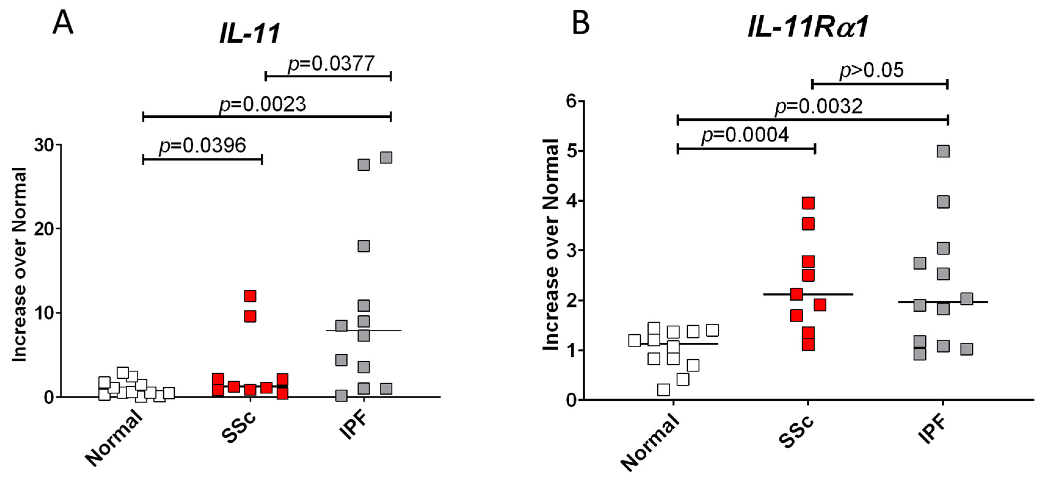
3.1. IL-11 and Its Receptor, IL-11Rα1, Have Increased Expression in SSc and IPF Fibroblasts

Gene expression of IL-11 and its receptor (IL-11Rα1) were evaluated in fibroblasts isolated from diseased lung tissues from patients with SSc and IPF and compared to fibroblasts derived from normal lungs. We used the IPF lung cell lines as a comparison for the SSc lung cell lines to determine if IL-11 and IL-11Rα1 expression were similar and a common feature of fibrosis. The relative expression of IL-11 was increased 3.35-fold in SSc (p = 0.0396) and 9.97-fold in IPF (p = 0.0023) (Figure 1A). We also saw that IPF fibroblasts expressed 2.97-fold more IL-11 than SSc fibroblasts (p = 0.0377, Figure 1A). Additionally, IL-11Rα1 was 2.00-fold higher in both SSc and IPF when compared to normal lung fibroblasts (p = 0.0004 and p = 0.0032, respectively, Figure 1B). However, there was no significant difference in the expression of IL-11Rα1 between SSc and IPF fibroblasts (p > 0.05, Figure 1B).

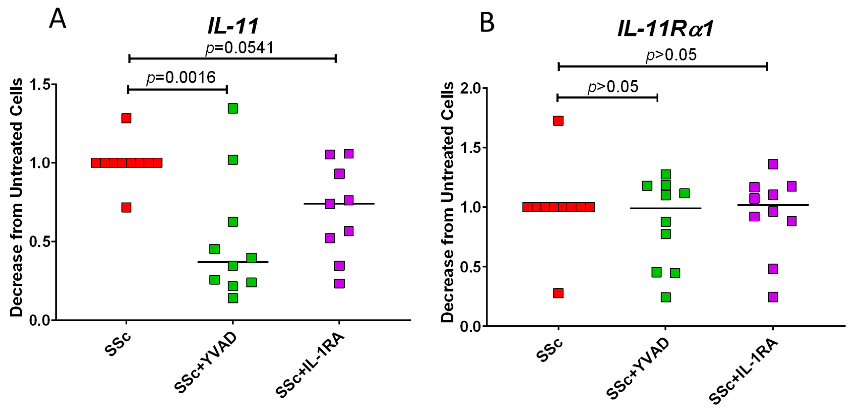
3.2. Inhibition of Caspase-1 Lowers IL-11 Expression in SSc Fibroblasts

Our prior studies have shown the role of caspase-1 in SSc fibrosis, and therefore, we investigated whether the expression of IL-11 was dependent on caspase-1 activity [32]. When caspase-1 was inhibited with the irreversible caspase-1 inhibitor, Z-YVAD-FMK, we found that IL-11 expression in SSc fibroblasts was significantly decreased by 49.59 ± 13.05% (Figure 2A; p = 0.0016). Because caspase-1 cleaves pro-IL-1β into its active form, we tested whether signaling from the IL-1 receptor was necessary for IL-11 expression. By blocking the IL-1 receptor with IL-1RA, we saw a non-significant trend for reduced IL-11 expression (Figure 2A; p = 0.0541). However, when analyzing the expression of IL-11Rα1, we found that neither YVAD nor IL-1RA could lower the transcripts of this gene in SSc fibroblasts (Figure 2B, p > 0.05 and p > 0.05, respectively).

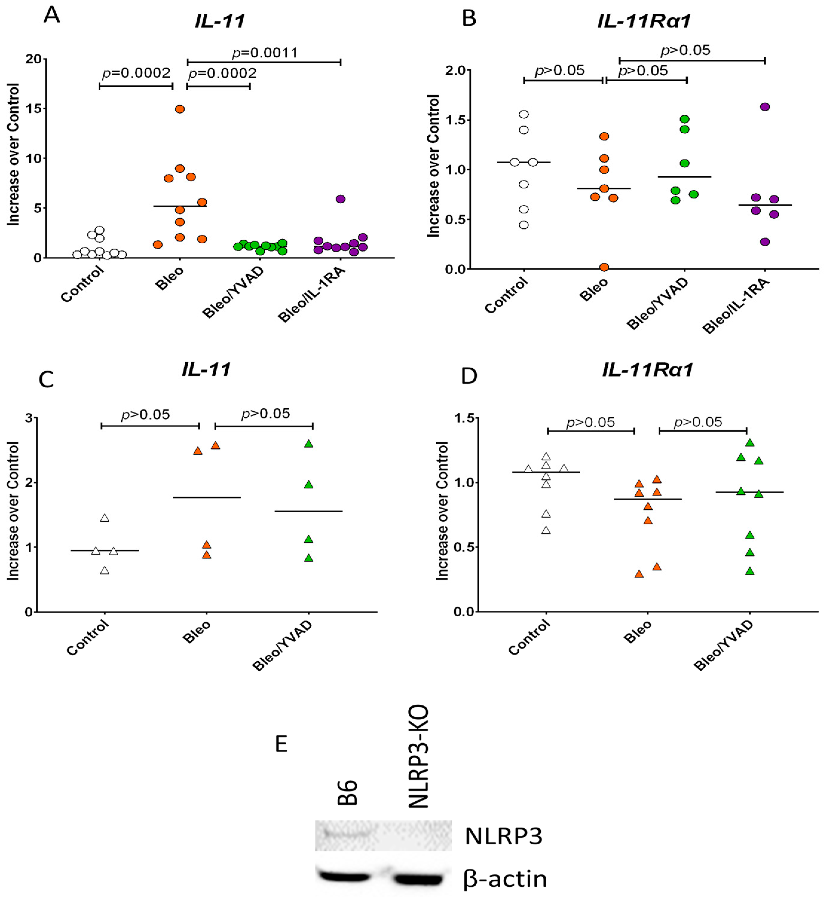
3.3. NLRP3 Inflammasome Activation of Caspase-1 Induces IL-11 Expression

Using bleomycin on normal fibroblasts, we confirmed the observation by Schafer et al., who showed that bleomycin can increase IL-11 expression [10]. We found a 5.92 +/− 4.2-fold increase in IL-11 expression (Figure 3A, p = 0.0002). This increase was completely abolished when caspase-1 activity was blocked with YVAD (Figure 3A, p = 0.0002). The increased IL-11 expression with bleomycin was also abolished with IL-1RA (Figure 3A, p = 0.0011). However, we observed no change in the expression of IL-11Rα1 when bleomycin was used (p > 0.05) and when YVAD or IL-1RA inhibitors were used (Figure 3B, p > 0.05 and p > 0.05, respectively). To confirm the role of the inflammasome activation of caspase-1 in IL-11 expression, we investigated if NLRP3-depleted fibroblasts could induce IL-11 transcripts when treated with bleomycin (Figure 3C). These studies show that the activation of the NLRP3 inflammasome is necessary for IL-11 expression by the inability of bleomycin to increase IL-11 transcription in NLRP3-deficient fibroblasts (Figure 3C, p > 0.05). We also saw that IL-11Rα1 expression was not dependent on this pathway (Figure 3D, p > 0.05).

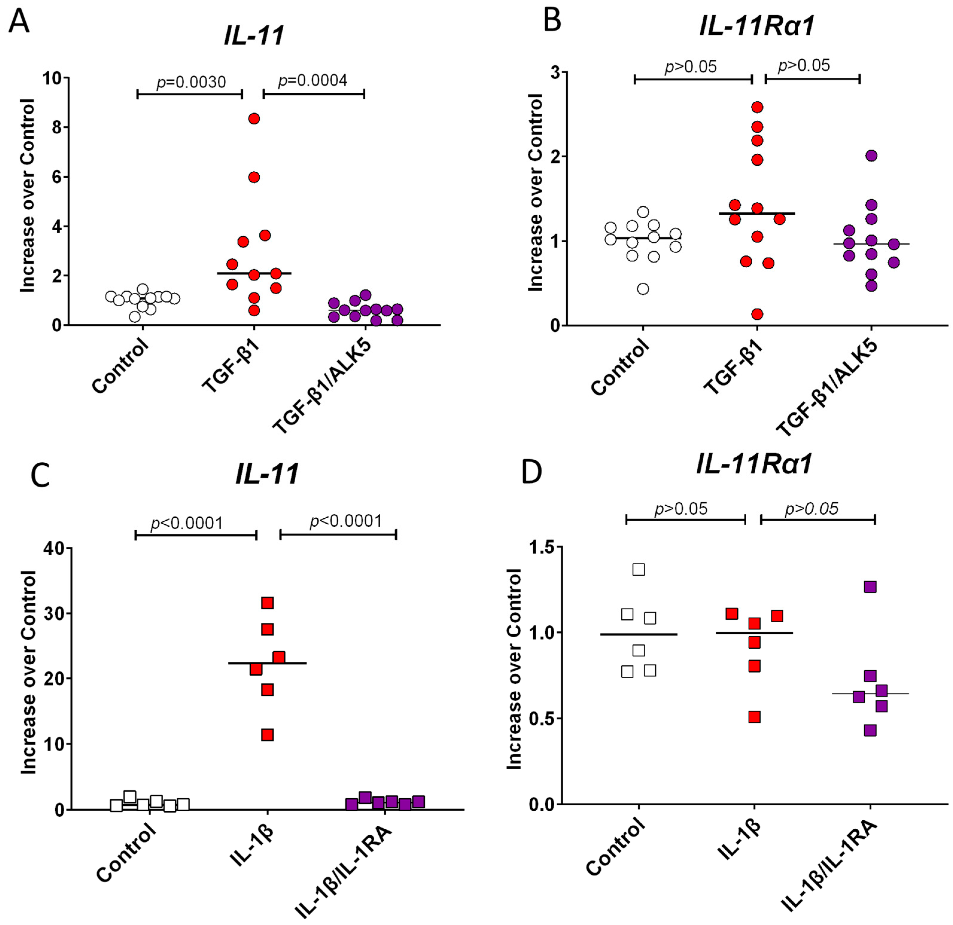
3.4. TGF-β1 and IL-1β Induce IL-11 Expression but Do Not Change the Expression of IL-11Ra1

TGF-β1 was previously shown to upregulate IL-11 expression [10]. We confirmed this and found IL-11 expression increased by 2.97 ± 2.31-fold with TGF-β1 (Figure 4A, p = 0.0030). The blockade of the TGF-β receptor with ALK5 confirmed the role of TGF-β receptor signaling in IL-11 expression (Figure 4A, p = 0.0004). However, we did not see any change in the expression of IL-11Rα1 with TGF-β1 or ALK5 (Figure 4B, p > 0.05 and p > 0.05, respectively). IL-1β also caused an increase in IL-11 expression 22.40 ± 7.89-fold (p < 0.0001), and this was abolished when the receptor was blocked with IL-1RA (Figure 4C, p < 0.0001). The expression of IL-11Rα1 was unchanged with IL-1β (Figure 4D, p > 0.05 and p > 0.05). When we compared the increase in IL-11 expression between IL-1β and TGF-β1, we noted that IL-1β induced significantly more IL-11 gene transcripts than TGF-β1 (22.40 vs. 2.97 respectively, p < 0.0001).

3.5. TLR4 Signaling Induces IL-11 and IL-11Rα1 Expression

LPS is known to activate TLR4 to induce gene expression, but it can also activate the NLRP3 inflammasome [34,35,36]. To further elaborate on the NLRP3 inflammasome activity in the IL-11 pathway, we investigated the role of TLR4 signaling in IL-11 expression by stimulating normal fibroblasts with LPS. We found that LPS significantly increased IL-11 transcripts by 1.52 ± 0.38-fold (Figure 5A, p = 0.0042), and this was abolished with YVAD (Figure 5A, p < 0.0001), confirming the role of the inflammasome in IL-11 expression. LPS also increased the expression of IL-11Rα1 by 1.51 ± 0.20-fold (Figure 5B, p = 0.0132); however, this could not be reduced with YVAD (Figure 5B, p > 0.05).

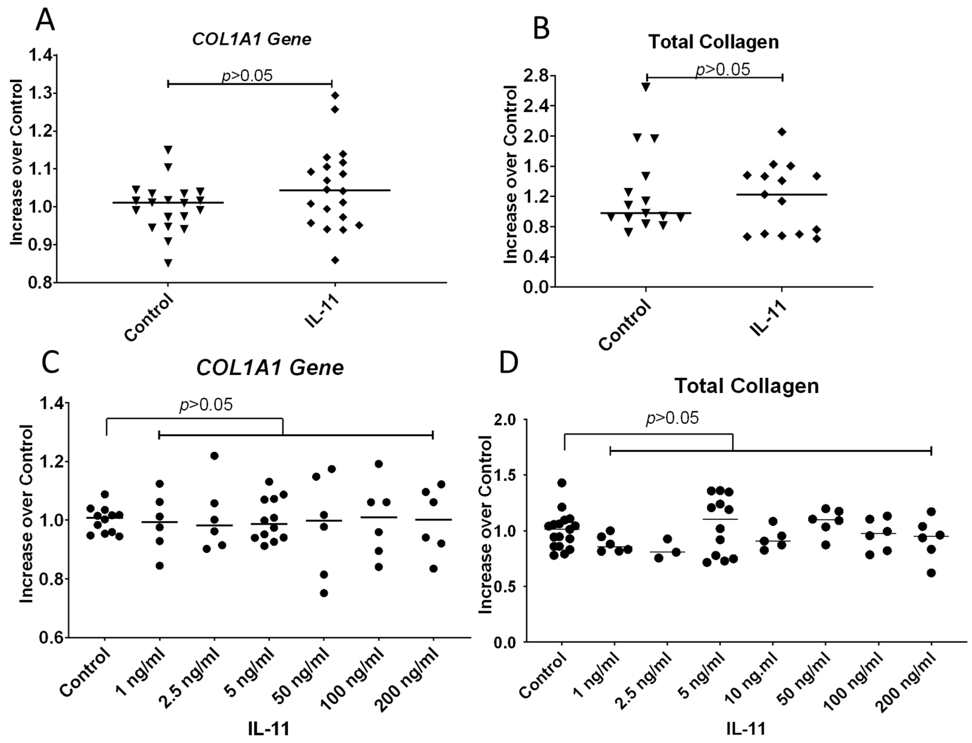
3.6. IL-11 Does Not Increase COL1A1 Gene Expression or Total Collagen Protein Secreted by Fibroblasts

Because IL-11 has been reported to be profibrotic, we investigated whether IL-11 could induce the expression of the COL1A1 gene and increase the amounts of collagen secreted by normal human fibroblasts. Many studies have reported using human recombinant IL-11 at 5 ng/mL [9,10,12]. Therefore, we used this concentration on normal human fibroblasts and evaluated for COL1A1 gene expression and total collagen protein secreted into the culture media over 48 h. We could not induce COL1A1 gene expression with 5 ng/mL (Figure 6A, p > 0.05), nor was there an increase in the total amount of collagen protein secreted into the culture media (Figure 6B, p > 0.05). Believing that the amount of IL-11 used might not be optimal, we then assessed a titration of IL-11 from 1 ng/mL to 200 ng/mL. We could not induce the expression of the COL1A1 gene with any of these concentrations, nor were we able to capture an increase in the secretion of total collagen protein into the media (Figure 6C, p > 0.05 and 5D, p > 0.05). This finding suggests that IL-11 does not induce collagen protein in normal fibroblasts.

3.7. ERK1/2 (p44/p42) Is Weakly Phosphorylated by Human Recombinant IL-11

As we did not see any increase in collagen protein accumulation over time during our experimental conditions, we wanted to confirm whether IL-11 was active and could phosphorylate ERK1/2 (p44/p42). In these studies, we used TGF-β1 as a comparison cytokine as it is known to phosphorylate ERK1/2. We found that IL-11 nominally increased the phosphorylation of ERK1/2, but it was not found to be as robust as that which was observed in TGF-β1 treated cells (Figure 7).

4. Discussion

These studies show that IL-11 and IL-11Rα1 gene expression is increased in SSc and IPF fibroblasts. We can induce IL-11 expression by activating the NLRP3 inflammasome and show this mechanism is separate from increased expression of its receptor IL-11Rα1. We show that IL-11 transcripts are increased with bleomycin and LPS, an effect mitigated with caspase-1 inhibition. This increase in IL-11 is not seen in NLRP-3 deficient fibroblasts. Further, IL-11 expression is increased with TGF-β1 and IL-1β, with those effects ablated with their respective inhibitors. We could not show that IL-11 increases collagen expression in normal fibroblasts, suggesting a more complicated mechanism in fibrotic disease.
IL-11 and IL-11Rα1 transcripts are increased in primary pulmonary fibroblasts from SSc and the fibrotic control, IPF. Interestingly, we found that IPF fibroblasts have significantly higher IL-11 gene expression than SSc fibroblasts. We speculate that this difference in IL-11 expression is because the fibrotic process in IPF is more profound than the interstitial lung disease observed in SSc. Following a diagnosis of IPF, patients have poor long-term prognosis with a disabling course involving mortality within 3–5 years, while SSc patients with interstitial lung disease have 50–70% survival at 10 years [37,38]. This could indicate a more aggressive process in IPF and, therefore, a more prominent role for IL-11.
Simultaneously, IL-11Rα1 transcripts in both fibrotic diseases were significantly increased, but the levels of expression were not different between the two diseases. The elevated expression of IL-11Rα1 suggests it may also be involved in the pathogenesis of fibrosis. However, its expression was not altered with an inhibition of caspase-1 or when the IL-1 and TGF-β receptors were inhibited, nor did its expression change with bleomycin. We did see, however, a concurrent increase in IL-11Rα1 and IL-11 transcripts when LPS was used. Overall, this suggests that the mechanism inducing the expression of the IL-11 receptor is likely different from the mechanism regulating the expression of IL-11.
Previous studies show that collagen in SSc fibroblasts is decreased with the caspase-1 inhibitor YVAD, and here, we show that IL-11 expression is dependent on caspase-1 activity (Figure 2) [32]. Given that caspase-1 activates IL-1β, we were able to show that IL-11 expression is dependent on the IL-1 receptor and show decreased IL-11 expression with IL-1RA in SSc fibroblasts. However, the decrease in IL-11 expression was more dramatic with YVAD than IL-1RA (Figure 3A). This may be explained by the insufficient saturation of the receptor antagonist on the IL-1 receptor on the cell membrane or by additional cytokines dependent on caspase-1 activity that could potentially induce IL-11.
Both caspase-1 and IL-1β are downstream effectors of the inflammasome, which can be activated with bleomycin and LPS via TLR4 signaling [30,34]. The NLRP3 inflammasome is overexpressed in fibrosis, and we found increased caspase-1 activity in the fibroblasts of patients with SSc [32]. The blockade of caspase-1 with YVAD lowers fibrotic mediators such as collagen, and this was confirmed in NLRP3-deficient mouse models of bleomycin-induced fibrosis [34,39]. Here, we show the link between the NLRP3 inflammasome and IL-11 expression, as fibroblasts deficient in NLRP3 could not induce IL-11 transcripts (Figure 3C). We also showed that the activation of the inflammasome via bleomycin (Figure 3A) and LPS (Figure 5A) led to increased IL-11 expression, which was abrogated with YVAD. However, the expression of the receptor was not dependent on the inflammasome, and an increase in its expression could only be induced by LPS (Figure 5B). These findings suggest that the NF-kB pathway is involved in IL-11Rα1 expression. At the same time, downstream signaling mediated by an activated inflammasome drives IL-11 expression, but more studies are needed to unveil this mechanism.
Consistent with earlier accounts in the literature, we were able to demonstrate increased IL-11 expression with TGF-β1 and IL-1β [10,16]. This increase in expression was mitigated with an ALK5 inhibitor, implicating the ALK5 signaling cascade from the TGF-β receptor in the upregulation of the IL-11 gene (Figure 4). Like TGF-β1, IL-1β can increase IL-11 expression in normal fibroblasts, which was abolished with IL-1RA (Figure 4C). The implication of these cytokines in the upregulation of IL-11 transcripts emphasizes the importance of crosstalk between these two pathways.
The limitations of this study are related to the in vitro nature of the experiments. Gene expression can only capture so much of the cellular signaling of a short-acting cytokine. In addition, detecting a cytokine with very low expression can be challenging. Some studies detect IL-11 by ELISA [8,16,18], and we have been unable to detect IL-11 protein in media or cell lysates of normal or fibrotic cells using immunoblotting. Furthermore, building on prior studies [10], we noted that SSc patients had picogram quantities of IL-11 in their serum [8,18]. Steadman and O’Reilly [8] reported elevated IL-11 levels in SSc sera to be 58 pg/mL, while the Cook group [18] reported much lower amounts of IL-11 (27.2 pg/mL). The amounts of recombinant IL-11 (5–10 ng/mL) used to stimulate fibroblasts in the in vitro studies by us and others are, therefore, not physiologically relevant [9,10,12] and are approximately 100 to 400 times more than that found in the sera from fibrotic patients. Further, our observations cannot fully discount the effect of causation. More studies should be performed to confirm these results.
Overall, our findings support Tan et al., whose elegant and thorough studies suggest that IL-11 might be a minor profibrotic cytokine secondary to TGF-β1 signaling [16]. They reported increased IL-11 transcripts in fibrotic cells; like us, they could not detect IL-11 protein. They also found that using 100 ng/mL of IL-11 only induced a modest response in a subset of the TGF-β1 profibrotic gene signature. Their studies also demonstrated increased IL-11 transcripts with bleomycin, and they could not detect the protein in the murine lung tissues.

5. Conclusions

In summary, our data from human fibroblasts derived from lung tissue of patients with SSc show that both the IL-11 and IL-11Rα1 transcripts were elevated but did not appear to be involved in the fibrosis pathophysiology, defined in our studies as increased collagen secretion. Given the numerous studies in the literature implicating IL-11 as a driver or an inhibitor of fibrosis, understanding both the upstream and downstream mechanisms that promote the expression of this cytokine is crucial. Further investigation is needed to strengthen the relationship between IL-11 expression and activation of the NLRP3 inflammasome in fibrosis.

Author Contributions

C.M.M. and C.M.A. conceptualized this study. C.M.M., L.M.C., A.G.F., and C.M.A. performed the methodology and data acquisition. C.M.M., A.G.F., and C.M.A. validated the data and conducted formal analyses. C.M.M. and C.M.A. prepared the original draft of this manuscript. C.M.M., L.M.C., A.G.F., and C.M.A. undertook the final review and editing. All authors have read and agreed to the published version of the manuscript.

Funding

This work was supported by the NIH, NIAMS R21073947, to C.M.A. and the Alpha Omega Alpha Carolyn L. Kuckein Student Research Fellowship to C.M.M.

Institutional Review Board Statement

Ethical review and approval were waived for this study because this research used cell lines derived from surgical tissue resected for clinical purposes that would otherwise be discarded. In addition, some of the cell lines were established from deceased individuals. No identifiers were associated with the cell lines, and it is not considered to be human-subject research.

Informed Consent Statement

The studies involved human cell lines established from discarded surgical tissues that were not collected by the persons involved in this study. The cell lines were deidentified. This is not considered Human Subjects research.

Data Availability Statement

Aggregated gene expression studies data will be available upon request.

Conflicts of Interest

The authors declare no conflicts of interest.

References

  1. Mayes, M.D.; Lacey, J.V., Jr.; Beebe-Dimmer, J.; Gillespie, B.W.; Cooper, B.; Laing, T.J.; Schottenfeld, D. Prevalence, incidence, survival, and disease characteristics of systemic sclerosis in a large US population. Arthritis Rheum. 2003, 48, 2246–2255. [Google Scholar] [CrossRef] [PubMed]
  2. Kelemen, B.W.; Mathai, S.C.; Tedford, R.J.; Damico, R.L.; Corona-Villalobos, C.; Kolb, T.M.; Chaisson, N.F.; Harris, T.H.; Zimmerman, S.L.; Kamel, I.R.; et al. Right ventricular remodeling in idiopathic and scleroderma-associated pulmonary arterial hypertension: Two distinct phenotypes. Pulm. Circ. 2015, 5, 327–334. [Google Scholar] [CrossRef] [PubMed]
  3. McNearney, T.A.; Reveille, J.D.; Fischbach, M.; Friedman, A.W.; Lisse, J.R.; Goel, N.; Tan, F.K.; Zhou, X.; Ahn, C.; Feghali-Bostwick, C.A.; et al. Pulmonary involvement in systemic sclerosis: Associations with genetic, serologic, sociodemographic, and behavioral factors. Arthritis Rheum. 2007, 57, 318–326. [Google Scholar] [CrossRef] [PubMed]
  4. Rosendahl, A.H.; Schönborn, K.; Krieg, T. Pathophysiology of systemic sclerosis (scleroderma). Kaohsiung J. Med. Sci. 2022, 38, 187–195. [Google Scholar] [CrossRef] [PubMed]
  5. Mei, Q.; Liu, Z.; Zuo, H.; Yang, Z.; Qu, J. Idiopathic Pulmonary Fibrosis: An Update on Pathogenesis. Front. Pharmacol. 2021, 12, 797292. [Google Scholar] [CrossRef]
  6. Swarnakar, R.; Garje, Y.; Markandeywar, N.; Mehta, S. Exploring the common pathophysiological links between IPF, SSc-ILD and post-COVID fibrosis. Lung India 2022, 39, 279–285. [Google Scholar] [CrossRef]
  7. Wynn, T.A. Cellular and molecular mechanisms of fibrosis. J. Pathol. 2008, 214, 199–210. [Google Scholar] [CrossRef]
  8. Steadman, T.; O’Reilly, S. Elevated interleukin-11 in systemic sclerosis and role in disease pathogenesis. J. Dermatol. 2023, 50, 1255–1261. [Google Scholar] [CrossRef]
  9. Ng, B.; Dong, J.; D’Agostino, G.; Viswanathan, S.; Widjaja, A.A.; Lim, W.W.; Ko, N.S.J.; Tan, J.; Chothani, S.P.; Huang, B.; et al. Interleukin-11 is a therapeutic target in idiopathic pulmonary fibrosis. Sci. Transl. Med. 2019, 11, eaaw1237. [Google Scholar] [CrossRef]
  10. Schafer, S.; Viswanathan, S.; Widjaja, A.A.; Lim, W.W.; Moreno-Moral, A.; DeLaughter, D.M.; Ng, B.; Patone, G.; Chow, K.; Khin, E.; et al. IL-11 is a crucial determinant of cardiovascular fibrosis. Nature 2017, 552, 110–115. [Google Scholar] [CrossRef]
  11. Ng, B.; Viswanathan, S.; Widjaja, A.A.; Lim, W.W.; Shekeran, S.G.; Goh, J.W.T.; Tan, J.; Kuthubudeen, F.; Lim, S.Y.; Xie, C.; et al. IL11 Activates Pancreatic Stellate Cells and Causes Pancreatic Inflammation, Fibrosis and Atrophy in a Mouse Model of Pancreatitis. Int. J. Mol. Sci. 2022, 23, 3549. [Google Scholar] [CrossRef] [PubMed]
  12. Widjaja, A.A.; Singh, B.K.; Adami, E.; Viswanathan, S.; Dong, J.; D’Agostino, G.A.; Ng, B.; Lim, W.W.; Tan, J.; Paleja, B.S.; et al. Inhibiting Interleukin 11 Signaling Reduces Hepatocyte Death and Liver Fibrosis, Inflammation, and Steatosis in Mouse Models of Nonalcoholic Steatohepatitis. Gastroenterology 2019, 157, 777–792.e714. [Google Scholar] [CrossRef] [PubMed]
  13. Milara, J.; Roger, I.; Montero, P.; Artigues, E.; Escrivá, J.; Del Río, R.; Cortijo, J. Targeting IL-11 to reduce fibrocyte circulation and lung accumulation in animal models of pulmonary hypertension-associated lung fibrosis. Br. J. Pharmacol. 2024, 181, 2991–3009. [Google Scholar] [CrossRef] [PubMed]
  14. Bai, X.; Zhao, G.; Chen, Q.; Li, Z.; Gao, M.; Ho, W.; Xu, X.; Zhang, X.Q. Inhaled siRNA nanoparticles targeting IL11 inhibit lung fibrosis and improve pulmonary function post-bleomycin challenge. Sci. Adv. 2022, 8, eabn7162. [Google Scholar] [CrossRef] [PubMed]
  15. Allanki, S.; Strilic, B.; Scheinberger, L.; Onderwater, Y.L.; Marks, A.; Günther, S.; Preussner, J.; Kikhi, K.; Looso, M.; Stainier, D.Y.R.; et al. Interleukin-11 signaling promotes cellular reprogramming and limits fibrotic scarring during tissue regeneration. Sci. Adv. 2021, 7, eabg6497. [Google Scholar] [CrossRef] [PubMed]
  16. Tan, Y.; Mosallanejad, K.; Zhang, Q.; O’Brien, S.; Clements, M.; Perper, S.; Wilson, S.; Chaulagain, S.; Wang, J.; Abdalla, M.; et al. IL11-mediated stromal cell activation may not be the master regulator of pro-fibrotic signaling downstream of TGFβ. Front. Immunol. 2024, 15, 1293883. [Google Scholar] [CrossRef]
  17. Widjaja, A.A.; Viswanathan, S.; Jinrui, D.; Singh, B.K.; Tan, J.; Wei Ting, J.G.; Lamb, D.; Shekeran, S.G.; George, B.L.; Schafer, S.; et al. Molecular Dissection of Pro-Fibrotic IL11 Signaling in Cardiac and Pulmonary Fibroblasts. Front. Mol. Biosci. 2021, 8, 740650. [Google Scholar] [CrossRef]
  18. Adami, E.; Viswanathan, S.; Widjaja, A.A.; Ng, B.; Chothani, S.; Zhihao, N.; Tan, J.; Lio, P.M.; George, B.L.; Altunoglu, U.; et al. IL11 is elevated in systemic sclerosis and IL11-dependent ERK signalling underlies TGFβ-mediated activation of dermal fibroblasts. Rheumatology 2021, 60, 5820–5826. [Google Scholar] [CrossRef]
  19. Liu, Z.; Wang, Y.; Yan, J.; Liu, J.; Chen, B.; Zhang, L.; Cheng, L. Efficacy and safety of recombinant human interleukin-11 in the treatment of acute leukaemia patients with chemotherapy-induced thrombocytopenia: A systematic review and meta-analysis. J. Eval. Clin. Pract. 2020, 26, 262–271. [Google Scholar] [CrossRef]
  20. Chen, M.; Li, L.; Xia, Q.; Chen, X.; Liao, Z.; Wang, C.; Shen, B.; Zhou, M.; Zhang, Q.; Zhang, Y.; et al. A real-world observation on thrombopoietic agents for patients with cancer treatment-induced thrombocytopenia in China: A multicenter, cross-sectional study. Cancer 2024, 130, 1524–1538. [Google Scholar] [CrossRef]
  21. Bhatia, M.; Davenport, V.; Cairo, M.S. The role of interleukin-11 to prevent chemotherapy-induced thrombocytopenia in patients with solid tumors, lymphoma, acute myeloid leukemia and bone marrow failure syndromes. Leuk. Lymphoma 2007, 48, 9–15. [Google Scholar] [CrossRef] [PubMed]
  22. Wang, J.C.; Chen, C.; Lou, L.H.; Mora, M. Blood thrombopoietin, IL-6 and IL-11 levels in patients with agnogenic myeloid metaplasia. Leukemia 1997, 11, 1827–1832. [Google Scholar] [CrossRef] [PubMed]
  23. Nakagawa, M.; Owada, Y.; Izumi, Y.; Nonin, S.; Sugioka, K.; Nakatani, D.; Iwata, S.; Mizutani, K.; Nishimura, S.; Ito, A.; et al. Four cases of investigational therapy with interleukin-11 against acute myocardial infarction. Heart Vessel. 2016, 31, 1574–1578. [Google Scholar] [CrossRef] [PubMed]
  24. Obana, M.; Maeda, M.; Takeda, K.; Hayama, A.; Mohri, T.; Yamashita, T.; Nakaoka, Y.; Komuro, I.; Takeda, K.; Matsumiya, G.; et al. Therapeutic activation of signal transducer and activator of transcription 3 by interleukin-11 ameliorates cardiac fibrosis after myocardial infarction. Circulation 2010, 121, 684–691. [Google Scholar] [CrossRef] [PubMed]
  25. Zhu, M.; Lu, B.; Cao, Q.; Wu, Z.; Xu, Z.; Li, W.; Yao, X.; Liu, F. IL-11 Attenuates Liver Ischemia/Reperfusion Injury (IRI) through STAT3 Signaling Pathway in Mice. PLoS ONE 2015, 10, e0126296. [Google Scholar] [CrossRef]
  26. Cook, S.A. Understanding interleukin 11 as a disease gene and therapeutic target. Biochem. J. 2023, 480, 1987–2008. [Google Scholar] [CrossRef]
  27. Sweeney, M.; O’Fee, K.; Villanueva-Hayes, C.; Rahman, E.; Lee, M.; Vanezis, K.; Andrew, I.; Lim, W.W.; Widjaja, A.; Barton, P.J.R.; et al. Cardiomyocyte-Restricted Expression of IL11 Causes Cardiac Fibrosis, Inflammation, and Dysfunction. Int. J. Mol. Sci. 2023, 24, 12989. [Google Scholar] [CrossRef]
  28. Lokau, J.; Nitz, R.; Agthe, M.; Monhasery, N.; Aparicio-Siegmund, S.; Schumacher, N.; Wolf, J.; Möller-Hackbarth, K.; Waetzig, G.H.; Grötzinger, J.; et al. Proteolytic Cleavage Governs Interleukin-11 Trans-signaling. Cell Rep. 2016, 14, 1761–1773. [Google Scholar] [CrossRef]
  29. Kolb, M.; Margetts, P.J.; Anthony, D.C.; Pitossi, F.; Gauldie, J. Transient expression of IL-1beta induces acute lung injury and chronic repair leading to pulmonary fibrosis. J. Clin. Investig. 2001, 107, 1529–1536. [Google Scholar] [CrossRef]
  30. Gurcel, L.; Abrami, L.; Girardin, S.; Tschopp, J.; van der Goot, F.G. Caspase-1 activation of lipid metabolic pathways in response to bacterial pore-forming toxins promotes cell survival. Cell 2006, 126, 1135–1145. [Google Scholar] [CrossRef]
  31. Hussein, M.R.; Hassan, H.I.; Hofny, E.R.M.; Elkholy, M.; Fatehy, N.A.; Abd Elmoniem, A.E.A.; Ezz El-Din, A.M.; Afifi, O.A.; Rashed, H.G. Alterations in mononuclear inflammatory cells, CD4/CD8+ T cells, interleukin-1á, and tumor necrosis factor à in the bronchoalveolar lavage fluid, peripheral blood, and skin of patients with systemic sclerosis. J. Clin. Pathol. 2005, 58, 178–184. [Google Scholar] [CrossRef] [PubMed]
  32. Artlett, C.M.; Sassi-Gaha, S.; Rieger, J.L.; Boesteanu, A.C.; Feghali-Bostwick, C.A.; Katsikis, P.D. The inflammasome activating caspase-1 mediates fibrosis and myofibroblast differentiation in systemic sclerosis. Arthritis Rheum. 2011, 63, 3563–3574. [Google Scholar] [CrossRef] [PubMed]
  33. Luo, D.D.; Fielding, C.; Phillips, A.; Fraser, D. Interleukin-1 beta regulates proximal tubular cell transforming growth factor beta-1 signalling. Nephrol. Dial. Transplant. 2009, 24, 2655–2665. [Google Scholar] [CrossRef] [PubMed]
  34. Gurung, P.; Li, B.; Subbarao Malireddi, R.K.; Lamkanfi, M.; Geiger, T.L.; Kanneganti, T.D. Chronic TLR Stimulation Controls NLRP3 Inflammasome Activation through IL-10 Mediated Regulation of NLRP3 Expression and Caspase-8 Activation. Sci. Rep. 2015, 5, 14488. [Google Scholar] [CrossRef] [PubMed]
  35. Mezzasoma, L.; Schmidt-Weber, C.B.; Fallarino, F. In Vitro Study of TLR4-NLRP3-Inflammasome Activation in Innate Immune Response. Methods Mol. Biol. 2023, 2700, 163–176. [Google Scholar] [CrossRef] [PubMed]
  36. Huo, M.; Guo, W.; Ding, L. Benidipine Hydrochloride Inhibits NLRP3 Inflammasome Activation by Inhibiting LPS-Induced NF-κB Signaling in THP-1 Macrophages. J. Inflamm. Res. 2024, 17, 6307–6316. [Google Scholar] [CrossRef]
  37. Fernández Fabrellas, E.; Peris Sánchez, R.; Sabater Abad, C.; Juan Samper, G. Prognosis and Follow-Up of Idiopathic Pulmonary Fibrosis. Med. Sci. 2018, 6, 51. [Google Scholar] [CrossRef]
  38. Volkmann, E.R.; Fischer, A. Update on Morbidity and Mortality in Systemic Sclerosis-Related Interstitial Lung Disease. J. Scleroderma Relat. Disord. 2021, 6, 11–20. [Google Scholar] [CrossRef]
  39. Martínez-Godínez, M.A.; Cruz-Domínguez, M.P.; Jara, L.J.; Domínguez-López, A.; Jarillo-Luna, R.A.; Vera-Lastra, O.; Montes-Cortes, D.H.; Campos-Rodríguez, R.; López-Sánchez, D.M.; Mejía-Barradas, C.M.; et al. Expression of NLRP3 inflammasome, cytokines and vascular mediators in the skin of systemic sclerosis patients. Isr. Med. Assoc. J. IMAJ 2015, 17, 5–10. [Google Scholar]
Figure 1. Fibrotic fibroblasts express higher levels of IL-11 and IL-11Ra1 gene transcripts than normal healthy fibroblasts. (A) Expression of IL-11 and (B) IL-11R⍺1 was measured in RNA extracted from 650,000 SSc fibroblasts (n = 9 independent lung fibroblast lines), the fibrotic control, idiopathic pulmonary fibrosis IPF, (n = 12 replicates from 8 independent lung fibroblast lines), and normal healthy lung fibroblasts (n = 9 replicates from 7 independent fibroblast lines) by qPCR. Data are shown as a fold increase over the average expression found in the normal healthy fibroblasts. Statistical analyses: two-tailed, unpaired t-test.
Rheumato 04 00013 g001
Figure 2. Inhibition of caspase-1 decreases IL-11 expression but does not affect IL-11Rα1 expression. SSc fibroblasts (squares, n = 10, from 7 independent lung fibroblast lines) were cultured with 10 μM YVAD or 2 mg/mL IL-1RA. IL-11 (A) and IL-11Ra1 (B) gene expression were measured by qPCR. Statistical analysis: one-way ANOVA with Tukey’s correction for multiple comparisons.
Rheumato 04 00013 g002
Figure 3. Activation of the inflammasome with bleomycin induces the expression of the IL-11 gene via IL-1 but does not affect the expression of IL-11Ra1. Normal healthy fibroblasts were treated with 10 mM bleomycin (Bleo) +/− 10 μM YVAD or 2 mg/mL IL-1RA (circles, n = 10 replicates, from 4 independent fibroblast lines), and IL-11 (A) and IL-11Ra1 (B) gene expression was measured by qPCR. IL-11 (C) and IL-11Ra1 (D) gene expression was evaluated in NLRP3-KO fibroblasts (triangles, n = 4–8) were treated with 10 μM Bleo +/− 10 μM YVAD. (E) NLRP3 protein in wild-type (B6) and NLRP3-KO fibroblast cell lines. Statistical analysis: one-way ANOVA with Tukey’s correction for multiple comparisons.
Rheumato 04 00013 g003
Figure 4. TGF-β1 and IL-1β increase IL-11 expression while IL-11R⍺1 expression remains unchanged. Normal healthy fibroblasts were treated for 24 h with TGF-β1, +/− ALK5 (circles, n = 12 replicates, from 3 independent experiments), and IL-11 (A) and IL-11Rα1 (B) measured by qPCR. Normal healthy fibroblasts were also incubated with 1 ng/mL IL-1β +/− 2 mg/mL IL-1RA (squares, n = 6) in serum-free media, and IL-11 (C) and IL-11Rα1 (D) measured by qPCR. Statistical analyses: one-way ANOVA with Tukey’s correction for multiple comparisons.
Rheumato 04 00013 g004
Figure 5. LPS upregulates the genetic expression of IL-11 and IL-11Rα1. Normal healthy fibroblasts (n = 6–18, from 4 independent fibroblast cell lines) were treated with 1 ng/mL LPS +/− 10 μM YVAD. RNA was extracted, and IL-11 (A) and IL-11Rα1 (B) were measured by qPCR. Statistical analyses: one-way ANOVA with Tukey’s correction for multiple comparisons.
Rheumato 04 00013 g005
Figure 6. IL-11 does not increase COL1A1 gene expression or increase total collagen protein secreted by normal healthy fibroblasts. (A) Normal fibroblasts were treated with 5 ng/mL IL-11 (circles, n = 20), and the COL1A1 gene was assessed by qPCR. (B) Culture media from these experiments (squares, n = 15) were assessed for total collagen protein by Sirius Red. (C) An IL-11 titration was studied for COL1A1 gene expression (circles, n = 6–12), and (D) total collagen (squares, n = 3–12) was measured in the culture media with Sirius Red. Statistical analysis: for (A,B), a two-tailed paired t-test was used, and for (C,D), a one-way ANOVA with Tukey’s correction was used.
Rheumato 04 00013 g006
Figure 7. IL-11 is a weaker ERK1/2 (p44/p42) phosphorylator than TGF-β1. We compared the phosphorylation capabilities between IL-11 (5 ng/mL) and TGF-β1 (5 ng/mL) in normal fibroblasts after 48 h of exposure to the cytokine. A total of 40 μg protein/condition was electrophoresed and transferred to nitrocellulose. TGF-β1 was used as a positive control for ERK1/2 phosphorylation. β-actin was used for a loading control.
Rheumato 04 00013 g007
Table 1. Primers that were used to amplify the gene transcripts.
SpeciesGeneForward PrimerReverse Primer
Humanβ-actin5′–TTGCCGACAGGATGCAGAA–3′5′–GCCGATCCACACGGAGTACTT–3′
IL-115′–GGGGACATGAACTGTGTTTGC–3′5′–GGGCGACAGCTGTATCTGG–3′
IL-11Rα15′–CTGGGCTAGGGCATGAACTG–3′5′–CTGGGACTCCAAGTGCAAGA–3′
Mouseβ-actin5′–ACGGCCAGGTCATCACTATTG–3′5′–CAAGAAGGAAGGCTGGAAAAGA–3′
IL-115′–TTGGGATCTTTGCAGCCTTCCT–3′5′–CATGCCGGAGGTAGGACATC–3′
IL-11Rα15′–CAACTCAGTGGAGCGGGAG–3′5′–AGCTGCTGCTCATCTTGATAAT–3′
Disclaimer/Publisher’s Note: The statements, opinions and data contained in all publications are solely those of the individual author(s) and contributor(s) and not of MDPI and/or the editor(s). MDPI and/or the editor(s) disclaim responsibility for any injury to people or property resulting from any ideas, methods, instructions or products referred to in the content.

