Article

Stoichiometric Multiprotein Assembly Scaffolded by a Heterotrimeric DNA Clamp for Enzyme Colocalization and DNA Functionalization

by
Arabella Essert
and
Kathrin Castiglione
*
Institute of Bioprocess Engineering, Friedrich-Alexander-Universität Erlangen-Nürnberg, 91052 Erlangen, Germany
*
Author to whom correspondence should be addressed.
SynBio 2025, 3(4), 16; https://doi.org/10.3390/synbio3040016
Submission received: 22 April 2025 / Revised: 7 July 2025 / Accepted: 25 September 2025 / Published: 6 November 2025

Abstract

Researchers strive to exploit kinetic potentials of multistep reactions by positioning enzymes in a regulated fashion. Therein, the proliferating cell nuclear antigen (PCNA) from Sulfolobus solfataricus is a promising biomolecular tool due to its extraordinary architecture. PCNA is a circular DNA sliding clamp, which can bind and move along DNA and thus, be applied for the immobilization and transport of biomolecules on versatile DNA scaffolds. Additionally, its heterotrimeric character facilitates the colocalization of enzyme cascades with defined stoichiometry. This study provides insights into the in vitro binding behavior of PCNA and its potential as protein scaffold for DNA functionalization and controlled biocatalysis: (1) PCNA was capable of binding circular DNA and wireframe DNA nanostructures. (2) DNA binding was predominantly mediated by the PCNA1 subunit. (3) PCNA assembly around DNA was compromised when cysteines were introduced at the PCNA–PCNA interfaces to stabilize the ring via disulfide bonds. (4) A two-enzyme cascade, comprising a pseudo-monomeric cytochrome P450 BM3 monooxygenase and a monomeric alcohol dehydrogenase (ADH), was successfully fused to PCNA, retaining catalytic activity. (5) When immobilized on DNA, the cascade performance was not assessable, due to nearly complete loss of ADH activity in proximity to DNA.

1. Introduction

The way living organisms master physiological reactions has inspired researchers for decades and has laid the foundation for the development of innovative biosynthetic technologies [1,2]. Mimicking in vivo molecular nanomachineries is the main objective, combining positive aspects of conventional one-pot multistep reaction systems and enzyme immobilization in the most effective manner [3,4]. Such ‘metabolons’ are supramolecular complexes of several enzymes that are positioned in a tight sequence [5]. This can further reduce waste generation and fine-tune thermodynamics by shifting the reaction equilibrium to the product side if the consecutive enzyme catalyzes an irreversible reaction [6,7]. Moreover, when enzymes are artificially immobilized on scaffolds, biocatalyst recycling and thus, further downstream processing is simplified [8]. Additionally, catalytic or physical characteristics are modifiable by the type of immobilization [9,10] or by the microenvironment of the carrier [11]. Finally, co-immobilization brings different enzymes into close proximity and thus, promotes the special phenomenon termed ‘substrate channeling’ under certain conditions [12]. In serial reactions, it boosts the cascade performance by direct transfer of the intermediate from one active site to the next without its diffusion into the bulk solution [13]. This colocalization of enzymes protects labile cofactors (e.g., nicotinamide adenine dinucleotide, NADH) [14] and prevents the accumulation of inhibitory intermediates or byproducts [15]. Additionally, high local intermediate concentrations occur between co-acting enzymes, which reduce the apparent Michaelis constant (Km) [16].
To fully exploit these effects by fine-tuning the key parameters—spacing, order, orientation, and ratio—of the contributing enzymes, nanoscale precision is mandatory [17]. Among the many versatile immobilization materials [18,19,20], biomolecular scaffolds are of particular interest, as they are biocompatible, biodegradable, and applicable within host cells as well [21]. A high degree of site-specificity can be achieved with protein scaffolds, making them ideal candidates for the positional co-immobilization of enzymes that catalyze reactions in a defined order [22]. The core elements of protein scaffolds are protein binding modules, which are either connected by covalent peptide linkers (I) or self-assemble via noncovalent complementary interactions (II). Cascade enzymes are fused to the corresponding binding partner of a specific module (I) or are directly fused to an adapter domain (II) [23]. With the growing number of known protein interaction partners, exhibiting high specificity, this field is already well advanced. A paradigm in this category is the ‘designer cellulosome’. Its protein backbone—the ‘scaffoldin’—consists of cohesin modules that specifically bind their dockerin partners, each fused to a single enzyme molecule or complete enzyme cluster (e.g., via EutM). Combining cohesin-dockerin pairs from different species—each characterized by specific binding interactions—facilitates colocalization in a regulated fashion [24]. Other potentially site-specific protein scaffolds include the GBD-SH3-PDZ system [25], affibodies [26], and coiled-coil peptides [27].
An alternative to linear protein scaffolds is a ring-shaped assembly of proteins on the proliferating cell nuclear antigen (PCNA). Across all domains of life, PCNA plays a central role as a transport vehicle that recruits auxiliary proteins for DNA replication, DNA repair, and cell proliferation [28]. Due to its unique ring-shaped topology, enzymes can be arranged in a circle, creating a molecular nanofactory. Six structurally homologous globular domains, arranged with conserved pseudohexameric symmetry, form the foundation and are divided into either two or three monomers [29]. Typically, PCNA subunits are identical, though some archaeal variants form heterotrimers, providing additional control over enzyme stoichiometry [30]. This also applies to PCNA from the thermophilic organism Sulfolobus solfataricus, which has been used for biotechnological purposes in previous studies [31]. This protein formed the center of an artificial electron transfer system designed by Hirakawa and Nagamune [32]. Each of the three redox components of the P450 system from Pseudomonas putida was fused to the C-termini of the three PCNA monomers, which are located on the same side of the ring. After self-assembly of all three PCNA fusion proteins, the system exhibited a 50-fold increase in activity compared to the free equimolar P450 system.
Another highlight of PCNA is its ability to encircle and slide along double-stranded DNA (dsDNA), earning it the term ‘DNA sliding clamp’ [33]. Due to the robust complementary Watson–Crick base pairing, DNA is highly stable and spatially addressable [34]. Therefore, a broad variety of multidimensional structures can be designed at the nanoscale [35]. Rothemund developed the concept of ‘DNA origami’ [36], which opened the door to a versatile toolbox of DNA nanotechnologies [37]. The basic principle involves the self-assembly of a long single-stranded DNA scaffold (M13mp18 viral genome), guided by multiple short oligonucleotides (‘staples’) that match specific regions of the scaffold strand [38]. An innovative origami approach is the folding of polyhedral meshes by Benson et al. [39]. Unlike most origami algorithms, the scaffold DNA does not form self-crossovers, thereby expanding the repertoire of arbitrary, highly stable shapes. These ‘wireframe DNA nanostructures’ mainly comprise single double helices, granting them high flexibility, but also making them an interesting target for immobilization via PCNA.
PCNA can serve as a key component in novel DNA-based biosensors, such as the molecular DNA nanorobots, like ‘DNA walkers’ [40] or ‘DNA spiders’ [41]. These nanomachines move in a directed and controlled manner, driven by strand displacement mechanisms [42]. Alternatively, PCNA can be employed to co-immobilize enzymes on DNA scaffolds, generating three-dimensional mega enzyme clusters. Within these densely packed clusters, inter-enzyme distances are minimized, promoting substrate channeling, and thereby enhancing overall catalytic efficiency [43]. Typically, DNA origami staples are functionalized with affinity binding partners, such as the (strept)avidin/biotin pair [44] or aptamer/antibody combinations [45]. Directed by sequence complementarity, these functionalized staples hybridize with specific regions of the origami scaffold, allowing for precise spatial organization of biomolecules [46]. However, each DNA origami architecture requires a unique set of staples, which limits the versatility of this approach across different origami structures. In contrast, PCNA provides a universal, sequence-independent platform for immobilization of biomolecules on DNA scaffolds. This flexibility has the potential to overcome the limitations imposed by sequence-specific staple hybridization, offering a broadly applicable strategy for DNA functionalization.
Our objective was to harness the dual functionality of PCNA: (1) its ability to immobilize protein cargo through DNA encirclement, and (2) its intrinsic stoichiometry, which enables precise spatial control of enzyme colocalization. In nature, PCNA binds via a complex clamp loading mechanism, which is assisted by the clamp loader Replication Factor C (RFC) [47,48]. Previous findings reveal that PCNA can bind DNA in vitro without relying on these auxiliary proteins [49,50,51]. Thus, we expanded these studies to investigate the binding behavior of PCNA from Sulfolobus solfataricus (Figure 1A) across distinct DNA scaffolds and at varying PCNA multimerization states. Based on these results, we utilized PCNA as a scaffold for immobilization of a biocatalytic model system on DNA—a two-enzyme system with partial in situ NADH regeneration: A monomeric alcohol dehydrogenase from Thermococcus kodakarensis (ADH) [52] and a thermostable, dimer-stabilized cytochrome P450 BM3 monooxygenase variant (P450) [53] for enantioselective alcohol production [54]. The two enzymes were intended to be (1) colocalized via PCNA (Figure 1C), and the resulting multienzyme complex was then to be (2) immobilized on the different DNA scaffolds, including an exemplary wireframe DNA nanostructure. Due to weak intermolecular interactions between PCNA monomers, the heterotrimer is not very stable [55]. Hence, we incorporated covalent disulfide bonds at the PCNA-PCNA interfaces, as described by Hirakawa et al., to aim for robust DNA binding [56]. To prepare the DNA-bound PCNA complex, we established a method using only the ADH fused to the ring-stabilized PCNASS (Figure 1B). This strategy has provided additional insights into the DNA-binding characteristics of PCNA. Finally, we investigated the effect of PCNA-mediated colocalization and DNA immobilization on the catalytic performance of the two-enzyme system.

2. Results and Discussion

2.1. Binding of PCNA on Different DNA Scaffolds

Previous in vitro studies have demonstrated that PCNA from different species can bind to DNA without the assistance of the molecular clamp loader [49,50,51]. We expanded these studies by the DNA binding analysis of heterotrimeric PCNA from Sulfolobus solfataricus on different DNA scaffolds (Figure 2). For this purpose, we selected plasmid DNA (Figure 3(A1,A2)) and a novel wireframe DNA nanostructure forming a blossom (Figure 3(A3)) [57]. Changes in the molar mass of DNA towards higher values (DNA shifts) were determined relative to a DNA standard. Hence, the obtained numbers may deviate from actual values due to geometric differences between PCNA and DNA. To enable a quantitative comparison of binding results between different PCNA species, we calculated the number of PCNA monomers bound per DNA scaffold (Method S1, Figures S1 and S2, Table S1). Artifacts caused by the mere presence of protein were excluded, as high concentrations of the non-DNA-binding protein bovine serum albumin (BSA) induced no DNA shift during the electrophoretic mobility shift assay (EMSA) (Figure S3).
Higher concentrations of PCNA resulted in increased DNA shifts, indicating functional DNA binding. However, the overall amount of bound protein was low. At maximum concentrations of 100 µM and 150 µM PCNA, only ~30 (plasmid) and ~40 (wireframe blossom) PCNA units were bound to DNA—well below the theoretical binding capacities. Higher PCNA concentrations were difficult to achieve due to protein precipitation. PCNA has a thickness of ~30 Å [29]. Hence, we assume that PCNA blocks one helix turn (3.4 nm) via binding, which corresponds to 10 bp. On this premise, the hypothetical binding capacities would be ~270 (plasmid) or ~725 (wireframe blossom), calculated from the plasmid size (2686 bp) or the scaffold DNA (7249 nucleotides, nt). However, plasmids, especially in the case of supercoiling or multimerization, adopt secondary or tertiary structures through self-crossovers, limiting the accessibility of binding sites for PCNA. The same applies to wireframe DNA with multiple hinge regions, suggesting that the actual binding capacity is likely lower.
The presumably low binding affinities correlate with the very high dissociation constants (Kd) of 0.7 mM (short dsDNA) or over 1 mM (plasmid) for the structurally homologous human PCNA [50]. We attempted to determine Kd values by, e.g., fluorescence polarization, thermal shift assay, or surface plasmon resonance spectroscopy. But none of these yielded conclusive results due to the high autofluorescence of DNA or the complexity of the PCNA-DNA assembly, involving several binding affinities. Nonetheless, our results have proven that PCNA had the ability to bind plasmid DNA as well as the wireframe DNA structure.

2.2. DNA Binding at Different PCNA Multimerization Levels

To evaluate the contribution of each monomer to DNA binding, both individually and in assembly, we analyzed single monomers as well as different combinations of dimers and trimers (Figure 3). The overall measurement uncertainty ranged between 25% and 55%, with a tendency towards the higher values for samples in which a few PCNA monomers were bound to DNA (e.g., dimer sample). In these cases, the resulting shifts in DNA mobility were relatively small, making them more susceptible to measurement inaccuracies. The reproducibility of technical replicates was higher with relative errors below 20% (Figures S6 and S7). Despite the variability in absolute measurements, the observed DNA shift patterns were clearly distinguishable and consistent across all experiments, allowing for robust and meaningful interpretation of the binding behavior.
Figure 3. DNA binding analysis of PCNA in different multimeric states. Schematic illustration and atomic force microscopy (AFM) visualization of relaxed (A1) and supercoiled plasmid (A2) as well as wireframe blossom with relative internal distances at the highlighted position (A3). AFM images of exemplary pBR322 plasmid (4361 bp) or wireframe blossom were taken by Witz and Stasiak [58] or Götzfried [57], respectively. EMSA of multimerized plasmid with 50 µM (B1) or 75 µM (B2) of a PCNA species, and of wireframe blossom with 50 µM of a PCNA species (B3). The measurement uncertainty ranged from 25% (PCNA1, trimer) to 55% (dimer). Full gel images and an additional biological replicate are shown in Figures S6 and S8 in the Supplementary Materials.
Synbio 03 00016 g003
Remarkable was the DNA shift, caused by PCNA1 on the agarose gel. With up to ~90 bound monomers per plasmid (75 µM PCNA), it indicates a notable DNA binding affinity (Figure 3(B1–B3)). Since PCNA1 and PCNA2 were used at equivalent concentrations in the dimer sample, the total number of PCNA monomers was double that in the single PCNA1 sample. PCNA1 and PCNA2 form a highly stable dimer with a Kd of 12 pM [55]. If the DNA binding affinity was unaffected by dimerization, the number of bound PCNA monomers would be expected to be twice that of PCNA1 alone. However, we observed a two- to threefold lower number of bound PCNA monomers—between 20 and 35 per DNA scaffold. This implies that PCNA1 exhibits a lower DNA binding affinity in the heterodimeric complex than as a monomer. The association of PCNA2 probably disoriented PCNA1 relative to the DNA double helix, reducing interaction points between PCNA1 and the DNA. Single PCNA2 and PCNA3 monomers caused no DNA shift, suggesting minimal or no DNA binding affinity. However, PCNA3 acted cooperatively with the PCNA dimer, effectively doubling the number of bound PCNA monomers.
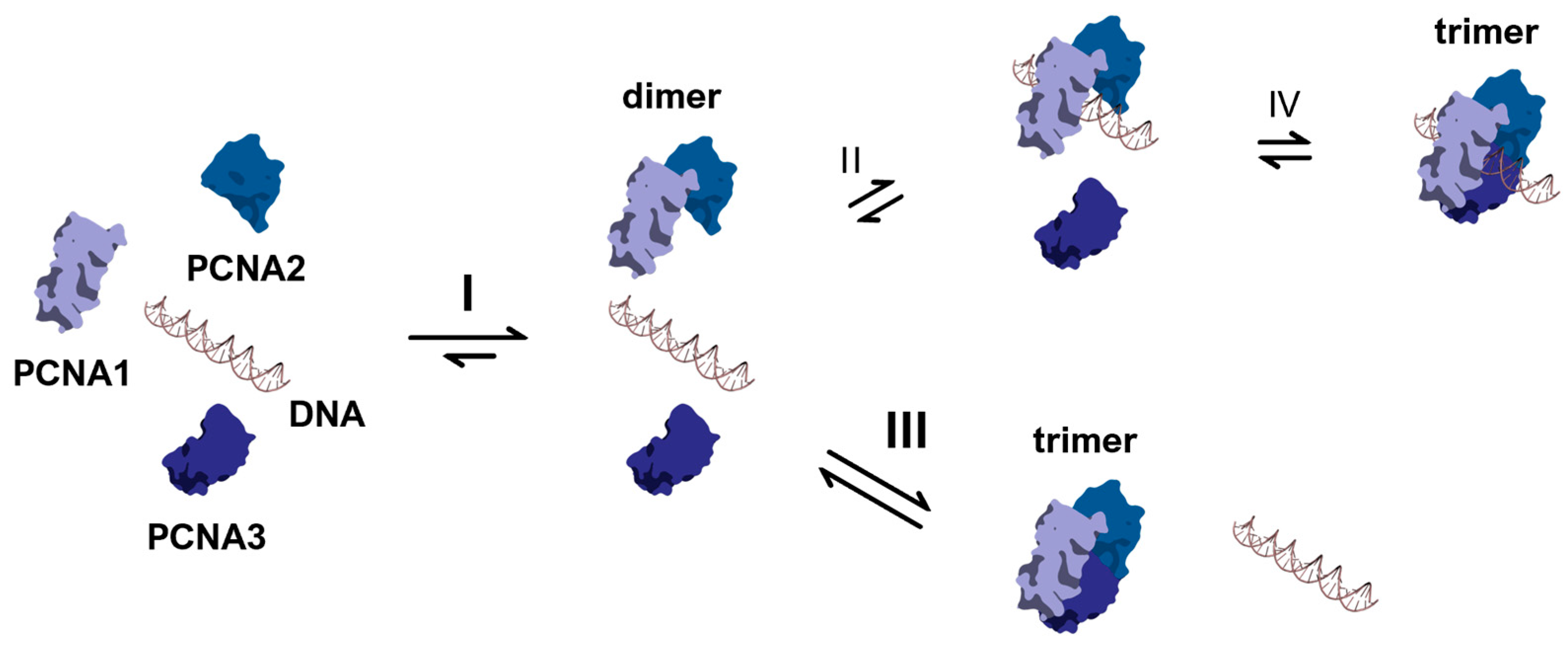
In summary, PCNA1 has been identified as the key player in DNA binding of PCNA, while its association with PCNA2 reduced the DNA binding affinity. This loss of binding affinity is crucial for enabling sufficient clamp mobility [59]. Finally, PCNA3 was likely recruited by the already DNA-bound PCNA dimer (Figure 4, II) [55], positioning the heterotrimeric ring in its native orientation around the DNA duplex.

2.3. Clamp Loader-Independent DNA Binding Mechanism of PCNA
DNA recognition of heterotrimeric PCNA mainly involves polar interactions between PCNA1 and DNA, whereas the PCNA2 and PCNA3 monomers did not induce DNA shifts during EMSA (Figure 3). This phenomenon arises from the distinct shapes and the degrees of positive charge at the PCNA-DNA interfaces. PCNA1 from Sulfolobus solfataricus (PDB ID: 2HII) and human PCNA (PDB ID: 6FCM) share a high degree of structural homology [60]. 6 residues (K20, K77, K80, R149, H153, K217) were identified as specific interaction points during DNA sliding of human PCNA, ordered in the form of a right-hand spiral, matching the pitch of B-DNA [50]. PCNA1 harbors 10 basic residues that may be involved in DNA binding. Among them, residues analogous to those in human PCNA include K77, K81, and K210. The PCNA1-DNA interface is probably located on the N-terminal side of PCNA1, facing the central cavity of the complete ring. There, K85, K86 and K106 may serve a role equivalent to that of R149 and H153 in human PCNA, which are situated in the C-terminal region of the inner surface of the ring. Additional basic residues K10, K76, K206, and R213 may also be crucial for DNA binding. In contrast, PCNA2 has only 6 putative DNA-binding residues (R17, K81, R82, K207, R209, R210), located on the inner side of the ring. Combined with its sharper structural curvature, which presumably limits the number of polar contacts, PCNA2 had no or very low DNA binding affinity. PCNA2 and PCNA3 share the strongly curved shape, similar to that of PCNA2 from Sulfolobus tokodaii, which imparts an asymmetric character to the full-closed heterotrimeric ring [61]. For PCNA3, we identified 7 putative DNA binding residues on the inner surface of the ring (R9, K12, R20, K76, K79, K140, K209). Like PCNA2, monomeric PCNA3 did not appear to interact with DNA (Figure 3(B1–B3)).
In summary, two main factors could explain the potentially higher DNA binding affinity of PCNA1: (1) PCNA1 harbors the most basic amino acids on the inner surface of the ring, which may be involved in DNA recognition. (2) The sharper curvature of PCNA2 and PCNA3 likely limits the number of polar interactions with DNA. Based on these results, we propose the in vitro DNA binding mechanism of PCNA in the absence of a clamp loader in Figure 4. After heterodimerization of PCNA1 and PCNA2 (I), some dimers bind to DNA via the electrostatic interaction between positively charged residues on the inner side of the partial ring and the negatively charged phosphate backbone of dsDNA (II). Given the potentially low DNA binding affinity of PCNA in the µM to mM range [50], compared to the Kd of PCNA3 (270 nM) for the PCNA dimer [55], most dimers will recruit PCNA3 directly to form the trimer without prior DNA binding (III). Only a subset of DNA-bound dimers will associate with PCNA3 to complete the ring while encircling the DNA (IV).
Figure 4. Clamp loader-independent DNA binding mechanism of PCNA. Formation of the stable PCNA heterodimer (I). PCNA1-mediated DNA binding of the heterodimer (II). Heterotrimerization of either free heterodimer and PCNA3 (III), or of DNA-bound heterodimer and PCNA3 (IV).
Synbio 03 00016 g004

2.4. DNA Binding of Cys-Mutated PCNA
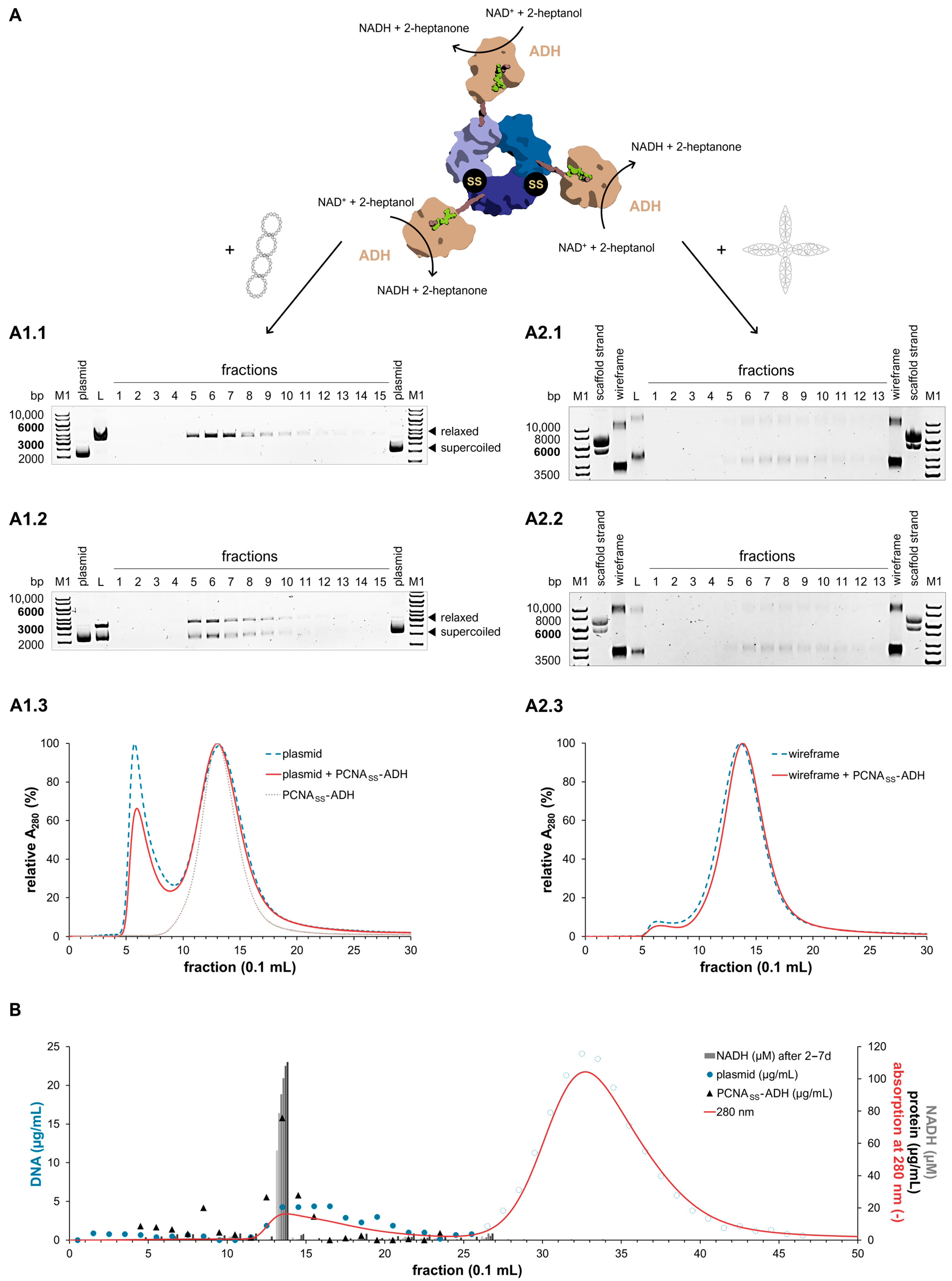
After verifying the DNA binding capability of PCNA in vitro and demonstrating successful binding of PCNA to various DNA species, including a wireframe DNA nanostructure, we next applied this functionality in a two-enzyme model system. First, we established a method based on multimodal chromatography using a Capto Core 700 column to isolate the colocalized two-enzyme system when bound to DNA. To create a stable heterotrimer and therefore, facilitate permanent DNA binding of PCNA, the loose association of PCNA3 and the heterodimer [55] was stabilized by covalent connections. We introduced cysteines at the PCNA1-PCNA3 and PCNA2-PCNA3 interfaces to generate PCNACYS mutants (Figure 5) [56]. Given its monomeric structure, we initially fused only the ADH to PCNACYS (Figure 6A). DNA binding needed to occur prior to the formation of the disulfide bonds via oxidation to initiate ring closure. Therefore, we verified that the concentrations of NaCl, DTT, or EDTA, applied to prepare the protein-DNA complex, did not affect DNA binding (Figure S7).
In fact, we observed a DNA shift for the collected fractions of the PCNASS-ADH-plasmid complex (Figure 6(A1.1–A1.3)). In the negative control (Figure 6(A1.2)), two bands appeared without the addition of the fusion protein: one for supercoiled plasmid and one for relaxed plasmid. Supercoiled DNA was probably resolved by visible light irradiation during the disulfide bond oxidation [62], which was enhanced by contact with highly concentrated protein (Figure 6(A1.1)). No significant DNA shift was detected for the fractions collected during purification of the PCNASS-ADH-wireframe complex either (Figure 6(A2.1–A2.3)). Nonetheless, DNA binding took place prior to purification of the protein-DNA complex, as the loading sample containing wireframe DNA and PCNASS-ADH exhibited a significant DNA shift during EMSA. Although freely assembled trimers were the dominant species in the loading samples (Figure S9), the primary contributors were apparently PCNACYS-ADH dimers, most of which likely dissociated from DNA during chromatography due to their potentially low DNA binding affinity [50]. Low concentrations of PCNA3CYS-ADH were also monitored in the purified fraction (Figure S10), implicating a small amount of full-closed PCNASS-ADH bound to DNA.
Additional EMSA experiments of PCNACYS without fusion to the ADH revealed that the cysteine mutations had a negative impact on DNA binding (Figures S4 and S11). All PCNACYS monomers and the PCNACYS dimer caused DNA shifts to a similar degree as the equivalent PCNA mixtures. The only exception was the PCNACYS trimer mixture, which induced no DNA shift (Figure S6). SDS-PAGE and size exclusion chromatography (SEC) experiments confirmed that the cysteine mutations did not interfere with PCNA assembly in the absence of DNA (Figures S12 and S13). The modified PCNA1-PCNA3 and PCNA2-PCNA3 interfaces might have disoriented PCNA3 within the heterotrimeric complex. As a result, correct PCNA assembly was compromised around DNA [61]. This effect may have been further exacerbated by the potential influence of the fused ADH molecules on DNA binding.
Figure 5. PCNA1-PCNA3 and PCNA2-PCNA3 interfaces of PCNA (PDB ID: 2HII) with reduced cysteines (PCNACYS) and PCNA with covalent disulfide bonds (PCNASS) [61]. Dashed lines display polar interactions. Cysteines were incorporated at position 108 (PCNA1), 171 (PCNA2), 112 (PCNA3) and 180 (PCNA3) via mutagenesis, and disulfide bonds formed via oxidation. In silico mutagenesis included energy minimizing steps of residues within a radius of 5 Å using YASARA 13.9.8 [63].
Synbio 03 00016 g005
Based on these findings, we conclude that DNA binding was impaired for the full-closed PCNACYS and PCNACYS-ADH (before disulfide formation), or PCNASS and PCNASS-ADH (after disulfide formation). Cysteine mutations at the PCNA3 interface did not affect single PCNACYS monomers or PCNACYS dimers. Only in combination with PCNA3CYS, DNA binding was hampered by interfacial cysteine mutations. It remains unclear whether the isolated PCNASS-ADH-DNA complex contained exclusively heterotrimers or also included dimers or monomers still bound to DNA. Further investigations should elucidate the proportion of DNA-bound heterotrimers relative to dimeric or monomeric species following complex isolation.
Figure 6. Isolation of the PCNASS-ADH-plasmid complex (A1.1A1.3,B) and PCNASS-ADH-wireframe complex (A2.1A2.3) via Capto Core 700 chromatography. (A) Isolation via one chromatography column. PCNASS-ADH is illustrated with nicotinamide adenine dinucleotide (NAD+/NADH, green) and 2-heptanol/2-heptanone (brown) in the active site of the ADH, which oxidized 2-heptanol to 2-heptanone while reducing NAD+ to NADH. EMSA of loading sample (L) and collected fractions for 50 nM plasmid or 20 nM wireframe blossom, in presence (A1.1,A2.1) and absence (A1.2,A2.2) of 40 µM PCNASS-ADH. Absorption at 280 nm (A280) during chromatography for PCNASS-ADH in combination with plasmid (A1.3) or wireframe blossom (A2.3). Controls were 50 nM plasmid or 20 nM wireframe blossom, without addition of PCNASS-ADH (negative control 1) as well as 40 µM PCNASS-ADH, without addition of DNA (negative control 2). Full gel images are shown in Figure S14. (B) Isolation of the PCNASS-ADH-plasmid complex (75 nM plasmid and 50 µM PCNASS-ADH) via three interconnected Capto Core 700 columns. Additional to the A280 (red lines), we measured DNA concentrations (blue circles), protein concentrations (black triangles) as well as NADH formation (grey columns) by the ADH fused to PCNA in the collected fractions. Increasing greyscale of the columns denotes reaction time: the first measurement was taken 2 days after reaction initiation, and each darker shade represents one additional day. Open blue circles denote artifacts arising from the light absorption of oxidized glutathione. Corresponding non-reducing SDS-PAGE of loading samples, EMSA of the fractions and negative controls are displayed in Figures S9 and S15. Additional experiments with plasmid (Figures S15 and S17) and with wireframe blossom (Figure S18) at varying DNA and protein concentrations are included in the Supplementary Materials.
Synbio 03 00016 g006

2.5. Activity of PCNASS-ADH Fusion Proteins on DNA

While examining the chromatograms for the purification of the PCNASS-ADH-DNA complexes, we observed two peaks (Figure 6): The first peak was attributed to either DNA without proteins (negative control 1) or to the PCNASS-ADH-DNA complex (Figure 6(A1.3,A2.3). The second peak represented remaining oxidized glutathione, which absorbs UV light [64]. Unbound proteins remained in the column. We considered the sophisticating effects of oxidized glutathione, reacting with redox agents in the protein concentration assay or absorbing UV light, by subtracting the base line. To further minimize these basal effects and improve glutathione removal, we used three interconnected chromatography columns instead of one (Figure 6B). The corresponding EMSA experiment is displayed in the Supplementary Materials (Figure S15). To exclude the possibility of protein aggregates potentially eluting during the first peak and reacting with NAD+ in the activity assay afterwards, we analyzed the fractions of negative control 2 that contained only the PCNASS-ADH fusion protein but no DNA. Even after four weeks, no NAD+ consumption was detectable in negative control 2. This indicates that only DNA-bound PCNASS-ADH was present in the first peak fractions.
We measured an activity of approximately 0.07 min−1 in fraction 14 (Figure 6B). At pH 9 and 30 °C, the expected catalytic constant for the ADH oxidizing 1-phenylethanol is ~60 min−1 (1.8 U/mg) [52], implicating that the PCNASS-ADH complexed with plasmid DNA has lost nearly all its activity. Additional complex isolation experiments using 2-heptanol as substrate, including one with wireframe blossom DNA, confirmed these results (Table S2, Figures S16–S18). An activity loss solely due to the fusion of the ADH to PCNA was not observed. Competitive or allosteric inhibition can also be excluded, because PCNA1-ADH and PCNA2-ADH exhibited the same activity as unfused ADH in the presence or absence of highly concentrated plasmid DNA of up to 1 mg/mL (Figure S19). Contrary to our initial assumption, the microenvironment of DNA might have manipulated catalysis of the fusion enzyme located in close proximity to the DNA duplex [65]. In this context, pH was reduced, decelerating the activity of the ADH, which has an optimal pH above 10 [52]. We had anticipated that PCNA, with a ring height over 2 nm, combined with the PCNASS-ADH-connecting peptide linker, would create sufficient distance between the DNA and fused enzymes [66]. However, the enzyme was linked to PCNA via a (GGGGS)2 linker, whose considerable flexibility might have allowed the ADH to get close to the DNA. These findings suggest that the ADH is not suitable for this particular co-immobilization setup on DNA scaffolds.

2.6. Colocalization of a Two-Enzyme System

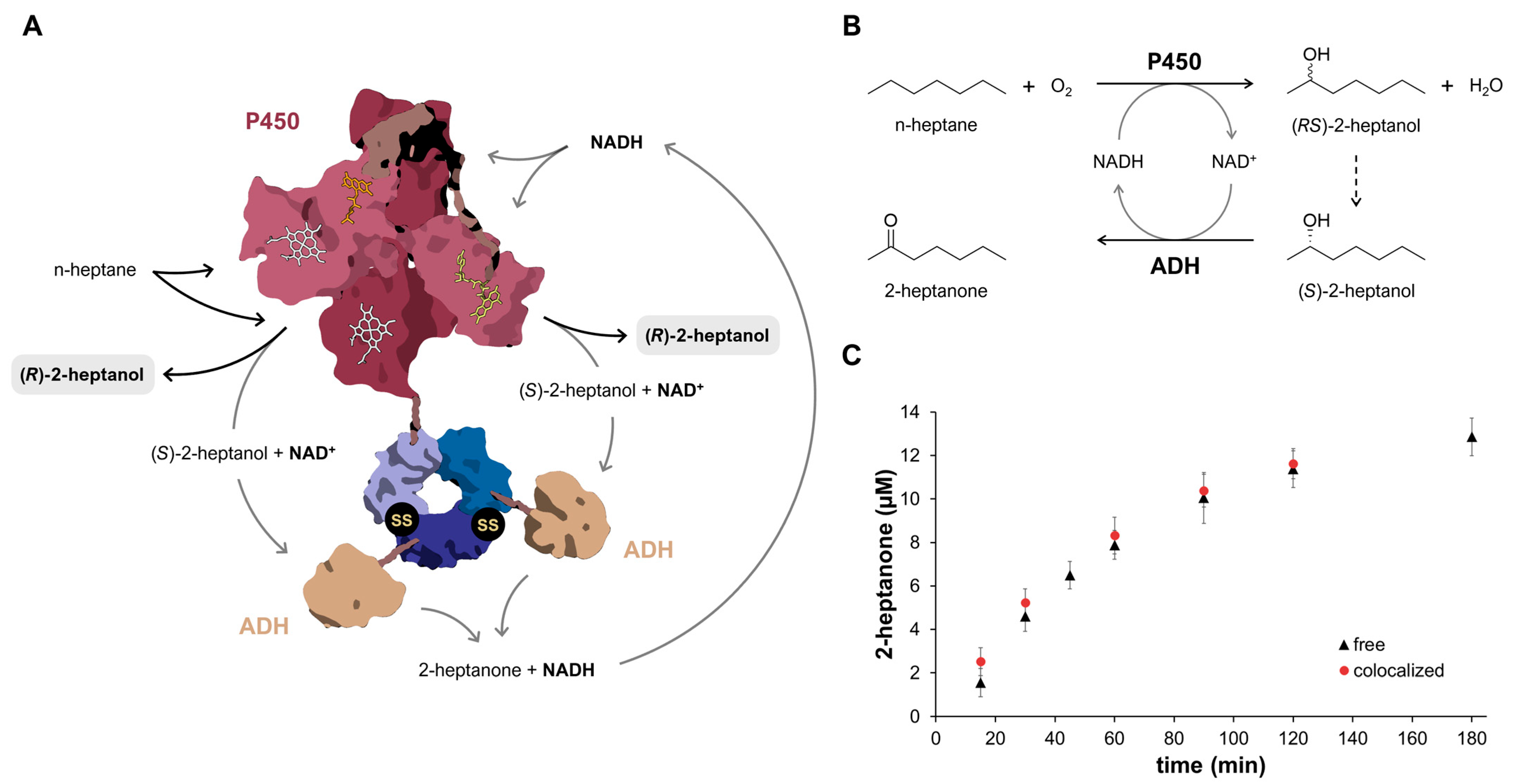
In the final stage of our research, we sought to integrate both potential applications of PCNA: enzyme colocalization and immobilization on DNA. To this end, we designed a two-enzyme model system by fusing each enzyme to one subunit of the heterotrimeric PCNASS variant. We fused a pseudo-monomeric cytochrome P450 variant [53] to the C-terminus of PCNA1CYS and two molecules of an ADH variant with enhanced activity [52] to the C-termini of PCNA2CYS and PCNA3CYS, respectively. As illustrated in Figure 7A, the heterotrimeric complex was stabilized via covalent disulfide bonds. Upon PCNA-mediated immobilization on DNA, however, the ADH exhibited a drastic reduction in catalytic activity (Section 2.5). Due to the potentially low levels of product formation, we did not immobilize the two-enzyme system on a DNA scaffold. Instead, we focused on investigating the effects of PCNA fusion on enzyme activity. Although previous enzyme engineering efforts had improved ADH activity [52], the catalytic rates remained insufficient to enable effective substrate channeling. Since the ADH catalyzes the second, rate-limiting reaction of the cascade, accumulation of intermediates occurred. Hence, our investigation was confined to evaluating the performance of the assembled fusion proteins relative to the equivalent free, unfused enzymes.
The catalyzed reaction cascade is depicted in Figure 7B. The starting alkane (n-heptane) was hydroxylated into an alcohol (primarily 2-heptanol and 3-heptanol) (1. P450 reaction). The alcohol intermediate was then S-selectively converted into a ketone (primarily 2-heptanone and 3-heptanone) (2. ADH reaction). The final product was the remaining R-alcohol (primarily (R)-2-heptanol) [54]. We measured comparable enzyme activities of 0.14 ± 0.026 min−1 (free) and 0.16 ± 0.003 min−1 (colocalized) 2-heptanone per ADH (Figure 7C), which were significantly lower than its maximal activity of ~40 min−1 (1.2 U/mg) at pH 9.0 and 30 °C [52]. This discrepancy originated from the very high Km of the ADH for the alcohol intermediate (primarily 2-heptanol), which was likely in the range of the wild type ADH (~150 mM) (Figure S20). Hence, the Km was greater than the intermediate concentration of ~120 µM 2-heptanol by approximately three orders of magnitude. Furthermore, the free (85 ± 2%) and colocalized (81 ± 5%) enzyme systems displayed similar enantiomeric excess values for the (S)-3-heptanol intermediate. It is important to note that the intermediate concentration of 3-heptanol (~50 µM) was significantly higher than the product yield of 3-heptanone (~5 µM) over the monitored reaction period. Thus, the values primarily reflected the enantioselectivity of the cytochrome P450 variant [54]. Based on these observations, we conclude that neither the ADH nor the cytochrome P450 variant was affected by fusion to PCNA.

3. Materials and Methods

3.1. Cloning and Site-Directed Mutagenesis

All genes, encoding the PCNA fusion proteins (Figure 1) as well as unfused enzymes, were cloned into the pET28a expression vector. The corresponding protein sequences (Table S3), applied primers (Table S4), and details on molecular cloning procedures (Method S1) can be found in the supplementary material. Generally, the enzymes were fused to the C-termini of each PCNA monomer, with a GGGGSGGGGS spacer between them, respectively. To prevent dissociation of the heterotrimer, we inserted cysteines at the interfaces between PCNA1 and PCNA3 as well as PCNA2 and PCNA3 to generate PCNACYS variants according to Hirakawa et al. [56]. All PCNA-enzyme constructs were based on the ring-stabilized PCNASS variant.

3.2. Protein Expression and Purification

A detailed description of the procedure is written in the Supplementary Materials (Method S2). All genes were expressed in Escherichia coli (E. coli) using auto-induction media [67], containing kanamycin. To produce the cytochrome P450 variant and its fusion proteins, thiamine, aminolevulinic acid, as well as other trace minerals were added to the media. The genes of the PCNA monomers and their fusion proteins were expressed separately. After expression overnight, the cells were lysed by sonication, while cooling, and the cell lysates, then, clarified. All PCNA proteins were purified by Ni-NTA affinity chromatography. Only the fusion proteins and unfused cytochrome P450 monooxygenase underwent a second purification step via StrepTactin XT affinity chromatography (Iba Lifesciences, Göttingen, Germany). After desalting, the proteins were frozen in liquid nitrogen and stored at −80 °C. Protein concentrations were determined using the PierceTM BCA Protein Assay (Thermo Fisher Scientific, Waltham, MA, USA) [68].

3.3. SDS-PAGE

To prepare samples for non-reducing SDS-PAGE, we used loading buffer (50 mM Tris-HCl, pH 6.8, 50 mM SDS, 1.4 M glycerol, 0.75 mM bromophenol blue). For reducing SDS-PAGE, we used loading buffer containing 0.35 M β-mercaptoethanol. The samples were heated for 10 min at 120 °C. 12% polyacrylamide gels were prepared according to the manufacturer’s protocol for the omniPAGE Vertical System (Cleaver Scientific, Rugby, UK). After electrophoresis, the SDS gels were stained with a dye solution (0.25 g/L coomassie brillant blue G-250, 10 % (v/v) acetic acid, 15% (v/v) isopropanol) for 10 min. Then, the gels were washed and stored in water. Visual analysis was performed under white light using the ChemiDocTM MP Imaging System (Bio-Rad, Hercules, CA, USA). For reference, we applied the peqGOLD Protein Marker I, 14–116 kDa, peqGOLD Protein Marker II, 10–200 kDa (VWR, Radnor, PA, USA), or the Prosieve QuadColor Protein Marker, 4.6–300 kDa (Biozym Scientific, Hessisch Oldendorf, Germany). SDS gels of purified PCNA variants are presented in Figure S21.

3.4. Preparation of the DNA Scaffolds

The pUC19 plasmid was produced in E. coli DH5α cells, cultivated in LB media [69], which contained 100 µg/mL carbenicillin, under different conditions at 37 °C. For the preparation of single plasmids, the cultivation and purification took place according to the protocol of the NucleoSpin Plasmid Mini Kit (Macherey-Nagel, Düren, Germany). In the last step, plasmid DNA was eluted with sterile, distilled water. Multimerized plasmid DNA was prepared by cultivating cells until they reached the stationary phase, then using high cell densities in plasmid isolation.
The wireframe blossom DNA nanostructure was developed by Dr. Marisa Seiwald [57] and provided by Prof. Dr. Friedrich Simmel (Chair of Physics of Synthetic Biological Systems, Technical University of Munich, Germany). The origami structure was folded based on a 7249 nt long single-stranded scaffold DNA, derived from the bacteriophage genome M13mp18, using 162 short staple strands (Figure S22, Table S5). The staple strands were used in a fourfold molar excess compared to the scaffold strand. The wireframe structures were folded in wireframe folding buffer (10 mM Tris-HCl, pH 8.5, 1 mM EDTA, 12 mM MgCl2) after the following procedure: 80 °C (5 min), 80 °C to 60 °C (20 min), 60 °C to 24 °C (14 h), 4 °C. Excess of unbound staple strands was removed by PEG precipitation according to Stahl et al. [70]. After repeating this step three times in total, the wireframe structure was redissolved in wireframe folding buffer and stored at −20 °C.
Final DNA concentrations were determined by measuring the absorption at 260 nm using the NanoDropTM 2000 spectral photometer (Thermo Fisher Scientific, Waltham, MA, USA).

3.5. DNA Binding Assay

To analyze the DNA binding of PCNA, we added different concentrations of its trimeric mixture to the EMSA binding buffer (50 mM Tris-HCl, 150 mM NaCl, 1× Gel Loading Dye, Purple from NEB, Ipswich, MA, USA), which contained either plasmid DNA or wireframe DNA. Applied DNA concentrations are depicted in the figure descriptions (Figure 2, Figures S4 and S5). PCNA3 was added in twofold excess over equimolar amounts of PCNA1 and PCNA2. Each DNA scaffold was titrated over a concentration spectrum from 0 up to 150 µM PCNA. The titration experiments were performed twice for each DNA scaffold while applying different DNA concentrations.
To evaluate the DNA binding of PCNA at different oligomerization states, we mixed either plasmid DNA (15 nM) or wireframe DNA (5 nM) with 50 µM or 75 µM of each monomer, dimer, or trimer. In dimer samples, PCNA1 and PCNA2 were added in an equimolar ratio and in trimer samples, PCNA3 variants were added at a twofold excess compared to the equimolar amount of PCNA1 or PCNA2. We performed this binding assay at least three times—two experiments with plasmid and one experiment with wireframe blossom.
All samples, with a total volume of 12 µL each, were incubated for at least 30 min at room temperature before undergoing agarose gel electrophoresis, as described for the electrophoretic mobility shift assay (EMSA). Based on the RF values, we calculated the number of bound PCNA monomers, considering the molar masses and net charges of each monomer in relation to the net charge of DNA nucleotides (Table S1, Method S1). Measurement uncertainties were calculated as described in Method S1 in the Supplementary Materials and are presented either as error bars (Figure 2 and Figure S6) or reported in the respective figure captions (Figure 3). Reproducibility is illustrated as the relative error of replicate measurements in Figures S6 and S7.

3.6. Electrophoretic Mobility Shift Assay (EMSA)

The samples were loaded onto a 1% (w/v) agarose gel pre-stained with peqGREEN (VWR, Radnor, PA, USA). Electrophoresis was performed using the PerfectBlueTM Horizontal Maxi Gel System (VWR, Radnor, PA, USA) in TAE buffer (40 mM Tris-acetate, pH 8.5, 1 mM EDTA) at 5.2 V/cm. The agarose gels were analyzed under UV light using the ChemiDocTM MP Imaging System (Bio-Rad, Hercules, CA, USA) afterwards. For reference, we applied the peqGOLD DNA Ladder, 1 kb (VWR, Radnor, PA, USA) (marker 1, M1) as well as the GeneRulerTM High Range DNA Ladder (Thermo Fisher Scientific, Waltham, MA, USA) (marker 2, M2). Besides the control samples of DNA, without the addition of PCNA variants, we prepared another sample containing only 5 nM single-stranded scaffold DNA for the wireframe binding assays. The contrast and brightness of all EMSA gels were increased by 20–40%. The gels were cropped and evaluated using ImageJ 1.54f (see full images in Figures S2, S8 and S14).

3.7. Preparation of the PCNASS-ADH-DNA Complex

The isolation of DNA functionalized with PCNASS-ADH was conducted in three steps: (1) DNA binding, (2) ring closing by disulfide formation, and (3) purification of the PCNASS-DNA complex via Capto Core 700 chromatography to remove excess of unbound PCNASS variant. A detailed description of the method is included in the Supplementary Materials (Method S3). Applied concentrations of PCNASS variants as well as DNA (plasmid or wireframe DNA) are depicted in the figure description of Figure 6. For the loading sample and each fraction, we performed an EMSA, determined DNA concentrations, protein concentrations, as well as the ADH activity. The results of all PCNASS-ADH-DNA complex isolation experiments are displayed in Figure 6 and the Supplementary Materials—three with plasmid DNA (Figures S15–S17) and one with wireframe blossom (Figure S18).

3.8. ADH Activity Assay

The enzyme activities of the Capto Core 700 fractions were measured photometrically by monitoring the absorption of NADH at 340 nm. The reaction conditions were 50 mM Tris-HCl (pH 9.0), 100 mM NaCl, 2 mM NAD+, 1% (v/v) 1-phenylethanol, with the pH adjusted to the reaction temperature of 30 °C—considering the temperature coefficient of −0.028/°C [71]. NAD+ reduction was followed daily for 7 days. The total reaction volume was 200 µL, containing 50 µL of each fraction, respectively. The enzyme activity (catalytic constant) was calculated as the number of NAD+ molecules reduced by one enzyme molecule per minute. Unlike the fractions, the controls were measured as triplicates. Control 1 comprised 50 µL Capto Core buffer instead of 50 µL fraction. Control 2 was used to detect evaporation and contained 200 µL Capto Core buffer. Control 3 represented NADH decay and contained 0.5 mM NADH instead of 2 mM NAD+ in reaction buffer. The positive control was the loading sample, diluted 1:50 in the reaction mixture. NADH production was corrected by subtracting the absorption of control 1 and considering the NADH decay (control 3), with a half-life time of 660 ± 14 h (Figure S23).

3.9. Preparation of the Colocalized Enzyme System

Analogous to the preparation of the PCNASS-ADH-DNA complex, three steps were necessary to prepare the colocalized enzyme system: (1) self-assembly, (2) ring closing via disulfide formation, and (3) purification via size exclusion chromatography to remove heterodimers or monomers. For step 1, PCNA1CYS-P450 (7.5 µM), PCNA2CYS-ADH (15 µM), and PCNA3CYS-ADH (45 µM) were combined and incubated in a reducing buffer (50 mM Tris-HCl, pH 8.0, 150 mM NaCl, 10 mM DTT) for 1 h at 4 °C. Step 2 was conducted as with the PCNASS-ADH-DNA complex. Step 3 consisted of SEC using the SEC buffer (50 mM Tris-HCl, pH 8.0, 150 mM NaCl) and a Superdex 200 Increase 10/300 GL column (Cytiva, Marlborough, MA, USA) at a flow rate of 0.5 mL/min. The fractions containing the full colocalized enzyme system were combined afterwards (Figure S24). We measured the concentration of active P450 enzyme, using the Pyridine Hemochromagen Assay [72] and the molar extinction coefficient ε of 86 ± 2 L mmol−1 cm−1 for the PCNA1CYS-P450 fusion protein (Figure S25) as well as the total protein concentration using the PierceTM BCA Protein Assay (Thermo Fisher Scientific, Waltham, MA, USA) [68].

3.10. Analysis of the Two-Enzyme System by Gas Chromatography

For the colocalized two-enzyme system, 0.5 µM of active P450 concentration was applied, while for the free two-enzyme system, 0.5 µM active unfused cytochrome P450 (ε = 67 ± 1 L mmol−1 cm−1) [53] and 1.1 µM ADH with enhanced activity were used. The reaction took place in a total volume of 500 µL, comprising 50 mM Tris-HCl (pH 9.0), 100 mM NaCl, 2 mM NADH, and 50 mM n-heptane. The pH was adjusted at the reaction temperature of 30 °C—considering the temperature coefficient of −0.028/°C [71]. At regular intervals, the reaction was stopped with 100 µL concentrated HCl (37%, (v/v)). The samples were extracted in 400 µL methyl tert-butyl ether (MTBE), containing 1 mM n-decane (internal standard), and then dried over Na2SO4. The reaction products were detected by gas chromatography via flame ionization (GC-FID) using a FS-Hydrodex β-TBDAc column (Macherey-Nagel, Düren, Germany). The oven temperature program was set as follows: 50 °C, 5 °C/min to 95 °C, 9.5 min [53]. We measured triplicates except for the single control measurement at defined intervals. The control was the free two-enzyme system, containing storage buffer (50 mM Tris-HCl, pH 8.0, 100 mM NaCl) instead of the ADH protein solution. Exemplary GC spectra are displayed in the supplementary material (Figure S26). The product was quantified by calibration curves over a concentration range of 5 µM to 500 µM. Catalytic constants were calculated based on the initial reaction rates, representing the number of 2-heptanol molecules oxidized by one ADH enzyme molecule per minute.

4. Conclusions and Prospect

In this study, we investigated the potential of the heterotrimeric PCNA from Sulfolobus solfataricus as a molecular tool for sequence-independent DNA immobilization of biomolecules. By fusion to PCNA, we created a stoichiometrically defined multienzyme complex intended for attachment to a model wireframe DNA nanostructure. Our findings extended the current understanding of functional DNA binding of the heterotrimeric DNA sliding clamp without the assistance of clamp loaders: (1) PCNA was capable of binding not only circular DNA, but also synthetic wireframe DNA nanostructures. (2) Among the three subunits, PCNA1 played the predominant role in DNA binding, while PCNA2 showed no detectable DNA interaction. (3) Ring-stabilizing cysteines at both the interfaces between the PCNA1/PCNA2 dimer and PCNA3 impaired proper self-assembly around DNA, likely due to structural disfigurations. It remains unclear whether a low amount of ring-stabilized PCNASS variants were bound to DNA, or whether only monomeric and dimeric species were loosely associated with DNA. In either case, the DNA binding by the PCNA variants was consistently weak, limiting the applicability of PCNA as DNA binding platform. In addition to that, we observed that DNA-induced microenvironments significantly decreased the activity of an ADH, catalyzing the second reaction of the enzymatic model system. Thus, we focused on the fusion of both the cascade enzymes—a cytochrome P450 variant and the ADH—to PCNA, which did not affect their overall kinetic characteristics.
In conclusion, the applicability of PCNA as DNA binding tool is currently limited by two key factors: (1) Due to its inherently low affinity for DNA, functionalization via PCNA is highly inefficient. Achieving adequate loading of DNA scaffolds requires large quantities of highly concentrated PCNA fusion proteins. To render PCNA a more effective tool for DNA binding, it would be necessary to engineer the PCNA-DNA interface—specifically the surface on the inner face of the ring, thereby increasing the binding affinity to DNA. Among the three subunits, PCNA1 appears to be the most promising target due to its naturally higher DNA binding affinity. Moreover, modifying only a single subunit may reduce the risk of disrupting the heterotrimeric ring structure, which implies the next consideration. (2) Conformational alterations of assembled PCNA, such as those observed in the ring-stabilized variants, prevent efficient DNA binding. Alternative strategies for subunit interconnection should be explored to ensure correct heterotrimerization around DNA. Greater conformational flexibility and spatial accommodation should be addressed to allow proper subunit orientation. Furthermore, the design of linkers used to couple enzymes to PCNA can play a critical role in maintaining DNA binding functionality as well. The peptide spacer connecting PCNA to its protein cargo should possess sufficient flexibility or be oriented away from the DNA strand engaged by PCNA, thereby allowing adequate space for correct assembly around the DNA duplex.
At present, native PCNA is not readily suited for efficient immobilization of biomolecules on DNA scaffolds without the support of clamp loaders. Overcoming its inherently low DNA binding affinity is essential to establish PCNA as viable platform for sequence-independent DNA functionalization. Furthermore, DNA interactions can be modulated by the specific type of monomer and its oligomeric state. For instance, DNA binding can be initiated by a PCNA dimer engineered for enhanced DNA affinity. Upon subsequent addition of the third subunit to complete the ring, DNA interactions are diminished due to conformational reorganization of the heterotrimeric complex. This mechanism enables controlled switching between DNA-bound and sliding states, which holds considerable potential for DNA-based applications (e.g., biosensing). However, the necessary structural optimizations pose significant challenges, as they carry the risk of disrupting the ring formation, specifically during its assembly around DNA. In this regard, ring stabilization through disulfide bonds has proven unsuitable, due to its restrictive impact on conformational dynamics.
Realizing the potential of PCNA in the context of enzyme immobilization has proven to be more complex than initially anticipated, as well. Although enzyme functionality generally remains intact upon fusion with PCNA, only specific enzymatic setups benefit from the catalytic or DNA binding advantages offered by this system: (1) Enzyme activity can be significantly affected when immobilized on DNA, due to DNA-induced microenvironments that can interfere with catalysis. In this respect, PCNA, as a bridging element between DNA and the enzyme, seemed to be unable to modify or shield the immediate DNA microenvironment. This imposes constraints on the choice and design of attached enzymes for immobilization on DNA. Effective systems include enzymes with high activity at low pH (e.g., GOx/HRP system) [73] or enzymes equipped with optimized linkers that provide sufficient spatial separation from the DNA scaffold. Linker optimization is also an essential approach, leveraging the second major function of heterotrimeric PCNA—its well-defined stoichiometry, enabling controlled colocalization of multiple enzymes. (2) In multienzyme cascades, having the rate-limiting reactions in preceding steps, catalytically favorable substrate channeling may be possible [74]. By fine-tuning the linker architecture—adjusting both orientation and distance between PCNA and the attached enzymes—a spatially defined and catalytically favorable enzyme arrangement could be achieved [75]. However, implementing such configurations remains technically challenging, and adds another layer of complexity, underscoring the importance of careful multienzyme system selection. PCNA is particularly promising as scaffold for stoichiometric multiprotein assembly, when the intermediate transfer necessitates direct contact between catalytic components. A representative example is the electron transfer within the three-component P450 system from Pseudomonas putida [32].
In summary, PCNA has unique structural characteristics that could be advantageous in specific biotechnological applications. Nonetheless, its use as general protein scaffold for sequence-independent DNA functionalization or stoichiometric multiprotein assembly may require substantial system-specific optimization.

Supplementary Materials

The following supporting information can be downloaded at https://www.mdpi.com/article/10.3390/synbio3040016/s1: Figure S1: Reciprocal calibration curve; Figure S2: Titration of DNA against different PCNA concentrations (Full images); Figure S3: EMSA of pUC19 plasmid in presence of 30 mg/mL bovine serum albumin; Figure S4: Titration of pUC19 against different PCNA and PCNACYS concentrations; Figure S5: Titration of wireframe blossom against different PCNA concentrations; Figure S6: EMSA of PCNA and PCNACYS in different multimeric states; Figure S7: EMSA of PCNA in presence of salts and reaction components; Figure S8: EMSA of PCNA in different multimeric states (Full images); Figure S9: Non-reducing SDS-PAGE of PCNASS-ADH; Figure S10: SDS-PAGE of the isolated PCNASS-ADH-plasmid complex; Figure S11: Titration of DNA against different PCNACYS concentrations; Figure S12: Disulfide formation of PCNASS; Figure S13: SEC of PCNASS and PCNASS-ADH in different multimeric states; Figure S14: EMSA of the isolated PCNASS-ADH-DNA complexes (Full images); Figure S15: Isolation of the PCNASS-ADH-plasmid complex (Experiment 1); Figure S16: Isolation of the PCNASS-ADH-plasmid complex (Experiment 2); Figure S17: Isolation of the PCNASS-ADH-plasmid complex (Experiment 3); Figure S18: Isolation of the PCNASS-ADH-wireframe complex (Experiment 2); Figure S19: Activity of the PCNA1-ADH and PCNA2-ADH fusion proteins; Figure S20: Michaelis–Menten kinetics of the ADH with 2-heptanol; Figure S21: SDS-PAGE of purified PCNA variants; Figure S22: UV map of wireframe blossom; Figure S23: Inactivation curve of NADH; Figure S24: SDS-PAGE of the colocalized two-enzyme system; Figure S25: Pyridine Hemochromagen Assay of the heterodimeric PCNA1-P450; Figure S26: GC spectra of the reaction catalyzed by the two-enzyme system; Method S1: Quantification of DNA-bound PCNA units via EMSA; Method S2: Cloning and site-directed mutagenesis; Method S3: Protein expression and purification; Method S4: Preparation of the PCNASS-ADH-DNA complex; Table S1: Molar masses, isoelectric points (pI) and net charges of PCNA monomers; Table S2: Activity of PCNASS-ADH immobilized on DNA; Table S3: Protein sequences; Table S4: Oligonucleotides for golden gate cloning and for site-directed mutagenesis; Table S5: Oligonucleotides of the wireframe blossom DNA nanostructure. References [76,77,78,79,80] are cited in the Supplementary Materials.

Author Contributions

Conceptualization, A.E. and K.C.; Methodology, A.E.; Validation, A.E.; Formal Analysis, A.E.; Investigation, A.E. and K.C.; Resources, K.C.; Data Curation, A.E.; Writing—Original Draft Preparation, A.E. and K.C.; Writing—Review and Editing, K.C.; Visualization, A.E.; Supervision, K.C.; Project Administration, A.E.; Funding Acquisition, K.C. All authors have read and agreed to the published version of the manuscript.

Funding

This research received no external funding.

Data Availability Statement

The original contributions presented in this study are included in the article/Supplementary Materials. Further inquiries can be directed to the corresponding author.

Acknowledgments

The authors gratefully acknowledge Friedrich Simmel, Jonathan List, and Marisa Seiwald (Chair of Physics of Synthetic Biological Systems, TU München) for providing the wireframe DNA nanostructure and its AFM image as well as for helpful discussions. Furthermore, we thank Johannes Schweininger (Institute of Bioengineering, FAU Erlangen-Nürnberg) and Simon Dolles (Department of Chemistry and Pharmacy, FAU Erlangen-Nürnberg) for experimental advice and support, as well as Elke Heidenreich (Institute of Bioprocess Engineering, FAU Erlangen-Nürnberg) for technical assistance.

Conflicts of Interest

The authors declare no conflicts of interest.

Abbreviations

The following abbreviations are used in this manuscript:
ADHAlcohol dehydrogenase
AFMAtomic force microscopy
E. coliEscherichia coli
GC-FIDGas chromatography—flame ionization detector
KdDissociation constant/binding constant
KmMichaelis constant
MTBEMethyl tert-butyl ether
NAD+/NADHNicotinamide adenine dinucleotide
P450Cytochrome P450 monooxygenase
PCNAProliferating cell nuclear antigen
SECSize exclusion chromatography

References

  1. Hwang, E.T.; Lee, S. Multienzymatic cascade reactions via enzyme complex by immobilization. ACS Catal. 2019, 9, 4402–4425. [Google Scholar] [CrossRef]
  2. Rabe, K.S.; Müller, J.; Skoupi, M.; Niemeyer, C.M. Cascades in compartments: En route to machine-assisted biotechnology. Angew. Chem. Int. Ed. 2017, 56, 13574–13589. [Google Scholar] [CrossRef]
  3. Conrado, R.J.; Varner, J.D.; DeLisa, M.P. Engineering the spatial organization of metabolic enzymes: Mimicking nature’s synergy. Curr. Opin. Biotechnol. 2008, 19, 492–499. [Google Scholar] [CrossRef] [PubMed]
  4. Ellis, G.A.; Klein, W.P.; Lasarte-Aragones, G.; Thakur, M.; Walper, S.A.; Medintz, I.L. Artificial multienzyme scaffolds: Pursuing in vitro substrate channeling with an overview of current progress. ACS Catal. 2019, 9, 10812–10869. [Google Scholar] [CrossRef]
  5. Jiang, Y.; Zhang, X.; Yuan, H.; Huang, D.; Wang, R.; Liu, H.; Wang, T. Research progress and the biotechnological applications of multienzyme complex. Appl. Microbiol. Biotechnol. 2021, 105, 1759–1777. [Google Scholar] [CrossRef] [PubMed]
  6. Fessner, W.-D.; Badia, J.; Eyrisch, O.; Schneider, A.; Sinerius, G. Enzymatic syntheses of rare ketose 1-phosphates. Tetrahedron Lett. 1992, 33, 5231–5234. [Google Scholar] [CrossRef]
  7. Wang, Z.; Sekar, B.S.; Li, Z. Recent advances in artificial enzyme cascades for the production of value-added chemicals. Bioresour. Technol. 2021, 323, 124551. [Google Scholar] [CrossRef]
  8. Bié, J.; Sepodes, B.; Fernandes, P.C.B.; Ribeiro, M.H.L. Enzyme immobilization and co-immobilization: Main framework, advances and some applications. Processes 2022, 10, 494. [Google Scholar] [CrossRef]
  9. Bilal, M.; Zhao, Y.; Noreen, S.; Shah, S.Z.H.; Bharagava, R.N.; Iqbal, H.M.N. Modifying bio-catalytic properties of enzymes for efficient biocatalysis: A review from immobilization strategies viewpoint. Biocatal. Biotransformation 2019, 37, 159–182. [Google Scholar] [CrossRef]
  10. Garcia-Galan, C.; Berenguer-Murcia, Á.; Fernandez-Lafuente, R.; Rodrigues, R.C. Potential of different enzyme immobilization strategies to improve enzyme performance. Adv. Synth. Catal. 2011, 353, 2885–2904. [Google Scholar] [CrossRef]
  11. Hoarau, M.; Badieyan, S.; Marsh, E.N.G. Immobilized enzymes: Understanding enzyme–surface interactions at the molecular level. Org. Biomol. Chem. 2017, 15, 9539–9551. [Google Scholar] [CrossRef]
  12. Zhang, Y.-H.P. Substrate channeling and enzyme complexes for biotechnological applications. Biotechnol. Adv. 2011, 29, 715–725. [Google Scholar] [CrossRef]
  13. Dubey, N.C.; Tripathi, B.P. Nature inspired multienzyme immobilization: Strategies and concepts. ACS Appl. Bio Mater. 2021, 4, 1077–1114. [Google Scholar] [CrossRef] [PubMed]
  14. Geck, M.K.; Kirsch, J.F. A novel, definitive test for substrate channeling illustrated with the aspartate aminotransferase/malate dehydrogenase system. Biochemistry 1999, 38, 8032–8037. [Google Scholar] [CrossRef] [PubMed]
  15. Dueber, J.E.; Wu, G.C.; Malmirchegini, G.R.; Moon, T.S.; Petzold, C.J.; Ullal, A.V.; Prather, K.L.J.; Keasling, J.D. Synthetic protein scaffolds provide modular control over metabolic flux. Nat. Biotechnol. 2009, 27, 753–759. [Google Scholar] [CrossRef] [PubMed]
  16. Svedružić, Ž.M.; Odorčić, I.; Chang, C.H.; Svedružić, D. Substrate channeling via a transient protein-protein complex: The case of D-glyceraldehyde-3-phosphate dehydrogenase and L-lactate dehydrogenase. Sci. Rep. 2020, 10, 10404. [Google Scholar] [CrossRef]
  17. Wang, X.; Jiang, Y.; Liu, H.; Yuan, H.; Huang, D.; Wang, T. Research progress of multi-enzyme complexes based on the design of scaffold protein. Bioresour. Bioprocess. 2023, 10, 72. [Google Scholar] [CrossRef]
  18. Zdarta, J.; Kołodziejczak-Radzimska, A.; Bachosz, K.; Rybarczyk, A.; Bilal, M.; Iqbal, H.M.N.; Buszewski, B.; Jesionowski, T. Nanostructured supports for multienzyme co-immobilization for biotechnological applications: Achievements, challenges and prospects. Adv. Colloid Interface Sci. 2023, 315, 102889. [Google Scholar] [CrossRef]
  19. Xu, K.; Chen, X.; Zheng, R.; Zheng, Y. Immobilization of multi-enzymes on support materials for efficient biocatalysis. Front. Bioeng. Biotechnol. 2020, 8, 660. [Google Scholar] [CrossRef]
  20. Meena, J.; Gupta, A.; Ahuja, R.; Singh, M.; Panda, A.K. Recent advances in nano-engineered approaches used for enzyme immobilization with enhanced activity. J. Mol. Liq. 2021, 338, 116602. [Google Scholar] [CrossRef]
  21. Gad, S.; Ayakar, S. Protein scaffolds: A tool for multi-enzyme assembly. Biotechnol. Rep. 2021, 32, e00670. [Google Scholar] [CrossRef]
  22. Kazenwadel, F.; Franzreb, M.; Rapp, B.E. Synthetic enzyme supercomplexes: Co-immobilization of enzyme cascades. Anal. Methods 2015, 7, 4030–4037. [Google Scholar] [CrossRef]
  23. Vanderstraeten, J.; Briers, Y. Synthetic protein scaffolds for the colocalisation of co-acting enzymes. Biotechnol. Adv. 2020, 44, 107627. [Google Scholar] [CrossRef]
  24. Artzi, L.; Bayer, E.A.; Moraïs, S. Cellulosomes: Bacterial nanomachines for dismantling plant polysaccharides. Nat. Rev. Microbiol. 2017, 15, 83–95. [Google Scholar] [CrossRef] [PubMed]
  25. Moon, T.S.; Dueber, J.E.; Shiue, E.; Prather, K.L.J. Use of modular, synthetic scaffolds for improved production of glucaric acid in engineered E. coli. Metab. Eng. 2010, 12, 298–305. [Google Scholar] [CrossRef] [PubMed]
  26. Nygren, P.-Å. Alternative binding proteins: Affibody binding proteins developed from a small three-helix bundle scaffold. FEBS J. 2008, 275, 2668–2676. [Google Scholar] [CrossRef] [PubMed]
  27. Mason, J.M.; Müller, K.M.; Arndt, K.M. Considerations in the Design and Optimization of Coiled Coil Structures. In Protein Engineering Protocols, 1st ed.; Arndt, K.M., Müller, K.M., Eds.; Humana Press: Totowa, NJ, USA, 2007; Volume 352, pp. 35–70. [Google Scholar]
  28. Moldovan, G.-L.; Pfander, B.; Jentsch, S. PCNA, the maestro of the replication fork. Cell 2007, 129, 665–679. [Google Scholar] [CrossRef]
  29. Kelman, Z. PCNA: Structure, functions and interactions. Oncogene 1997, 14, 629–640. [Google Scholar] [CrossRef]
  30. Pan, M.; Kelman, L.M.; Kelman, Z. The archaeal PCNA proteins. Biochem. Soc. Trans. 2011, 39, 20–24. [Google Scholar] [CrossRef]
  31. Williams, G.J.; Johnson, K.; Rudolf, J.; McMahon, S.A.; Carter, L.; Oke, M.; Liu, H.; Taylor, G.L.; White, M.F.; Naismith, J.H. Structure of the heterotrimeric PCNA from Sulfolobus solfataricus. Acta Crystallogr. F Struct. Biol. Commun. 2006, 62, 944–948. [Google Scholar] [CrossRef]
  32. Hirakawa, H.; Nagamune, T. Molecular assembly of P450 with ferredoxin and ferredoxin reductase by fusion to PCNA. ChemBioChem 2010, 11, 1517. [Google Scholar] [CrossRef] [PubMed]
  33. De Biasio, A.; de Opakua, A.I.; Mortuza, G.B.; Molina, R.; Cordeiro, T.N.; Castillo, F.; Villate, M.; Merino, N.; Delgado, S.; Gil-Cartón, D.; et al. Structure of p15(PAF)-PCNA complex and implications for clamp sliding during DNA replication and repair. Nat. Commun. 2015, 6, 6439. [Google Scholar] [CrossRef] [PubMed]
  34. Yang, Y.R.; Liu, Y.; Yan, H. DNA Nanostructures as Programmable Biomolecular Scaffolds. Bioconjug. Chem. 2015, 26, 1381–1395. [Google Scholar] [CrossRef] [PubMed]
  35. Wang, P.; Meyer, T.A.; Pan, V.; Dutta, P.K.; Ke, Y. The Beauty and Utility of DNA Origami. Chem 2017, 2, 359–382. [Google Scholar] [CrossRef]
  36. Rothemund, P.W.K. Folding DNA to create nanoscale shapes and patterns. Nature 2006, 440, 297–302. [Google Scholar] [CrossRef]
  37. Nummelin, S.; Kommeri, J.; Kostiainen, M.A.; Linko, V. Evolution of Structural DNA Nanotechnology. Adv. Mater. 2018, 30, e1703721. [Google Scholar] [CrossRef]
  38. Saccà, B.; Niemeyer, C.M. DNA origami: The art of folding DNA. Angew. Chem. Int. Ed. Engl. 2012, 51, 58–66. [Google Scholar] [CrossRef]
  39. Benson, E.; Mohammed, A.; Gardell, J.; Masich, S.; Czeizler, E.; Orponen, P.; Högberg, B. DNA rendering of polyhedral meshes at the nanoscale. Nature 2015, 523, 441–444. [Google Scholar] [CrossRef]
  40. Muscat, R.A.; Bath, J.; Turberfield, A.J. A programmable molecular robot. Nano Lett. 2011, 11, 982–987. [Google Scholar] [CrossRef]
  41. Lund, K.; Manzo, A.J.; Dabby, N.; Michelotti, N.; Johnson-Buck, A.; Nangreave, J.; Taylor, S.; Pei, R.; Stojanovic, M.N.; Walter, N.G.; et al. Molecular robots guided by prescriptive landscapes. Nature 2010, 465, 206–210. [Google Scholar] [CrossRef]
  42. Bath, J.; Turberfield, A.J. DNA nanomachines. Nat. Nanotech 2007, 2, 275–284. [Google Scholar] [CrossRef] [PubMed]
  43. Sweetlove, L.J.; Fernie, A.R. The role of dynamic enzyme assemblies and substrate channelling in metabolic regulation. Nat. Commun. 2018, 9, 2136. [Google Scholar] [CrossRef] [PubMed]
  44. Wilchek, M.; Bayer, E.A. The avidin-biotin complex in bioanalytical applications. Anal. Biochem. 1988, 171, 1–32. [Google Scholar] [CrossRef] [PubMed]
  45. Bock, L.C.; Griffin, L.C.; Latham, J.A.; Vermaas, E.H.; Toole, J.J. Selection of single-stranded DNA molecules that bind and inhibit human thrombin. Nature 1992, 355, 564–566. [Google Scholar] [CrossRef]
  46. Ngo, T.A.; Dinh, H.; Nguyen, T.M.; Liew, F.F.; Nakata, E.; Morii, T. Protein adaptors assemble functional proteins on DNA scaffolds. Chem. Commun. 2019, 55, 12428–12446. [Google Scholar] [CrossRef]
  47. Dionne, I.; Brown, N.J.; Woodgate, R.; Bell, S.D. On the mechanism of loading the PCNA sliding clamp by RFC. Mol. Microbiol. 2008, 68, 216–222. [Google Scholar] [CrossRef]
  48. Kelman, Z.; Hurwitz, J. Biochemical characterization of a clamp-loader complex homologous to eukaryotic replication factor C from the hyperthermophilic archaeon Sulfolobus solfataricus. J. Mol. Biol. 2000, 298, 779–789. [Google Scholar]
  49. Roberts, J.A.; White, M.F. An archaeal endonuclease displays key properties of both eukaryal XPF-ERCC1 and Mus81. J. Biol. Chem. 2005, 280, 5924–5928. [Google Scholar] [CrossRef]
  50. De March, M.; Merino, N.; Barrera-Vilarmau, S.; Crehuet, R.; Onesti, S.; Blanco, F.J.; de Biasio, A. Structural basis of human PCNA sliding on DNA. Nat. Commun. 2017, 8, 13935. [Google Scholar] [CrossRef]
  51. Georgescu, R.E.; Kim, S.-S.; Yurieva, O.; Kuriyan, J.; Kong, X.-P.; O’Donnell, M. Structure of a sliding clamp on DNA. Cell 2008, 132, 43–54. [Google Scholar] [CrossRef]
  52. Essert, A.; Castiglione, K. Enhancing the activity of a monomeric alcohol dehydrogenase for site-specific applications by site-directed mutagenesis. Protein Eng. Des. Sel. 2023, 36, gzad006. [Google Scholar] [CrossRef] [PubMed]
  53. Essert, A.; Castiglione, K. Dimer Stabilization by SpyTag/SpyCatcher Coupling of the Reductase Domains of a Chimeric P450 BM3 Monooxygenase from Bacillus spp. Improves its Stability, Activity, and Purification. ChemBioChem 2024, 25, e202300650. [Google Scholar] [CrossRef] [PubMed]
  54. Müller, C.A.; Akkapurathu, B.; Winkler, T.; Staudt, S.; Hummel, W.; Gröger, H.; Schwaneberg, U. In Vitro Double Oxidation of n-Heptane with Direct Cofactor Regeneration. Adv. Synth. Catal. 2013, 355, 1787–1798. [Google Scholar] [CrossRef]
  55. Dionne, I.; Nookala, R.K.; Jackson, S.P.; Doherty, A.J.; Bell, S.D. A heterotrimeric PCNA in the hyperthermophilic archaeon Sulfolobus solfataricus. Mol. Cell 2003, 11, 275–282. [Google Scholar] [CrossRef]
  56. Hirakawa, H.; Kakitani, A.; Nagamune, T. Introduction of selective intersubunit disulfide bonds into self-assembly protein scaffold to enhance an artificial multienzyme complex’s activity. Biotechnol. Bioeng. 2013, 110, 1858–1864. [Google Scholar] [CrossRef]
  57. Götzfried, M.A. Dynamic DNA Nanostructures Actuated by Hydrophobic and Magnetic Stimuli. Ph.D. Thesis, Technical University of Munich, Garching, Germany, 2021. [Google Scholar]
  58. Witz, G.; Stasiak, A. DNA supercoiling and its role in DNA decatenation and unknotting. Nucleic Acids Res. 2010, 38, 2119–2133. [Google Scholar] [CrossRef]
  59. Li, H.; Zheng, F.; O’Donnell, M. Water skating: How polymerase sliding clamps move on DNA. FEBS J. 2021, 288, 7256–7262. [Google Scholar] [CrossRef]
  60. Doré, A.S.; Kilkenny, M.L.; Jones, S.A.; Oliver, A.W.; Roe, S.M.; Bell, S.D.; Pearl, L.H. Structure of an archaeal PCNA1-PCNA2-FEN1 complex: Elucidating PCNA subunit and client enzyme specificity. Nucleic Acids Res. 2006, 34, 4515–4526. [Google Scholar] [CrossRef]
  61. Pascal, J.M.; Tsodikov, O.V.; Hura, G.L.; Song, W.; Cotner, E.A.; Classen, S.; Tomkinson, A.E.; Tainer, J.A.; Ellenberger, T. A flexible interface between DNA ligase and PCNA supports conformational switching and efficient ligation of DNA. Mol. Cell 2006, 24, 279–291. [Google Scholar] [CrossRef]
  62. Horiuchi, H.; Takagi, M.; Yano, K. Relaxation of supercoiled plasmid DNA by oxidative stresses in Escherichia coli. J. Bacteriol. 1984, 160, 1017–1021. [Google Scholar] [CrossRef]
  63. Land, H.; Humble, M.S. YASARA: A tool to obtain structural guidance in biocatalytic investigations. In Protein Engineering: Methods and Protocols, 1st ed.; Bornscheuer, U.T., Höhne, M., Eds.; Springer: New York, NY, USA, 2018; Volume 1685, pp. 43–67. [Google Scholar]
  64. Anslow, G.A.; Lyman, E.R. A Spectrophotometric Study of Glutathione. J. Opt. Soc. Am. 1941, 31, 114. [Google Scholar] [CrossRef]
  65. Kröll, S.; Niemeyer, C.M. Nucleic Acid-based Enzyme Cascades-Current Trends and Future Perspectives. Angew. Chem. Int. Ed. Engl. 2024, 63, e202314452. [Google Scholar] [CrossRef] [PubMed]
  66. Zhang, Y.; Tsitkov, S.; Hess, H. Proximity does not contribute to activity enhancement in the glucose oxidase-horseradish peroxidase cascade. Nat. Commun. 2016, 7, 13982. [Google Scholar] [CrossRef] [PubMed]
  67. Studier, F.W. Protein production by auto-induction in high density shaking cultures. Protein Expr. Purif. 2005, 41, 207–234. [Google Scholar] [CrossRef] [PubMed]
  68. Walker, J.M. The Bicinchoninic Acid (BCA) Assay for Protein Quantitation. In Protein Protocols Handbook, 3rd ed.; Walker, J.M., Ed.; Humana Press: Totowa, NJ, USA, 2009; Volume 1, pp. 11–14. [Google Scholar]
  69. Sambrook, J.; Russell, D.W. Bacterial Growth Media. In Molecular Cloning: A Laboratory Manual, 3rd ed.; Sambrook, J., Russell, D.W., Eds.; Cold Spring Harbor Laboratory Press: Cold Spring Harbor, NY, USA, 2001; Volume 3, pp. 717–727. [Google Scholar]
  70. Stahl, E.; Martin, T.G.; Praetorius, F.; Dietz, H. Facile and Scalable Preparation of Pure and Dense DNA Origami Solutions. Angew. Chem. 2014, 126, 12949–12954. [Google Scholar] [CrossRef]
  71. Mandaji, M.; Rübensam, G.; Hoff, R.B.; Hillebrand, S.; Carrilho, E.; Kist, T.L. Sample stacking in CZE using dynamic thermal junctions I. Analytes with low dpKa/dT crossing a single thermally induced pH junction in a BGE with high dpH/dT. Electrophoresis 2009, 30, 1501–1509. [Google Scholar] [CrossRef]
  72. Barr, I.; Guo, F. Pyridine Hemochromagen Assay for Determining the Concentration of Heme in Purified Protein Solutions. Bio-protocol 2015, 5, e1594. [Google Scholar] [CrossRef]
  73. Müller, J.; Niemeyer, C.M. DNA-directed assembly of artificial multienzyme complexes. Biochem. Biophys. Res. Commun. 2008, 377, 62–67. [Google Scholar] [CrossRef]
  74. Kuzmak, A.; Carmali, S.; von Lieres, E.; Russell, A.J.; Kondrat, S. Can enzyme proximity accelerate cascade reactions? Sci. Rep. 2019, 9, 455. [Google Scholar] [CrossRef]
  75. Bauler, P.; Huber, G.; Leyh, T.; McCammon, J.A. Channeling by proximity: The catalytic advantages of active site colocalization using Brownian dynamics. J. Phys. Chem. Lett. 2010, 1, 1332–1335. [Google Scholar] [CrossRef]
  76. Voet, D.; Voet, J.G. Electrophoretic Mobility and Stoke’s Law. In Biochemistry, 4th ed.; Voet, D., Voet, J.G., Eds.; John Wiley & Sons: Hoboken, NJ, USA, 2011; Volume 1, pp. 165–172. [Google Scholar]
  77. Rill, R.L.; Beheshti, A.; Van Winkle, D.H. DNA electrophoresis in agarose gels: Effects of field and gel concentration on the exponential dependence of reciprocal mobility on DNA length. Electrophoresis 2002, 23, 2710–2719. [Google Scholar] [CrossRef]
  78. Kozlowski, L.P. Proteome-pI 2.0: Proteome isoelectric point database update. Nucleic Acids Res. 2022, 50, D1535–D1540. [Google Scholar] [CrossRef]
  79. Liu, H.; Naismith, J.H. An efficient one-step site-directed deletion, insertion, single and multiple-site plasmid mutagenesis protocol. BMC Biotechnol. 2008, 8, 91. [Google Scholar] [CrossRef] [PubMed]
  80. Winter, J.A.; Bunting, K.A. Rings in the extreme: PCNA interactions and adaptations in the archaea. Archaea 2012, 2012, 951010. [Google Scholar] [CrossRef]
Figure 1. Different PCNA constructs for (1) DNA binding studies (A,B) as well as for (2) investigations of enzyme catalysis after DNA binding (B), and by colocalization of a two-enzyme system (C). All fusion proteins had an intramolecular GGGGSGGGGS linker between an enzyme and the corresponding PCNA monomer. Disulfide bonds were reduced during DNA binding and oxidized for isolation of the stable PCNASS-ADH-DNA complex as well as colocalized enzyme system. ADH: Alcohol dehydrogenase. P450: Cytochrome P450 monooxygenase. PCNA: Proliferating cell nuclear antigen consisting of the three different monomers PCNA1, PCNA2 and PCNA3.
Synbio 03 00016 g001
Figure 2. Binding analysis of PCNA with plasmid (A) and wireframe blossom (B), monitored via EMSA (left) and with their corresponding binding curves (right). (A) Titration of 1 nM plasmid. (B) Titration of 5 nM wireframe blossom against different PCNA concentrations. Full gel images as well as additional experiments are shown in Figures S2, S4 and S5 in the Supplementary Materials.
Synbio 03 00016 g002
Figure 7. Colocalized two-enzyme system consisting of heterodimeric PCNA1CYS-P450, as well as monomeric PCNA2CYS-ADH and PCNA3CYS-ADH (both ADHs were variants with enhanced activity) [52]. The PCNA1CYS-fused P450 is dimer-stabilized via a SpyTag003/SpyCatcher003 pair (brown) and harbors the heme group (white), flavine mononucleotide (orange), and the flavin adenine dinucleotide (yellow) as cofactors. (A) Hypothetical intermediate flux. (B) Two-step reaction cascade, including in silico NADH regeneration, catalyzed by a cytochrome P450 and ADH. The initial substrate, n-alkane is oxidized by the cytochrome P450 enzyme under consumption of NADH (1. P450 reaction). Intermediates of the two-step reaction are (R)- and (S)-heptanol [54] as well as NAD+. In the second oxidation step, the ADH S-selectively oxidizes heptanol (preferably 2-(S)-heptanol) into heptanone, while NADH is regenerated (2. ADH reaction). The main products are then 2-heptanone and the remaining (R)-2-heptanol. (C) Product formation of the free two-enzyme system (unfused enzymes) and colocalized two-enzyme system (PCNA-mediated) at pH 9 and 30 °C. Error bars symbolize the deviation of three technical replicates.
Synbio 03 00016 g007
Disclaimer/Publisher’s Note: The statements, opinions and data contained in all publications are solely those of the individual author(s) and contributor(s) and not of MDPI and/or the editor(s). MDPI and/or the editor(s) disclaim responsibility for any injury to people or property resulting from any ideas, methods, instructions or products referred to in the content.