Review

A Guide in Synthetic Biology: Designing Genetic Circuits and Their Applications in Stem Cells

1
Biotechnology/Biomolecular Chemistry Program, Faculty of Science, Cairo University, Giza 12613, Egypt
2
Biotechnology Department, Faculty of Science, Cairo University, Giza 12613, Egypt
*
Author to whom correspondence should be addressed.
These authors contributed equally to this work.
SynBio 2025, 3(3), 11; https://doi.org/10.3390/synbio3030011
Submission received: 12 June 2025 / Revised: 6 July 2025 / Accepted: 17 July 2025 / Published: 22 July 2025

Abstract

Stem cells, unspecialized cells with regenerative and differentiation capabilities, hold immense potential in regenerative medicine, exemplified by hematopoietic stem cell transplantation. However, their clinical application faces significant limitations, including their tumorigenic risk due to uncontrolled proliferation and cellular heterogeneity. This review explores how synthetic biology, an interdisciplinary approach combining engineering and biology, offers promising solutions to these challenges. It discusses the concepts, toolkit, and advantages of synthetic biology, focusing on the design and integration of genetic circuits to program stem cell differentiation and engineer safety mechanisms like inducible suicide switches. This review comprehensively examines recent advancements in synthetic biology applications for stem cell engineering, including programmable differentiation circuits, cell reprogramming strategies, and therapeutic cell engineering approaches. We highlight specific examples of genetic circuits that have been successfully implemented in various stem cell types, from embryonic stem cells to induced pluripotent stem cells, demonstrating their potential for clinical translation. Despite these advancements, the integration of synthetic biology with mammalian cells remains complex, necessitating further research, standardized datasets, open access repositories, and interdisciplinary collaborations to build a robust framework for predicting and managing this complexity.

1. Introduction

Stem cells represent a diverse family of unspecialized cells that exist throughout the human body, characterized by their remarkable ability to self-renew and differentiate into specialized cell types. Based on their developmental potential, stem cells can be classified into four major categories: totipotent stem cells (capable of forming an entire organism, including extraembryonic tissues), pluripotent stem cells (able to differentiate into all three germ layers), multipotent stem cells (restricted to specific lineages), and unipotent stem cells (committed to a single cell type). Embryonic stem cells (ESCs) derived from the inner cell mass of blastocysts represent the gold standard of pluripotency, while induced pluripotent stem cells (iPSCs) offer similar potential without the ethical concerns associated with embryonic sources. Adult stem cells, including hematopoietic stem cells (HSCs), mesenchymal stem cells (MSCs), and neural stem cells (NSCs), provide more limited but clinically relevant differentiation potential [1].
Stem cells possess the capability to regenerate themselves and differentiate extensively. Stem cells can originate from either an embryo or a mature adult. Furthermore, they offer various levels of specialization, and the differentiation potential decreases gradually as they become more restricted and specialized [1]. As mentioned above, the four different levels are totipotent, pluripotent, multipotent, and unipotent; totipotent has the maximum development potency and can give rise to an entire embryo, whereas unipotent can differentiate into only one specific cell type [2]. As a result of their immense potential, stem cells have drawn significant attention and are being extensively employed globally in regenerative medicine applications that seek to repair damaged tissue [2]. One of the most globally widespread applications is hematopoietic stem cell (HSC) transplantation. Because HSCs can generate all of the blood’s functioning hematopoietic lineages, such as erythrocytes, leukocytes, and platelets, HSC transplantation addresses problems originating from improper hematopoietic system functioning, including disorders such as leukemia and anemia [3]. Furthermore, various types of stem cell-based therapies are available, including those for spinal cord injury, heart failure [3], retinal and macular degeneration [4], tendon ruptures, and type 1 diabetes [5].
However, despite the significant advantages provided by stem cells, they nonetheless have several limitations. One of the most serious limitations is their extensive reproduction ability, which is considered a double-edged sword since if the cells continue proliferating continuously after transplantation, this may lead to malignancy [6]. A recent systematic investigation revealed that more than 20% of human pluripotent stem cells (hPSCs) and their in vitro descendants possessed cancer-associated mutations. Surprisingly, 64% of these samples included mutations within the TP53 gene, a widely recognized tumor suppressor gene whose malfunction is strongly associated with tumorigenesis and cancer progression [7]. The study’s findings underline the importance of frequently monitoring cancer-associated mutations in stem cells, particularly when they are being assessed for clinical application. Cellular heterogeneity is considered another important hurdle. Pluripotent stem cells (PSCs) have two key characteristics: self-renewal and unlimited differentiation. However, each PSC line is unique, with variations in their morphology, gene expression profile, growth curve, and ability to differentiate into distinct cell types [6]. This heterogeneity is a critical concern since it can result in inconsistent results and unexpected therapy effectiveness.
In recent years, synthetic biology (SynBio) has emerged as a transformative interdisciplinary approach that combines engineering and biology to design and re-engineer biomolecular components or pathways, which are then introduced into organisms for reprogramming. SynBio provides cells with entirely novel functions by assembling genetic components into increasingly complex genomic circuits [8]. SynBio aims to redesign existing biological components or construct novel genetic circuits to control cellular behavior and deliver new therapies. The key aspects of SynBio show significant potential when integrated with stem cells, allowing them to overcome their complications. Stem cells naturally undergo differentiation by controlling when and in what amounts their transcription factors (TFs) are expressed [9]. However, the challenge is that differentiation outcomes frequently result in inadequate cell yields of the intended cell type as well as heterogeneity [10]. Fortunately, SynBio implementation can overcome this issue by programming stem cells with genetic circuits and driving differentiation into the desired lineages [8]. Furthermore, SynBio provides promising solutions to additional challenges, such as tumorigenic risk, by engineering stem cells with inducible suicide or elimination switches, which are designed to eliminate cells if abnormal behavior is detected [11,12].
Therefore, this review will highlight the concepts, toolkit, and advantages offered by SynBio as an emerging science and discuss the concept of genetic circuits and the required fundamentals for their design, their integration, and how they are applied in the context of stem cells; the challenges encountered; and future aspects for the establishment of SynBio integration with stem cells.

2. Synthetic Biology Concepts and Toolkit

Among the various definitions of SynBio is the application of engineering principles and frameworks to biological systems. SynBio can be identified as a unique branch of engineering, rather than biology, representing a fusion between engineering concepts and biological toolkits. Because this integration necessitates a multidisciplinary approach, SynBio combines various scientific disciplines such as bioinformatics, molecular biology, electrical engineering, biotechnology, biophysics, computer science, and biochemistry [13]. Its multidisciplinary nature merges the tools, concepts, and capabilities offered by each field separately, opening new horizons of control, manipulation, and re-engineering at all levels of biological systems. SynBio enables the redesign or creation of synthetic oligonucleotides to whole microbial genomes, logic genetic circuits, biomolecules, and metabolic pathways to produce the desired biochemicals and networks [14,15]. As a result, it permits the engineering of existing organisms so that they acquire novel behaviors, properties, or functions, as well as allowing for the construction of biological components and devices for the design of genetic circuits with interchangeable inputs and outputs [13]. Owing to SynBio’s interdisciplinary nature, applications, and power, a new level of complexity has emerged, requiring tools and concepts such as DNA synthesis, standardization, and abstraction hierarchy to facilitate the handling of this complexity.

2.1. Synthetic DNA

One of the cornerstones of SynBio is the ability to synthesize oligonucleotide sequences, which are the building blocks of life, base by base, to achieve the required function. Synthetic DNA provides freedom to SynBio. It can be designed to encode regulatory parts such as synthetic promoters, ribosomal binding sites (RBSs), and terminators, and consequently a whole genetic circuit, or functional coding sequences such as re-engineered or synthetic proteins and enzymes [16]. As SynBio is based on the Design, Build, Test, and Learn cycle (DBTL), it necessitates continuous trial and error when optimizing complex systems. As a result, synthetic DNA is regarded as the foundation of SynBio, as it enables the creation of designed biological systems that use synthetic sequences rather than requiring researchers to isolate DNA from or utilize pre-existing natural sequences [17]. Thus, as novel de novo applications develop, the demand for synthetic DNA grows.
Codon optimization is a critical application of the synthetic DNA concept that is essential in heterologous recombinant protein production. Low expression levels of heterologous proteins outside their native chassis pose a major challenge for recombinant proteins with valuable uses, including industrial enzymes and biopharmaceuticals. Due to differences in codon usage between expression chassis, gene sequences are often not translated as efficiently in a foreign chassis as they are in their native host. This phenomenon is known as codon usage bias [18]. To overcome this challenge, synthetic DNA sequences are designed using codon optimization. Modifying codon sequences allows for control over desired protein expression by replacing native codons with synonymous codons favored by the host chassis with the aim of encoding the same amino acids and aligning with their translational machinery and codon usage bias [18].

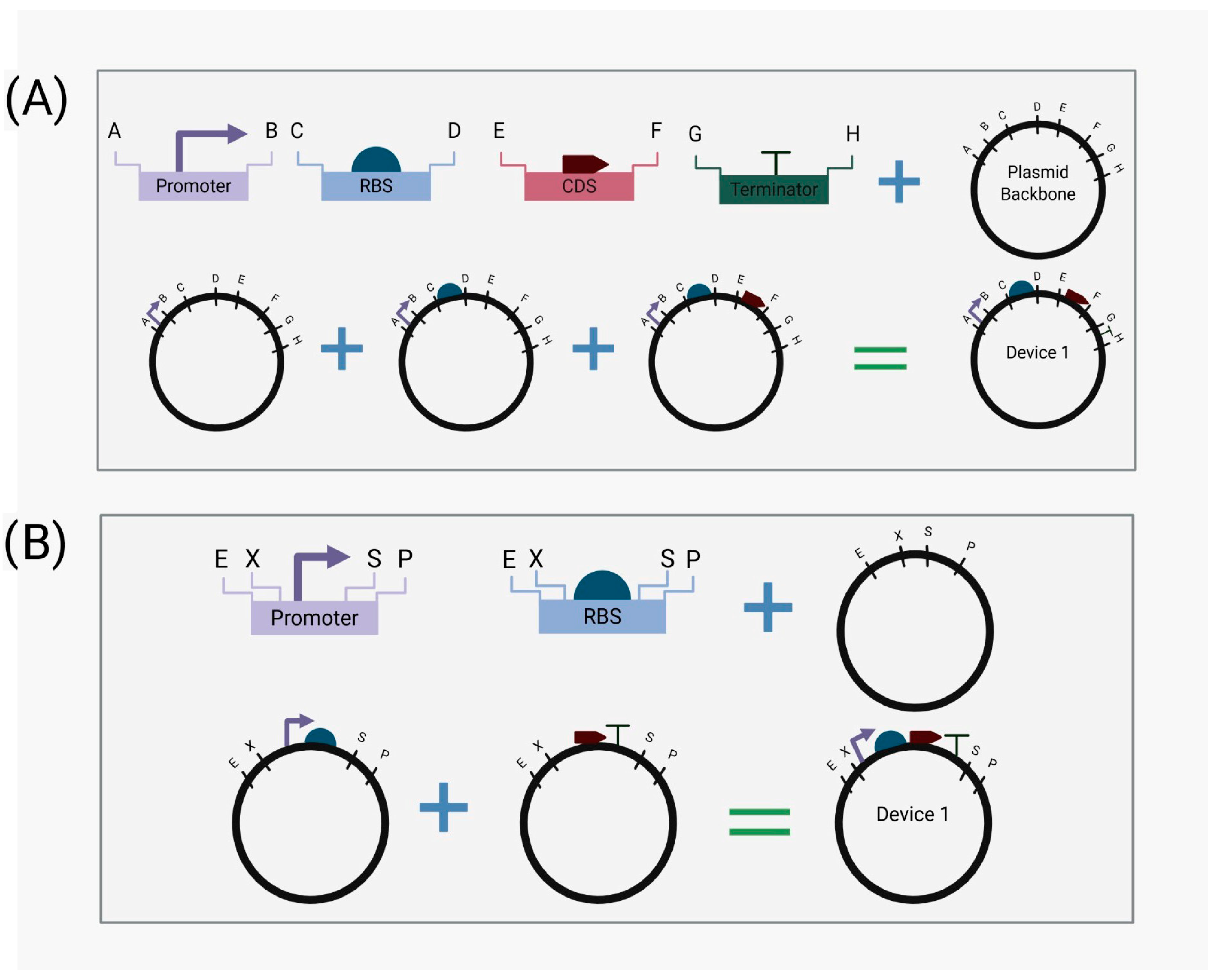
2.2. Standardization

Building sophisticated genetic circuits and leveraging DBTL for fine-tuning, optimization, and high-throughput processing with unreliable and inconsistent parts will take years of trial and error, cost significant resources, and limit the applicability of complex applications. Therefore, standardization is considered a crucial aspect of SynBio, serving as a focal point for integrating engineering disciplines with SynBio. The engineering standards equivalent in SynBio, when designing genetic circuits, are standardized biological parts, as they are the genetic circuits’ building blocks. They are re-engineered genetic sequences that encode a regulatory (promoters, RBS, terminators) or functional (CDS) feature with precise design and performance [19]. When various biological parts are integrated, a biological device is developed to execute a more sophisticated function. Parts standardization can be accomplished through functional and physical standardization [20]. The first entails characterizing, understanding, and predicting the part’s behavior. The latter entails utilizing a standard physical cloning strategy for biological parts [21].
Hence, the concept of BioBricks arose (Figure 1), which incorporates prefix and suffix restriction sites (EcoRI, XbaI, SpeI, and PstI) into each part [22]. BioBricks are extensively used in International Genetically Engineered Machine (IGEM) projects and are included in the Registry of Standard Biological Parts. This type of standardization allows for modular use of standard parts, overcomes traditional molecular biology cloning limitations, and facilitates the creation of high-throughput circuit libraries through the reproducibility, reliable compatibility, interchangeability, and predictable behavior of the standard parts. Despite all the advantages of standardization, it primarily seeks to characterize parts, devices, and potentially systems while ignoring the complex, sophisticated nuances of each. Therefore, the concept of abstraction evolved.

2.3. Abstraction Hierarchy

The human brain cannot effectively handle dealing with highly complex systems while keeping every aspect in mind, including the sequences used, the mechanisms behind regulatory parts, the interaction between the chassis and the devices, and how the system responds to induced changes. Abstraction hierarchy is a fundamental concept to follow while using SynBio tools and disciplines. It helps design complex systems, devices, and parts regardless of detailed knowledge of the finest details, such as cellular processes or every pathway involved, while constructing the initial design [20]. The abstraction hierarchy enables synthetic biologists to follow a logical, sequential framework. At the highest level of abstraction, we focus on the desired final system function or output, which is the endpoint of our design. Moving down through the hierarchy, we break down the system’s function into the needed devices’ subfunctions and behaviors. Then, we choose the parts constructing each device according to their nature and the desired output. At the lowest level of abstraction, the genetic sequences of individual parts must be investigated and designed. Combining SynBio tools with the abstraction hierarchy increases design flexibility, improves predictability, and makes DBTL easier to implement.
Synthetic Biology Open Language (SBOL) is one of the tools that facilitates following the hierarchy while designing genetic circuits. It is a data standard that provides SynBio designs with a standard format to facilitate design utility and exchange between research groups and different software [23]. It enables researchers to reuse DNA sequences and parts, with defined descriptions and functionality, in their hierarchical form to design various devices and systems [23].

3. Designing Genetic Circuits

Combining the aforementioned principles and toolkits provided by SynBio, particularly standardization and synthetic DNA synthesis, enables the ability to control, tune, and freely design genetic circuits interchangeably. They are the fundamental components of designed systems in SynBio, and they play a critical role in achieving the intended functionality. Genetic circuits can control gene expression, sense different inputs, and encode numerous outputs (fluorescent, colorimetric, biochemical compounds, and others), resulting in the complex intended function. Before beginning to construct circuits, it is critical to become familiar with the basic biological parts that can be used, how they can work together through understanding common circuit architectures, and what bioinformatics tools and wet lab techniques facilitate genetic circuit design.

3.1. Fundamental Biological Parts

3.1.1. Gene Expression Regulatory Parts

Previously, only natural cis-elements, enhancers, silencers, and promoters were known to control gene expression [24]. However, SynBio has discovered and utilized a variety of regulators, including synthetic promoters, riboswitches, toeholds, and synthetic transcription factors (SynTFs) [25].
Promoters/Synthetic Promoters
The gene promoter sequence, along with the transcription start site (TSS), is crucial in gene expression regulation [26]. The promoter’s main function is to specifically bind and correctly position the transcription initiation complex, enabling the activation or repression of gene expression by TFs [26,27]. Thus, promoters and transcription machinery are viewed as attractive biological parts to be engineered so that their functionality can be tuned and their gene expression controlled.
Promoters are generally classified into constitutive and inducible promoters depending on whether they are constantly active or whether their activity depends on specific stimuli, respectively [27]. Examples of inducible promoters include PBAD and PSAL, which are activated in response to arabinose and salicylate, respectively, and their integration into a functional genetic circuit will be further discussed in the Logic Gates Section [28].
Native promoters often exhibit weak expression and are large in size; as a result, the necessity of engineering synthetic promoters that are compact and exhibit strong expression patterns has been increasing, in addition to minimizing background expression [27]. Synthetic promoter engineering typically includes core promoter elements such as the TATA box, initiator, and CAAT box, in addition to cis-regulatory elements such as activators, enhancers, operators, and repressors from different sources [27]. By modifying or eliminating these cis-regulatory elements, as well as the sequence of the core promoter region, scientists can control promoter sensitivity and output strength [14,27,29,30]. Since synthetic promoters can be re-engineered to respond to different inducers or to alter their expression strength, designing novel promoters that can respond to the microenvironment of injured tissues (such as pH, specific metabolites, or enzymes) to induce tissue regeneration is a promising approach for regenerative medicine [31,32]. In stem cells, synthetic promoters can be precisely engineered to control gene expression in response to specific cellular signals or microenvironmental conditions, allowing for fine-tuned regulation of stem cell differentiation, proliferation, and therapeutic output for regenerative medicine applications [33,34].
Riboswitches
Riboswitches are a type of non-coding RNA that regulate gene expression upon sensing small ligand molecules [14]. Their structure is composed of two main functional domains, which are the aptamer domain that specifically binds to the ligand molecule and the expression domain that undergoes conformational changes, resulting in the regulation of gene expression either at the transcriptional or translational level [35,36,37].
In translational regulation mechanisms, riboswitches typically regulate the accessibility of the RBS and the start codon and thus control ribosome binding based on the thermodynamic equilibrium [38]. For transcriptional regulation, riboswitches form a ligand-dependent terminator or anti-terminator structure, creating a state of OFF/ON mode based on ligand presence [37,39,40]. The absence of the riboswitch inducer ligand, indicating the OFF state, forms a stable hairpin terminator structure followed by uracil residues, which terminate the transcription of mRNA. However, the interaction of the inducer with the riboswitch, indicating the ON state, prevents the formation of the hairpin terminator, allowing the transcription process to proceed [41].
In genetic circuits, riboswitches integrated into the 5’UTR of mRNAs provide ligand-dependent gene expression that offers an efficient biological part [14,42]. Since glutamine plays a critical role in controlling cancer stem cells’ (CSCs) fate, differentiation, and epigenetic modulation [43], a glutamine riboswitch can be utilized to encode desired output in a glutamine-dependent manner in the tumor microenvironment. In stem cells, riboswitches can offer a powerful tool for precise, ligand-inducible control over gene expression, enabling researchers to manipulate stem cell behavior, differentiation pathways, and therapeutic protein production in a highly regulated and context-dependent manner [44,45,46].
Toehold Switches
Toehold switches are RNA regulatory components that respond to RNA sequences known as trigger RNA to regulate gene expression [41]. Their structure consists of an RNA hairpin structure in which the RBS and start codon are contained, thereby silencing the translation process. The upstream hairpin structure contains a complementary sequence for the trigger RNA, known as a single-strand toehold domain [41]. In response to the trigger RNA molecule, it binds to the toehold domain, initiating a strand displacement reaction, resulting in the opening of the hairpin structure and exposing the RBS and start codon region, enabling ribosome access and translation process initiation [47]. Hence, toehold switches can be designed as complements to the targeted trigger RNA.
Based on the evidence of the role of microRNAs (miRNAs) such as miR-19 and miR-21 in the development of CSCs [48], the cancer microenvironment can be monitored using toehold-regulated genetic circuits that can monitor the activity of these miRNAs as an early detection sensor for rapid therapeutic intervention. In stem cells, this capability allows for precise control over gene expression in response to specific cellular cues, enabling the engineering of stem cell behavior for therapeutic purposes, such as directing differentiation or detecting aberrant states [49,50,51].
Synthetic Transcription Factors
SynTFs are synthetic proteins that regulate gene expression with remarkable specificity. While initial experiments in SynBio used naturally occurring TFs, the number was limited, and each required substantial tuning and characterization. SynTFs have been utilized as an alternative to address these constraints, as they can be engineered to respond to an intended inducer and bind to a specific genomic sequence [52]. This is achievable through the natural structure of SynTFs, which comprises an effector binding domain (EBD) (transcriptional activation or repression domain) linked with a DNA binding domain [29]. These domains work together to make SynTFs tunable and capable of regulating gene expression in a specific effector-dependent manner, which can be a small ligand, metabolite, or even protein.
The tunability and engineering of these TFs can be achieved through a variety of methods, including random mutation, single-point mutation of natural TFs, designing chimeric TFs by linking two naturally occurring domains with a linker, or de novo designing one or both domains from scratch to bind to the desired effector or DNA sequence [53,54]. SynTFs can be designed and validated using common protein bioinformatics tools such as AlphaFold and Rosetta, as well as established wet laboratory methods [55]. A breakthrough in SynTF design was the use of zinc finger proteins, which can be engineered to recognize any nucleotide triplet in DNA and tweaked to recognize longer unique sequences [56]. When integrated with genetic circuits, SynTFs can be designed to control and regulate the genetic circuit or to be the circuit’s output, performing another function in the system. In stem cell applications, SynTFs can be precisely engineered to bind with specific proteins or surface markers, thereby enabling the targeted production of cell toxins or therapeutic proteins and, crucially, providing fine-tuned control over cell fate and differentiation for various therapeutic applications [57].

3.1.2. Other Parts: Ribosomal Binding Sites, Coding Sequences, and Terminators

Another significant biological part of genetic circuits is the RBS, which is critical for translation initiation and ribosome recruitment by mRNA. In prokaryotic systems, the RBS is known as the Shine–Dalgarno (SD) sequence, which is located a few nucleotides upstream of the start codon [58]. This sequence precisely positions the start codon within the small subunit of the ribosome, resulting in accurate and efficient protein synthesis [59,60,61]. Because of its critical regulatory function, the RBS is a prominent target for SynBio, where it can be manipulated to affect system responsiveness, translation efficiency, and protein expression levels, thereby optimizing genetic circuits for desired outcomes [62].
Coding sequences are another significant component of genetic circuits, as they provide clear indicators of circuit function that are frequently monitored through the expression of reporter genes. Commonly utilized reporter genes include green fluorescent protein (GFP), luciferase, and β-galactosidase, which provide quantitative output signals linked to transcriptional or translational activity [63,64]. Furthermore, the coding sequence can produce any desired output, including proteins, enzymes, metabolites, and even SynTF, which can control other circuits or systems. These outputs are regulated by the previously described biological components, allowing for precise monitoring of circuit behavior in response to specific input signals [63].
Terminators are important genetic elements that enable the proper disengagement of RNA polymerase from the DNA template during transcription. They are used to isolate transcriptional units from one another, and their inclusion in genetic circuits helps to control the overall gene expression level [65].

3.2. Circuit Architectures
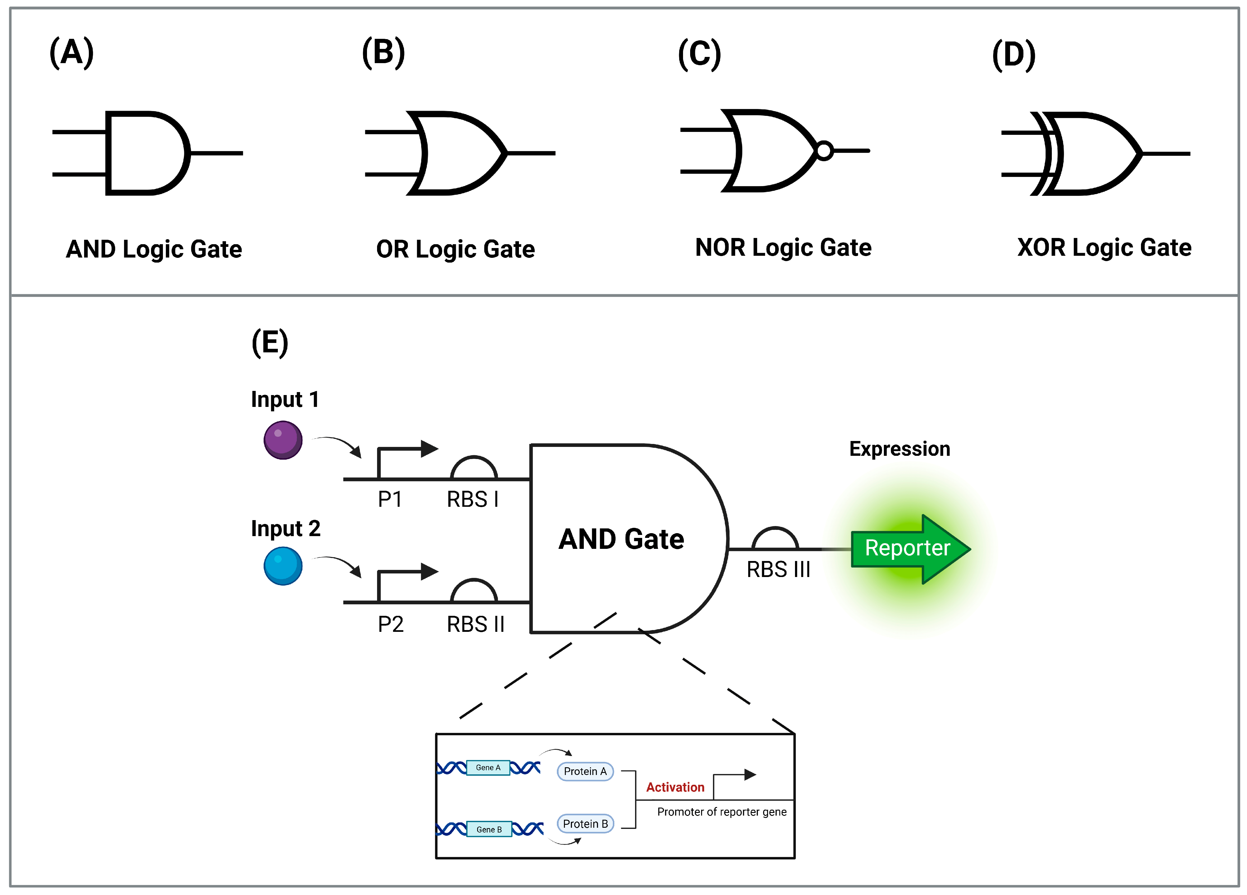
3.2.1. Logic Gates

Synthetic biologists have integrated digital electronic principles with cells to control how they process input information. Electronic systems use logic gates, which take binary inputs (0 or 1) to generate a predetermined output signal; similarly, biological logic gates are constructed utilizing the fundamental parts of genetic circuits to control gene expression, either activating or repressing it as an output signal [66]. Biological parts can be combined to construct different types of logic gates, including but not limited to AND, OR, NOR, and XOR gates (Figure 2). AND gates require the presence of both inputs to be turned on, whereas OR gates require at least one of the two inputs to be turned on, NOR gates are turned on only if both two inputs are absent, and XOR gates require only one of the two inputs to be turned on [67].
As an example of an AND gate, two inducible promoters, PBAD and PSAL, were employed, which are activated by arabinose and salicylate, respectively [28]. This genetic circuit requires the activation of two promoters in order to express T7 RNA polymerase as an output signal. The circuit assembly functions such that at the first gate, PBAD activates transcription of T7 RNA polymerase, but translation is blocked by the presence of amber stop codons; therefore, the function of the second gate is critical, with PSAL activating transcription of supD, an amber suppressor tRNA, and thus the two environmental stimuli are required to successfully express T7 RNA polymerase [14]. In stem cells, logic gates can be used to regulate the output of genetic circuits in a multi-input-dependent manner, enabling precise control over cell fate, differentiation, and therapeutic functions based on complex environmental cues or internal cellular states [68].

3.2.2. Toggle Switches

A toggle switch is an example of a bistable gene regulatory circuit, as it allows the cell to maintain two stable transcriptional states, similar to the reset–set latch in electronics, which uses a NOR gate to regulate two stable states and can be toggled depending on the specific input it receives. When the input is removed, it retains the memory of the current state [69].
Timothy S. Gardner designed a toggle switch that consists of two repressive transcriptional units, each containing a promoter and a repressor gene that blocks the other promoter if expressed, ensuring that only one state is activated at a time [69]. An output signal, such as GFP, can serve as an indicator of the active state [70].

3.2.3. Oscillators

Biological oscillators are regulatory circuits or devices that control periodic oscillations in gene expression, which are necessary for biological timekeeping. One example of a naturally occurring oscillator is the circadian clock in cyanobacteria, which uses positive and negative feedback loops to coordinate functions such as photosynthesis and nitrogen fixation [71]. Oscillators function as peacemakers in virtually all living organisms, helping to maintain the periodicity of physiological activities and environmental cycles such as day–night transitions [71,72,73].
An oscillator circuit known as a repressilator was designed [63], consisting of three transcriptional repressor genes arranged in a cyclic negative feedback loop, with each repressor gene used to repress the next in sequence to maintain periodic gene expression [14]. Such an architecture can be widely used in stem cells to dynamically control cell functions by establishing feedback loops in response to the periodic changes in biological systems or microenvironments [74].

3.3. Implementing Genetic Circuits

The DBTL cycle, on which SynBio is based, necessitates the integration of both computational and experimental methodologies when building genetic circuits to ensure cycle reproducibility. As a result, various computational tools (Table 1) and wet lab methodologies have emerged to implement genetic circuits while navigating the DBTL cycle; some of them will be discussed in this section [75].

3.3.1. Computational Tools

The use of computational tools in SynBio enables the rational design, modeling, and optimization of genetic circuits prior to their implementation in biological systems. In stem cell applications, where gene regulation must be finely tuned, computational tools are essential to stimulate circuit behavior and predict system dynamics. Among the extensively used platforms are COPASI, Cello, and iBioSim, each of which offers distinct capabilities, advantages, and limitations [119].
COPASI
COPASI is sophisticated software tool designed for the simulation and analysis of biochemical networks and their dynamics [120]. It implements multiple modeling approaches, including deterministic, stochastic, and hybrid simulation methods, to accommodate various biological systems. The software enables researchers to construct detailed mathematical representations of metabolic and signaling pathways, which facilitate in silico experimentation to predict system behaviors under various conditions. COPASI offers a comprehensive suite of analytical capabilities, including time course simulations, parameter scanning and estimation, steady-state analysis, and metabolic control analysis [121]. The dual-interface approach features both a graphical user interface (GUI) for accessibility and command line/API options for advanced users, which makes COPASI versatile across different skill levels [122]. Despite its capabilities, COPASI exhibits specific constraints, especially in stochastic simulation. The software cannot perform stochastic simulations for models containing species or compartments defined by an ordinary differential equation (ODE) rule [123]. These limitations restrict the application of stochastic approaches to certain complex model types.
Cello
Cello represents a groundbreaking framework that functions essentially as a programming language for designing computational circuits in living cells. The most recent version is Cello 2.0, which generates a complete DNA sequence for genetic circuits based on high-level software descriptions and libraries of characterized DNA parts that represent Boolean logic gates. Cello’s design process follows a structured workflow starting with logic synthesis (converting a Verilog description to an abstract Boolean network), technology mapping (assigning biological parts to network nodes), placement (arranging parts into a coherent DNA sequence), and finally export (generating annotated sequence representations suitable for fabrication) [112,124]. Cello 2.0 significantly improves upon its predecessor by offering flexible logic gate descriptions, formal genome placement rules, a new graphical interface, enhanced Verilog 2005 support, and SynBioHub integration [112]. These upgrades extend its usability beyond E. coli plasmids to various organisms and genetic contexts. It provides an automated workflow platform that simplifies circuit design and makes it accessible to users with limited computational skills. The software also generates performance predictions through histograms. The earlier version, Cello 1.0, was restricted to a single gate type (NOR) with a fixed architecture comprising two input promoters in a tandem arrangement [124]. Its modeling capabilities were limited and assumed gate inputs combined linearly without accounting for potential “roadblocking” effects between promoters [125].
iBioSim
iBioSim was developed primarily for modeling, analyzing, and designing genetic circuits; however, it also incorporates metabolic networks, cell signaling pathways, and other biological systems [126]. The platform provides comprehensive support for multicellular models and offers visualization tools that enhance our understanding of complex biological dynamics [121]. iBioSim facilitates interoperability through robust support for the Systems Biology Markup Language (SBML), which enables model exchange across different platforms. iBioSim demonstrates exceptional standards compliance and supports all levels and versions of the SBML for import. The software’s reliability has been demonstrated by its distinction as the first tool to produce correct results for all examples in the SBML benchmark suite, with successful testing on stochastic benchmark suites and curated models in the BioModels database [127]. iBioSim is one of the few tools to support SBOL, positioning it at the forefront of standardization efforts in SynBio. This early adoption reflects the platform’s commitment to interoperability and data exchange, which are critical factors for collaborative SynBio research [128].

3.3.2. Wet Lab Assembly Approaches

Once a genetic circuit has been computationally designed, it must be physically implemented in the laboratory using efficient and accurate DNA assembly methods [129]. Several modular cloning approaches have emerged to facilitate the construction of complex genetic circuits while using SynBio toolkits for DNA synthesis and standardization, as well as to overcome the limitations of traditional molecular biology techniques (Figure 3).
BioBrick Standard Assembly
The BioBrick standard provides a foundational approach to modular cloning. It is based on defined prefix and suffix sequences flanking genetic parts (as mentioned previously), which allows for standardized assembly and part exchangeability [130]. In early SynBio applications, this method facilitated the development of reusable genetic elements, which laid the groundwork for the more advanced assembly techniques used today [131]. This assembly approach is less commonly used in mammalian systems due to limitations in scalability and sequence constraints [132].
Golden Gate Assembly
Golden Gate is a type IIS restriction enzyme-based method that enables the seamless and directional assembly of multiple DNA fragments in a one-pot reaction [133]. Type IIS enzymes cut outside their recognition sequences and generate custom overhangs that guide the assembly of fragments in a predefined order. This method is highly efficient for building multigene constructs and combinatorial libraries, making it well suited for assembling synthetic transcriptional networks that regulate stem cell behavior [134]. Golden Gate has been widely adopted in modular cloning systems like MoClo and GoldenBraid, which offer standardized parts and compatibility with high-throughput workflows [133].
Gibson Assembly
Gibson assembly is a sequence overlap-based technique that joins DNA fragments with complementary ends in a single isothermal reaction using a mixture of exonuclease, DNA polymerase, and DNA ligase [135]. It allows for scarless assembly of large constructs and is particularly useful when precise control over the sequence context is required, which is an important feature for circuits operating in mammalian stem cells, where regulatory elements such as insulators, enhancers, and 3’ UTRs must be preserved without introducing artifacts [135].
Sequence and Ligation-Independent Cloning (SLIC)
SLIC is a flexible method for assembling DNA without restriction enzymes, relying on a homologous recombination-based mechanism in vitro [136]. It allows for the assembly of multiple fragments with overlapping regions and is compatible with synthetic gene fragments, making it suitable for assembling custom-designed synthetic transcriptional units for stem cell applications [137].
CRISPR-Based In Situ Integration
In addition to in vitro DNA assembly, genome engineering tools such as CRISPR-Cas9 allow for the direct integration of synthetic circuits into defined genomic loci of stem cells [138]. This ensures stable and predictable expression of circuit components and reduces the variability associated with episomal vectors or random integration [139]. Targeted knock-in strategies using homology-directed repair (HDR) or non-homologous end joining (NHEJ) are commonly employed to introduce synthetic transcriptional regulators or reporter systems into PSCs [140].
Therefore, the integration of computational and wet lab design tools enables the rapid prototyping and refinement of genetic circuits that are responsive, robust, and adaptable to the dynamic environment of stem cells. These tools form the backbone of SynBio applications in stem cell engineering, from controlling lineage commitment to programming therapeutic behaviors [141].

4. Integrating Genetic Circuits with Stem Cells

Recent advances in synthetic biology have resulted in increasingly complex genetic circuits, allowing for progressively sophisticated control of cellular behavior. Synthetic biology has shifted its attention from microorganisms to mammalian systems, particularly stem cells and their derivatives, as technologies for gene editing and genetic circuit design have advanced and our understanding of systems biology has grown [64]. This shift addresses the missing piece of the stem cell control puzzle—genetic circuits—and opens up exciting opportunities to develop a broad range of stem cell applications, as well as control cell differentiation, reprogram cells, engineer tissues, and develop cell therapies [142].
The integration of synthetic biology with stem cell research represents a paradigm shift in regenerative medicine and cellular engineering. Stem cells, with their inherent plasticity and therapeutic potential, provide an ideal platform for implementing sophisticated genetic circuits that can respond to environmental cues, control differentiation pathways, and produce therapeutic outputs. This convergence has led to the development of programmable stem cells that can be precisely controlled for various biomedical applications, from tissue engineering to cell-based therapies for degenerative diseases.
SynBio and genetic circuits allow researchers to approach these processes using control theory concepts, such as treating biological signals as inputs and stem cell responses as outputs. In this context, for example, “fate regulation” is frequently synonymous with “feedback control” [143], an integral part of control systems theory. Just as machines rely on feedback for consistent performance, cells use biochemical and biophysical cues to control responses and maintain desired states [144]. These feedback mechanisms integrate signals from multiple biological layers, ranging from systemic signals to gene regulation networks within individual stem cells. Closed-loop feedback systems adjust for disturbances and maintain output stability, as opposed to open-loop systems, which have a one-way information flow and are susceptible to noise [145]. A simple open-loop circuit would be an inducer that causes a TF to activate the expression of a target gene, whereas, in a closed-loop circuit, the gene product provides feedback to regulate its own expression [144]. For example, genetic toggle switches and oscillators can both be utilized to control or modulate gene expression heterogeneity, allowing for greater control over stem cell differentiation for therapeutic applications [146].

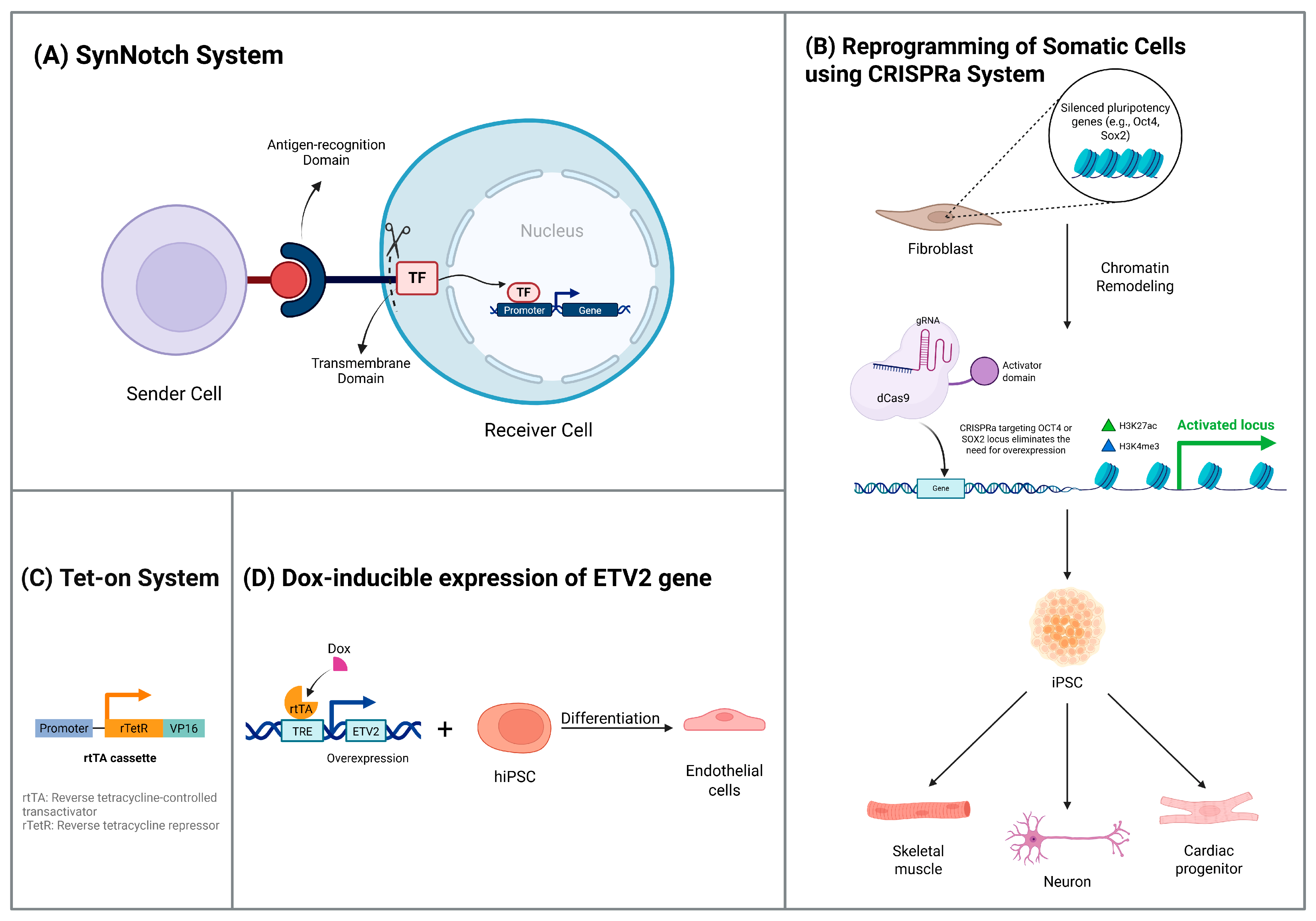
4.1. Cell Differentiation

Genetic circuits can direct stem cell differentiation into desired lineages by dynamically controlling gene expression, such as transcription factor activity, to better recapitulate the patterns of gene expression observed throughout embryonic development [147]. Recent advances in this field have demonstrated the power of synthetic circuits to overcome the limitations of traditional differentiation protocols, which often suffer from low efficiency, heterogeneous outcomes, and a lack of temporal control.
A novel genetic switch was engineered by integrating components from Neurospora crassa and the Escherichia coli Lac repressor system to create an orthogonal regulatory system for use in mammalian cells [148]. After initial validation in immortalized cell lines, this circuit demonstrated tight and tunable gene expression in pluripotent stem cells, highlighting the potential for synthetic circuits to endow cells with artificial decision-making capabilities for precise lineage specification [149].
SynBio has been utilized to direct engineered stem cell differentiation in vivo using genetic circuits, with doxycycline (Dox)-inducible genetic switches being used for controlling ectopic TFs [150,151,152]. In fact, a recent study developed a platform for generating hPSC lines for stable Dox-inducible expression of essential TFs, targeting safe harbor sites in the human genome such as AAVS1, hROSA26, and CLYBL [153]. Controlled overexpression of lineage-specific TFs has been shown to improve the efficiency of hPSC differentiation into myocytes, neurons, and oligodendrocytes [153]. Another study emphasized the necessity for controlled timing of gene expression during cell fate decisions with the pulsing of a key TF. Induced pluripotent stem cells (iPSCs) were found to form three-dimensional multicellular tissue that displayed characteristics similar to liver tissue, due to the early pulsing of the TF, GATA6 [151].
A particularly innovative approach involves the use of temporal control circuits that can precisely time the expression of differentiation factors. These circuits have been designed to mimic the natural developmental cascades observed during embryogenesis, where specific transcription factors are expressed in defined temporal windows. For instance, researchers have developed circuits that can sequentially activate different transcription factors over time, leading to more efficient and homogeneous differentiation outcomes compared to traditional methods that rely on simultaneous factor expression.
The Synthetic Notch-based system (SynNotch) has been developed to control organogenesis using synthetic receptors and is an effective tool for modifying cell-to-cell interactions [154]. A SynNotch-expressing recipient cell recognizes a sender cell. SynNotch consists of an intracellular TF, a Notch core transmembrane domain with proteolytic cleavage sites, and an extracellular antigen recognition domain [154]. When SynNotch receptors recognize their target antigen, they release an engineered TF, resulting in specific transcriptional regulation. This strategy was used for mouse embryonic stem cells (ESCs) that were programmed to use SynNotch to activate the neural differentiation factor Neurogenin1 [155]. Contact between receiver ESCs and sender cells resulted in neuronal differentiation at the interface between the two [154]. SynNotch could control the expression of adhesion molecules that cause cellular rearrangements, the expression of morphogens or receptors, and other differentiation processes [156].
The SynNotch system represents a breakthrough in synthetic biology applications for stem cells because it enables spatial control of differentiation. Unlike traditional chemical induction methods that affect all cells uniformly, SynNotch allows for precise spatial patterning of cell fates within a population. This capability is particularly valuable for tissue engineering applications where different cell types need to be organized in specific spatial arrangements to recapitulate native tissue architecture.

4.2. Cell Reprogramming

Cellular reprogramming represents one of the most transformative applications of synthetic biology in stem cell research. The ability to convert one cell type into another through the controlled expression of specific transcription factors has revolutionized regenerative medicine and opened new avenues for disease modeling and drug discovery.
Stem cells’ fate is primarily determined by a complex, endogenous network of transcription factors that constantly send and respond to physiological signals, altering gene expression in a cell type-specific manner [157]. The landmark discovery that overexpression of four key transcription factors, Oct4, Sox2, Klf4, and c-Myc, can override established cell identities and induce somatic cells to pluripotency [8] has paved the way for synthetic biology approaches to cellular reprogramming. Based on this principle, SynBio can reprogram cells by controlling these TFs using various genetic circuits for different uses. Genetic circuits can specifically target specific damaged cell types, express these TFs and inducing their transformation into iPSCs as a regeneration strategy [158].
Recent advances in synthetic biology have led to the development of more sophisticated reprogramming strategies that go beyond the simple overexpression of Yamanaka factors. These include the use of synthetic transcription factors, epigenetic modifiers, and small-molecule-inducible systems that can provide more precise temporal and spatial control over the reprogramming process.
Large-scale chromatin modification is necessary for cellular reprogramming [159]. To induce transcription and epigenetic remodeling, synthetic transcription factors such as CRISPR activators (CRISPRa) have been employed to promote modifications to chromatin structure by targeting specific loci [159]. When reprogramming fibroblasts into iPSCs, CRISPRa targeting Oct4 or Sox2 eliminates the necessity for Oct4 or Sox2 overexpression, respectively [160,161]. CRISPRa (Figure 4) has been used to replace native factors for the generation of neurons, skeletal muscle, and cardiac progenitors [162,163,164].
Another strategy for controlling reprogramming is to target native signaling pathways in donor cells. Small-molecule inhibitors that target pro-inflammatory transforming growth factor beta (TGF-β) signaling can enhance reprogramming efficiency [165,166,167,168], as TGF-β signalling may hinder reprogramming by promoting fibrosis and senescence [169]. Inhibiting the inflammatory cytokine interleukin 1-beta (IL-1β) and its downstream effectors in adult mouse fibroblasts promotes reprogramming into cardiomyocytes [170]. This suppression of the pro-inflammatory cascade was reported to enhance reprogramming in postnatal and adult fibroblasts but not in embryonic fibroblasts [159].
Recent experimental data have demonstrated the effectiveness of synthetic biology approaches in improving reprogramming efficiency. For example, a study by Weltner et al. (2018) [171] showed that using synthetic promoters to drive reprogramming factor expression increased iPSC generation efficiency three-fold compared to viral methods. Additionally, the use of synthetic circuits that can sense cellular stress and adjust factor expression accordingly has been shown to reduce the formation of partially reprogrammed cells, a major challenge in traditional reprogramming protocols.

4.3. Cell Therapies

The integration of SynBio with stem cell research has resulted in the development of cell therapies with genetically engineered properties. These engineered cell therapies represent a new paradigm in medicine, where cells are programmed to perform specific therapeutic functions in response to disease-related cues or external stimuli.
Genetic circuits have shown promise in generating pancreatic β cells, which play a crucial role in insulin secretion and glucose regulation [172]. Type 1 diabetes is caused by the autoimmune destruction of these cells, which impairs the body’s ability to regulate blood glucose levels. One therapeutic strategy involves overexpressing three TFs, Pdx1, Ngn3, and Mafa, to differentiate pancreatic progenitor stem cells into mature β cells [173,174]. Recently, a genetic band-pass filter circuit was designed to temporally synchronize the expression of these TFs [175]. This sophisticated regulation resulted in a more homogenous population of insulin-producing β cells, outperforming those generated using traditional chemical induction approaches. The effectiveness of this method emphasizes the significance of dynamic, time-sensitive gene regulation during differentiation [8], which can be precisely controlled by genetic circuits.
Experimental validation of these synthetic β-cell circuits has shown remarkable results. In a recent study, engineered β-cells produced using temporal control circuits demonstrated glucose-responsive insulin secretion that closely mimicked native pancreatic β-cells. When transplanted into diabetic mouse models, these cells successfully restored glucose homeostasis for over 6 months, with no evidence of tumor formation or immune rejection. This represents a significant advancement over previous approaches that often resulted in cells with poor glucose responsiveness or limited survival after transplantation.
A key focus in stem cell research is the creation of therapeutic-grade hPSCs using genetically designed fail-safe switches that selectively destroy hPSCs but not differentiated cells during transplantation [11,176]. For example, a recently designed switch employs a resistance gene regulated by an endogenous miRNA-302, which is exclusively expressed in hPSCs and not differentiated cells [11]. Furthermore, the study assessed the efficiency of the inducible caspase-9 (iCaspase9) gene as a fail-safe switch to prevent the unintended tumorigenic transformation of iPSCs derived from somatic cells [11]. A lentiviral vector was utilized to transduce iCaspase9 into two iPSC lines, and the efficacy was assessed both in vitro and in vivo. Apoptosis was induced in approximately 95% of both iPSCs and iPSC-derived neural stem/progenitor cells (iPSC-NS/PCs) in vitro. For in vivo assessment, they transplanted iPSC-NS/PCs into NOD/SCID mice’s damaged spinal cords to evaluate in vivo functionality. All transplanted cells that impaired motor function recovery due to mass effect were eliminated after iCaspase9 activation [11]. These findings suggest that the iCaspase9 system could be an important countermeasure against adverse post-transplantation events in stem cell transplant therapy.
Additional safety mechanisms have been developed to address the tumorigenic risk associated with stem cell therapies. These include synthetic circuits that can detect abnormal proliferation patterns and trigger cell death, as well as circuits that can respond to external signals to eliminate transplanted cells if adverse effects occur. Such safety systems are crucial for the clinical translation of stem cell therapies and have been shown to be highly effective in preclinical models.

4.4. Tissue Engineering

The use of biological circuits and their expression can result in functionalized cells for tissue engineering [177]. Tissue engineering applications of synthetic biology have shown particular promise in creating tissues with enhanced functionality and improved integration with host tissues.
For example, Gersbach designed a Tet-off system that regulates Runx2 factors to control in vivo osteogenic processes [178]. The Tet-on system consists of two components: a transcriptional activator protein that is constitutively expressed and responds to doxycycline (dox), called the reverse tetracycline transactivator (rtTA), and an inducible promoter regulated by a Tet-responsive element, rtTA. The rtTA drives transgene expression [179]. Yao employed a Tet-on system in engineered rat chondrocytes to produce Sox9, a crucial factor in chondrocyte viability, which activated the production of proteins associated with type II collagen and aggrecan in cartilage tissue engineering [180]. Upon injection of Dox (Tet system inducer) in an implanted cell scaffold, chondrocyte apoptosis was inhibited [180].
Recent advances in tissue engineering have demonstrated the potential of synthetic biology to create more sophisticated tissue constructs. For instance, researchers have developed circuits that can sense mechanical stress and respond by producing appropriate extracellular matrix proteins, leading to tissues that can adapt to their mechanical environment. This represents a significant advancement over static tissue engineering approaches.
Cells use the NF-κB pathway to regulate the production of inflammatory cytokines, which activate inflammatory responses [34]. A study developed a synthetic inducible promoter and virally transduced it into an iPSC cell line, which was found to possess self-regulating and inflammation-attenuating properties in vitro via the controlled release of anti-inflammatory molecules, specifically driving the expression of IL-1Ra. Subsequently, the cells were differentiated into chondrocytes, the cells that comprise cartilage tissue [34]. Furthermore, the Tet-on system was employed to overexpress the interleukin-1 receptor antagonist IL-1Ra gene, which modulates inflammatory cytokines during the chondrogenesis process in cartilage repair [181].
A related technique is to engineer stem cells to produce master transcription factors under the control of drug-inducible promoters [182]. ETV2, for example, when overexpressed, generates endothelial cells from hiPSCs [183,184,185,186]. These ETV2-expressing cells can then be combined with hiPSCs, differentiating into other cell types, such as neurons, in a brain cortical organoid protocol [182]. The ETV2-expressing fraction of engineered cells differentiates into endothelial precursors that can organize to form microvascular structures [187].
A two-way cell communication genetic circuit, developed to mimic natural angiogenic signaling during blood vessel formation [188], can be utilized for tissue communication. “Sender” cells were engineered to express tryptophan synthase (TrpB26), an E. coli enzyme that synthesizes L-tryptophan from indole. In contrast, “receiver” cells were designed with a circuit that can detect L-tryptophan and induce the expression of a reporter gene, secreted alkaline phosphatase. Using this synthetic communication system, the researchers recapitulated natural angiogenesis by stimulating mature blood vessel formation sequentially with vascular endothelial growth factor (VEGF) and Angiopoietin-1 [189].
Experimental data from tissue engineering applications have shown promising results. In a recent study, engineered cartilage tissues created using synthetic biology approaches demonstrated superior mechanical properties and better integration with host tissue compared to conventional tissue engineering methods. The engineered tissues maintained their functionality for over 12 months in animal models, with evidence of continued matrix production and cellular viability.

5. Challenges and Limitations of Integrating Synthetic Biology with Stem Cells

Before delving into the specific challenges, it is important to understand the major types of stem cells and their unique characteristics, as these properties directly influence the design and implementation of synthetic biology approaches.

5.1. Major Types of Stem Cells and Their Characteristics

Stem cells can be broadly classified based on their developmental potential and origin, each presenting unique advantages and challenges for synthetic biology applications:
Embryonic Stem Cells (ESCs) are derived from the inner cell mass of blastocysts and represent the gold standard of pluripotency. They can differentiate into all three germ layers (ectoderm, mesoderm, and endoderm) and theoretically any cell type in the adult body. ESCs have unlimited self-renewal capacity and maintain stable karyotypes over extended culture periods. However, their use is associated with ethical concerns, potential immune rejection, and a risk of teratoma formation [190].
Induced Pluripotent Stem Cells (iPSCs) are generated by reprogramming somatic cells through the expression of specific transcription factors (typically Oct4, Sox2, Klf4, and c-Myc). iPSCs share many characteristics with ESCs, including pluripotency and self-renewal capacity, while avoiding ethical issues associated with embryonic sources. They can be generated from patient-specific cells, potentially eliminating immune rejection concerns. However, iPSCs may retain epigenetic memory from their cell of origin and can exhibit genomic instability [191].
Hematopoietic Stem Cells (HSCs) are multipotent stem cells that give rise to all blood cell lineages. They are well characterized and have been successfully used in clinical applications for decades. HSCs can be isolated from bone marrow, peripheral blood, or umbilical cord blood. They demonstrate excellent engraftment potential and have established protocols for expansion and differentiation [192].
Mesenchymal Stem Cells (MSCs) are multipotent cells that can differentiate into bone, cartilage, fat, and other connective tissues. They can be isolated from various sources, including bone marrow, adipose tissue, and dental pulp. MSCs have immunomodulatory properties and low immunogenicity, making them attractive for therapeutic applications. However, they have limited proliferative capacity, and their differentiation potential decreases with age [193].
Neural Stem Cells (NSCs) are multipotent cells that can generate neurons, astrocytes, and oligodendrocytes. They are found in specific niches in the adult brain and can be derived from pluripotent stem cells. NSCs are particularly relevant for treating neurodegenerative diseases and spinal cord injuries. However, they are difficult to isolate and expand, and their differentiation is highly dependent on environmental cues [194].
Each stem cell type presents unique opportunities and challenges for synthetic biology applications. The choice of stem cell type depends on the intended application, with considerations including differentiation potential, safety profile, scalability, and regulatory requirements.

5.2. SynBio Challenges

Although SynBio has promising applications in many fields, such as stem cell research, many hurdles must be overcome, especially in designing and implementing genetic circuits [195]. As biological systems are dynamic and tend to be unpredictable, the complexity of intended genetic circuits can be a significant issue. Functional instability and decreased reliability of the synthetic constructs can result from the inconsistent performance of the circuits in different hosts or environmental conditions [195,196]. For instance, off-target effects and context-dependent behavior of genetic circuits in complex mammalian cellular environments, particularly in stem cells, pose significant challenges in predicting outcomes [66].
Another significant limitation of synthetic circuits is the metabolic load they impose on their hosts. Engineered pathways tend to demand large quantities of cellular resources, which can interfere with natural metabolic processes and have unintended physiological effects [197]. This metabolic burden can lead to reduced cell viability, altered cellular functions, and inefficient production of desired outputs, which is especially critical in resource-sensitive stem cell populations [198].
Furthermore, the operation of such circuits is also compromised by the thermodynamic constraints of molecule synthesis in biological organisms, especially when attempting to synthesize complex or energy-intensive molecules [197]. The evolutionary instability of synthetic circuits is another issue. Mutation, horizontal gene transfer, and genetic drift can all reduce circuit function over time or cause off-target characteristics to arise in engineered organisms [196]. Although several strategies, such as the use of genetic safeguards, have been developed to mitigate these effects, they are not yet in common use or broadly effective [197]. The long-term stability of engineered genetic circuits within rapidly dividing or differentiating stem cell populations remains a key concern, as genetic drift can lead to a loss of function or unintended phenotypes over time [199].
Synthetic circuits also pose environmental and biosafety risks, as well as having direct technological and biological implications. Accidental spills of genetically modified organisms into the environment or intentional release threaten contamination of the natural gene pool, loss of biodiversity, and uncontrolled gene flow [196]. An engineered microbe for bioremediation, for instance, may interact in unexpected ways with natural organisms or compounds and produce adverse ecological impacts. Therefore, synthetic organisms that are released outside of the lab should be engineered with mechanisms for containing their life cycle and controlling their spread [200].
Furthermore, SynBio circuits pose biosecurity threats through the possibility of misuse. The dual use of SynBio technology is a concern with regard to the synthesis of dangerous pathogens for bioterrorism. The synthesis of the entire poliovirus genome in the laboratory as a proof of principle illustrates SynBio’s potential for the synthesis of harmful materials [195]. Synthetic circuit construction is also limited by legal and ethical constraints. Current legal and ethical paradigms are challenged when natural organisms are redesigned or completely new types of life are created [201,202]. Such innovations need to be carefully controlled and regulated. With the promise of increased accessibility through SynBio technologies comes the risk of misuse by malicious actors or hobbyists, and thus the necessity for international ethical guidelines, regulatory frameworks, and monitoring policies [202,203,204,205]. The ethical considerations are particularly pronounced when dealing with human stem cells, raising questions about germline editing, human enhancement, and the definition of life itself, necessitating robust public discourse and clear regulatory boundaries [206].

5.3. Stem Cell Challenges

Despite the enormous potential of stem cell therapies, there are several significant limitations preventing their widespread clinical application. The primary concern in the field is the development of robust and scalable differentiation protocols, alongside ensuring the safety of ESC and iPSC-based transplants. Safety issues include the occurrence of genetic abnormalities and the risk of uncontrolled differentiation [207]. Achieving homogeneous differentiation into desired cell types with high purity and yield remains a major hurdle, often leading to heterogeneous cell populations that can compromise therapeutic efficacy and safety [208].
ESCs are associated with several drawbacks, including immune rejection, the potential to differentiate into inappropriate cell types, tumorigenicity, and the risk of contamination. Similarly, germline stem cells, while pluripotent, are limited by their limited availability and the possibility of forming embryonic teratoma in vivo [209]. In contrast, adult stem cells exhibit multipotency and are less likely to elicit immune rejection. They can also be activated in situ by pharmacological agents. However, their therapeutic use is constrained by several factors: they are limited in number, difficult to isolate and expand in vitro, show limited differentiation potential in culture, and are challenging to handle and scale up for transplantation. Moreover, adult stem cells may exhibit telomere shortening and source-dependent variability and may carry inherited genetic abnormalities [209]. The inherent variability between different stem cell lines and donor sources further complicates the standardization and reproducibility of therapeutic outcomes [210].
Immunological rejection remains a critical barrier. The concept of achieving specific transplantation tolerance has eluded researchers despite decades of progress in immunology. Histocompatibility mismatches at polymorphic loci lead to rejection with acute antibody-mediated responses resulting from donor antigen recognition and interaction with the allograft vascular endothelium, which constitutes a major cause of graft failure [211,212]. Nevertheless, clinical transplantation of both organs and hematopoietic stem cells has demonstrated that practical, empirically derived methods can effectively mitigate immunogenicity and alloreactivity in many cases [211]. Developing universal donor stem cell lines or strategies to induce immune tolerance without broad immunosuppression are active areas of research crucial for widespread clinical adoption [213].
Beyond biological challenges, economic and infrastructural limitations also impede the application of stem cell therapies, particularly in developing countries. These therapies are highly expensive, and nations with limited financial and technological resources struggle to sustain research initiatives. Poor infrastructure and minimal governmental support contribute to stalled progress. Additionally, the socioeconomic status of many patients limits the adoption of advanced treatments, even when they become available. Enhanced private sector investment, international collaborations, and the promotion of exchange programs are essential to overcoming these barriers and facilitating the broader adoption of stem cell technologies [214]. The high cost of Good Manufacturing Practice (GMP)-compliant production of stem cell therapies and the complex regulatory approval processes further contribute to their limited accessibility and affordability globally [215].

6. Future Aspects and Conclusions

Machine learning (ML) and deep learning (DL) have witnessed significant advancements in recent years by providing significant implications for synthetic biology [216]. The synergy between these technologies and synthetic biology is bidirectional, with synthetic biology providing extensive datasets from DNA synthesis, which in turn enrich and train ML models, while ML and DL help improve and optimize experimental design, system optimization, protein structure prediction, automated microscopy analysis, and novel biological part generation, as previously explained in our review [216,217]. As previously explained in our review, the integration of AI-driven approaches with synthetic biology has shown particular promise in stem cell applications, where machine learning algorithms can predict optimal circuit designs, identify potential off-target effects, and optimize differentiation protocols.
Recent developments in AI-assisted synthetic biology have led to several breakthrough applications in stem cell research. For example, deep learning models have been used to predict the behavior of genetic circuits in different stem cell types, reducing the need for extensive experimental validation. Additionally, machine learning algorithms have been employed to design synthetic promoters with improved specificity and reduced leakage, addressing one of the major challenges in stem cell engineering.
As synthetic biology continues to apply engineering principles in constructing biological components in various domains, this will enhance ML-based method efficiency and data richness [218]. The increasing availability of high-quality datasets from synthetic biology experiments, combined with advances in computational power and algorithm development, is expected to accelerate the pace of discovery in stem cell engineering.
To overcome the current limitations in this field and to fully utilize ML/DL models in synthetic biology, future advancements should be adopted, the first of which is the generation and standardization of high-quality datasets, especially for biological parts that lack sufficient data for enriching ML models, such as non-coding elements and regulatory motifs [219]. Second is developing open access and public repositories with well-annotated data to facilitate ML/DL model training [220]. Third, the integration of explainable AI approaches with DL models will help researchers understand and overcome the black box nature of DL; integrating ML models with automated DNA synthesis and high-throughput screening platforms will provide a faster and more efficient experimental validation process [221]. Finally, increasing interdisciplinary collaborations and training between synthetic biology, computer science, and engineering will enhance the development of robust tools that are biologically effective and computationally well structured [222].
The future of synthetic biology in stem cell research also depends on addressing several key technical and regulatory challenges. These include developing standardized protocols for circuit design and validation, establishing safety guidelines for clinical applications, and creating regulatory frameworks that can keep pace with technological advances. International collaboration will be essential to ensure that these technologies are developed responsibly and made accessible to researchers and patients worldwide.
Engineering stem cells using synthetic biology is a double-edged sword. It offers flexibility, control, predictability, and continuous optimization; however, at the same time, it creates a new level of complexity by combining both stem cell and synthetic biology complexities. Therefore, applying synthetic biology to mammalian cells remains a challenging area of science, which requires creativity through overcoming the limitations and decades of research to build a well-established framework that predicts and facilitates dealing with such complexity [223].
Despite these challenges, the potential benefits of synthetic biology applications in stem cell research are enormous. The ability to precisely control stem cell behavior, enhance therapeutic efficacy, and reduce safety risks represents a paradigm shift in regenerative medicine. As the field continues to mature, we can expect to see increasingly sophisticated applications that will transform how we approach the treatment of degenerative diseases, tissue engineering, and personalized medicine.
The integration of synthetic biology with stem cell research represents one of the most promising frontiers in biomedical science. While significant challenges remain, the rapid pace of technological advancement and increasing interdisciplinary collaboration suggest that many of these obstacles will be overcome in the coming years. The ultimate goal of creating programmable, safe, and effective stem cell therapies is within reach, promising to revolutionize medicine and improve the lives of millions of patients worldwide.

Author Contributions

Conceptualization, N.H. (Nourhan Hassan); writing—original draft preparation, K.S.E., O.G., N.H. (Nouran Hesham), S.A., N.M. and A.M.; writing—review and editing, N.H. (Nourhan Hassan); supervision, E.M.E. and N.H. (Nourhan Hassan). All authors have read and agreed to the published version of the manuscript.

Funding

This research received no external funding.

Acknowledgments

We express our gratitude to the CU iGEM 2019 team and the CU_Egypt iGEM 2022 team (Pharaohs), who generously shared their knowledge and experience with us. They provided us with guidance, inspiration, mentorship, and support over the past few years; most importantly, they cultivated our passion for synthetic biology.

Conflicts of Interest

The authors declare no conflicts of interest.

References

  1. Zakrzewski, W.; Dobrzyński, M.; Szymonowicz, M.; Rybak, Z. Stem cells: Past, present, and future. Stem Cell Res. Ther. 2019, 10, 68. [Google Scholar] [CrossRef] [PubMed]
  2. Aly, R.M. Current state of stem cell-based therapies: An overview. Stem Cell Investig. 2020, 7, 8. [Google Scholar] [CrossRef] [PubMed]
  3. Menasché, P.; Vanneaux, V.; Hagège, A.; Bel, A.; Cholley, B.; Cacciapuoti, I.; Parouchev, A.; Benhamouda, N.; Tachdjian, G.; Tosca, L.; et al. Human embryonic stem cell-derived cardiac progenitors for severe heart failure treatment: First clinical case report: Figure 1. Eur. Heart J. 2015, 36, 2011–2017. [Google Scholar] [CrossRef] [PubMed]
  4. Schwartz, S.D.; Regillo, C.D.; Lam, B.L.; Eliott, D.; Rosenfeld, P.J.; Gregori, N.Z.; Hubschman, J.-P.; Davis, J.L.; Heilwell, G.; Spirn, M.; et al. Human embryonic stem cell-derived retinal pigment epithelium in patients with age-related macular degeneration and Stargardt’s macular dystrophy: Follow-up of two open-label phase 1/2 studies. Lancet 2015, 385, 509–516. [Google Scholar] [CrossRef] [PubMed]
  5. Ilic, D.; Ogilvie, C. Concise Review: Human Embryonic Stem Cells—What Have We Done? What Are We Doing? Where Are We Going? Stem Cells 2016, 35, 17–25. [Google Scholar] [CrossRef] [PubMed]
  6. Yamanaka, S. Pluripotent Stem Cell-Based Cell Therapy—Promise and Challenges. Cell Stem Cell 2020, 27, 523–531. [Google Scholar] [CrossRef] [PubMed]
  7. Lezmi, E.; Jung, J.; Benvenisty, N. High prevalence of acquired cancer-related mutations in 146 human pluripotent stem cell lines and their differentiated derivatives. Nat. Biotechnol. 2024, 42, 1667–1671. [Google Scholar] [CrossRef] [PubMed]
  8. Healy, C.P.; Deans, T.L. Genetic circuits to engineer tissues with alternative functions. J. Biol. Eng. 2019, 13, 39. [Google Scholar] [CrossRef] [PubMed]
  9. Ferreira, R.; Ohneda, K.; Yamamoto, M.; Philipsen, S. GATA1 Function, a Paradigm for Transcription Factors in Hematopoiesis. Mol. Cell. Biol. 2005, 25, 1215–1227. [Google Scholar] [CrossRef] [PubMed]
  10. Wobus, A.M.; Boheler, K.R. Embryonic Stem Cells: Prospects for Developmental Biology and Cell Therapy. Physiol. Rev. 2005, 85, 635–678. [Google Scholar] [CrossRef] [PubMed]
  11. Itakura, G.; Kawabata, S.; Ando, M.; Nishiyama, Y.; Sugai, K.; Ozaki, M.; Iida, T.; Ookubo, T.; Kojima, K.; Kashiwagi, R.; et al. Fail-Safe System against Potential Tumorigenicity after Transplantation of iPSC Derivatives. Stem Cell Rep. 2017, 8, 673–684. [Google Scholar] [CrossRef] [PubMed]
  12. Kojima, K.; Miyoshi, H.; Nagoshi, N.; Kohyama, J.; Itakura, G.; Kawabata, S.; Ozaki, M.; Iida, T.; Sugai, K.; Ito, S.; et al. Selective Ablation of Tumorigenic Cells Following Human Induced Pluripotent Stem Cell-Derived Neural Stem/Progenitor Cell Transplantation in Spinal Cord Injury. Stem Cells Transl. Med. 2018, 8, 260–270. [Google Scholar] [CrossRef] [PubMed]
  13. Kurtoğlu, A.; Yıldız, A.; Arda, B. The view of synthetic biology in the field of ethics: A thematic systematic review. Front. Bioeng. Biotechnol. 2024, 12, 1397796. [Google Scholar] [CrossRef] [PubMed]
  14. Khalil, A.S.; Collins, J.J. Synthetic biology: Applications come of age. Nat. Rev. Genet. 2010, 11, 367–379. [Google Scholar] [CrossRef] [PubMed]
  15. Gibson, D.G.; Glass, J.I.; Lartigue, C.; Noskov, V.N.; Chuang, R.-Y.; Algire, M.A.; Benders, G.A.; Montague, M.G.; Ma, L.; Moodie, M.M.; et al. Creation of a Bacterial Cell Controlled by a Chemically Synthesized Genome. Science 2010, 329, 52–56. [Google Scholar] [CrossRef] [PubMed]
  16. Hughes, R.A. Synthetic DNA; Humana: New York, NY, USA, 2017; Volume 1472. [Google Scholar] [CrossRef]
  17. Hughes, R.A.; Ellington, A.D. Synthetic DNA Synthesis and Assembly: Putting the Synthetic in Synthetic Biology. Cold Spring Harb. Perspect. Biol. 2017, 9, a023812. [Google Scholar] [CrossRef] [PubMed]
  18. Demissie, E.A.; Park, S.-Y.; Moon, J.H.; Lee, D.-Y. Comparative Analysis of Codon Optimization Tools: Advancing toward a Multi-Criteria Framework for Synthetic Gene Design. J. Microbiol. Biotechnol. 2025, 35, e2411066. [Google Scholar] [CrossRef] [PubMed]
  19. Canton, B.; Labno, A.; Endy, D. Refinement and standardization of synthetic biological parts and devices. Nat. Biotechnol. 2008, 26, 787–793. [Google Scholar] [CrossRef] [PubMed]
  20. Kuldell, N.; Bernstein, R.; Ingram, K.; Hart, K. BioBuilder: Synthetic Biology in the Lab; O’Reilly Media: Sebastopol, CA, USA, 2015; Volume 237. [Google Scholar]
  21. Arkin, A. Setting the standard in synthetic biology. Nat. Biotechnol. 2008, 26, 771–774. [Google Scholar] [CrossRef] [PubMed]
  22. Müller, K.M.; Arndt, K.M. Standardization in synthetic biology. Methods Mol. Biol. 2012, 813, 23–43. [Google Scholar] [CrossRef] [PubMed]
  23. Galdzicki, M.; Clancy, K.P.; Oberortner, E.; Pocock, M.; Quinn, J.Y.; Rodriguez, C.A.; Roehner, N.; Wilson, M.L.; Adam, L.; Anderson, J.C.; et al. The Synthetic Biology Open Language (SBOL) provides a community standard for communicating designs in synthetic biology. Nat. Biotechnol. 2014, 32, 545–550. [Google Scholar] [CrossRef] [PubMed]
  24. Spitz, F.; Furlong, E.E.M. Transcription factors: From enhancer binding to developmental control. Nat. Rev. Genet. 2012, 13, 613–626. [Google Scholar] [CrossRef] [PubMed]
  25. Nielsen, A.A.; Voigt, C.A. Multi-input CRISPR/Cas genetic circuits that interface host regulatory networks. Mol. Syst. Biol. 2014, 10, 763. [Google Scholar] [CrossRef] [PubMed]
  26. Lenhard, B.; Sandelin, A.; Carninci, P. Metazoan promoters: Emerging characteristics and insights into transcriptional regulation. Nat. Rev. Genet. 2012, 13, 233–245. [Google Scholar] [CrossRef] [PubMed]
  27. Ali, S.; Kim, W.C. A Fruitful Decade Using Synthetic Promoters in the Improvement of Transgenic Plants. Front. Plant Sci. 2019, 10, 493712. [Google Scholar] [CrossRef] [PubMed]
  28. Anderson, J.C.; Voigt, C.A.; Arkin, A.P. Environmental signal integration by a modular AND gate. Mol. Syst. Biol. 2007, 3, 133. [Google Scholar] [CrossRef] [PubMed]
  29. Liu, W.; Stewart, C.N., Jr. Plant synthetic promoters and transcription factors. Curr. Opin. Biotechnol. 2016, 37, 36–44. [Google Scholar] [CrossRef] [PubMed]
  30. Srivastava, R.; Rai, K.M.; Srivastava, M.; Kumar, V.; Pandey, B.; Singh, S.P.; Bag, S.K.; Singh, B.D.; Tuli, R.; Sawant, S.V. Distinct Role of Core Promoter Architecture in Regulation of Light-Mediated Responses in Plant Genes. Mol. Plant 2014, 7, 626–641. [Google Scholar] [CrossRef] [PubMed]
  31. Artemyev, V.; Gubaeva, A.; Paremskaia, A.I.; Dzhioeva, A.A.; Deviatkin, A.; Feoktistova, S.G.; Mityaeva, O.; Volchkov, P.Y. Synthetic Promoters in Gene Therapy: Design Approaches, Features and Applications. Cells 2024, 13, 1963. [Google Scholar] [CrossRef] [PubMed]
  32. Yasmeen, E.; Wang, J.; Riaz, M.; Zhang, L.; Zuo, K. Designing artificial synthetic promoters for accurate, smart, and versatile gene expression in plants. Plant Commun. 2023, 4, 100558. [Google Scholar] [CrossRef] [PubMed]
  33. Sadiq, I.Z.; Abubakar, F.S.; Katsayal, B.S.; Ibrahim, B.; Adamu, A.; Usman, M.A.; Aliyu, M.; Suleiman, M.A.; Muhammad, A. Stem Cells in Regenerative Medicine: Unlocking Therapeutic Potential Through Stem Cell Therapy, 3D Bioprinting, Gene Editing, and Drug Discovery. Biomed. Eng. Adv. 2025, 9, 100172. [Google Scholar] [CrossRef]
  34. Vogel, A.M.; Persson, K.M.; Seamons, T.R.; Deans, T.L.; Bayley, H. Synthetic biology for improving cell fate decisions and tissue engineering outcomes. Emerg. Top. Life Sci. 2019, 3, 631–643. [Google Scholar] [CrossRef] [PubMed]
  35. Findeiß, S.; Etzel, M.; Will, S.; Mörl, M.; Stadler, P.F. Design of Artificial Riboswitches as Biosensors. Sensors 2017, 17, 1990. [Google Scholar] [CrossRef] [PubMed]
  36. Serganov, A.; Nudler, E. A Decade of Riboswitches. Cell 2013, 152, 17–24. [Google Scholar] [CrossRef] [PubMed]
  37. Jang, S.; Jang, S.; Yang, J.; Seo, S.W.; Jung, G.Y. RNA-based dynamic genetic controllers: Development strategies and applications. Curr. Opin. Biotechnol. 2017, 53, 1–11. [Google Scholar] [CrossRef] [PubMed]
  38. Machtel, P.; Bąkowska-Żywicka, K.; Żywicki, M. Emerging applications of riboswitches—From antibacterial targets to molecular tools. J. Appl. Genet. 2016, 57, 531–541. [Google Scholar] [CrossRef] [PubMed]
  39. Zhang, J.; Jensen, M.K.; Keasling, J.D. Development of biosensors and their application in metabolic engineering. Curr. Opin. Chem. Biol. 2015, 28, 1–8. [Google Scholar] [CrossRef] [PubMed]
  40. Hallberg, Z.F.; Su, Y.; Kitto, R.Z.; Hammond, M.C. Engineering and In Vivo Applications of Riboswitches. Annu. Rev. Biochem. 2017, 86, 515–539. [Google Scholar] [CrossRef] [PubMed]
  41. Chau, T.H.T.; Mai, D.H.A.; Pham, D.N.; Le, H.T.Q.; Lee, E.Y. Developments of Riboswitches and Toehold Switches for Molecular Detection—Biosensing and Molecular Diagnostics. Int. J. Mol. Sci. 2020, 21, 3192. [Google Scholar] [CrossRef] [PubMed]
  42. Winkler, W.C.; Breaker, R.R. Regulation of bacterial gene expression by riboswitches. Annu. Rev. Microbiol. 2005, 59, 487–517. [Google Scholar] [CrossRef] [PubMed]
  43. Pacifico, F.; Leonardi, A.; Crescenzi, E. Glutamine Metabolism in Cancer Stem Cells: A Complex Liaison in the Tumor Microenvironment. Int. J. Mol. Sci. 2023, 24, 2337. [Google Scholar] [CrossRef] [PubMed]
  44. Wieland, M.; Ausländer, D.; Fussenegger, M. Engineering of ribozyme-based riboswitches for mammalian cells. Methods 2012, 56, 351–357. [Google Scholar] [CrossRef] [PubMed]
  45. Topp, S.; Gallivan, J.P. Emerging Applications of Riboswitches in Chemical Biology. ACS Chem. Biol. 2010, 5, 139–148. [Google Scholar] [CrossRef] [PubMed]
  46. Kim, N.; Yokobayashi, Y. Scalable control of stem cell fate by riboswitch-regulated RNA viral vector without genomic integration. Mol. Ther. 2025, 33, 1213–1225. [Google Scholar] [CrossRef] [PubMed]
  47. Green, A.A.; Silver, P.A.; Collins, J.J.; Yin, P. Toehold Switches: De-Novo-Designed Regulators of Gene Expression. Cell 2014, 159, 925–939. [Google Scholar] [CrossRef] [PubMed]
  48. Khan, A.Q.; Ahmed, E.I.; Elareer, N.R.; Junejo, K.; Steinhoff, M.; Uddin, S. Role of miRNA-Regulated Cancer Stem Cells in the Pathogenesis of Human Malignancies. Cells 2019, 8, 840. [Google Scholar] [CrossRef] [PubMed]
  49. Guilak, F.; Cohen, D.M.; Estes, B.T.; Gimble, J.M.; Liedtke, W.; Chen, C.S. Control of stem cell fate by physical interactions with the extracellular matrix. Cell Stem Cell 2009, 5, 17–26. [Google Scholar] [CrossRef] [PubMed]
  50. Ferrai, C.; Schulte, C. Mechanotransduction in stem cells. Eur. J. Cell Biol. 2024, 103, 151417. [Google Scholar] [CrossRef] [PubMed]
  51. Brown, P.T.; Handorf, A.M.; Bae Jeon, W.; Li, W.-J. Stem Cell-based Tissue Engineering Approaches for Musculoskeletal Regeneration. Curr. Pharm. Des. 2013, 19, 3429–3445. [Google Scholar] [CrossRef] [PubMed]
  52. Heiderscheit, E.A.; Eguchi, A.; Spurgat, M.C.; Ansari, A.Z. Reprogramming cell fate with artificial transcription factors. FEBS Lett. 2018, 592, 888–900. [Google Scholar] [CrossRef] [PubMed]
  53. Hersey, A.N.; Kay, V.E.; Lee, S.; Realff, M.J.; Wilson, C.J. Engineering allosteric transcription factors guided by the LacI topology. Cell Syst. 2023, 14, 645–655. [Google Scholar] [CrossRef] [PubMed]
  54. Eguchi, A.; Lee, G.O.; Wan, F.; Erwin, G.S.; Ansari, A.Z. Controlling gene networks and cell fate with precision-targeted DNA-binding proteins and small-molecule-based genome readers. Biochem. J. 2014, 462, 397–413. [Google Scholar] [CrossRef] [PubMed]
  55. Jumper, J.; Evans, R.; Pritzel, A.; Green, T.; Figurnov, M.; Ronneberger, O.; Tunyasuvunakool, K.; Bates, R.; Žídek, A.; Potapenko, A.; et al. Highly accurate protein structure prediction with AlphaFold. Nature 2021, 596, 583–589. [Google Scholar] [CrossRef] [PubMed]
  56. Slusarczyk, A.L.; Lin, A.; Weiss, R. Foundations for the design and implementation of synthetic genetic circuits. Nat. Rev. Genet. 2012, 13, 406–420. [Google Scholar] [CrossRef] [PubMed]
  57. Lohmueller, J.J.; Armel, T.Z.; Silver, P.A. A tunable zinc finger-based framework for Boolean logic computation in mammalian cells. Nucleic Acids Res. 2012, 40, 5180–5187. [Google Scholar] [CrossRef] [PubMed]
  58. Shine, J.; Dalgarno, L. The 3′-Terminal Sequence of Escherichia coli 16S Ribosomal RNA: Complementarity to Nonsense Triplets and Ribosome Binding Sites. Proc. Natl. Acad. Sci. USA 1974, 71, 1342–1346. [Google Scholar] [CrossRef] [PubMed]
  59. Alberts, B.; Johnson, A.; Lewis, J.; Raff, M.; Roberts, K.; Walter, P. Molecular Biology of the Cell, 4th ed.; Garland Science: New York, NY, USA, 2002. Available online: https://www.ncbi.nlm.nih.gov/books/NBK21054/ (accessed on 10 June 2025).
  60. Laursen, B.S.; Sørensen, H.P.; Mortensen, K.K.; Sperling-Petersen, H.U. Initiation of Protein Synthesis in Bacteria. Microbiol. Mol. Biol. Rev. 2005, 69, 101–123. [Google Scholar] [CrossRef] [PubMed]
  61. Chen, H.; Pomeroy-Cloney, L.; Bjerknes, M.; Tam, J.; Jay, E. The Influence of Adenine-rich Motifs in the 3′ Portion of the Ribosome Binding Site on Human IFN-γ Gene Expression in Escherichia coli. J. Mol. Biol. 1994, 240, 20–27. [Google Scholar] [CrossRef] [PubMed]
  62. Mutalik, V.K.; Guimaraes, J.C.; Cambray, G.; Lam, C.; Christoffersen, M.J.; Mai, Q.-A.; Tran, A.B.; Paull, M.; Keasling, J.D.; Arkin, A.P.; et al. Precise and reliable gene expression via standard transcription and translation initiation elements. Nat. Methods 2013, 10, 354–360. [Google Scholar] [CrossRef] [PubMed]
  63. Elowitz, M.B.; Leibler, S. A synthetic oscillatory network of transcriptional regulators. Nature 2000, 403, 335–338. [Google Scholar] [CrossRef] [PubMed]
  64. Cameron, D.E.; Bashor, C.J.; Collins, J.J. A brief history of synthetic biology. Nat. Rev. Microbiol. 2014, 12, 381–390. [Google Scholar] [CrossRef] [PubMed]
  65. Chen, Y.-J.; Liu, P.; Nielsen, A.A.K.; Brophy, J.A.N.; Clancy, K.; Peterson, T.; Voigt, C.A. Characterization of 582 natural and synthetic terminators and quantification of their design constraints. Nat. Methods 2013, 10, 659–664. [Google Scholar] [CrossRef] [PubMed]
  66. Brophy, J.A.N.; Voigt, C.A. Principles of genetic circuit design. Nat. Methods 2014, 11, 508–520. [Google Scholar] [CrossRef] [PubMed]
  67. Nielsen, A.A.K.; Der, B.S.; Shin, J.; Vaidyanathan, P.; Paralanov, V.; Strychalski, E.A.; Ross, D.; Densmore, D.; Voigt, C.A. Genetic circuit design automation. Science 2016, 352, aac7341. [Google Scholar] [CrossRef] [PubMed]
  68. Bashor, C.J.; Helman, N.C.; Yan, S.; Lim, W.A. Using Engineered Scaffold Interactions to Reshape MAP Kinase Pathway Signaling Dynamics. Science 2008, 319, 1539–1543. [Google Scholar] [CrossRef] [PubMed]
  69. Gardner, T.S.; Cantor, C.R.; Collins, J.J. Construction of a genetic toggle switch in Escherichia coli. Nature 2000, 403, 339–342. [Google Scholar] [CrossRef] [PubMed]
  70. Garner, K.L. Principles of synthetic biology. Essays Biochem. 2021, 65, 791–811. [Google Scholar] [CrossRef] [PubMed]
  71. Dunlap, J.C. Molecular Bases for Circadian Clocks. Cell 1999, 96, 271–290. [Google Scholar] [CrossRef] [PubMed]
  72. Bell-Pedersen, D.; Cassone, V.M.; Earnest, D.J.; Golden, S.S.; Hardin, P.E.; Thomas, T.L.; Zoran, M.J. Circadian rhythms from multiple oscillators: Lessons from diverse organisms. Nat. Rev. Genet. 2005, 6, 544–556. [Google Scholar] [CrossRef] [PubMed]
  73. Gallego, M.; Virshup, D.M. Post-translational modifications regulate the ticking of the circadian clock. Nat. Rev. Mol. Cell Biol. 2007, 8, 139–148. [Google Scholar] [CrossRef] [PubMed]
  74. Purcell, O.; Savery, N.J.; Grierson, C.S.; di Bernardo, M. A comparative analysis of synthetic genetic oscillators. J. R. Soc. Interface 2010, 7, 1503–1524. [Google Scholar] [CrossRef] [PubMed]
  75. MacDonald, J.T.; Barnes, C.; Kitney, R.I.; Freemont, P.S.; Stan, G.-B.V. Computational design approaches and tools for synthetic biology. Integr. Biol. 2011, 3, 97–108. [Google Scholar] [CrossRef] [PubMed]
  76. Villalobos, A.; Ness, J.E.; Gustafsson, C.; Minshull, J.; Govindarajan, S. Gene Designer: A synthetic biology tool for constructing artificial DNA segments. BMC Bioinform. 2006, 7, 285. [Google Scholar] [CrossRef] [PubMed]
  77. Radha KesavanNair, L. Computational design of guide RNAs and vector to knockout LasR gene of Pseudomonas aeruginosa. Gene Genome Ed. 2023, 6, 100028. [Google Scholar] [CrossRef]
  78. Boeing, P.; Ozdemir, T.; Barnes, C.P. Design Tools for Synthetic Biology. Synthetic Biology Handbook; CRC Press: Boca Raton, FL, USA, 2016; pp. 259–279. [Google Scholar] [CrossRef]
  79. Liu, B.; Åberg, C.; van Eerden, F.J.; Marrink, S.J.; Poolman, B.; Boersma, A.J. Design and Properties of Genetically Encoded Probes for Sensing Macromolecular Crowding. Biophys. J. 2017, 112, 1929–1939. [Google Scholar] [CrossRef] [PubMed]
  80. Zulkower, V.; Rosser, S.; Ponty, Y. DNA Chisel, a versatile sequence optimizer. Bioinformatics 2020, 36, 4508–4509. [Google Scholar] [CrossRef] [PubMed]
  81. Diez, M.; Medina-Muñoz, S.G.; Castellano, L.A.; Pescador, G.d.S.; Wu, Q.; Bazzini, A.A. iCodon customizes gene expression based on the codon composition. Sci. Rep. 2022, 12, 1–16. [Google Scholar] [CrossRef] [PubMed]
  82. Grote, A.; Hiller, K.; Scheer, M.; Münch, R.; Nörtemann, B.; Hempel, D.C.; Jahn, D. JCat: A novel tool to adapt codon usage of a target gene to its potential expression host. Nucleic Acids Res. 2005, 33, W526–W531. [Google Scholar] [CrossRef] [PubMed]
  83. McLaughlin, J.A.; Beal, J.; Mısırlı, G.; Grünberg, R.; Bartley, B.A.; Scott-Brown, J.; Vaidyanathan, P.; Fontanarrosa, P.; Oberortner, E.; Wipat, A.; et al. The Synthetic Biology Open Language (SBOL) Version 3: Simplified Data Exchange for Bioengineering. Front. Bioeng. Biotechnol. 2020, 8, 567377. [Google Scholar] [CrossRef] [PubMed]
  84. Golebiewski, M.; Bader, G.; Gleeson, P.; Gorochowski, T.E.; Keating, S.M.; König, M.; Myers, C.J.; Nickerson, D.P.; Sommer, B.; Waltemath, D.; et al. Specifications of standards in systems and synthetic biology: Status, developments, and tools in 2024. J. Integr. Bioinform. 2024, 21, 20240015. [Google Scholar] [CrossRef]
  85. Shetty, R.P.; Endy, D.; Knight, T.F., Jr. Engineering BioBrick vectors from BioBrick parts. J. Biol. Eng. 2008, 2, 5. [Google Scholar] [CrossRef] [PubMed]
  86. McLaughlin, J.A.; Myers, C.J.; Zundel, Z.; Mısırlı, G.; Zhang, M.; Ofiteru, I.D.; Goñi-Moreno, A.; Wipat, A. SynBioHub: A Standards-Enabled Design Repository for Synthetic Biology. ACS Synth. Biol. 2018, 7, 682–688. [Google Scholar] [CrossRef] [PubMed]
  87. Ham, T.S.; Dmytriv, Z.; Plahar, H.; Chen, J.; Hillson, N.J.; Keasling, J.D. Design, implementation and practice of JBEI-ICE: An open source biological part registry platform and tools. Nucleic Acids Res. 2012, 40, e141. [Google Scholar] [CrossRef] [PubMed]
  88. Liu, W.; Wang, P.; Zhuang, X.; Ling, Y.; Liu, H.; Wang, S.; Yu, H.; Ma, L.; Jiang, Y.; Zhao, G.; et al. RDBSB: A database for catalytic bioparts with experimental evidence. Nucleic Acids Res. 2024, 53, D709–D716. [Google Scholar] [CrossRef] [PubMed]
  89. Seiler, C.Y.; Park, J.G.; Sharma, A.; Hunter, P.; Surapaneni, P.; Sedillo, C.; Field, J.; Algar, R.; Price, A.; Steel, J.; et al. DNASU plasmid and PSI:Biology-Materials repositories: Resources to accelerate biological research. Nucleic Acids Res. 2013, 42, D1253–D1260. [Google Scholar] [CrossRef] [PubMed]
  90. Cox, R.S.; Nishikata, K.; Shimoyama, S.; Yoshida, Y.; Matsui, M.; Makita, Y.; Toyoda, T. PromoterCAD: Data-driven design of plant regulatory DNA. Nucleic Acids Res. 2013, 41, W569–W574. [Google Scholar] [CrossRef] [PubMed]
  91. Dudek, C.-A.; Jahn, D. PRODORIC: State-of-the-art database of prokaryotic gene regulation. Nucleic Acids Res. 2021, 50, D295–D302. [Google Scholar] [CrossRef] [PubMed]
  92. Liu, H.; Wei, Z.; Dominguez, A.; Li, Y.; Wang, X.; Qi, L.S. CRISPR-ERA: A comprehensive design tool for CRISPR-mediated gene editing, repression and activation: Fig. 1. Bioinformatics 2015, 31, 3676–3678. [Google Scholar] [CrossRef] [PubMed]
  93. Fornace, M.E.; Huang, J.; Newman, C.T.; Porubsky, N.J.; Pierce, M.B.; Pierce, N.A. NUPACK: Analysis and Design of Nucleic Acid Structures, Devices, and Systems. ChemRxiv 2022. [Google Scholar] [CrossRef]
  94. Varenyk, Y.; Spicher, T.; Hofacker, I.L.; Lorenz, R.; Cowen, L. Modified RNAs and predictions with the ViennaRNA Package. Bioinformatics 2023, 39, btad696. [Google Scholar] [CrossRef] [PubMed]
  95. Borujeni, A.E.; Mishler, D.M.; Wang, J.; Huso, W.; Salis, H.M. Automated physics-based design of synthetic riboswitches from diverse RNA aptamers. Nucleic Acids Res. 2015, 44, 1–13. [Google Scholar] [CrossRef] [PubMed]
  96. Wu, M.J.; Andreasson, J.O.L.; Kladwang, W.; Greenleaf, W.; Das, R. Automated Design of Diverse Stand-Alone Riboswitches. ACS Synth. Biol. 2019, 8, 1838–1846. [Google Scholar] [CrossRef] [PubMed]
  97. Lee, J.; Kladwang, W.; Lee, M.; Cantu, D.; Azizyan, M.; Kim, H.; Limpaecher, A.; Gaikwad, S.; Yoon, S.; Treuille, A.; et al. RNA design rules from a massive open laboratory. Proc. Natl. Acad. Sci. USA 2014, 111, 2122–2127. [Google Scholar] [CrossRef] [PubMed]
  98. To, A.C.-Y.; Chu, D.H.-T.; Wang, A.R.; Li, F.C.-Y.; Chiu, A.W.-O.; Gao, D.Y.; Choi, C.H.J.; Kong, S.-K.; Chan, T.-F.; Chan, K.-M.; et al. A comprehensive web tool for toehold switch design. Bioinformatics 2018, 34, 2862–2864. [Google Scholar] [CrossRef] [PubMed]
  99. Cisneros, A.F.; Rouleau, F.D.; Bautista, C.; Lemieux, P.; Dumont-Leblond, N. Toeholder: A software for automated design and in silico validation of toehold riboswitches. PeerJ Phys. Chem. 2023, 5, e28. [Google Scholar] [CrossRef]
  100. Magana Gomez, P.G.; Kovalevskiy, O. AlphaFold A Practical Guide Online Tutorial–EBI-EMBL. Available online: https://doi.org/10.6019/TOL.AlphaFold-w.2024.00001.1 (accessed on 10 June 2025).
  101. Lyskov, S.; Chou, F.-C.; Conchúir, S.Ó.; Der, B.S.; Drew, K.; Kuroda, D.; Xu, J.; Weitzner, B.D.; Renfrew, P.D.; Sripakdeevong, P.; et al. Serverification of Molecular Modeling Applications: The Rosetta Online Server That Includes Everyone (ROSIE). PLoS ONE 2013, 8, e63906. [Google Scholar] [CrossRef] [PubMed]
  102. Mandell, J.G.; Barbas, C.F. Zinc Finger Tools: Custom DNA-binding domains for transcription factors and nucleases. Nucleic Acids Res. 2006, 34, W516–W523. [Google Scholar] [CrossRef] [PubMed]
  103. Heigwer, F.; Kerr, G.; Walther, N.; Glaeser, K.; Pelz, O.; Breinig, M.; Boutros, M. E-TALEN: A web tool to design TALENs for genome engineering. Nucleic Acids Res. 2013, 41, e190. [Google Scholar] [CrossRef] [PubMed]
  104. Minniti, J.; Checler, F.; Duplan, E.; Alves da Costa, C. TFinder: A Python Web Tool for Predicting Transcription Factor Binding Sites. J. Mol. Biol. 2025, 437, 168921. [Google Scholar] [CrossRef] [PubMed]
  105. Wingender, E.; Kel, A.; Krull, M. Transcription factor databases. Encycl. Bioinform. Comput. Biol. ABC Bioinform. 2018, 2, 134–141. [Google Scholar] [CrossRef]
  106. Bryne, J.C.; Valen, E.; Tang, M.-H.E.; Marstrand, T.; Winther, O.; da Piedade, I.; Krogh, A.; Lenhard, B.; Sandelin, A. JASPAR, the open access database of transcription factor-binding profiles: New content and tools in the 2008 update. Nucleic Acids Res. 2007, 36, D102–D106. [Google Scholar] [CrossRef] [PubMed]
  107. Salis, H.M. The Ribosome Binding Site Calculator. Methods Enzymol. 2011, 498, 19–42. [Google Scholar] [CrossRef] [PubMed]
  108. Na, D.; Lee, D. RBSDesigner: Software for designing synthetic ribosome binding sites that yields a desired level of protein expression. Bioinformatics 2010, 26, 2633–2634. [Google Scholar] [CrossRef] [PubMed]
  109. Seo, S.W.; Yang, J.-S.; Kim, I.; Yang, J.; Min, B.E.; Kim, S.; Jung, G.Y. Predictive design of mRNA translation initiation region to control prokaryotic translation efficiency. Metab. Eng. 2013, 15, 67–74. [Google Scholar] [CrossRef] [PubMed]
  110. Bonde, M.T.; Pedersen, M.; Klausen, M.S.; Jensen, S.I.; Wulff, T.; Harrison, S.; Nielsen, A.T.; Herrgård, M.J.; Sommer, M.O. Predictable tuning of protein expression in bacteria. Nat. Methods 2016, 13, 233–236. [Google Scholar] [CrossRef] [PubMed]
  111. Farasat, I.; Kushwaha, M.; Collens, J.; Easterbrook, M.; Guido, M.; Salis, H.M. Efficient search, mapping, and optimization of multi-protein genetic systems in diverse bacteria. Mol. Syst. Biol. 2014, 10, 731. [Google Scholar] [CrossRef] [PubMed]
  112. Jones, T.S.; Oliveira, S.M.D.; Myers, C.J.; Voigt, C.A.; Densmore, D. Genetic circuit design automation with Cello 2.0. Nat. Protoc. 2022, 17, 1097–1113. [Google Scholar] [CrossRef] [PubMed]
  113. Watanabe, L.; Nguyen, T.; Zhang, M.; Zundel, Z.; Zhang, Z.; Madsen, C.; Roehner, N.; Myers, C. iBioSim 3: A Tool for Model-Based Genetic Circuit Design. ACS Synth. Biol. 2018, 8, 1560–1563. [Google Scholar] [CrossRef] [PubMed]
  114. Kaznessis, Y.N. SynBioSS-Aided Design of Synthetic Biological Constructs. Methods Enzymol. 2011, 498, 137–152. [Google Scholar] [CrossRef] [PubMed]
  115. Chandran, D.; Bergmann, F.T.; Sauro, H.M. Computer-aided design of biological circuits using tinkercell. Bioeng. Bugs 2010, 1, 276–283. [Google Scholar] [CrossRef] [PubMed]
  116. Czar, M.J.; Cai, Y.; Peccoud, J. Writing DNA with GenoCADTM. Nucleic Acids Res. 2009, 37, W40–W47. [Google Scholar] [CrossRef] [PubMed]
  117. Jelemenská, K.; Siebert, M.; Macko, D.; Čičák, P. Logic circuit design verification support tool-Fit Board. Procedia-Soc. Behav. Sci. 2011, 28, 305–310. [Google Scholar] [CrossRef]
  118. Beal, J.; Weiss, R.; Densmore, D.; Adler, A.; Babb, J.; Bhatia, S.; Davidsohn, N.; Haddock, T.; Yaman, F.; Schantz, R.; et al. TASBE: A Tool-Chain to Accelerate Synthetic Biological Engineering. In Proceedings of the 3rd International Workshop on Bio-Design Automation, San Diego, CA, USA, 6–7 January 2011. [Google Scholar]
  119. Marchisio, M.A.; Stelling, J.; Papin, J.A. Automatic Design of Digital Synthetic Gene Circuits. PLoS Comput. Biol. 2011, 7, e1001083. [Google Scholar] [CrossRef] [PubMed]
  120. Hoops, S.; Sahle, S.; Gauges, R.; Lee, C.; Pahle, J.; Simus, N.; Singhal, M.; Xu, L.; Mendes, P.; Kummer, U. COPASI—A COmplex PAthway SImulator. Bioinformatics 2006, 22, 3067–3074. [Google Scholar] [CrossRef] [PubMed]
  121. Matzko, R.; Konur, S. Technologies for design-build-test-learn automation and computational modelling across the synthetic biology workflow: A review. Netw. Model. Anal. Health Inform. Bioinform. 2024, 13, 1–23. [Google Scholar] [CrossRef]
  122. Tiwari, K.; Kananathan, S.; Roberts, M.G.; Meyer, J.P.; Shohan, M.U.S.; Xavier, A.; Maire, M.; Zyoud, A.; Men, J.; Ng, S.; et al. Reproducibility in systems biology modelling. Mol. Syst. Biol. 2021, 17, e9982. [Google Scholar] [CrossRef] [PubMed]
  123. Alves, R.; Antunes, F.; Salvador, A. Tools for kinetic modeling of biochemical networks. Nat. Biotechnol. 2006, 24, 667–672. [Google Scholar] [CrossRef] [PubMed]
  124. Buecherl, L.; Myers, C.J. Engineering genetic circuits: Advancements in genetic design automation tools and standards for synthetic biology. Curr. Opin. Microbiol. 2022, 68, 102155. [Google Scholar] [CrossRef] [PubMed]
  125. Del Vecchio, D.; Ninfa, A.J.; Sontag, E.D. Modular cell biology: Retroactivity and insulation. Mol. Syst. Biol. 2008, 4, 161. [Google Scholar] [CrossRef] [PubMed]
  126. Myers, C.J.; Barker, N.; Jones, K.; Kuwahara, H.; Madsen, C.; Nguyen, N.-P.D. iBioSim: A tool for the analysis and design of genetic circuits. Bioinformatics 2009, 25, 2848–2849. [Google Scholar] [CrossRef] [PubMed]
  127. Hucka, M.; Finney, A.; Sauro, H.M.; Bolouri, H.; Doyle, J.C.; Kitano, H.; Arkin, A.P.; Bornstein, B.J.; Bray, D.; Cornish-Bowden, A. The systems biology markup language (SBML): A medium for representation and exchange of biochemical network models. Bioinformatics 2003, 19, 524–531. [Google Scholar] [CrossRef] [PubMed]
  128. Roehner, N.; Beal, J.; Clancy, K.; Bartley, B.; Misirli, G.; Grünberg, R.; Oberortner, E.; Pocock, M.; Bissell, M.; Madsen, C.; et al. Sharing Structure and Function in Biological Design with SBOL 2.0. ACS Synth. Biol. 2016, 5, 498–506. [Google Scholar] [CrossRef] [PubMed]
  129. Gibson, D.G.; Young, L.; Chuang, R.-Y.; Venter, J.C.; Hutchison, C.A., III; Smith, H.O. Enzymatic assembly of DNA molecules up to several hundred kilobases. Nat. Methods 2009, 6, 343–345. [Google Scholar] [CrossRef] [PubMed]
  130. Knight, T. Idempotent Vector Design for Standard Assembly of Biobricks; MIT Artificial Intelligence Laboratory: Cambridge, MA, USA, 2003. [Google Scholar]
  131. Shetty, R.; Lizarazo, M.; Rettberg, R.; Knight, T.F. Assembly of BioBrick Standard Biological Parts Using Three Antibiotic Assembly. Methods Enzymol. 2011, 498, 311–326. [Google Scholar] [CrossRef] [PubMed]
  132. Casini, A.; Storch, M.; Baldwin, G.S.; Ellis, T. Bricks and blueprints: Methods and standards for DNA assembly. Nat. Rev. Mol. Cell Biol. 2015, 16, 568–576. [Google Scholar] [CrossRef] [PubMed]
  133. Sikkema, A.P.; Tabatabaei, S.K.; Lee, Y.; Lund, S.; Lohman, G.J.S. High-Complexity One-Pot Golden Gate Assembly. Curr. Protoc. 2023, 3, e882. [Google Scholar] [CrossRef] [PubMed]
  134. Weber, E.; Engler, C.; Gruetzner, R.; Werner, S.; Marillonnet, S.; Peccoud, J. A Modular Cloning System for Standardized Assembly of Multigene Constructs. PLoS ONE 2011, 6, e16765. [Google Scholar] [CrossRef] [PubMed]
  135. Avilan, L. Assembling Multiple Fragments: The Gibson Assembly. In DNA Manipulation and Analysis; Methods in Molecular Biology; Humana Press: New York, NY, USA, 2023; Volume 2633, pp. 45–53. [Google Scholar] [CrossRef]
  136. Li, M.Z.; Elledge, S.J. SLIC: A Method for Sequence-and Ligation-Independent Cloning. In Gene Synthesis; Methods in Molecular Biology; Humana Press: New York, NY, USA, 2012; Volume 852. [Google Scholar] [CrossRef]
  137. Jeong, J.-Y.; Yim, H.-S.; Ryu, J.-Y.; Lee, H.S.; Lee, J.-H.; Seen, D.-S.; Kang, S.G. One-Step Sequence- and Ligation-Independent Cloning as a Rapid and Versatile Cloning Method for Functional Genomics Studies. Appl. Environ. Microbiol. 2012, 78, 5440–5443. [Google Scholar] [CrossRef] [PubMed]
  138. Jinek, M.; Chylinski, K.; Fonfara, I.; Hauer, M.; Doudna, J.A.; Charpentier, E. A Programmable dual-RNA-guided DNA endonuclease in adaptive bacterial immunity. Science 2012, 337, 816–821. [Google Scholar] [CrossRef] [PubMed]
  139. Moreno, A.M.; Fu, X.; Zhu, J.; Katrekar, D.; Shih, Y.-R.V.; Marlett, J.; Cabotaje, J.; Tat, J.; Naughton, J.; Lisowski, L.; et al. In Situ Gene Therapy via AAV-CRISPR-Cas9-Mediated Targeted Gene Regulation. Mol. Ther. 2018, 26, 1818–1827. [Google Scholar] [CrossRef] [PubMed]
  140. Damage, G.; Centre, S.; O’DRiscoll, M.; Jeggo, P.A. The role of double-strand break repair—Insights from human genetics. Nat. Rev. Genet. 2006, 7, 45–54. [Google Scholar] [CrossRef]
  141. Lienert, F.; Lohmueller, J.J.; Garg, A.; Silver, P.A. Synthetic biology in mammalian cells: Next generation research tools and therapeutics. Nat. Rev. Mol. Cell Biol. 2014, 15, 95–107. [Google Scholar] [CrossRef] [PubMed]
  142. Ruder, W.C.; Lu, T.; Collins, J.J. Synthetic Biology Moving into the Clinic. Science 2011, 333, 1248–1252. [Google Scholar] [CrossRef] [PubMed]
  143. Cosentino, C.; Bates, D. Feedback Control in Systems Biology; CRC Press: Boca Raton, FL, USA, 2011; pp. 1–278. [Google Scholar] [CrossRef]
  144. Tewary, M.; Shakiba, N.; Zandstra, P.W. Stem cell bioengineering: Building from stem cell biology. Nat. Rev. Genet. 2018, 19, 595–614. [Google Scholar] [CrossRef] [PubMed]
  145. Becskei, A.; Serrano, L. Engineering stability in gene networks by autoregulation. Nature 2000, 405, 590–593. [Google Scholar] [CrossRef] [PubMed]
  146. Balázsi, G.; van Oudenaarden, A.; Collins, J.J. Cellular Decision Making and Biological Noise: From Microbes to Mammals. Cell 2011, 144, 910–925. [Google Scholar] [CrossRef] [PubMed]
  147. Takahashi, K.; Yamanaka, S. Induction of Pluripotent Stem Cells from Mouse Embryonic and Adult Fibroblast Cultures by Defined Factors. Cell 2006, 126, 663–676. [Google Scholar] [CrossRef] [PubMed]
  148. Fitzgerald, M.; Gibbs, C.; Shimpi, A.A.; Deans, T.L. Adoption of the Q Transcriptional System for Regulating Gene Expression in Stem Cells. ACS Synth. Biol. 2017, 6, 2014–2020. [Google Scholar] [CrossRef] [PubMed]
  149. Kemmer, C.; Gitzinger, M.; Baba, M.D.-E.; Djonov, V.; Stelling, J.; Fussenegger, M. Self-sufficient control of urate homeostasis in mice by a synthetic circuit. Nat. Biotechnol. 2010, 28, 355–360. [Google Scholar] [CrossRef] [PubMed]
  150. Busskamp, V.; Lewis, N.E.; Guye, P.; Ng, A.H.; Shipman, S.L.; Byrne, S.M.; E Sanjana, N.; Murn, J.; Li, Y.; Li, S.; et al. Rapid neurogenesis through transcriptional activation in human stem cells. Mol. Syst. Biol. 2014, 10, 760. [Google Scholar] [CrossRef] [PubMed]
  151. Guye, P.; Ebrahimkhani, M.R.; Kipniss, N.; Velazquez, J.J.; Schoenfeld, E.; Kiani, S.; Griffith, L.G.; Weiss, R. Genetically engineering self-organization of human pluripotent stem cells into a liver bud-like tissue using Gata6. Nat. Commun. 2016, 7, 10243. [Google Scholar] [CrossRef] [PubMed]
  152. Ran, D.; Shia, W.-J.; Lo, M.-C.; Fan, J.-B.; Knorr, D.A.; Ferrell, P.I.; Ye, Z.; Yan, M.; Cheng, L.; Kaufman, D.S.; et al. RUNX1a enhances hematopoietic lineage commitment from human embryonic stem cells and inducible pluripotent stem cells. Blood 2013, 121, 2882–2890. [Google Scholar] [CrossRef] [PubMed]
  153. Pawlowski, M.; Ortmann, D.; Bertero, A.; Tavares, J.M.; Pedersen, R.A.; Vallier, L.; Kotter, M.R. Inducible and Deterministic Forward Programming of Human Pluripotent Stem Cells into Neurons, Skeletal Myocytes, and Oligodendrocytes. Stem Cell Rep. 2017, 8, 803–812. [Google Scholar] [CrossRef] [PubMed]
  154. Trentesaux, C.; Yamada, T.; Klein, O.D.; Lim, W.A. Harnessing synthetic biology to engineer organoids and tissues. Cell Stem Cell 2023, 30, 10–19. [Google Scholar] [CrossRef] [PubMed]
  155. Malaguti, M.; Migueles, R.P.; Annoh, J.; Sadurska, D.; Blin, G.; Lowell, S. SyNPL: Synthetic Notch pluripotent cell lines to monitor and manipulate cell interactions in vitro and in vivo. Development 2022, 149, dev200226. [Google Scholar] [CrossRef] [PubMed]
  156. Williams, C.G.; Lee, H.J.; Asatsuma, T.; Vento-Tormo, R.; Haque, A. An introduction to spatial transcriptomics for biomedical research. Genome Med. 2022, 14, 1–18. [Google Scholar] [CrossRef] [PubMed]
  157. Graf, T.; Enver, T. Forcing cells to change lineages. Nature 2009, 462, 587–594. [Google Scholar] [CrossRef] [PubMed]
  158. Vierbuchen, T.; Ostermeier, A.; Pang, Z.P.; Kokubu, Y.; Südhof, T.C.; Wernig, M. Direct conversion of fibroblasts to functional neurons by defined factors. Nature 2010, 463, 1035–1041. [Google Scholar] [CrossRef] [PubMed]
  159. Wang, N.B.; Beitz, A.M.; Galloway, K. Engineering cell fate: Applying synthetic biology to cellular reprogramming. Curr. Opin. Syst. Biol. 2020, 24, 18–31. [Google Scholar] [CrossRef] [PubMed]
  160. Balboa, D.; Weltner, J.; Eurola, S.; Trokovic, R.; Wartiovaara, K.; Otonkoski, T. Conditionally Stabilized dCas9 Activator for Controlling Gene Expression in Human Cell Reprogramming and Differentiation. Stem Cell Rep. 2015, 5, 448–459. [Google Scholar] [CrossRef]
  161. Liu, P.; Chen, M.; Liu, Y.; Qi, L.S.; Ding, S. CRISPR-Based Chromatin Remodeling of the Endogenous Oct4 or Sox2 Locus Enables Reprogramming to Pluripotency. Cell Stem Cell 2018, 22, 252–261.e4. [Google Scholar] [CrossRef] [PubMed]
  162. Chavez, A.; Tuttle, M.; Pruitt, B.W.; Ewen-Campen, B.; Chari, R.; Ter-Ovanesyan, D.; Haque, S.J.; Cecchi, R.J.; Kowal, E.J.K.; Buchthal, J.; et al. Comparison of Cas9 activators in multiple species. Nat. Methods 2016, 13, 563–567. [Google Scholar] [CrossRef] [PubMed]
  163. Chakraborty, S.; Ji, H.; Kabadi, A.M.; Gersbach, C.A.; Christoforou, N.; Leong, K.W. A CRISPR/Cas9-Based System for Reprogramming Cell Lineage Specification. Stem Cell Rep. 2014, 3, 940–947. [Google Scholar] [CrossRef] [PubMed]
  164. Wang, J.; Jiang, X.; Zhao, L.; Zuo, S.; Chen, X.; Zhang, L.; Lin, Z.; Zhao, X.; Qin, Y.; Zhou, X.; et al. Lineage reprogramming of fibroblasts into induced cardiac progenitor cells by CRISPR/Cas9-based transcriptional activators. Acta Pharm. Sin. B 2020, 10, 313–326. [Google Scholar] [CrossRef] [PubMed]
  165. Babos, K.N.; Galloway, K.E.; Kisler, K.; Zitting, M.; Li, Y.; Shi, Y.; Quintino, B.; Chow, R.H.; Zlokovic, B.V.; Ichida, J.K. Mitigating Antagonism between Transcription and Proliferation Allows Near-Deterministic Cellular Reprogramming. Cell Stem Cell 2019, 25, 486–500.e9. [Google Scholar] [CrossRef] [PubMed]
  166. Zhao, Y.; Londono, P.; Cao, Y.; Sharpe, E.J.; Proenza, C.; O’rOurke, R.; Jones, K.L.; Jeong, M.Y.; Walker, L.A.; Buttrick, P.M.; et al. High-efficiency reprogramming of fibroblasts into cardiomyocytes requires suppression of pro-fibrotic signalling. Nat. Commun. 2015, 6, 8243. [Google Scholar] [CrossRef] [PubMed]
  167. Jia, Y.; Chang, Y.; Sun, P.; Li, H.; Guo, Z. Inhibition of profibrotic signalling enhances the 5-azacytidine-induced reprogramming of fibroblasts into cardiomyocytes. Int. J. Biochem. Cell Biol. 2020, 122, 105733. [Google Scholar] [CrossRef] [PubMed]
  168. Vidal, S.E.; Amlani, B.; Chen, T.; Tsirigos, A.; Stadtfeld, M. Combinatorial Modulation of Signaling Pathways Reveals Cell-Type-Specific Requirements for Highly Efficient and Synchronous iPSC Reprogramming. Stem Cell Rep. 2014, 3, 574–584. [Google Scholar] [CrossRef] [PubMed]
  169. Tominaga, K.; Suzuki, H.I. TGF-β Signaling in Cellular Senescence and Aging-Related Pathology. Int. J. Mol. Sci. 2019, 20, 5002. [Google Scholar] [CrossRef] [PubMed]
  170. Muraoka, N.; Nara, K.; Tamura, F.; Kojima, H.; Yamakawa, H.; Sadahiro, T.; Miyamoto, K.; Isomi, M.; Haginiwa, S.; Tani, H.; et al. Role of cyclooxygenase-2-mediated prostaglandin E2-prostaglandin E receptor 4 signaling in cardiac reprogramming. Nat. Commun. 2019, 10, 1–15. [Google Scholar] [CrossRef] [PubMed]
  171. Weltner, J.; Balboa, D.; Katayama, S.; Bespalov, M.; Krjutškov, K.; Jouhilahti, E.-M.; Trokovic, R.; Kere, J.; Otonkoski, T. Human pluripotent reprogramming with CRISPR activators. Nat. Commun. 2018, 9, 2643. [Google Scholar] [CrossRef] [PubMed]
  172. Pagliuca, F.W.; Millman, J.R.; Gürtler, M.; Segel, M.; Van Dervort, A.; Ryu, J.H.; Peterson, Q.P.; Greiner, D.; Melton, D.A. Generation of Functional Human Pancreatic β Cells In Vitro. Cell 2014, 159, 428–439. [Google Scholar] [CrossRef] [PubMed]
  173. Zhou, Q.; Brown, J.; Kanarek, A.; Rajagopal, J.; Melton, D.A. In vivo reprogramming of adult pancreatic exocrine cells to β-cells. Nature 2008, 455, 627–632. [Google Scholar] [CrossRef] [PubMed]
  174. Ariyachet, C.; Tovaglieri, A.; Xiang, G.; Lu, J.; Shah, M.S.; Richmond, C.A.; Verbeke, C.; Melton, D.A.; Stanger, B.Z.; Mooney, D.; et al. Reprogrammed Stomach Tissue as a Renewable Source of Functional β Cells for Blood Glucose Regulation. Cell Stem Cell 2016, 18, 410–421. [Google Scholar] [CrossRef] [PubMed]
  175. Saxena, P.; Heng, B.C.; Bai, P.; Folcher, M.; Zulewski, H.; Fussenegger, M. A programmable synthetic lineage-control network that differentiates human IPSCs into glucose-sensitive insulin-secreting beta-like cells. Nat. Commun. 2016, 7, 11247. [Google Scholar] [CrossRef] [PubMed]
  176. Parr, C.J.C.; Katayama, S.; Miki, K.; Kuang, Y.; Yoshida, Y.; Morizane, A.; Takahashi, J.; Yamanaka, S.; Saito, H. MicroRNA-302 switch to identify and eliminate undifferentiated human pluripotent stem cells. Sci. Rep. 2016, 6, 32532. [Google Scholar] [CrossRef] [PubMed]
  177. Langer, R.; Vacanti, J.P. Tissue engineering. Science 1993, 260, 920–926. [Google Scholar] [CrossRef] [PubMed]
  178. Gersbach, C.A.; Le Doux, J.M.; Guldberg, R.E.; García, A.J. Inducible regulation of Runx2-stimulated osteogenesis. Gene Ther. 2006, 13, 873–882. [Google Scholar] [CrossRef] [PubMed]
  179. Baron, U.; Bujard, H. Tet repressor-based system for regulated gene expression in eukaryotic cells: Principles and advances. Methods Enzymol. 2000, 327, 401–421. [Google Scholar] [CrossRef] [PubMed]
  180. Jo, A.; Denduluri, S.; Zhang, B.; Wang, Z.; Yin, L.; Yan, Z.; Kang, R.; Shi, L.L.; Mok, J.; Lee, M.J.; et al. The versatile functions of Sox9 in development, stem cells, and human diseases. Genes Dis. 2014, 1, 149–161. [Google Scholar] [CrossRef] [PubMed]
  181. Glass, K.A.; Link, J.M.; Brunger, J.M.; Moutos, F.T.; Gersbach, C.A.; Guilak, F. Tissue-engineered cartilage with inducible and tunable immunomodulatory properties. Biomaterials 2014, 35, 5921–5931. [Google Scholar] [CrossRef] [PubMed]
  182. Ho, C.; Morsut, L. Novel synthetic biology approaches for developmental systems. Stem Cell Rep. 2021, 16, 1051–1064. [Google Scholar] [CrossRef] [PubMed]
  183. Elcheva, I.; Brok-Volchanskaya, V.; Kumar, A.; Liu, P.; Lee, J.-H.; Tong, L.; Vodyanik, M.; Swanson, S.; Stewart, R.; Kyba, M.; et al. Direct induction of haematoendothelial programs in human pluripotent stem cells by transcriptional regulators. Nat. Commun. 2014, 5, 1–11. [Google Scholar] [CrossRef] [PubMed]
  184. Lange, L.; Hoffmann, D.; Schwarzer, A.; Ha, T.-C.; Philipp, F.; Lenz, D.; Morgan, M.; Schambach, A. Inducible Forward Programming of Human Pluripotent Stem Cells to Hemato-endothelial Progenitor Cells with Hematopoietic Progenitor Potential. Stem Cell Rep. 2020, 14, 122–137. [Google Scholar] [CrossRef] [PubMed]
  185. Liu, F.; Li, D.; Yu, Y.Y.L.; Kang, I.; Cha, M.-J.; Kim, J.Y.; Park, C.; Watson, D.K.; Wang, T.; Choi, K. Induction of hematopoietic and endothelial cell program orchestrated by ETS transcription factor ER 71/ETV. EMBO Rep. 2015, 16, 654–669. [Google Scholar] [CrossRef] [PubMed]
  186. Veldman, M.B.; Zhao, C.; Gomez, G.A.; Lindgren, A.G.; Huang, H.; Yang, H.; Yao, S.; Martin, B.L.; Kimelman, D.; Lin, S.; et al. Transdifferentiation of Fast Skeletal Muscle Into Functional Endothelium in Vivo by Transcription Factor Etv2. PLoS Biol. 2013, 11, e1001590. [Google Scholar] [CrossRef] [PubMed]
  187. Cakir, B.; Xiang, Y.; Tanaka, Y.; Kural, M.H.; Parent, M.; Kang, Y.-J.; Chapeton, K.; Patterson, B.; Yuan, Y.; He, C.-S.; et al. Engineering of human brain organoids with a functional vascular-like system. Nat. Methods 2019, 16, 1169–1175. [Google Scholar] [CrossRef] [PubMed]
  188. Bacchus, W.; Lang, M.; El-Baba, M.D.; Weber, W.; Stelling, J.; Fussenegger, M. Synthetic two-way communication between mammalian cells. Nat. Biotechnol. 2012, 30, 991–996. [Google Scholar] [CrossRef] [PubMed]
  189. Bao, X.; Lian, X.; Hacker, T.A.; Schmuck, E.G.; Qian, T.; Bhute, V.J.; Han, T.; Shi, M.; Drowley, L.; Plowright, A.T.; et al. Long-term self-renewing human epicardial cells generated from pluripotent stem cells under defined xeno-free conditions. Nat. Biomed. Eng. 2016, 1, 0003. [Google Scholar] [CrossRef] [PubMed]
  190. Thomson, J.A.; Itskovitz-Eldor, J.; Shapiro, S.S.; Waknitz, M.A.; Swiergiel, J.J.; Marshall, V.S.; Jones, J.M. Embryonic Stem Cell Lines Derived from Human Blastocysts. Science 1998, 282, 1145–1147. [Google Scholar] [CrossRef] [PubMed]
  191. Takahashi, K.; Tanabe, K.; Ohnuki, M.; Narita, M.; Ichisaka, T.; Tomoda, K.; Yamanaka, S. Induction of Pluripotent Stem Cells from Adult Human Fibroblasts by Defined Factors. Cell 2007, 131, 861–872. [Google Scholar] [CrossRef] [PubMed]
  192. Weissman, I.L. Stem Cells. Cell 2000, 100, 157–168. [Google Scholar] [CrossRef] [PubMed]
  193. Pittenger, M.F.; Mackay, A.M.; Beck, S.C.; Jaiswal, R.K.; Douglas, R.; Mosca, J.D.; Moorman, M.A.; Simonetti, D.W.; Craig, S.; Marshak, D.R. Multilineage Potential of Adult Human Mesenchymal Stem Cells. Science 1999, 284, 143–147. [Google Scholar] [CrossRef] [PubMed]
  194. Gage, F.H. Mammalian Neural Stem Cells. Science 2000, 287, 1433–1438. [Google Scholar] [CrossRef] [PubMed]
  195. Safaei, M.; Mobini, G.-R.; Abiri, A.; Shojaeian, A. Synthetic biology in various cellular and molecular fields: Applications, limitations, and perspective. Mol. Biol. Rep. 2020, 47, 6207–6216. [Google Scholar] [CrossRef] [PubMed]
  196. Trump, B.D.; Cegan, J.C.; Wells, E.; Keisler, J.; Linkov, I. A critical juncture for synthetic biology: Lessons from nanotechnology could inform public discourse and further development of synthetic biology. EMBO Rep. 2018, 19, e46153. [Google Scholar] [CrossRef] [PubMed]
  197. Zakeri, B.; Carr, P.A. The limits of synthetic biology. Trends Biotechnol. 2015, 33, 57–58. [Google Scholar] [CrossRef] [PubMed]
  198. Wu, G.; Yan, Q.; Jones, J.A.; Tang, Y.J.; Fong, S.S.; Koffas, M.A. Metabolic Burden: Cornerstones in Synthetic Biology and Metabolic Engineering Applications. Trends Biotechnol. 2016, 34, 652–664. [Google Scholar] [CrossRef] [PubMed]
  199. Lynch, M. Evolution of the mutation rate. Trends Genet. 2010, 26, 345–352. [Google Scholar] [CrossRef] [PubMed]
  200. Gaj, T.; Gersbach, C.A.; Barbas, C.F., III. ZFN, TALEN, and CRISPR/Cas-based methods for genome engineering. Trends Biotechnol. 2014, 31, 397–405. [Google Scholar] [CrossRef] [PubMed]
  201. Serrano, L. Synthetic biology: Promises and challenges. Mol. Syst. Biol. 2007, 3, 158. [Google Scholar] [CrossRef] [PubMed]
  202. Wang, F.; Zhang, W. Synthetic biology: Recent progress, biosafety and biosecurity concerns, and possible solutions. J. Biosaf. Biosecurity 2019, 1, 22–30. [Google Scholar] [CrossRef]
  203. Ehni, H.-J. Dual use and the ethical responsibility of scientists. Arch. Immunol. Ther. Exp. 2008, 56, 147–152. [Google Scholar] [CrossRef] [PubMed]
  204. Kuhlau, F.; Eriksson, S.; Evers, K.; HÖglund, A.T. Taking due care: Moral obligations in dual use research. Bioethics 2008, 22, 477–487. [Google Scholar] [CrossRef] [PubMed]
  205. Nordmann, B.D. Issues in biosecurity and biosafety. Int. J. Antimicrob. Agents 2010, 36, S66–S69. [Google Scholar] [CrossRef] [PubMed]
  206. Gyngell, C.; Douglas, T.; Savulescu, J. The Ethics of Germline Gene Editing. J. Appl. Philos. 2016, 34, 498–513. [Google Scholar] [CrossRef] [PubMed]
  207. Ronen, D.; Benvenisty, N. Genomic stability in reprogramming. Curr. Opin. Genet. Dev. 2012, 22, 444–449. [Google Scholar] [CrossRef] [PubMed]
  208. Murry, C.E.; Keller, G. Differentiation of Embryonic Stem Cells to Clinically Relevant Populations: Lessons from Embryonic Development. Cell 2008, 132, 661–680. [Google Scholar] [CrossRef] [PubMed]
  209. Liras, A. Future research and therapeutic applications of human stem cells: General, regulatory, and bioethical aspects. J. Transl. Med. 2010, 8, 131. [Google Scholar] [CrossRef] [PubMed]
  210. Brons, I.G.M.; Smithers, L.E.; Trotter, M.W.B.; Rugg-Gunn, P.; Sun, B.; de Sousa Lopes, S.M.C.; Howlett, S.K.; Clarkson, A.; Ahrlund-Richter, L.; Pedersen, R.A.; et al. Derivation of pluripotent epiblast stem cells from mammalian embryos. Nat. Cell Biol. 2007, 448, 191–195. [Google Scholar] [CrossRef] [PubMed]
  211. Charron, D.; Suberbielle-Boissel, C.; Al-Daccak, R. Immunogenicity and Allogenicity: A Challenge of Stem Cell Therapy. J. Cardiovasc. Transl. Res. 2008, 2, 130–138. [Google Scholar] [CrossRef] [PubMed]
  212. Gloor, J.; Cosio, F.; Lager, D.J.; Stegall, M.D.; Stegall, M.D. The Spectrum of Antibody-Mediated Renal Allograft Injury: Implications for Treatment. Am. J. Transplant. 2008, 8, 1367–1373. [Google Scholar] [CrossRef] [PubMed]
  213. Pearl, J.I.; Lee, A.S.; Leveson-Gower, D.B.; Sun, N.; Ghosh, Z.; Lan, F.; Ransohoff, J.; Negrin, R.S.; Davis, M.M.; Wu, J.C. Short-Term Immunosuppression Promotes Engraftment of Embryonic and Induced Pluripotent Stem Cells. Cell Stem Cell 2011, 8, 309–317. [Google Scholar] [CrossRef] [PubMed]
  214. Trounson, A.; McDonald, C. Stem Cell Therapies in Clinical Trials: Progress and Challenges. Cell Stem Cell 2015, 17, 11–22. [Google Scholar] [CrossRef] [PubMed]
  215. Mason, C.; Dunnill, P. A Brief Definition of Regenerative Medicine. Regen. Med. 2007, 3, 1–5. [Google Scholar] [CrossRef] [PubMed]
  216. Goshisht, M.K. Machine Learning and Deep Learning in Synthetic Biology: Key Architectures, Applications, and Challenges. ACS Omega 2024, 9, 9921–9945. [Google Scholar] [CrossRef] [PubMed]
  217. Beardall, W.A.; Stan, G.-B.; Dunlop, M.J. Deep Learning Concepts and Applications for Synthetic Biology. GEN Biotechnol. 2022, 1, 360–371. [Google Scholar] [CrossRef] [PubMed]
  218. Meng, F.; Ellis, T. The second decade of synthetic biology: 2010–2020. Nat. Commun. 2020, 11, 5174. [Google Scholar] [CrossRef] [PubMed]
  219. Angermueller, C.; Pärnamaa, T.; Parts, L.; Stegle, O. Deep learning for computational biology. Mol. Syst. Biol. 2016, 12, 878. [Google Scholar] [CrossRef] [PubMed]
  220. Wilkinson, M.D.; Dumontier, M.; Aalbersberg, I.J.; Appleton, G.; Axton, M.; Baak, A.; Blomberg, N.; Boiten, J.W.; da Silva Santos, L.B.; Bourne, P.E.; et al. The FAIR Guiding Principles for scientific data management and stewardship. Sci. Data 2016, 3, 160018. [Google Scholar] [CrossRef] [PubMed]
  221. King, R.D.; Rowland, J.; Oliver, S.G.; Young, M.; Aubrey, W.; Byrne, E.; Liakata, M.; Markham, M.; Pir, P.; Soldatova, L.N.; et al. The Automation of Science. Science 2009, 324, 85–89. [Google Scholar] [CrossRef] [PubMed]
  222. Carbonell, P.; Radivojevic, T.; García Martín, H. Opportunities at the Intersection of Synthetic Biology, Machine Learning, and Automation. ACS Synth. Biol. 2019, 8, 1474–1477. [Google Scholar] [CrossRef] [PubMed]
  223. Lim, W.A.; June, C.H. The Principles of Engineering Immune Cells to Treat Cancer. Cell 2017, 168, 724–740. [Google Scholar] [CrossRef] [PubMed]
Figure 1. The BioBrick assembly system. This figure illustrates the difference between the traditional molecular approach and the BioBrick concept for genetic circuit assembly. (A) When utilizing the traditional molecular approach while assembling the four essential biological parts, promoter (P), ribosomal binding site (RBS), coding sequence (CDS), and terminator (T), into a plasmid backbone to construct one device, each part may have restriction sites different from the other parts, requiring the same restriction sites in the plasmid backbone and their absence in the parts’ sequences, which is practically challenging. (B) When utilizing standard BioBricks parts along with DNA synthesis technology, devices can be constructed using the standard restriction sites (EcoRI, XbaI, SpeI, and PstI) in both the parts and plasmid backbone, which is achievable by synthesizing their sequences, allowing for device construction with reduced effort and time. The prefix and suffix sequences (shown in colored boxes) enable standardized assembly and part interchangeability. Created with Biorender.com.
Synbio 03 00011 g001
Figure 2. Logic gates in synthetic biology. This figure illustrates the logic gates from an engineering perspective and their implementation in synthetic biology. (A) An AND gate produces output signals only when both input signals are present. (B) An OR gate produces output signals when at least one input signal, or both, is present. (C) A NOR gate is activated only when both signals are absent. (D) An XOR gate is activated when only a single signal is present. (E) A synthetic biology implementation of the AND gate concept, where inputs 1 and 2 represent environmental stimuli. Both inputs activate promoters 1 and 2, which drive the expression of proteins A and B, respectively. In this AND gate configuration, proteins A and B must cooperate to activate the third promoter, resulting in expression of a reporter gene such as green fluorescent protein (GFP) or other desired outputs. The circuit demonstrates how multiple environmental cues can be integrated to control cellular responses. Created with Biorender.com.
Synbio 03 00011 g002
Figure 3. Synthetic biology assembly approaches and their advantages. (A) Advanced synthetic biology assembly technologies enable the construction of multiple complex devices into a single plasmid backbone. Multiple devices can be combined and further integrated with additional devices, providing interchangeable use of biological components in various systems and applications, ultimately enabling the construction of sophisticated systems with complex multi-device genetic circuits. (B) These technologies facilitate the creation of high-throughput circuit libraries through the interchangeable use and combination of different parts with varying strengths and characteristics. In contrast, traditional cloning approaches cannot construct complex multi-device plasmids in a single step, requiring multiple sequential cloning procedures. The modular nature of modern assembly methods significantly reduces time and effort while increasing design flexibility and scalability. Created with Biorender.com.
Synbio 03 00011 g003
Figure 4. Applications of synthetic biology and genetic circuits in stem cell engineering and reprogramming. (A) The SynNotch system consists of a sender cell presenting a specific antigen, which is recognized by a receiver cell expressing the antigen recognition domain of the SynNotch receptor. Upon antigen recognition, the transmembrane domain of the SynNotch receptor undergoes proteolytic cleavage and releases an engineered transcription factor into the cytoplasm, which then translocates to the nucleus and initiates targeted gene expression programs. (B) Fibroblasts with silenced pluripotency genes (such as OCT4 and SOX2) can be epigenetically reprogrammed into iPSCs when catalytically inactive Cas9 (dCas9) fused to transcriptional activator domains binds to the target gene loci via guide RNA (gRNA), activating the expression of endogenous pluripotency genes. The resulting iPSCs retain their pluripotent characteristics and can subsequently be differentiated into various specialized cell lineages, including skeletal muscle cells, neurons, or cardiac progenitors, depending on the specific differentiation protocols employed. (C) The Tet-on inducible system comprises a constitutively expressed tetracycline reverse transactivator (rtTA) and an inducible promoter containing tetracycline-responsive elements (TREs). The rtTA consists of a reverse tet repressor (rTetR) domain fused to the VP16 transactivation domain. In the presence of doxycycline, rtTA binds to TRE sequences and activates transcription of the downstream gene of interest. (D) A doxycycline-inducible genetic circuit designed to overexpress the ETV2 transcription factor. In the presence of doxycycline, rtTA binds to TRE sequences and activates ETV2 transcription. When this circuit is introduced into human-induced pluripotent stem cells (hiPSCs), it promotes efficient differentiation into functional endothelial cells, which can be used for vascular tissue engineering and therapeutic angiogenesis applications. Created with Biorender.com.
Synbio 03 00011 g004
Table 1. Synthetic biology tools.
A. Nucleic Acid Design and Assembly Tools
ToolDescriptionTool LinkReference
Gene DesignerA tool to design synthetic DNA sequences.https://www.atum.bio/gene-designer/, accessed on 10 June 2025[76]
BenchlingDesign and analyze DNA, RNA, and amino acid sequences using smart software.https://www.benchling.com/molecular-biology, accessed on 10 June 2025[77]
SnapGeneSoftware for DNA editing, cloning simulation, sequence alignment, and plasmid visualization.https://www.snapgene.com/, accessed on 10 June 2025[78]
GeneArtIt is a reliable and cost-effective method for obtaining custom DNA constructs with 100% sequence accuracy.https://www.thermofisher.com/eg/en/home/life-science/cloning/gene-synthesis/geneart-gene-synthesis.html, accessed on 10 June 2025[79]
DNAChiselA Python library that optimizes DNA sequences based on a set of restrictions and objectives. Version 3.2.6.https://pypi.org/project/dnachisel/, accessed on 10 June 2025[80]
IDT Codon Optimization
Tool
Transfers the protein sequence, or DNA, from one organism to another for expression. Through sequence screening and filtering to reduce complexity and minimize secondary structures, the IDT algorithm offers the optimal sequence alternative.https://eu.idtdna.com/pages/tools/codon-optimization-tool, accessed on 10 June 2025[81]
jCatA method to modify a target gene’s codon use according to its possible expression host.https://www.jcat.de/, accessed on 10 June 2025[82]
B. Standardization and Abstraction
ToolDescriptionTool LinkReference
SBOLDescribes and shares details about synthetic biology parts, devices, and systems. Its companion, SBOL Visual, provides a clear set of symbols and guidelines for drawing genetic circuits, making complex designs easier to understand.https://sbolstandard.org/, accessed on 10 June 2025[83]
SBMLAn XML-based format was created to help computers interpret models of biological processes.https://sbml.org/, accessed on 10 June 2025[84]
C. Biological Parts and Repositories
ToolDescriptionTool LinkReference
Registry of Standard Biological PartsAn online catalog of devices, systems, and DNA parts, each of which is configured as a BioBrick, allowing for standardized assembly.https://parts.igem.org/, accessed on 10 June 2025[85]
SynBioHubA design repository provides computational access for software and data integration and a graphical user interface that enables users to browse, upload, and share synthetic biology designs.https://synbiohub.org/, accessed on 10 June 2025[86]
JBEI-ICEsAn open-source platform for managing biological part data, including plasmids and DNA parts in various assembly standards.https://public-registry.jbei.org/, accessed on 10 June 2025[87]
RDBSBA comprehensive resource containing experimentally validated catalytic bioparts. It offers detailed qualitative and quantitative catalytic information, including activities, substrates, optimal pH and temperature, and chassis specificity.https://www.biosino.org/rdbsb/, accessed on 10 June 2025[88]
DNASUA central repository for high-quality plasmid clones and online plasmid resources.https://dnasu.org/, accessed on 10 June 2025[89]
D. Promoter and Regulatory Design
ToolDescriptionTool LinkReference
PromoterCADAn online tool to develop synthetic promoters with modified transcriptional regulation.http://promotercad.org, accessed on 10 June 2025[90]
PRODORICOne of the biggest databases of prokaryotic transcription factor binding sites from various bacterial sources, it offers tools for interpretation and visualization.https://www.prodoric.de/, accessed on 10 June 2025[91]
CRISPR-ERAA guide RNA design tool for wide-ranging CRISPR applications in gene repression, activation, and genome editing.http://crispr-era.stanford.edu/, accessed on 10 June 2025[92]
E. Riboswitches and RNA Devices
ToolDescriptionTool LinkReference
NUPACKA software suite for designing and analyzing nucleic acid systems, devices, and structures.https://www.nupack.org/, accessed on 10 June 2025[93]
ViennaRNAA collection of stand-alone applications and libraries for secondary structure analysis and prediction of RNA nucleic acids.http://rna.tbi.univie.ac.at/, accessed on 10 June 2025[94]
Riboswitch CalculatorA statistical thermodynamic model that predicts the role of riboswitches that regulate translation.https://salislab.net/software/, accessed on 10 June 2025[95]
RiboLogicAn algorithm for creating RNA molecule sequences that adopt specific secondary structures.https://github.com/wuami/RiboLogic, accessed on 10 June 2025[96]
EternaBotAn algorithm for determining an RNA sequence for a specific base pairing arrangement or secondary structure.http://eternabot.org/, accessed on 10 June 2025[97]
F. Toehold Switches
ToolDescriptionTool LinkReference
Toehold Switch
Web Tool
A web tool for designing toehold switches while also predicting the efficacy of designed toehold switches.https://yiplab.cse.cuhk.edu.hk/toehold/, accessed on 10 June 2025[98]
ToeholderOpen-source software for automated design and in silico comparison of toehold riboswitches.https://github.com/igem-ulaval/toeholder, accessed on 10 June 2025[99]
G. Synthetic Transcription Factor Design
ToolDescriptionTool LinkReference
AlphafoldAn AI-based tool developed by DeepMind to predict 3D protein structures from amino acid sequences with near-experimental accuracy.https://deepmind.google/science/alphafold/, accessed on 10 June 2025[100]
RosettaA versatile molecular modeling software for 3D structure prediction and high-resolution design of proteins, nucleic acids, and synthetic polymers. https://rosettacommons.org/software/, accessed on 10 June 2025[101]
ZincFingerToolsTools used for identifying target sites and designing custom zinc finger proteins (ZFPs).http://www.zincfingers.org/, accessed on 10 June 2025[102]
E-TALENA web-based tool for designing TALENs targeting single or multiple genes across various experimental scales.http://www.e-talen.org/, accessed on 10 June 2025[103]
TFinderA Python-based web tool for identifying transcription factor binding sites and regulatory motifs. It extracts promoter or gene terminal regulatory regions using NCBI APIs and searches for Individual Motifs in different formats. It also provides binding scores and p-values.https://tfinder-ipmc.streamlit.app/, accessed on 10 June 2025[104]
TRANSFACA database of transcription factors, their binding sites, and DNA binding specificity models like PWMs and DBDs.https://genexplain.com/transfac-product/, accessed on 10 June 2025[105]
JASPARAn open-access database of matrix models representing transcription factor and DNA binding motif preferences. https://jaspar.elixir.no/, accessed on 10 June 2025[106]
H. Ribosome Binding Site (RBS) Design
ToolDescriptionTool LinkReference
RBS CalculatorA design tool for predicting and managing bacterial translation initiation and protein expression. The tool might potentially optimize a synthetic RBS sequence to achieve the desired translation initiation.https://salislab.net/software/predict_rbs_calculator, accessed on 10 June 2025[107]
RBSDesignerPredicts translation efficiency and designs a synthetic RBS.http://ssbio.cau.ac.kr/web/?page_id=195, accessed on 10 June 2025[108]
UTR DesignerA prediction tool for designing sequences around the TIR and predicting translation efficiency for the logical regulation of protein synthesis.https://sbi.postech.ac.kr/utr_designer/, accessed on 10 June 2025[109]
EMOPECA tool used to modulate protein expression in E. coli species.http://emopec.biosustain.dtu.dk, accessed on 10 June 2025[110]
SalisLab RBS Library CalculatorIt finds the smallest degenerate RBS sequence with the highest search coverage for an input coding sequence by combining a genetic algorithm and a biophysical model of translation.https://salislab.net/software/design_rbs_library_calculator, accessed on 10 June 2025[111]
I. Genetic Circuits Design
ToolDescriptionTool LinkReference
Cello 2.0Automated design of logic genetic circuits using biological parts into DNA sequences.https://www.cellocad.org/, accessed on 10 June 2025[112]
iBioSimEnables circuit design, synthesis, and analysis through a model-based strategy.https://async.ece.utah.edu/tools/ibiosim/, accessed on 10 June 2025[113]
SynBioSSModeling and simulation of gene regulatory networks and biological systems.http://www.synbioss.org/, accessed on 10 June 2025[114]
TinkerCellA visual modeling tool for constructing plasmids, artificial gene networks, and other synthetic genetic systems composed of standard genetic parts.https://www.tinkercell.com/, accessed on 10 June 2025[115]
GenoCADComputer-aided design software for synthetic biology is used to design genetic parts and constructs.http://www.genocad.org/, accessed on 10 June 2025[116]
LogicGate DesignerA software tool for designing, simulating, and testing digital logic circuits using logic gates.https://logic.ly/, accessed on 10 June 2025[117]
TASBEA toolchain to accelerate synthetic biological engineering, which allows researchers to incorporate their own design tools.https://tasbe.github.io/, accessed on 10 June 2025[118]
Disclaimer/Publisher’s Note: The statements, opinions and data contained in all publications are solely those of the individual author(s) and contributor(s) and not of MDPI and/or the editor(s). MDPI and/or the editor(s) disclaim responsibility for any injury to people or property resulting from any ideas, methods, instructions or products referred to in the content.

