
Article

Rebound Response in Food Intake to Light–Dark Reversal Stress Is Not Established in Young Adult Female Rats

1
Department of Human Life Environments, Kyoto Notre Dame University, 1 Minami-Nonogami-Cho, Shimogamo, Sakyo-Ku, Kyoto 606-0847, Japan
2
Department of Obstetrics and Gynecology, Keio University School of Medicine, Shinjuku, Tokyo 160-8582, Japan
3
Department of Food Science and Nutrition, Nara Women’s University, Nara 630-8506, Japan
4
Division of Animal Disease Model, Research Center for Experimental Modeling of Human Disease, Kanazawa University, Kanazawa 920-8641, Japan
5
Department of Cellular and Molecular Function Analysis, Graduate School of Medical Science, Kanazawa University, Kanazawa 920-8641, Japan
6
Ochi Yume Clinic Nagoya, Nagoya 460-0002, Japan
7
School of Veterinary Medicine, Azabu University, Sagamihara 252-5201, Japan
*
Author to whom correspondence should be addressed.
Dietetics 2026, 5(3), 38; https://doi.org/10.3390/dietetics5030038
Submission received: 20 April 2026 / Revised: 11 June 2026 / Accepted: 22 June 2026 / Published: 1 July 2026

Abstract

Underweight in pregnant women adversely affects the next generation. Although young female underweight has become an important issue even in developed countries, the precise mechanisms that induce an underweight status in young women remains unknown. To examine the influence of feeding timing in young women on the following underweight conditions, we examined the effects of chronic light–dark cycle-reversed feeding restriction on post-restriction dietary behaviors using adult and young adult female rats. Eight- and 24-week-aged female Wistar rats were classified into three groups: (1) the control group (without time or calorie restriction), (2) the night-time-fed group that was fed only during the active phase, and (3) the daytime-fed group that was fed only during the non-active phase. After a 4-week feeding restriction, all groups were additionally fed ad libitum for 7 weeks with daily food intake and weight gain measurements. After sacrifice, mRNA expressions of neuropeptide Y (NPY), agouti-related protein (AgRP), orexin-A, pro-opiomelanocortin (POMC), and thyrotropin-releasing hormone (TRH) in the hypothalamus and leptin in the fatty tissues were examined by real-time PCR. Daytime-fed groups decreased food intake during restriction. After stress relief, adult rats showed a rebound increase in food intake beyond the level of the control group, whereas young adult rats showed no significant rebound response. At the end of the non-restricted period, both adult and young adult rats in the daytime-fed group reduced NPY expression in the hypothalamus. These findings indicate that recovery responses in food intake against chronic light–dark cycle-reversed stress are different between adult and young adult rats. The lower response in young adult rats may provide clues to elucidating a new mechanism for underweight status in young females.

1. Introduction

In developed countries, in addition to lifestyle-related diseases caused by obesity, measures to improve underweight conditions in young women have also become important issues [1,2]. Underweight in pregnant women is associated with low birth weight and can adversely affect the next generation [3]. Although the thinness of young women and the malnutrition of pregnant and lactating women are recognized as social problems [4,5], they have not been improved despite numerous attempts. A major reason for poor results may be the academic tendency to simply associate these symptoms with “young women’s desire to be thin” without clarifying the true mechanisms of onset [6,7]. In fact, it was reported that dietary habits were different between underweight women with and without a desire for thinness [8]. Therefore, the elucidation of other underlying mechanisms to explain the development of underweight status in young women has been warranted.
Humans have acquired adaptive behaviors and biological mechanisms to overcome environmental changes such as artificial lighting at night and the disruption of light–dark patterns [9]. Currently, artificial light–dark cycles such as night work have been suggested to induce various metabolic diseases by interfering with circadian clock systems [10]. Recent studies showed that a disrupted feeding rhythm was one of the factors to induce light–dark cycle-related disorders [11] and also had adverse effects on reproductive function [12].
Previously, we observed that ovulation was suppressed in young adult female rats when they were restricted to feeding only during the inactive (light) phase for 4 weeks, indicating that chronic light–dark cycle-reversed feeding restriction suppressed reproductive function [13]. During the feeding restriction, the daily food intake was significantly reduced as compared with the control group that was fed ad libitum [13]. In this rat model, after the light–dark cycle-reversed feeding restriction was lifted, we preliminarily observed no excessive increase in food intake than the non-restricted control group when the rats were continued to be fed ad libitum. In contrast, it has been widely accepted that refeeding after fasting stresses promotes food intake and sometimes leads to excess weight rebound [14,15,16,17,18]. In general, animal studies reporting stress-induced weight gain have used male rodents that are less affected by the reproductive cycle. This may be the reason for the discrepancy between previous reports and the results of our preliminary study.
During the adolescent period, the hypothalamic–pituitary–gonadal axis is established, being accompanied by the appearance of secondary sexual characteristics. In this phase, as secondary sexual characteristics, somatic organs in both sexes develop significantly, and the body grows into a reproductively active state with clear sexual differentiation as the secretion of each sex hormone increases [19]. In females, a new reproductive rhythm, so-called estrus/menstrual cycle, is incorporated into the biological rhythm that has been established prior to puberty [12]. The reorganized neural network in the female hypothalamus is more complex than that in males who do not develop a sexual cycle. Therefore, it is important to re-evaluate the adverse effects of chronic dietary stress on the reorganizing hypothalamus during puberty from the perspective of female life science.
Although the reorganized hypothalamic neural network stabilizes during the subsequent post-adolescent period, young adults in this period tend to change their lifestyles, including their eating habits because they become university students or members of society. Consequently, the post-adolescent period has the potential risk of disrupting the stabilization of the hypothalamic neural network in adulthood, which may influence the development of underweight status in young women. Based on the above background, focusing on rebound dietary behaviors after eating stress in the young adult period, we investigated the differences between adult and young adult female rats in the effects of chronic light–dark cycle-reversed feeding restriction on post-restriction eating behaviors.

2. Methods and Materials

2.1. Animals

Six-week-old female wild-type Wistar rats (n = 30) and 22-week-old female wild-type Wistar rats (n = 24) were purchased from Japan SLC Ltd. (Hamamatsu, Japan), and all rats were housed individually and gently using a rat feeding restriction apparatus (FDB-700/FDL-8D, Melquest Ltd., Toyama, Japan) on a normal 12 h light/dark schedule at the Laboratory Animal Research Center of Nara Women’s University. The rats were acclimated for two weeks, during which time they were fed a commercial laboratory diet (Certified Diet MF, Oriental Yeast Co., Ltd., Tokyo, Japan) in the first week and then fed a standard caloric diet (AIN93-G, 20.0% protein, 62.9% carbohydrate and 7.0% fat, 3.95 kcal/g, Oriental Yeast Co., Ltd, Tokyo, Japan) in the next week. Food and water were available ad libitum [12]. During the acclimating period, the stability of their estrus cycle, which corresponded to the post-adolescent (young adult) or adult stages, was confirmed by a vaginal smear method as described previously [13]. In this study, as a model of the post-adolescent (young adult) phase, we used 8-week-aged female rats as described elsewhere [20,21,22,23,24], whereas 24-week-aged female rats were used as the adult stage [21,25,26]. After the food restriction experiments started, the rats were fed AIN93-G. All experimental procedures and housing conditions were approved by Nara Women’s University Animal Care Committee (approval No. 22-05) and all animals were treated in accordance with the Institutional Guidelines for Experiments Using Animals.

2.2. Feeding Restriction

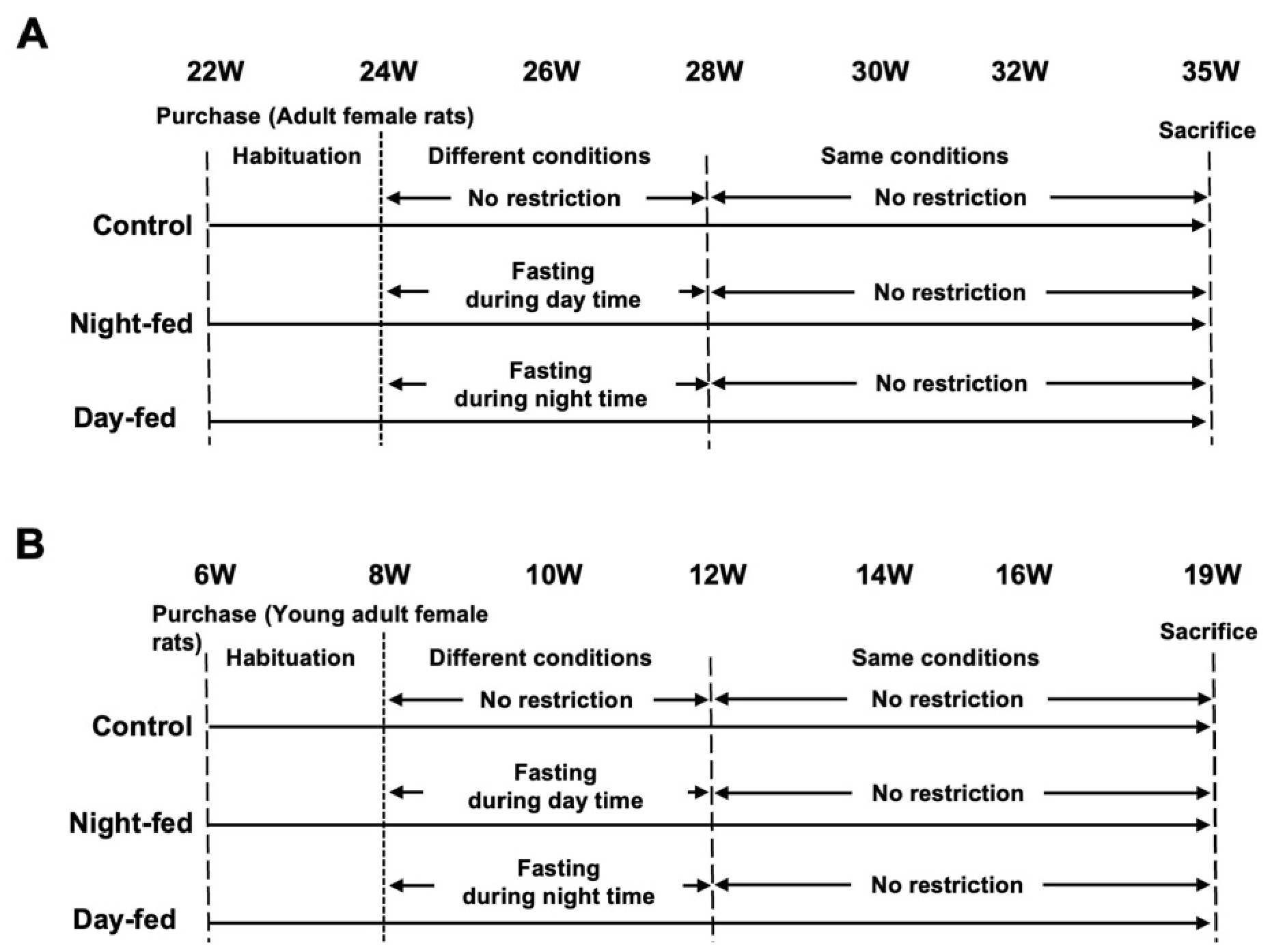
The light–dark cycle-converted feeding restriction was performed as previously described [13]. After pre-breeding for 2 weeks, healthy 8-week-aged young adult female (n = 30) and 24-week-aged adult female rats (n = 24) were assigned into 3 groups by randomly selecting housing apparatus: (1) the control group that was fed without time restriction (n = 8–10, free access to a standard caloric diet), (2) the night-time-fed group that was fed only night-time (19:00–7:00, fasting during non-active phase to standard caloric diet, n = 8–10), and (3) the daytime-fed group that was fed only during the daytime (7:00–19:00, fasting during active phase to standard caloric diet, n = 8–10) for 4 weeks. After the feeding restriction, all groups were additionally fed ad libitum for 7 weeks (Figure 1A,B). The rats were housed individually and separately in a restriction apparatus, and the experiment was repeated 8 times in adult female rats and 10 times in young adult female rats, with one rat from each of the three groups (total of three rats) as one unit. During the experimental period, the body weight and amount of food ingested by each rat were measured daily using a scale balance (GF-6100, A&D Company, Limited, Tokyo, Japan). The health status of the rats was also checked daily and the rats showing obvious abnormalities in activity or obvious facial expressions were excluded [27,28].

2.3. Tissue Preparation

After an unrestricted period of 7 weeks, the rats were sacrificed by exsanguination under deep inhalation anesthesia using isoflurane (FUJIFILM Wako Pure Chemical Corporation, Osaka, Japan). After sacrifice, the hypothalamus, peritoneal and perirenal white fatty tissues, and ovary were removed. Total weights of the peritoneal and perirenal white fatty tissues and ovary were measured. The hypothalamus and peritoneal white fatty tissues were stored in RNAlater solution (Thermo Fisher Scientific, Waltham, MA, USA) at −30 °C and further subjected to RNA analysis.

2.4. RNA Extraction and Real-Time PCR

As food ingestion stimulating factors, the mRNA expressions of NPY [29] and AgRP [30], and a modulating factor, orexin-A [31] in the hypothalamus were examined by real-time PCR. On the other hand, as food ingestion suppressing factors, the mRNA expressions of TRH [32] and POMC [33,34] in the hypothalamus and leptin [35] in the white fatty tissues were explored.
Total RNA was isolated from hypothalamus and white fatty tissues using the acid guanidinium thiocyanate procedure. The extracted RNA was reverse-transcribed into cDNA using the ReverTraAce®qPCR RTkit (Toyobo, Tokyo, Japan) in accordance with the manufacturer’s instructions. After converting total RNA (1 μg) to cDNA, real-time PCR was performed using SYBR green real-time PCR Master Mix-Plus (Toyobo, Japan) and the CronoSTARTM 96 Real-Time PCR system (Clontech, Kusatsu, Japan) under blinded managements. The expression levels of each mRNA were normalized to those of 18SrRNA mRNA. The PCR primers used in this study are listed in Table 1. Each PCR product was purified with a MinElute Gel Extraction Kit (Qiagen, Valencia, CA, USA) and the purified PCR products were sequenced to confirm their identity.

2.5. Statistical Analysis

One rat in the control group of young adult female rats was excluded by health status. All rats that reached the experimental endpoint were included in the analysis. The sample size was predetermined based on our previous report [13] and was further evaluated using a statistical power analysis program soft (G*Power 3.1), showing that n = 8 per group was sufficient to detect a significant treatment effect on the body weight gain and amount of food intake with 90% power and α = 0.05. After the data were checked for normal distribution using the Shapiro–Wilk test, the differences in the amount of food intake, body weight gain, organ weight, and differences in intensity of mRNA expressions among the 3 groups were analyzed by one-way ANOVA followed by Dunnett’s test using IBM SPSS Statistics for Windows, V. 29 (IBM Corp., Armonk, NY, USA). The data are shown as the mean ± standard error (SE) and values exceeding 3 SD were excluded from the analysis. A p-value of < 0.05 was considered significant.

3. Results

3.1. Changes in Body Weight and Food Intake in Adult Female Rats

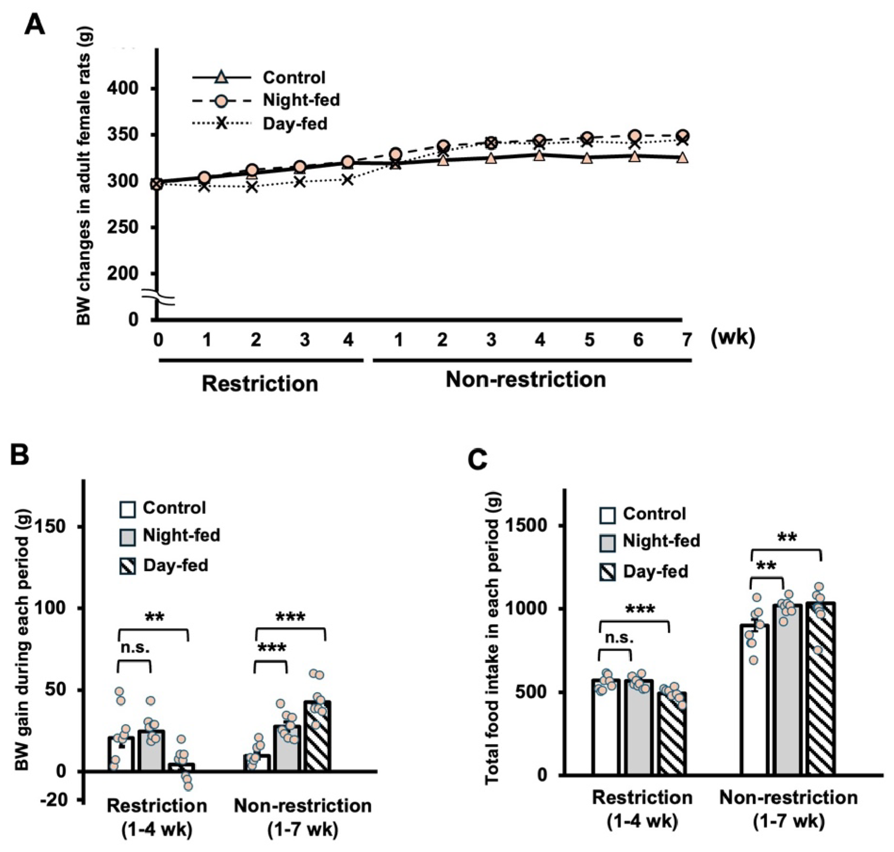
The experimental schedule of adult female rats is shown in Figure 1A. The total weekly changes in body weight in the three groups during food-restricted and subsequent non-restricted periods are plotted in Figure 2A. During the food-restricted period, the mean body weight was lower than that of the control group in the daytime-fed group, while the mean body weights in both daytime- and night-time-fed groups became higher than the control group after the restriction was lifted.
The mean of body weight gains in the daytime-fed group was significantly reduced during the restriction period, whereas those of the daytime-fed and night-time-fed groups were significantly elevated than that of the control group during the non-restricted periods (Figure 2B). Consistent with the above results, total food intake was significantly reduced during the restriction period in the daytime-fed group, whereas those of the daytime-fed and night-time-fed groups were significantly elevated than that of the control group during the non-restricted periods (Figure 2C).

3.2. Changes in Appetite-Related Gene Expressions and Organ Weights in Adult Female Rats

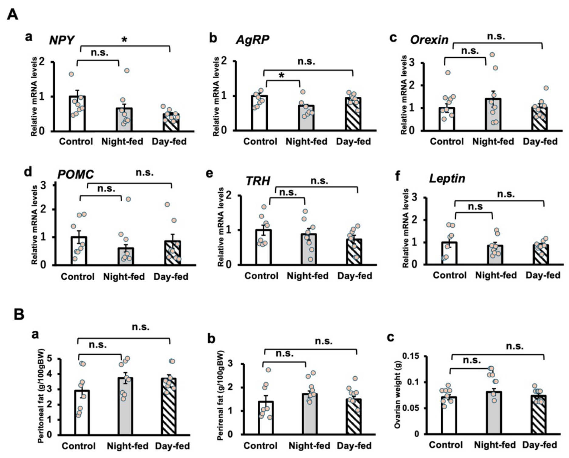
In adult female rats, NPY expression in the daytime-fed group and AgRP expression in the night-time-fed group were significantly reduced as compared with the control group (Figure 3A). There were no significant differences in the total weights of the peritoneal and perirenal fatty tissues and the ovary among the three groups (Figure 3B).

3.3. Changes in Body Weight and Food Intake in Young Adult Female Rats

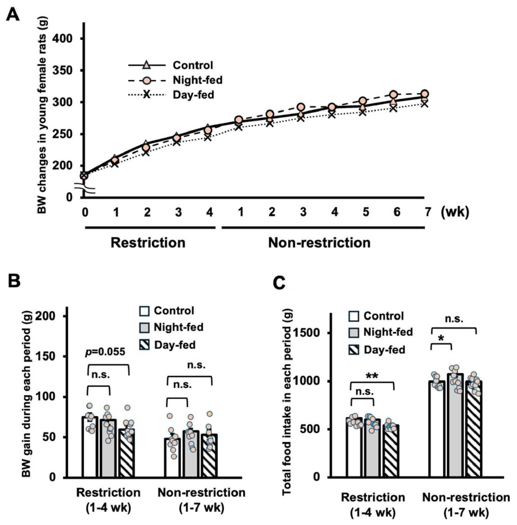
The experimental schedule of young adult female rats is shown in Figure 1B. The total weekly changes in body weight in the three groups during the food-restricted and subsequent non-restricted periods are plotted in Figure 4A. Similarly to adult female rats, the mean daily body weight in the daytime-fed group was lower than that of the control group during the food-restricted period. However, in contrast to adult female rats, its level remained under that of the control group after the restriction was lifted.
The mean of body weight gains in the daytime-fed group was reduced, but not significant during the restriction period (Figure 4B). On the other hand, there are no significant differences among the three groups in body weight gains per period after the restriction was lifted (Figure 4B). Consistent with the above results, total food intake was significantly reduced during the restriction period in the daytime-fed group, whereas there was no significant difference in total food intake between the daytime-fed and control groups during the non-restricted period (Figure 4C). In contrast, total food intake during the non-restricted period were significantly elevated in the night-time-fed group (Figure 4C).

3.4. Changes in Appetite-Related Gene Expressions and Organ Weights in Young Adult Female Rats

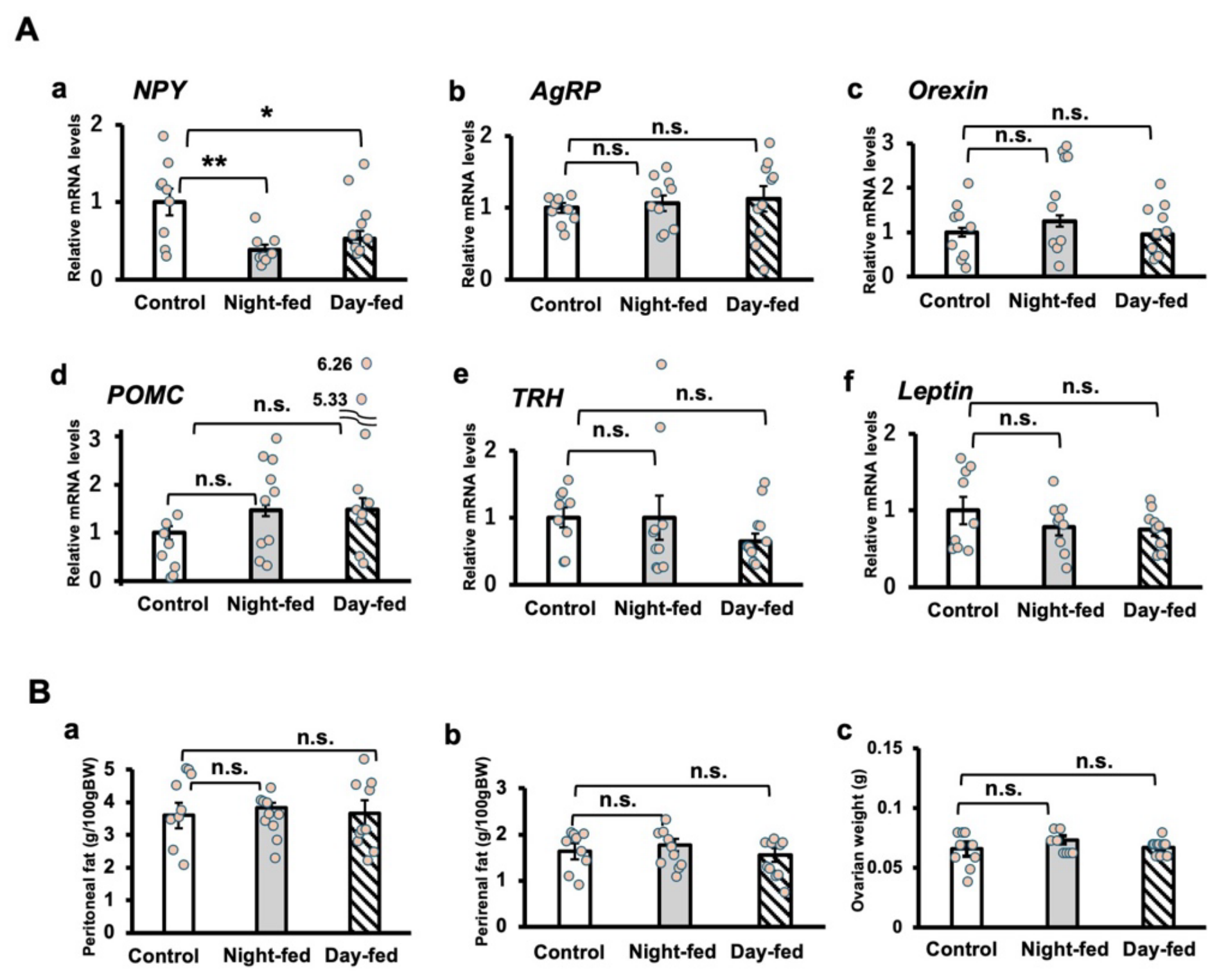
In young adult female rats, NPY expression in both night-time- and daytime-fed groups were significantly reduced as compared with the control group (Figure 5A). On the other hand, no significant differences were observed in the mRNA expressions of AgRP, orexin-A, POMC, TRH, and leptin (Figure 5A) among the groups. There were no significant differences in total weights of the peritoneal and perirenal fatty tissues and the ovary among the three groups (Figure 5B).

4. Discussion

In adult female rats, the hormonal and neural networks that regulate dietary behavior and reproductive rhythm have already been established. Therefore, it is presumed that they appropriately responded to stressful changes in the food supply environment and recovered their physical condition when the stress had been removed since they had to prepare for the possible recurrence of similarly difficult food supply conditions. In this study, the chronic stress of reversed day–night feeding suppressed food intake and weight gain, but when food intake was allowed to resume ad libitum, adult female rats increased both food intake and weight gain and recovered them beyond the control group, indicating that they were responding to the environmental changes. However, contrary to our expectations, even with ad libitum feeding, the hypothalamic expressions of NPY in the daytime-fed group and AgRP in the night-time-fed group, which are involved in stimulation of food ingestion [29,36], were lower than those of the control group. This suggests that mechanisms other than the NPY/AgRP system were mainly involved in the rebounding increase in food intake during the recovery period [37]. On the other hand, although the feedback system through the hypothalamus–adipose axis played an important role in regulating food ingestion, there was no significant change in adipose leptin expression.
In young adult female rats, food intake and weight gain are suppressed by light–dark cycle-reversed feeding. However, in contrast to adult female rats, there is no increase in food intake and weight gain during the following non-restricted period, showing the absence of a rebound response. Consequently, the chronic stress of reversed day–night feeding in this study caused no significant difference in body weights, food intake, and fatty tissue weights after stress relief. This may represent the possibility that young adult female rats reset their response in food intake to adjust them to a long-term severe food environment. At the end of the non-restricted period, the NPY expression in the hypothalamus was significantly decreased. Since food ingestion is promoted by the NPY/AgRP pathway, NPY may be involved in the above adaptation, suggesting certain changes in the hypothalamic network. In this regard, this finding may warn of a potential risk that the light–dark cycle-reversed feeding stress during the post-adolescent period has a persistent negative impact on hypothalamic functions, which are reorganized during puberty. If these changes are memorized, they may affect dietary behavior in adulthood.
As described above, this study using female rats showed that the recovery response to the chronic stress of reversed day–night feeding differed between young adulthood after puberty and mature adulthood. The model of reversed day–night feeding has two characteristics: one is eating during the inactive period, and the other is being exposed to hunger stress during the active period. The inactive phase is daytime in rodents, whereas it is night-time in humans. Therefore, feeding during the inactive period may correspond to night eating syndrome (NES), which is currently a problem in humans. NES is a disorder characterized by eating at bedtime or late at night [38]. NES patients were reported to have more sleep disorders and a higher body mass index (BMI) [39]. However, a recent review paper pointed out that the association between NES and a high BMI had not been consistent. In students, it was reported that there was no difference in BMI among students [40]. In addition, a more recent study has shown that among female students with a normal BMI, the NES group actually has a lower BMI [41]. These findings suggest that the impact of NES on BMI differs between young adults and mature adults. While the cause of this discrepancy remains unknown, it is consistent with the results of this study using rats. In 2014, it was already reported that the relationship between NES and BMI may differ with age; specifically, there is a positive correlation between NES and BMI in those aged 31–60, but no correlation in those under 30. The authors explained the age-related differences by suggesting that it took time for NES to manifest as BMI abnormalities [42]. In contrast, the present study suggests that it is caused by the differences in rebound response in food intake to light–dark reversal stress.
On the other hand, being exposed to hunger stress during the active period is similar to the issue of skipping breakfast, which is also a current problem in humans. NES was reported to be associated with skipping breakfast [43,44]. As evidence of the importance of skipping breakfast during the female post-adolescent period, we found that skipping breakfast worsened menstrual pain in female college students [45]. Afterwards, a positive correlation between menstrual pain and skipping breakfast in young women was reported worldwide [46,47,48,49,50]. At the same time, we also found that excessive dieting led to the development of dysmenorrhea and other menstrual disorders later in life [51,52], and that patients with a history of dysmenorrhea around the age of 20 years had a significantly higher risk of developing hypertensive disorders of pregnancy [53], suggesting that adverse dietary behavior around adolescence may induce obstetric and gynecological diseases in adulthood. By animal studies, we demonstrated that the discrepancy of the daily rhythms between food intake and the active phase inhibits peripheral clock functions in the uterus and that uterine clock suppression causes placental dysfunctions during pregnancy, leading to fetal loss [12,54,55,56]. Later, consistent with our above findings, based on large-scale epidemiological cohort studies, other groups reported that mothers who skipped breakfast from before conception through early pregnancy showed lower birth weight in newborns [57], a higher incidence of hypertensive disorders of pregnancy [58], and delayed early development of the infant after birth [59]. Based on these findings, we currently hypothesize that adverse dietary behavior around adolescence is a risk factor for gynecological diseases, proposing to name it adolescent dietary-habit-induced obstetric and gynecologic disease (ADHOGD) [60].
In recent years, the theory of developmental origins of health and disease (DOHaD) has attracted attention [61]. This concept, also known as the “fetal origins of adult disease,” proposes that exposure to specific environmental conditions, such as nutritional deficiencies, during development and growth periods determines the onset of various chronic metabolic diseases in adulthood, even though weight and other factors rapidly catch up once the environment improves [62,63]. The following studies identified “prenatal and neonatal periods” as critical stages when predictive adaptation could be established [64]. Therefore, “underweight in young women” is considered to be important issue that is directly related to low weight gain in pregnant women. On the other hand, in contrast to the DOHaD theory, the results of this study suggest that female adolescence and post-adolescent periods are other candidates of the critical phase when basic behaviors that can adapt to the food environment are determined. During adolescence, the hypothalamic neural network must incorporate a new system that regulates reproductive functions [65] and adapt to environmental changes in food supply because the body significantly grows into a reproductively active state, which can conceive next generations. These networks are presumed to mature during young adulthood. Therefore, as a human model of NES, this study suggests that the light–dark cycle-reversed food intake during adolescent and post-adolescent periods may induce suppressive changes in hypothalamic diet-regulating neural networks, which will potentially lead to low weight gain in young females and an increased risk of DOHaD. On the other hand, as a model of breakfast skipping, it may induce uterine dysfunction, leading to perinatal disorders (ADHOGD), which will also increase the risk of DOHaD. Accordingly, guidance on female dietary habits from adolescence to young adulthood should be provided with careful consideration of the potential for adaptation to the food supply environment during this period and the subsequent future risks of female disorders.
There are several limitations in this study. First, there is no information on the gene expression profiles immediately after stress relief. To understand the precise effects of time-restricted feeding stress during adolescence and post-adolescent periods, the longer-term effects of feeding stresses and the longitudinal changes in gene expressions after stress reduction should be further analyzed. Second, this study lacks direct evidence to support hypothalamic neural remodeling. To assess the precise changes in reorganization of diet-regulating neural networks, the profiles of local peptide or gene expression in the neural nucleus within the hypothalamus should be investigated using immunohistochemical or in situ hybridization studies. Third, this study lacks precise information on hormonal and physiological stress markers that can evaluate the potential influence of the estrus cycle and the status of stress. To clarify the biological significance of feeding stresses during young adult period on the following female health, the long-term relationship between post-adolescent dietary behaviors and reproductive functions should be investigated.

5. Conclusions

In conclusion, this study straightforwardly showed that the chronic light–dark cycle-reversed feeding stress during the post-adolescent period induced no significant rebound response in young adult female rats, indicating that the age-specific differences in feeding restriction-derived rebound responses. This issue should be further investigated to understand the novel mechanisms to explain the development of underweight status in young women.

Author Contributions

T.F., H.F. and R.N. conceived the study and study design; K.K. and R.N. performed the experiments and data analysis; T.F. and H.F. wrote the paper; T.F., M.O., T.D., H.A., H.F. and R.N. discussed the paper; T.F. contributed to funding acquisition; T.F. was the originator of the concept of this report. All authors have read and agreed to the published version of the manuscript.

Funding

This work was supported in part by Grants-in-Aid for Scientific Research (nos. 19H01617, 21K18297, and 24K00362).

Institutional Review Board Statement

All experimental procedures and housing conditions were approved by Nara Women’s University Animal Care Committee (approval No. 22-05, 1 April 2022) and all animals were treated in accordance with the Institutional Guidelines for Experiments Using Animals.

Informed Consent Statement

Not applicable.

Data Availability Statement

Data: All the detailed data in this paper are available from the lead contact; code: this paper does not use original code; other items: any additional information required to reanalyze the data reported in this paper is available from the lead contact upon request.

Acknowledgments

We thank Chiho Kasai and Shu Mizutani for their technical support.

Conflicts of Interest

The authors report no conflicts of interest.

Abbreviations

AgRPagouti-related protein
ADHOGDadolescent dietary habit-induced obstetric and gynecologic disease
BMIbody mass index
NESnight eating syndrome
NPYneuropeptide Y
POMCpro-opiomelanocortin
SDstandard deviation
TRHthyrotropin-releasing hormone

References

  1. Ogawa, M.; Nakazato, M.; Yokota, J.; Koga, K. Knowledge of the risks associated with being underweight and body shape differences among young Japanese women: A cross-sectional study. Biopsychosoc. Med. 2025, 19, 17. [Google Scholar] [CrossRef]
  2. Sicilia, A.; Fuller-Tyszkiewicz, M.; Rodgers, R.F.; Granero-Gallegos, A.; Lo Coco, G.; Dion, J.; McCabe, M.; Strodl, E.; Markey, C.H.; Aime, A.; et al. Cross-Country Measurement Invariance and Effects of Sociodemographic Factors on Body Weight and Shape Concern-Related Constructs in Eight Countries. Body Image 2020, 35, 288–299. [Google Scholar] [CrossRef]
  3. Chahal, N.; Qureshi, T.; Eljamri, S.; Catov, J.M.; Fazeli, P.K. Impact of Low Maternal Weight on Pregnancy and Neonatal Outcomes. J. Endocr. Soc. 2024, 9, bvae206. [Google Scholar] [CrossRef] [PubMed]
  4. Power, M.L.; Lott, M.L.; Mackeen, A.D.; DiBari, J.; Schulkin, J. A retrospective study of gestational weight gain in relation to the Institute of Medicine’s recommendations by maternal body mass index in rural Pennsylvania from 2006 to 2015. BMC Pregnancy Childbirth 2018, 18, 239. [Google Scholar] [CrossRef] [PubMed]
  5. Young, M.F.; Ramakrishnan, U. Maternal Undernutrition before and during Pregnancy and Offspring Health and Development. Ann. Nutr. Metab. 2021, 76, 41–53. [Google Scholar] [CrossRef] [PubMed]
  6. Yasuda, T. Desire for thinness among young Japanese women from the perspective of objective and subjective ideal body shape. Sci. Rep. 2023, 13, 14129. [Google Scholar] [CrossRef] [PubMed]
  7. Dou, G.; Wu, J.; Sun, S. Validation of two body scales for assessment of thin-ideal and muscularity-ideal body dissatisfaction among Chinese adult females. BMC Public Health 2025, 25, 1465. [Google Scholar] [CrossRef] [PubMed]
  8. Mori, N.; Asakura, K.; Sasaki, S. Differential dietary habits among 570 young underweight Japanese women with and without a desire for thinness: A comparison with normal weight counterparts. Asia Pac. J. Clin. Nutr. 2016, 25, 97–107. [Google Scholar] [CrossRef] [PubMed]
  9. Foster, R.G.; Roenneberg, T. Human responses to the geophysical daily, annual and lunar cycles. Curr. Biol. 2008, 18, R784–R794. [Google Scholar] [CrossRef] [PubMed]
  10. Melendez-Fernandez, O.H.; Liu, J.A.; Nelson, R.J. Circadian Rhythms Disrupted by Light at Night and Mistimed Food Intake Alter Hormonal Rhythms and Metabolism. Int. J. Mol. Sci. 2023, 24, 3392. [Google Scholar] [CrossRef] [PubMed]
  11. Ribas-Latre, A.; Fernandez-Veledo, S.; Vendrell, J. Time-restricted eating, the clock ticking behind the scenes. Front. Pharmacol. 2024, 15, 1428601. [Google Scholar] [CrossRef] [PubMed]
  12. Ono, M.; Dai, Y.; Fujiwara, T.; Fujiwara, H.; Daikoku, T.; Ando, H.; Kuji, N.; Nishi, H. Influence of lifestyle and the circadian clock on reproduction. Reprod. Med. Biol. 2025, 24, e12641. [Google Scholar] [CrossRef] [PubMed]
  13. Fujiwara, T.; Nakata, R.; Ono, M.; Mieda, M.; Ando, H.; Daikoku, T.; Fujiwara, H. Time Restriction of Food Intake During the Circadian Cycle Is a Possible Regulator of Reproductive Function in Postadolescent Female Rats. Curr. Dev. Nutr. 2019, 3, nzy093. [Google Scholar] [CrossRef] [PubMed]
  14. Wang, L.; Suyama, S.; Lee, S.A.; Ueta, Y.; Seino, Y.; Sharp, G.W.G.; Yada, T. Fasting inhibits excitatory synaptic input on paraventricular oxytocin neurons via neuropeptide Y and Y1 receptor, inducing rebound hyperphagia, and weight gain. Front. Nutr. 2022, 9, 994827. [Google Scholar] [CrossRef] [PubMed]
  15. Jeromson, S.; Akcan, M.; Baranowski, B.; Arbeau, M.; Bellucci, A.; Wright, D.C. Daily GDF15 treatment has sex-specific effects on body weight and food intake and does not enhance the effects of voluntary physical activity in mice. J. Physiol. 2024, 602, 6813–6826. [Google Scholar] [CrossRef] [PubMed]
  16. Reichenbach, A.; Stark, R.; Mequinion, M.; Lockie, S.H.; Lemus, M.B.; Mynatt, R.L.; Luquet, S.; Andrews, Z.B. Carnitine acetyltransferase (Crat) in hunger-sensing AgRP neurons permits adaptation to calorie restriction. FASEB J. 2018, 32, fj201800634R. [Google Scholar] [CrossRef] [PubMed]
  17. Chen, V.P.; Gao, Y.; Geng, L.; Brimijoin, S. Butyrylcholinesterase gene transfer in obese mice prevents postdieting body weight rebound by suppressing ghrelin signaling. Proc. Natl. Acad. Sci. USA 2017, 114, 10960–10965. [Google Scholar] [CrossRef] [PubMed]
  18. Morton, G.J.; Cummings, D.E.; Baskin, D.G.; Barsh, G.S.; Schwartz, M.W. Central nervous system control of food intake and body weight. Nature 2006, 443, 289–295. [Google Scholar] [CrossRef] [PubMed]
  19. Best, O.; Ban, S. Adolescence: Physical changes and neurological development. Br. J. Nurs. 2021, 30, 272–275. [Google Scholar] [CrossRef] [PubMed]
  20. Bello, N.T.; Yeh, C.Y.; James, M.H. Reduced Sensory-Evoked Locus Coeruleus-Norepinephrine Neural Activity in Female Rats With a History of Dietary-Induced Binge Eating. Front. Psychol. 2019, 10, 1966. [Google Scholar] [CrossRef] [PubMed]
  21. Yamamoto, K.; Kato, N.; Isogai, Y.; Kuroda, T.; Ishida, T.; Yamatodani, A. Induction and antagonism of pica induced by teriparatide in rats. Eur. J. Pharmacol. 2015, 764, 457–462. [Google Scholar] [CrossRef] [PubMed]
  22. Oyelowo, O.T.; Taire, E.O.; Ajao, O.I. Skipping the first active meal appears to adversely alter reproductive function in female than male rats. Curr. Res. Physiol. 2022, 5, 414–420. [Google Scholar] [CrossRef] [PubMed]
  23. Torrens, A.; Roy, P.; Lin, L.; Vu, C.; Grimes, D.; Inshishian, V.C.; Montesinos, J.S.; Ahmed, F.; Mahler, S.V.; Huestis, M.A.; et al. Comparative Pharmacokinetics of Delta(9)-Tetrahydrocannabinol in Adolescent and Adult Male and Female Rats. Cannabis Cannabinoid Res. 2022, 7, 814–826. [Google Scholar] [CrossRef] [PubMed]
  24. Oku, Y.; Noda, S.; Yamada, A.; Nakaoka, K.; Goseki-Sone, M. Twenty-eight days of vitamin D restriction and/or a high-fat diet influenced bone mineral density and body composition in young adult female rats. Ann. Anat. 2022, 243, 151945. [Google Scholar] [CrossRef] [PubMed]
  25. Ruvira, S.; Rodriguez-Rodriguez, P.; Canas, S.; Ramiro-Cortijo, D.; Aguilera, Y.; Munoz-Valverde, D.; Arribas, S.M. Evaluation of Parameters Which Influence Voluntary Ingestion of Supplements in Rats. Animals 2023, 13, 1827. [Google Scholar] [CrossRef] [PubMed]
  26. Ansari, A.; Walton, S.L.; Denton, K.M. Sex- and age-related differences in renal and cardiac injury and senescence in stroke-prone spontaneously hypertensive rats. Biol. Sex Differ. 2023, 14, 33. [Google Scholar] [CrossRef] [PubMed]
  27. Langford, D.J.; Bailey, A.L.; Chanda, M.L.; Clarke, S.E.; Drummond, T.E.; Echols, S.; Glick, S.; Ingrao, J.; Klassen-Ross, T.; Lacroix-Fralish, M.L.; et al. Coding of facial expressions of pain in the laboratory mouse. Nat. Methods 2010, 7, 447–449. [Google Scholar] [CrossRef] [PubMed]
  28. Sotocinal, S.G.; Sorge, R.E.; Zaloum, A.; Tuttle, A.H.; Martin, L.J.; Wieskopf, J.S.; Mapplebeck, J.C.; Wei, P.; Zhan, S.; Zhang, S.; et al. The Rat Grimace Scale: A partially automated method for quantifying pain in the laboratory rat via facial expressions. Mol. Pain 2011, 7, 55. [Google Scholar] [CrossRef] [PubMed]
  29. Mercer, R.E.; Chee, M.J.; Colmers, W.F. The role of NPY in hypothalamic mediated food intake. Front. Neuroendocrinol. 2011, 32, 398–415. [Google Scholar] [CrossRef] [PubMed]
  30. Sternson, S.M.; Atasoy, D. Agouti-related protein neuron circuits that regulate appetite. Neuroendocrinology 2014, 100, 95–102. [Google Scholar] [CrossRef] [PubMed]
  31. Rilling, F.L.; Reyes, M.; Blanco, E.; Burrows, R.; Peirano, P.; Algarin, C.; Merono, T.; Gahagan, S. Association of fasting Orexin-A levels with energy intake at breakfast and subsequent snack in Chilean adolescents. Psychoneuroendocrinology 2022, 140, 105718. [Google Scholar] [CrossRef] [PubMed]
  32. Zhang, X.; van den Pol, A.N. Thyrotropin-releasing hormone (TRH) inhibits melanin-concentrating hormone neurons: Implications for TRH-mediated anorexic and arousal actions. J. Neurosci. 2012, 32, 3032–3043. [Google Scholar] [CrossRef] [PubMed]
  33. Harno, E.; Gali Ramamoorthy, T.; Coll, A.P.; White, A. POMC: The Physiological Power of Hormone Processing. Physiol. Rev. 2018, 98, 2381–2430. [Google Scholar] [CrossRef] [PubMed]
  34. Minere, M.; Wilhelms, H.; Kuzmanovic, B.; Lundh, S.; Fusca, D.; Classen, A.; Shtiglitz, S.; Prilutski, Y.; Talpir, I.; Tian, L.; et al. Thalamic opioids from POMC satiety neurons switch on sugar appetite. Science 2025, 387, 750–758. [Google Scholar] [CrossRef] [PubMed]
  35. Tan, H.L.; Yin, L.; Tan, Y.; Ivanov, J.; Plucinska, K.; Ilanges, A.; Herb, B.R.; Wang, P.; Kosse, C.; Cohen, P.; et al. Leptin-activated hypothalamic BNC2 neurons acutely suppress food intake. Nature 2024, 636, 198–205. [Google Scholar] [CrossRef] [PubMed]
  36. Pratt, W.E.; Do, C.; Groome, A.M.; Smith, A.J.; Siegfried, A.C.; Calafiore, C.J. Homeostatic-related peptides injected into the rat nucleus accumbens alter palatable eating and impact the binge-like intake of a sweetened fat diet during simultaneous mu-opioid receptor stimulation. Front. Neurosci. 2025, 19, 1614819. [Google Scholar] [CrossRef] [PubMed]
  37. Hillebrand, J.J.; de Wied, D.; Adan, R.A. Neuropeptides, food intake and body weight regulation: A hypothalamic focus. Peptides 2002, 23, 2283–2306. [Google Scholar] [CrossRef] [PubMed]
  38. Sakthivel, S.J.; Hay, P.; Mannan, H. A Scoping Review on the Association between Night Eating Syndrome and Physical Health, Health-Related Quality of Life, Sleep and Weight Status in Adults. Nutrients 2023, 15, 2791. [Google Scholar] [CrossRef] [PubMed]
  39. Sakthivel, S.J.; Hay, P.; Touyz, S.; Currow, D.; Mannan, H. Association of participants who screened positive for night eating syndrome with physical health, sleep problems, and weight status in an Australian adult population. Eat. Weight. Disord.-Stud. Anorex. Bulim. Obes. 2023, 28, 77. [Google Scholar] [CrossRef] [PubMed]
  40. Yahia, N.; Brown, C.; Potter, S.; Szymanski, H.; Smith, K.; Pringle, L.; Herman, C.; Uribe, M.; Fu, Z.; Chung, M.; et al. Night eating syndrome and its association with weight status, physical activity, eating habits, smoking status, and sleep patterns among college students. Eat. Weight. Disord.-Stud. Anorex. Bulim. Obes. 2017, 22, 421–433. [Google Scholar] [CrossRef] [PubMed]
  41. Aslan, S.; Sozlu, S.; Bozkurt, O.; Camli, A.; Arslan-Tanis, M.; Kocaadam-Bozkurt, B. The interplay of night eating syndrome with resting metabolic rate, anthropometric measures, and sleep quality in normal-weight female university students: A case-control study. J. Eat. Disord. 2026, 14, 69. [Google Scholar] [CrossRef] [PubMed]
  42. Meule, A.; Allison, K.C.; Brahler, E.; de Zwaan, M. The association between night eating and body mass depends on age. Eat. Behav. 2014, 15, 683–685. [Google Scholar] [CrossRef] [PubMed]
  43. Hao, Z.; Guo, X.; Jing, Q.; Zhao, B.; Huang, M.; Ren, Z. Night-eating syndrome is associated with food consumption frequency among Chinese college students. Sci. Rep. 2026, 16, 5595. [Google Scholar] [CrossRef] [PubMed]
  44. Khan, M.S.I.; Paul, T.; Al Banna, M.H.; Hamiduzzaman, M.; Tengan, C.; Kissi-Abrokwah, B.; Tetteh, J.K.; Hossain, F.; Islam, M.S.; Brazendale, K. Skipping breakfast and its association with sociodemographic characteristics, night eating syndrome, and sleep quality among university students in Bangladesh. BMC Nutr. 2024, 10, 46. [Google Scholar] [CrossRef] [PubMed]
  45. Fujiwara, T. Skipping breakfast is associated with dysmenorrhea in young women in Japan. Int. J. Food Sci. Nutr. 2003, 54, 505–509. [Google Scholar] [CrossRef] [PubMed]
  46. Abu Helwa, H.A.; Mitaeb, A.A.; Al-Hamshri, S.; Sweileh, W.M. Prevalence of dysmenorrhea and predictors of its pain intensity among Palestinian female university students. BMC Womens Health 2018, 18, 18. [Google Scholar] [CrossRef] [PubMed]
  47. Hu, Z.; Tang, L.; Chen, L.; Kaminga, A.C.; Xu, H. Prevalence and Risk Factors Associated with Primary Dysmenorrhea among Chinese Female University Students: A Cross-sectional Study. J. Pediatr. Adolesc. Gynecol. 2020, 33, 15–22. [Google Scholar] [CrossRef] [PubMed]
  48. Wang, L.; Yan, Y.; Qiu, H.; Xu, D.; Zhu, J.; Liu, J.; Li, H. Prevalence and Risk Factors of Primary Dysmenorrhea in Students: A Meta-Analysis. Value Health 2022, 25, 1678–1684. [Google Scholar] [CrossRef] [PubMed]
  49. Mammo, M.; Alemayehu, M.; Ambaw, G. Prevalence of Primary Dysmenorrhea, Its Intensity and Associated Factors Among Female Students at High Schools of Wolaita Zone, Southern Ethiopia: Cross-Sectional Study Design. Int. J. Womens Health 2022, 14, 1569–1577. [Google Scholar] [CrossRef] [PubMed]
  50. Ghandour, R.; Hammoudeh, W.; Stigum, H.; Giacaman, R.; Fjeld, H.; Holmboe-Ottesen, G. Menstrual characteristics and dysmenorrhea among Palestinian adolescent refugee camp dwellers in the West Bank and Jordan: A cross-sectional study. Arch. Public Health 2023, 81, 47. [Google Scholar] [CrossRef] [PubMed]
  51. Fujiwara, T. Diet during adolescence is a trigger for subsequent development of dysmenorrhea in young women. Int. J. Food Sci. Nutr. 2007, 58, 437–444. [Google Scholar] [CrossRef] [PubMed]
  52. Fujiwara, T.; Ono, M.; Iizuka, T.; Sekizuka-Kagami, N.; Maida, Y.; Adachi, Y.; Fujiwara, H.; Yoshikawa, H. Breakfast Skipping in Female College Students Is a Potential and Preventable Predictor of Gynecologic Disorders at Health Service Centers. Diagnostics 2020, 10, 476. [Google Scholar] [CrossRef] [PubMed]
  53. Nakayama, M.; Ono, M.; Iizuka, T.; Kagami, K.; Fujiwara, T.; Sekizuka-Kagami, N.; Maida, Y.; Obata, T.; Yamazaki, R.; Daikoku, T.; et al. Hypertensive disorders of pregnancy are associated with dysmenorrhea in early adulthood: A cohort study. J. Obstet. Gynaecol. Res. 2020, 46, 2292–2297. [Google Scholar] [CrossRef] [PubMed]
  54. Hosono, T.; Ono, M.; Daikoku, T.; Mieda, M.; Nomura, S.; Kagami, K.; Iizuka, T.; Nakata, R.; Fujiwara, T.; Fujiwara, H.; et al. Time-Restricted Feeding Regulates Circadian Rhythm of Murine Uterine Clock. Curr. Dev. Nutr. 2021, 5, nzab064. [Google Scholar] [CrossRef] [PubMed]
  55. Nomura, S.; Hosono, T.; Ono, M.; Daikoku, T.; Michihiro, M.; Kagami, K.; Iizuka, T.; Chen, Y.; Shi, Y.; Morishige, J.I.; et al. Desynchronization between Food Intake and Light Stimulations Induces Uterine Clock Quiescence in Female Mice. J. Nutr. 2023, 153, 2283–2290. [Google Scholar] [CrossRef] [PubMed]
  56. Ono, M.; Toyoda, N.; Kagami, K.; Hosono, T.; Matsumoto, T.; Horike, S.I.; Yamazaki, R.; Nakamura, M.; Mizumoto, Y.; Fujiwara, T.; et al. Uterine Deletion of Bmal1 Impairs Placental Vascularization and Induces Intrauterine Fetal Death in Mice. Int. J. Mol. Sci. 2022, 23, 7637. [Google Scholar] [CrossRef] [PubMed]
  57. Aizawa, M.; Murakami, K.; Takahashi, I.; Onuma, T.; Noda, A.; Ueno, F.; Matsuzaki, F.; Ishikuro, M.; Obara, T.; Hamada, H.; et al. Association between frequency of breakfast intake before and during pregnancy and infant birth weight: The Tohoku Medical Megabank Project Birth and Three-Generation Cohort Study. BMC Pregnancy Childbirth 2023, 23, 268. [Google Scholar] [CrossRef] [PubMed]
  58. Aizawa, M.; Murakami, K.; Takahashi, I.; Onuma, T.; Noda, A.; Ueno, F.; Matsuzaki, F.; Ishikuro, M.; Obara, T.; Hamada, H.; et al. Skipping breakfast during pregnancy and hypertensive disorders of pregnancy in Japanese women: The Tohoku medical megabank project birth and three-generation cohort study. Nutr. J. 2022, 21, 71. [Google Scholar] [CrossRef] [PubMed]
  59. Aizawa, M.; Murakami, K.; Takahashi, I.; Ohseto, H.; Noda, A.; Shinoda, G.; Orui, M.; Ishikuro, M.; Obara, T.; Hamada, H.; et al. Association between frequency of breakfast intake before and during pregnancy and developmental delays in children: The Tohoku Medical Megabank Project Birth and Three-Generation Cohort Study. Nutr. J. 2023, 22, 66. [Google Scholar] [CrossRef] [PubMed]
  60. Fujiwara, T.; Ono, M.; Mieda, M.; Yoshikawa, H.; Nakata, R.; Daikoku, T.; Sekizuka-Kagami, N.; Maida, Y.; Ando, H.; Fujiwara, H. Adolescent Dietary Habit-induced Obstetric and Gynecologic Disease (ADHOGD) as a New Hypothesis-Possible Involvement of Clock System. Nutrients 2020, 12, 1294. [Google Scholar] [CrossRef] [PubMed]
  61. Itoh, H.; Ueda, M.; Suzuki, M.; Kohmura-Kobayashi, Y. Developmental Origins of Metaflammation; A Bridge to the Future Between the DOHaD Theory and Evolutionary Biology. Front. Endocrinol. 2022, 13, 839436. [Google Scholar] [CrossRef] [PubMed]
  62. Barker, D.J.; Winter, P.D.; Osmond, C.; Margetts, B.; Simmonds, S.J. Weight in infancy and death from ischaemic heart disease. Lancet 1989, 2, 577–580. [Google Scholar] [CrossRef] [PubMed]
  63. Godfrey, K.M.; Barker, D.J. Fetal nutrition and adult disease. Am. J. Clin. Nutr. 2000, 71, 1344S–1352S. [Google Scholar] [CrossRef] [PubMed]
  64. Simeoni, U.; Armengaud, J.B.; Siddeek, B.; Tolsa, J.F. Perinatal Origins of Adult Disease. Neonatology 2018, 113, 393–399. [Google Scholar] [CrossRef] [PubMed]
  65. Constantino, D.B.; Tonon, A.C.; de Oliveira, M.A.B.; Amando, G.R.; Freitas, J.J.; Xavier, N.B.; Ribeiro, R.J.; Idiart, M.; Hidalgo, M.P.L. Effects of lighting patterns in pubertal development and metabolism of female wistar rats. Physiol. Behav. 2022, 243, 113641. [Google Scholar] [CrossRef] [PubMed]
Figure 1. The experimental schedule of feeding restriction and longitudinal changes in daily body weight in adult and young adult female rats (A,B). The experimental schedule of adult and young adult female rats. After the acclimation period for 2 weeks, healthy 24-week-aged female rats (n = 24) (A) and 8-week-aged female rats (n = 30) (B) were assigned into 3 groups by randomly selecting housing apparatus: (1) the control group that was fed without time restriction (free access to a standard caloric diet), (2) the night-time-fed group that was fed only night-time (19:00–7:00, fasting during non-active phase to standard caloric diet), and (3) the daytime-fed group that was fed only during the daytime (7:00–19:00, fasting during active phase to standard caloric diet) for 4 weeks. After feeding restriction, all groups were additionally fed ad libitum for 7 weeks.
Dietetics 05 00038 g001
Figure 2. Changes in body weight and food intake in adult female rats. (A) Plots of the total changes in weekly body weight in three groups during food-restricted and subsequent non-restricted periods. (B) Body weight gain during restriction and non-restriction periods. (C) Total food intake during restriction and non-restriction periods. The differences were analyzed by one-way ANOVA followed by Dunnett’s test. The data are shown as the mean ± SE. n.s., not significant; ** p < 0.01, *** p < 0.001.
Dietetics 05 00038 g002
Figure 3. Changes in appetite-related gene expressions and organ weights in adult female rats. (A). Appetite-related gene expressions in adult female rats. (a) Npy, (b) Agrp, (c) Hcrt (Orexin-A), (d) Pomc, (e) Trh, (f) Lep. Expression levels of each mRNA were normalized to those of 18SrRNA mRNA. (B). Total weights of the peritoneal (a) and perirenal (b) fatty tissues and the ovary (c) removed from adult female rats. The differences were analyzed by one-way ANOVA followed by Dunnett’s test. The data are shown as the mean ± SE. n.s., not significant; * p < 0.05.
Dietetics 05 00038 g003
Figure 4. Changes in body weight and food intake in young adult female rats. (A). Plots of the total changes in weekly body weight in the three groups during food-restricted and subsequent non-restricted periods. (B). Body weight gain during restriction and non-restriction periods. (C). Total food intake during restriction and non-restriction periods. The differences were analyzed by one-way ANOVA followed by Dunnett’s test. The data are shown as the mean ± SE. n.s., not significant; * p < 0.05; ** p < 0.01.
Dietetics 05 00038 g004
Figure 5. Changes in appetite-related gene expressions and organ weights in young adult female rats. (A). Appetite-related gene expressions in young adult female rats. (a) Npy, (b) Agrp, (c) Hcrt (Orexin-A), (d) Pomc, (e) Trh, (f) Lep. Expression levels of each mRNA were normalized to those of 18SrRNA mRNA. (B). Total weights of the peritoneal (a) and perirenal (b) fatty tissues and the ovary (c) removed from young adult female rats. The differences were analyzed by one-way ANOVA followed by Dunnett’s test. The data are shown as the mean ± SE. n.s., not significant; * p < 0.05; ** p < 0.01.
Dietetics 05 00038 g005
Table 1. List of primers used in this study.
GeneAccession No.PrimerSequence (5′→3′)
NpyNC_051339ForwardTGTGGACTGACCCTCGCTCTAT
ReverseTGTAGTGTCGCAGAGCGGAGTA
AgrpNC_051354ForwardAGCTTTGGCAGAGGTGCTAGATC
ReverseTGCCAGTACCTAGCTTGCGG
PomcNM_139326.3ForwardGAGTTCAAGAGGGAGCTGGA
ReverseGGAAGTGCTCCACCCGATAG
Hcrt (Orexin-A)NM_013179.3ForwardCATCCTCACTCTGGGAAAG
ReverseAGGGATATGGCTCTAGCTC
TrhNM_013046.3ForwardGGACAAGTATTCATGGGC
ReverseCTCTTGGTGACATCAGAC
LepNM_013076ForwardCACCCCATTCTGAGTTTGTCC
ReverseCTCGCAGGTTCTCCAGGTC
18SrRNANW_023637849ForwardGGGTCGGGAGTGGGTAATTT
ReverseAAATTACCCACTCCCGACCC
Disclaimer/Publisher’s Note: The statements, opinions and data contained in all publications are solely those of the individual author(s) and contributor(s) and not of MDPI and/or the editor(s). MDPI and/or the editor(s) disclaim responsibility for any injury to people or property resulting from any ideas, methods, instructions or products referred to in the content.