Proceeding Paper

Density of SMI-32 Immunopositive Neurons in Eye-Specific Layers of Lateral Geniculate Nuclei in Kittens Reared with Monocular Deprivation and Unilateral Convergent Squint †

by
Polina Y. Shkorbatova
1,*,
Vsevolod A. Lyakhovetskii
2 and
Svetlana V. Alexeenko
3
1
Neuromorphology Laboratory, Pavlov Institute of Physiology RAS, 199034 St. Petersburg, Russia
2
Motor Physiology Laboratory, Pavlov Institute of Physiology RAS, 199034 St. Petersburg, Russia
3
Vision Physiology Laboratory, Pavlov Institute of Physiology RAS, 199034 St. Petersburg, Russia
*
Author to whom correspondence should be addressed.
Presented at the 2nd International Electronic Conference on Brain Sciences, 15–30 July 2021. Available online: https://sciforum.net/event/IECBS2021.
Med. Sci. Forum 2022, 8(1), 3; https://doi.org/10.3390/IECBS2021-10666
Published: 14 July 2021
(This article belongs to the Proceedings of The 2nd International Electronic Conference on Brain Sciences)

Abstract

:
To reveal the dynamics of the development of the morphological changes in lateral geniculate nuclei caused by binocular vision impairment, we study the changes in density of SMI-32 immunopositive neurons in the frontal sections of the LGNd of both hemispheres of 2- and 3-month-old kittens reared with monocular deprivation or unilateral convergent squint. We develop a custom software to divide the binocular part of the A-layers into 10 consecutive sectors and calculate the number of SMI-32 immunopositive neurons in each of them. The neuronal density was calculated and compared between groups in sectors with the same eccentricity. In monocularly deprived animals, a decline in the neuronal density relative to the control group was found in the layers innervated from the deprived eye in both age groups, regardless of eccentricity. However, in the strabismic kittens, the decrease in neuronal density was revealed only in the peripheral sectors of layer A1, driven by the deviated eye. The width of this area of reduced Y-neuron density was larger in the 3-month-old kittens, indicating that the development of the disorder has not yet stabilized at this age. The results may be interpreted as morpho-physiological correlates of different types of human amblyopia.

1. Introduction

Non-phosphorylated heavy neurofilament proteins, which can be labelled by SMI-32 antibodies, are characteristic for large, fast-conducting neurons with high-myelinated axones [1,2,3]. In the visual system, such properties are typical for Y-neurons—a population devoted to motion analysis [4,5,6]. Early physiological experiments have shown that the number of Y-neurons and their functional properties can be changed by altering the visual experience by monocular deprivation or squinting [7,8,9]. The loss of SMI-32-positive neurons in the A-layers of lateral geniculate nuclei (LGNd) in connection with a deprived/squinting eye has confirmed the morphological base of these changes [10,11,12,13]. However, whether this loss depends on the eccentricity of visual field projection is still unknown. To study this, as well as the time course of the developmental changes, we evaluate the density of SMI-32 immunopositive neurons in the LGNd of experimental models of monocular deprivation and unilateral convergent squint. Density measurements were carried out on 60-day-old kittens—the age of the onset of binocular vision [14,15]—and 90-day-old kittens, when the critical period for visual capacity ends [16] and the ratio of excitatory and inhibitory synapses in the LGN already corresponds to the level of adults [17].

2. Materials and Methods

The experimental procedures were approved by the Ethics Commission of the Pavlov Institute of Physiology, St. Petersburg, Russia (Protocol #30/03/2014), and were performed in accordance with the requirements of the European Community Council Directive (2010/63EU) on the protection of animals used in experimental and other scientific purposes.
The study was carried out on kittens who underwent surgery at postnatal day 7–10 (before natural eye opening). All surgeries were conducted under general anesthesia (Zoletil, 5 mg/kg and xylazine, 2 mg/kg, intramuscularly). The kittens of the MD groups were monocularly deprived by eyelid sutures: the margins of eyelids were trimmed and then lids were sutured together by closely spaced interrupted sutures [18]. In the kittens of the Strab groups, the skin and connective tissue incision was changed to a near lateral eye angle, and then the lateral rectus muscle [19] and the upper and lower lateral leaflets of the eye retractor were removed to produce a unilateral convergent squint; the wound was closed by interrupted sutures. The side of the surgery (right or left) was chosen randomly for each animal. The animals were left to grow for 60 or 90 days. Intact kittens of the same age were used as controls. The number of animals in each group is presented in Table 1.
At the end of this period, the animals were deeply anesthetized (Zoletil, 20 mg/kg and xylazine, 2 mg/kg, intramuscularly) and transcardially perfused with 4% paraformaldehyde. The frozen 50 μm thick frontal sections of the LGNd of both hemispheres were prepared and ones corresponding to AP 5.5–7.0 according to Horsley–Clark coordinates were then selected for histological processing and analysis. The SMI-32 primary antibodies were used to detect the heavy neurofilament protein with DAB-Ni as a chromogen.
After the images of the LGNd sections were acquired, the upper and lower boundaries of layers A and A1 and the location of the SMI-32 immunopositive neurons within these boundaries were marked manually (Figure 1A). To reveal changes in the density of the immunopositive neurons in layer A along the projection of the visual horizontal meridian, these layers were divided into 10 consecutive sectors (Figure 1B). This was performed using custom software which allowed us to divide the curves of the upper boundary of layer A and the lower boundary of layer A1 into segments of equal length.
LGNd layers A and A1 were innervated from different eyes (contralateral and ipsilateral, correspondingly). The differences between these layers in terms of the density of immunopositive neurons (the number of neurons per mm2) were calculated in kittens of all groups in all consecutive LGNd sectors using the formula of Michelson contrast:
D(i) = (NA(i) − NA1(i))/(NA(i) + NA1(i)),
where NA is the density of neurons in layer A; NA1 is the density of the neurons in layer A1; and i = 1 … 10 is the sector number.
To assess the strength of the deprivation effect in the hemispheres contralateral and ipsilateral to the deprived eye, we compared D-values between the normal and MD groups as:
Diff(i)MD = |D(i)MD − D(i)norm|
where i = 1 … 10 is the sector number.
Significant differences between the groups’ parameter values were determined at the p < 0.01 level using the method of hierarchical linear models [20]. Significant differences in distributions were determined at the level of p < 0.01 using the Kolmogorov–Smirnov test. Statistical data processing was carried out on the Matlab R2016b computing platform (Matworks Inc., Natick, MA, USA). All data are presented as mean ± standard deviation.

3. Results and Discussion

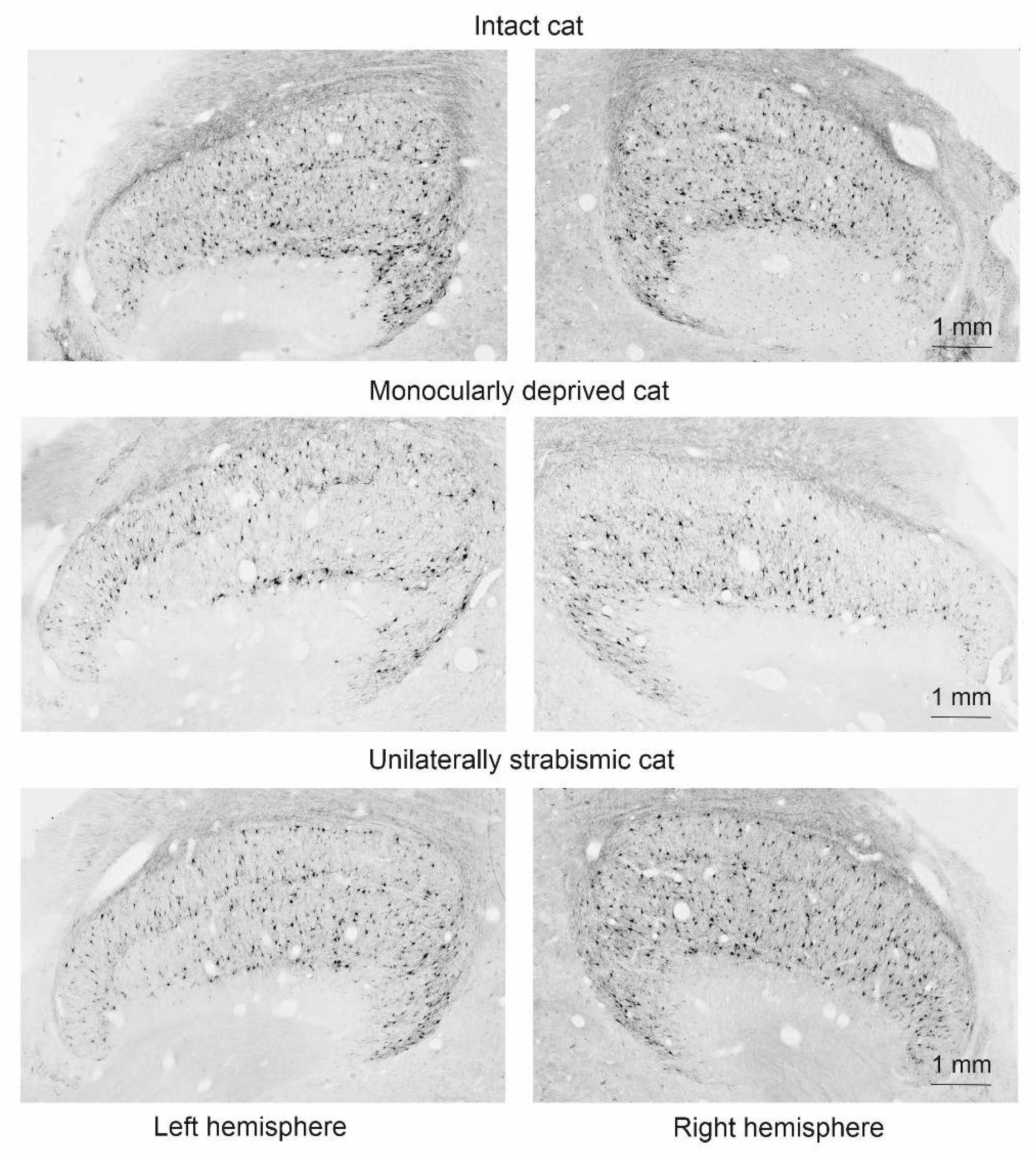
The examples of the histological sections of the LGN of both hemispheres of the intact cats, the cats deprived in the left eye, and the cats squinting in the left eye (aged 60 days) are shown in Figure 2. It is clearly seen that, in the intact cats, the SMI-32 immunopositive neurons were almost equally distributed in both the A and A1 layers. In the MD animals, they were almost absent in the layers innervated from the deprived eye: layer A of the right hemisphere and layer A1 of the left hemisphere. In the strabismic animals there were far fewer SMI-32 immunopositive neurons in layer A1, which receives input from the squinting eye, especially in the lateral (peripheral) part of this layer.
The results of the comparison of the D-values for all groups are presented in Figure 3.
In intact animals of both age groups (Norm-60, Norm-90) had a normalized density of SMI-32 immunopositive neurons in the A1 layer that exceeded that in the A layer in all sectors of the right and left hemispheres. Therefore, the relative differences in density (D) between layers A and A1, on average, had negative values. The distributions of the mean D-values did not differ across the sectors for the left and right hemispheres (Kolmogorov–Smirnov test, p > 0.01); thus, data from the right and left hemispheres of all animals of the same age were combined and used for comparison with the other groups.
In the MD-60 group, the D-values significantly differed from the D-values in the Norm-60 group in all 10 sectors in both hemispheres (p < 0.01). In the MD-90 group, D-values did not differ from the MD-60 group (p > 0.01). Note that the D-values were positive in the hemisphere ipsilateral to the deprived eye, and negative in the hemisphere contralateral to the deprived eye. This indicates a decrease in the density of SMI-32 immunopositive neurons in the layers innervated from the deprived eye in both hemispheres (in layer A of the hemisphere contralateral to the deprived eye and layer A1 of the hemisphere ipsilateral to the deprived eye).
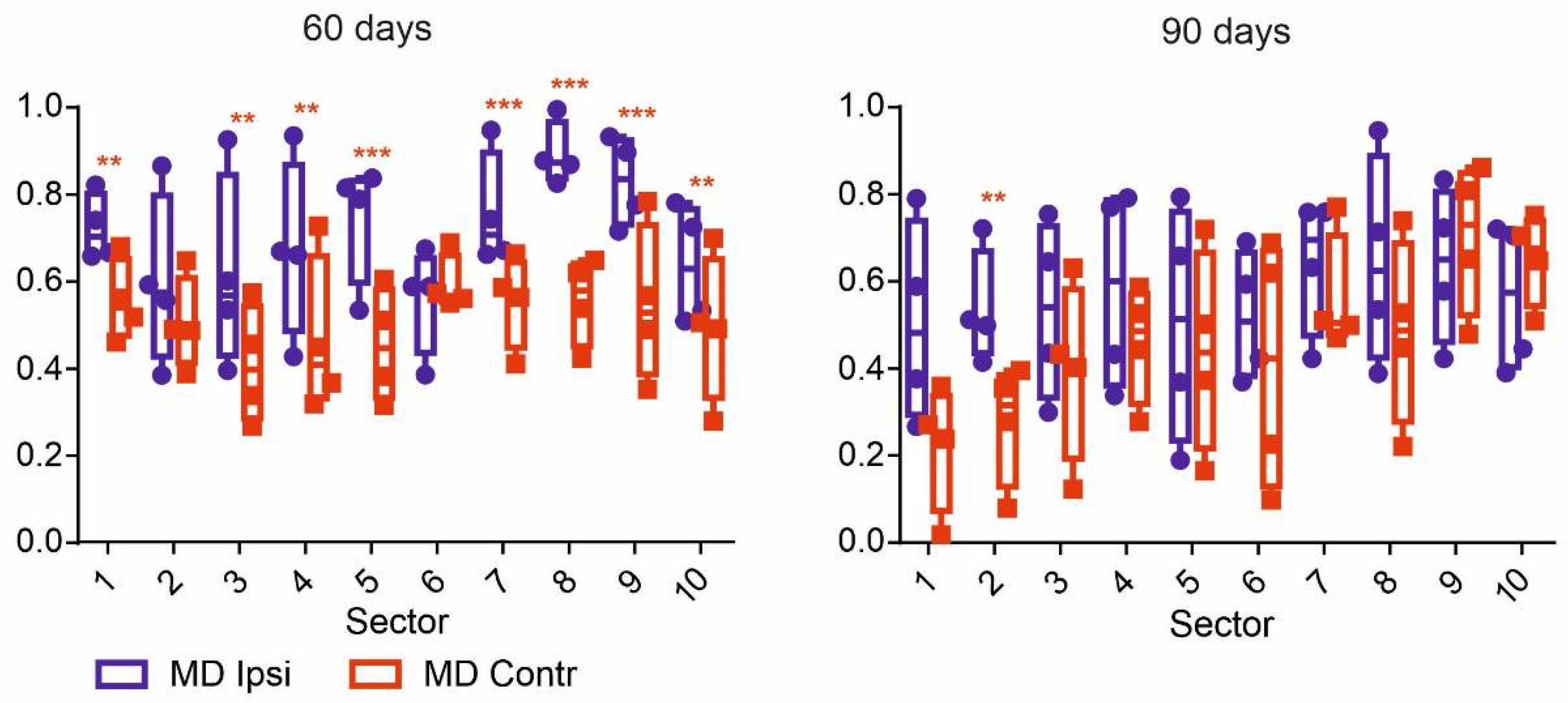
The strength of the deprivation effect in the hemispheres was different in the kittens of the MD-60 and MD-90 groups (Figure 4). At the age of 60 days (MD-60), the strength of the deprivation effect, DiffMD, was higher in the LGN ipsilateral to the deprived eye, and the nasal hemifield of the deprived eye was presented in the A1 layer. Hemispheric differences in this group of animals were found in 7/10 LGN sectors (namely, 1, 3–5, 7–9). However, at the age of 90 days (MD-90), such a difference was revealed in only one sector (number 2). This means that the increase in deprivation effect in the LGN was contralateral to the deprived eye where the temporal hemifield of the deprived eye was presented in the A layer. These data also indicate a delay in deprivation effect development in the temporal visual hemifield in relation to the nasal hemifield. This phenomenon has been described earlier for cortical and geniculate neurons [8,21] and is probably connected with the later development of ipsilateral projection pathways compared to contralateral ones [20,21].
In the kittens with the unilateral squint, the D-values did not differ from the normal D-values in either age group or in the sectors in the hemisphere contralateral to the deviated eye. However, in the hemisphere ipsilateral to the deviated eye, the D-values were increased in the peripheral part of the visual field projection. Moreover, such a statistically significant increase relative to the Norm-60 group was observed in sectors 8–10 in the Strab-60 group and in sectors 4–10 in the Strab-90 group. Hence, this decrease in cell density in the peripheral part of the A1 layer—driven by the squinted eye—is a physiological correlate of strabismic amblyopia and takes place in the area that receives input from the temporal retina and represents the projection of the nasal visual hemifield. This effect develops from the periphery to the center, and may display not only the difference between the developmental time courses of ipsilateral and contralateral visual pathways [22,23] and the later maturation of the visual periphery [24], but also the narrowing of the visual field of the deviated eye due to the partial deprivation of the temporal retina hidden behind the bridge of the nose, as supposed by Ikeda [25].
The received results may be interpreted as morpho-physiological correlates of different types of human amblyopia.

Supplementary Materials

The following supporting information can be downloaded at: https://www.mdpi.com/article/10.3390/IECBS2021-10666/s1.

Author Contributions

P.Y.S. and S.V.A. conceived the experiments, P.Y.S., V.A.L. and S.V.A. performed the research. P.Y.S. and V.A.L. analyzed the data. P.Y.S. and V.A.L. wrote the manuscript, P.Y.S., V.A.L. and S.V.A. edited the manuscript. All authors have read and agreed to the published version of the manuscript.

Funding

This work was supported by Russian Science Foundation grant 21-15-00235 (for the development of the software).

Institutional Review Board Statement

The study was conducted according to the guidelines of the Declaration of Helsinki, and approved by the Institutional Ethics Committee of Pavlov Institute of Physiology (#01/13 from 13.01.2014).

Informed Consent Statement

Not application.

Data Availability Statement

Data available on request from the authors.

Conflicts of Interest

The authors declare no competing or financial interests.

References

  1. Lawson, S.N.; Waddell, P.J. Soma neurofilament immunoreactivity is related to cell size and fibre conduction velocity in rat primary sensory neurons. J. Physiol. 1991, 435, 41–63. [Google Scholar] [CrossRef] [PubMed]
  2. Tsang, Y.M.; Chiong, F.; Kuznetsov, D.; Kasarskis, E.; Geula, C. Motor neurons are rich in non-phosphorylated neurofilaments: Cross-species comparison and alterations in ALS. Brain Res. 2000, 861, 45–58. [Google Scholar] [CrossRef]
  3. Kirkcaldie, M.T.; Dickson, T.C.; King, C.E.; Grasby, D.; Riederer, B.M.; Vickers, J.C. Neurofilament triplet proteins are restricted to a subset of neurons in the rat neocortex. J. Chem. Neuroanat. 2002, 24, 163–171. [Google Scholar] [CrossRef]
  4. Rowa, M.H.; Stone, J. Conduction velocity groupings among axons of cat retinal ganglion cells, and their relationship to retinal topography. Exp. Brain Res. 1976, 25, 339–357. [Google Scholar] [CrossRef]
  5. Hsiao, C.F.; Watanabe, M.; Fukuda, Y. The relation between axon diameter and axonal conduction velocity of Y, X and W cells in the cat retina. Brain Res. 1984, 309, 357–361. [Google Scholar] [CrossRef]
  6. Zeck, G.; Lambacher, A.; Fromherz, P. Axonal transmission in the retina introduces a small dispersion of relative timing in the ganglion cell population response. PLoS ONE 2011, 6, e20810. [Google Scholar] [CrossRef]
  7. Sherman, S.M.; Spear, P.D. Organization of visual pathways in normal and visually deprived cats. Physiol. Rev. 1982, 62, 738–855. [Google Scholar] [CrossRef]
  8. Mower, G.D.; Burchfiel, J.L.; Duffy, F.H. Animal models of strabismic amblyopia: Physiological studies of visual cortex and the lateral geniculate nucleus. Brain Res. 1982, 281, 311–327. [Google Scholar] [CrossRef]
  9. Mangel, S.C.; Wilson, J.R.; Sherman, S.M. Development of neuronal response properties in the cat dorsal lateral geniculate nucleus during monocular deprivation. J. Neurophysiol. 1983, 50, 240–264. [Google Scholar] [CrossRef]
  10. Bickford, M.E.; Guido, W.; Godwin, D.W. Neurofilament proteins in Y-cells of the cat lateral geniculate nucleus: Normal expression and alteration with visual deprivation. J. Neurosci. 1998, 18, 6549–6557. [Google Scholar] [CrossRef] [Green Version]
  11. Kutcher, M.R.; Duffy, K.R. Cytoskeleton alteration correlates with gross structural plasticity in the cat lateral geniculate nucleus. Vis. Neurosci. 2007, 24, 775–785. [Google Scholar] [CrossRef] [PubMed]
  12. Fennell, H.M. Strabismus-Induced Cytoskeletal Change in the Feline Lateral Geniculate Nucleus. Master’s Thesis, Dalhousie University, Halifax, NS, Canada, 2007. [Google Scholar]
  13. O’Leary, T.P.; Kutcher, M.R.; Mitchell, D.E.; Duffy, K.R. Recovery of neurofilament following early monocular deprivation. Front. Syst. Neurosci. 2012, 6, 22. [Google Scholar] [CrossRef] [PubMed] [Green Version]
  14. Timney, B. Development of binocular depth perception in kittens. Investig. Ophthalmol. Vis. Sci. 1981, 21, 493–496. [Google Scholar]
  15. Pettigrew, J.D. The effect of visual experience on the development of stimulus specificity by kitten cortical neurones. J. Physiol. 1974, 237, 49–74. [Google Scholar] [CrossRef] [Green Version]
  16. Hubel, D.H.; Wiesel, T.N. The period of susceptibility to the physiological effects of unilateral eye closure in kittens. J. Physiol. 1970, 206, 419–436. [Google Scholar] [CrossRef]
  17. Winfield, D.A.; Headon, M.P.; Powell, T.P. Postnatal development of the synaptic organisation of the lateral geniculate nucleus in the kitten with unilateral eyelid closure. Nature 1976, 263, 591–594. [Google Scholar] [CrossRef]
  18. Olson, C.R.; Freeman, R.D. Profile of the sensitive period for monocular deprivation in kittens. Exp. Brain Res. 1980, 39, 17–21. [Google Scholar] [CrossRef]
  19. Sireteanu, R.; Singer, W.; Fronius, M.; Greuel, J.M.; Best, J.; Fiorentini, A.; Bisti, S.; Schiavi, C.; Campos, E. Eye alignment and cortical binocularity in strabismic kittens: A comparison between tenotomy and recession. Vis. Neurosci. 1993, 10, 541–549. [Google Scholar] [CrossRef]
  20. Aarts, E.; Verhage, M.; Veenvliet, J.V.; Dolan, C.V.; van der Sluis, S. A solution to dependency: Using multilevel analysis to accommodate nested data. Nat. Neurosci. 2014, 17, 491–496. [Google Scholar] [CrossRef] [Green Version]
  21. Schmidt, K.E.; Stephan, M.; Singer, W.; Löwel, S. Spatial analysis of ocular dominance patterns in monocularly deprived cats. Cereb. Cortex 2002, 12, 783–796. [Google Scholar] [CrossRef] [Green Version]
  22. Crair, M.C.; Horton, J.C.; Antonini, A.; Stryker, M.P. Emergence of ocular dominance columns in cat visual cortex by 2 weeks of age. J. Comp. Neurol. 2001, 430, 235–249. [Google Scholar] [CrossRef]
  23. Blakemore, C.; Van Sluyters, R.C. Innate and environmental factors in the development of the kitten’s visual cortex. J. Physiol. 1975, 248, 663–716. [Google Scholar] [CrossRef] [PubMed] [Green Version]
  24. Burnat, K. Are visual peripheries forever young? Neural Plast. 2015, 2015, 307929. [Google Scholar] [CrossRef] [PubMed]
  25. Ikeda, H.; Plant, G.T.; Tremain, K.E. Nasal field loss in kittens reared with convergent squint: Neurophysiological and morphological studies of the lateral geniculate nucleus. J. Physiol. 1977, 270, 345–366. [Google Scholar] [CrossRef] [Green Version]
Figure 1. The method for obtaining the image of the LGN section: (A) drawing of the layer A and A1 borders with the positions of the SMI-32 immunopositive neurons marked; (B) dividing the LGN layers into 10 sectors.
Msf 08 00003 g001
Figure 2. The images of the histological sections of the LGN of the right and left hemispheres in intact monocularly deprived and unilaterally strabismic kittens aged 60 days.
Msf 08 00003 g002
Figure 3. D-values in kitten groups aged 60 and 90 days. ** p < 0.01, *** p < 0.001—significance level with respect to the normal groups.
Msf 08 00003 g003
Figure 4. DiffMD values (see Equation (2)) in MD kitten groups aged 60 and 90 days. ** p < 0.01, *** p < 0.001—significance level with respect to contralateral hemisphere.
Msf 08 00003 g004
Table 1. The number of animals in the experimental groups.
Group NameAge, DaysNumber of Animals
Norm-60604
Norm-90902
MD-60604
MD-90904
Strab-60604
Strab-90903
Publisher’s Note: MDPI stays neutral with regard to jurisdictional claims in published maps and institutional affiliations.