Proceeding Paper

Advancements in Nanotechnology for Orthopedic Applications: A Comprehensive Overview of Nanomaterials in Bone Tissue Engineering and Implant Innovation †

Nano Research Centre, Sylhet 3100, Bangladesh
*
Author to whom correspondence should be addressed.
Presented at the 1st International Online Conference on Clinical Reports, 19–20 March 2025; Available online: https://sciforum.net/event/IOCCR2025.
Med. Sci. Forum 2025, 32(1), 4; https://doi.org/10.3390/msf2025032004
Published: 26 June 2025
(This article belongs to the Proceedings of The 1st International Online Conference on Clinical Reports)

Abstract

Orthopedic implant technology has historically seen difficulties in attaining long-term stability and biological integration, leading to complications such as implant loosening, wear debris production, and heightened infection risk. Nanotechnology provides a revolutionary method for addressing these constraints through the introduction of materials characterized by exceptional biocompatibility, durability, and integration potential. Nanomaterials (NMs), characterized by distinctive surface topographies and elevated surface area-to-volume ratios, facilitate improved osseointegration and provide regulated medication release, thereby creating a localized therapeutic milieu surrounding the implant site. To overcome the long-standing constraints of conventional implants, such as poor osseointegration, low mechanical fixation, immunological rejection, and implant-related infections, nanotechnology is causing a revolution in the field of orthopedic research. NMs are ideally suited for orthopedic applications due to their exceptional features, including increased tribology, wear resistance, prolonged drug administration, and excellent tissue regeneration. Because of their nanoscale size, they can imitate the hierarchical structure of real bone, which in turn encourages the proliferation of cells, lowers the risk of infection, and helps with the mending of bone fractures. This article will investigate the wide-ranging possibilities of nanostructured ceramics, polymers, metals, and carbon materials in bone tissue engineering, diagnostics, and the treatment of implant-related infections, bone malignancies, and bone healing. In addition, this paper will provide a basic overview of the most recent discoveries in nanotechnology driving the future of translational orthopedic research. It will also highlight safety evaluations and regulatory requirements for orthopedic devices.

1. Introduction

Orthopedic implants have long faced challenges such as poor osseointegration, implant loosening, wear debris, and high infection risks, which often lead to implant failure and the need for revision surgeries. Traditional materials used in implants, namely, a mixture of Ti, stainless steel, and UHMWPE, while mechanically durable, struggle to fully integrate with natural bone tissue because of their bioinert surfaces and mismatched engineering properties [1]. These materials are prone to bacterial colonization, resulting in biofilm formation and persistent infections that compromise implant longevity. The growing demand for more effective and long-lasting orthopedic solutions has driven the exploration of nanotechnology as a revolutionary approach to overcoming these limitations [2]. Nanotechnology offers unprecedented opportunities to enhance implant performance through the design of advanced NMs with superior biocompatibility, biomimetic properties, and multifunctional capabilities. For instance, nanostructured coatings of hydroxyapatite (HA) and bioactive glass closely resemble the mineral composition of natural bone, significantly improving osteoblast adhesion and bone regeneration [3]. Similarly, gold nanoparticles (Au NPs) and carbon-based NMs (e.g., graphene oxide) have demonstrated exceptional antibacterial properties, reducing infection risks without relying on systemic antibiotics. Biodegradable polymers like PLA and PLGA further enable controlled drug delivery, allowing for the localized release of antimicrobial agents, anti-inflammatory drugs, or osteogenic growth factors directly at the implant site [4]. Various nano-biomaterials have been utilized in the development of orthopedic implants and frames to naturalize bone regeneration. Developing fully customizable orthopedic transplants and scaffolds with exactly tailored biological and mechanical properties is crucial [1]. Beyond material innovation, nanotechnology facilitates the development of smart implants with stimulus-responsive characteristics like pH- or temperature-triggered drug release, enhancing therapeutic precision. Additive manufacturing techniques, including 3D printing at the nanoscale, now allow for patient-specific implants with optimized porosity and mechanical strength, closely mimicking the natural bone architecture [5]. Metallic and metallic oxide nanoparticles have special characteristics rather than their bulk form. Some of them have good magnetic behavior which makes them good contrast agents for bioimaging. It is important to know where in the bone the fracture is for orthopedic studies. Some nanoparticles (NPs) can be used for orthopedic transplants to increase their microbicide efficacy, bioactivity, mechanical reinforcement, osteointegration, and imaging capabilities [6]. GelMA hydrogels, renowned for their intrinsic compatibility and facilitation of cellular growth in tissue regeneration, may be fortified and their imaging characteristics improved by the incorporation of Au NPs, with outcomes varying according to the size and concentration of the Au NPs used [7]. By enabling targeted drug delivery to specific sites in controlled doses, nanocarriers significantly reduce overall drug intake and associated side effects, thereby lowering both healthcare costs and adverse outcomes. Nanotechnology also plays a transformative role in tissue engineering by facilitating the regeneration and repair of damaged tissues, offering a potential alternative to traditional methods like artificial implants or organ transplants. Carbon nanotube-based scaffolds are useful for bone tissue growth [8]. NPs contribute to improved osseointegration by promoting cellular responses and creating a supportive environment for bone formation. They modulate crucial signaling pathways such as those involving transforming growth factor-beta (TGF-β) and fibroblast growth factor (FGF), both of which are vital in stem cell differentiation into osteoblasts. NPs are effective in localized drug delivery systems, ensuring high drug concentrations at the infection site while minimizing systemic exposure and promoting faster healing. Their antimicrobial activity is driven by mechanisms like electrostatic interaction with bacterial membranes, causing membrane disruption and leakage, as well as the generation of reactive oxygen species (ROS), which damage essential cellular structures and enhance antibacterial effectiveness, particularly against resistant strains [9]. This review comprehensively examines the transformative role of nanotechnology in orthopedic applications, focusing on its potential to revolutionize bone tissue engineering, implant design, and infection control while critically evaluating current limitations and future directions for clinical translation.

2. Nanoparticles in Orthopedic Implants and Osseointegration

Biologically inspired NMs are explored to enhance traditional implants, considering biocompatibility, chemical composition, surface and mechanical properties, and failure resistance to ensure bone-mimicking integration while maintaining structural integrity. A recent study showed that an auto-activating implant layered with hydroxyapatite (HA)/MoS2 successfully combats diseases caused by E. coli and S. aureus. This particle encourages bone tissue regeneration by inducing mesenchymal stem cells to convert into osteoblasts through modifications to cell membrane and mitochondrial membrane potentials [3]. There are different types of metallic NPs (Figure 1). Au NPs are highly useful materials as osteogenic agents. An investigation examined the osseointegration between Ti and bone implants with dual layers of Au NP stabilization. These studies included a layer of Au NPs and exhibited superior bone integration compared to those with hydroxyapatite (HA), making them a viable choice for individuals with osteoporosis. The amount of immobilized Au NPs on Ti samples was determined utilizing an indirect process. Various Ti models were placed into a 580 µM Au NP solution and the remaining non-stabilized Au NPs after the immobilization process were measured against a Au NP calibration curve to quantify the stabilized amount [10]. Spherical 30 nm Au NPs have been shown to effectively enhance osteogenic differentiation in osteoblasts [4].

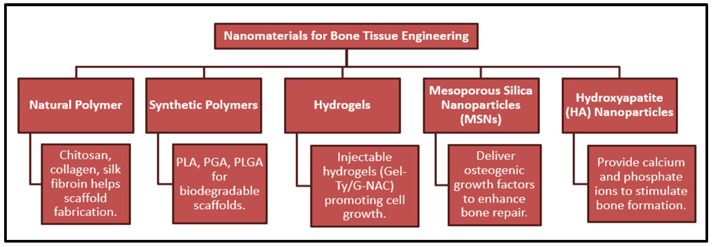
3. Nanomaterials for Bone Tissue Engineering

Nanobiotechnology (NBT) in bone tissue engineering (BTE) involves the nanoscale fabrication of frames utilizing both synthetic and natural polymers. These polymers can be integrated with various nanoscale metals, producing advanced nano-mixtures for antimicrobial drug delivery. This also improves growth factors by different mechanisms to obtain improved bone regeneration (Figure 2). NBT enables the utilization of NPs for imaging and cellular processes in vitro as well as in vivo [11]. For effective bone regeneration, biocompatibility is important for scaffolding, capable of promoting cell growth as well as sustaining osteogenic excitation at crack sites. An injectable hydrogel system was introduced in a study which consisted of enzymatically cross-linkable gelatin and functionalized Au NPs. For finding phenol crosslinking horseradish peroxidase (HRP), tyramine (Ty) was conjugated to the gelatin backbone, forming Gel-Ty. NAC was conjugated with the Au NP surface (Au-NAC) to enhance osteogenic differentiation. The Gel-Ty hydrogels incorporating Au-NAC (Gel-Ty/Au-NAC) exhibited outstanding strength as well as biocompatibility. Au-NAC promoted osteogenic specialization in the Gel-Ty matrix. So, phenol crosslinking is a proper approach for injectable hydrogels in tissue regeneration, while Au-NAC efficiently improves bone regeneration. Gel-Ty and Au-NAC hydrogels hold promise as biodegradable graft materials for bone defect treatment and as versatile platforms for tissue engineering applications, for example cell delivery, regenerative drugs, and drug delivery [12]. Different types of synthetic materials (polyethylene) and natural polymers (cellulose and collagen) were used in tissue engineering in another study, including tissue repair and bone regeneration. In implant biodegradation and scaffolds implantation (Figure 3), biocompatible and biodegradable polymers like PLA, PGA, and PHB, along with its co-polymer PHBV, can be used [13]. So, metal oxide/PLA or metal oxide/other biodegradable polymer nanocomposites can be synthesized to find new applications in tissue repair.

4. Nano–Bio Interactions: Biochemical and Biomechanical Perspectives

NPs can be broadly classified into four main types based on their composition: inorganic, carbon-based, organic, and composite NPs. Inorganic NPs, typically made from metals or metal oxides, are valued for their chemical stability and reactivity. Carbon-based NPs are notable for their excellent electrical and thermal properties, making them ideal for biosensing and drug delivery applications [14].
Figure 3. Essential components involved in bone healing process [15].
Msf 32 00004 g003
The interaction of NPs with biological systems depends on several factors, including dosage, solubility, and their stability in physiological environments. While some NPs dissolve and behave similarly to their chemical constituents, others remain structurally intact and may accumulate in tissues, potentially leading to toxicity (Figure 3). This raises concerns about their biocompatibility and necessitates further investigation into the toxicological effects associated with variations in size, shape, and surface properties [16]. Physicochemical characteristics like rigidity and surface texture greatly influence how NPs interact with cells. Studies suggest that stiffer NPs may be less easily internalized than softer, more flexible ones. However, cell–NP interactions are complex and depend on initial surface adhesion and cellular uptake pathways. The extracellular matrix plays a key role in this process by offering structural support and mediating NP transport across cell membranes. Transport mechanisms, such as transcytosis, are shaped by both the mechanical properties of NPs and the ECM [17]. NPs have unique thermal, optical, and magnetic properties, making them ideal for targeted cancer drug delivery. Their effectiveness depends on controlled cellular uptake, distribution, and localization, while biocompatibility and toxicity are influenced by their physicochemical traits. Mechanobiology helps analyze how NPs interact with cells through mechanical forces. NPs can alter the cytoskeleton, affecting cell stiffness, movement, and adhesion—key factors in cancer metastasis. These mechanical changes may lead to functional benefits or cellular dysfunction [18].

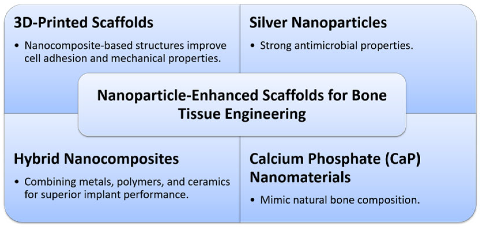
5. Nanoparticle-Enhanced Scaffolds for Bone Tissue Engineering

Metallic oxide NPs can be utilized in bone engineering by three main sections, cell labeling, bio-active-molecule delivery, and advancement [19]. Nanocomposites of metal oxide/chitosan have been synthesized and can be applied into scaffolding. Crosslinking chitosan (CS) frames with substances having two or more than two reactive groups can enhance their strength properties, resulting in better output than CS alone. Electrospinning and three-dimensional printing can be used to fabricate polymeric scaffolds [20]. NMs acquired from calcium phosphate (CaP), one reactive group of CS, can be incorporated into three-dimensionally printed scaffolds, as bone naturally consists of seventy percent CaP crystals and thirty percent organic collagen fibrils [5]. The scaffold must possess (i) sufficient mechanical strength to withstand external pressure, preserving the tissue’s shape and integrity; (ii) the ability to achieve improved cell adhesion and cell proliferation without swelling reactions, and exhibiting biocompatibility; and (iii) a suitable degradation rate [21]. A 3D-printable chitosan cryo-gel was developed by using difunctional polyurethane NPs as a network builder. The cryo-gel was printed on a liquid cryo-deposition medium and exhibited properties like water absorption, high compressibility, and elastic recovery (Figure 4). It was shown through cell-based experiments that the three-dimensionally printed chitosan cryo-gel scaffold was injectable, and provided solid mechanical strength, improving the proliferation of human adipose-derived adult stem cells [22]. Improved CT signals were found by utilizing the cyto-compatible size and compatible amount of AuNPs. GelMA-Au NPs and GelMA scaffolds were 3D printed, and further effects of in osteogenic differentiation. A bone defect was selected to place the GelMA-Au NPs and GelMA scaffolds. CT imaging demonstrated that GelMA-AuNPs have potential for bone tissue engineering, offering improved visual illustration for micro-CT imaging [7]. To develop the CT contrast agent with GelMA-based scaffolds, different sized Au NPs were merged into the GelMA pre-polymer solution at different concentrations. The metabolic activity of the L929 cell line was extensively minimized at a AuNP concentration of 0.0004 M for 40 nm and 60 nm NPs. Mechanical testing and CT imaging of GelMA-Au NPs were performed only within a concentration range which is cyto-compatible [23]. A study was carried out to examine the nature of human skin fibroblast (HSF) cells on porous scaffolds made from Polycaprolactone/chitosan and nano-hydroxyapatite/Polycaprolactone/chitosan, which were fabricated utilizing a freeze-drying technique [24]. Fabricated PLGA electrospun fibers with various diameters were assessed through cell seeding, showing that it has greater growth of HSF cells occurred on fiber diameters [25]. Growth factors, genetic materials, and drug potential can be enhanced through coating or encapsulation in biodegradable NPs, PLA, and PLGA, with non-degradable NPs such as Ag NPs and Au NPs [26]. The nanocomposites Ag/PLA NPs, Ag/PLGA NPs, Au/PLA NPs, and Au/PLGA NPs can be explored in this field.

6. Nanoparticles in Bone Regeneration

Copper oxide NPs were used to enhance the electrical conductivity of hydroxyapatite (HA) scaffolds, cell viability, and mechanical properties. To enhance its antimicrobial and osteoconductive properties, a Ti-6Al-4 V bone implant was biofunctionalized with silver and copper NPs [27]. An injectable hydrogel system with functionalized Au NPs was used to stimulate bone differentiation in hADSCs. Au NPs were incorporated in Ti implants to treat osteopenia and osteoporosis, with in vitro results showing a significant improvement in osteointegration. A scaffold incorporating Au NPs was three-dimensionally printed, assuring that it possessed appropriate biological properties and an imaging function [28]. Zi is a vital element found in all biological tissues. It plays a role in exciting bone mineralization, maintaining the structure of cell membranes, and assisting in pathological calcification. Approximately 30% of zinc is stored in the bone, making it the primary repository of zinc [29]. Alumina NPs were devoted to reinforcing PMMA nanocomposites. The volume of Al2O3 NPs increased, and improvements were observed in the hardness, flexural strength, and breaking strength of the PMMA nanocomposites [30]. A silk fibroin scaffold with incorporated Al2O3 NPs was created to prolong the osteogenic differentiation of rabbit adipose-derived stem cells (rADSCs) [31]. Customized magnesium NPs were incorporated into a PLGA biodegradable scaffold to provide diminished in vivo inflammatory responses, improved structural integrity, and increased biocompatibility [32].

7. Clinical Case Studies on Nanomaterials in Orthopedics

Recent clinical trials and case studies have validated the effectiveness of NMs in orthopedic applications. One in vivo study demonstrated that titanium implants coated with double layers of Au NPs showed significantly enhanced osseointegration compared to those coated with traditional hydroxyapatite [10]. Bone implants coated with silver and copper NPs using additive manufacturing exhibited self-defending antibacterial properties and promoted osteogenic activity in vivo [27]. A clinical trial evaluated injectable hydrogels embedded with Au NP-based complexes in human stem cells. Hydrogels enhanced bone differentiation and exhibited excellent biocompatibility, suggesting a promising approach for treating bone defects [12]. Such case studies reflect the growing transition of nanotechnology from the laboratory to real-world clinical practice, reinforcing its potential for future orthopedic innovations.

8. Conclusions

Nanotechnology has demonstrated significant potential in enhancing orthopedic implants by improving osseointegration, mechanical strength, and antimicrobial properties. The incorporation of NMs in bone tissue engineering and scaffold development opens new possibilities for treating bone defects and ensuring implant longevity. Further research is needed to optimize these technologies for clinical applications, address long-term safety concerns, and navigate regulatory challenges to ensure their successful integration. AuNPs show superior performance in orthopedic applications due to their excellent osseointegration, biocompatibility, and imaging capabilities. While silver and copper NPs offer strong antibacterial effects, AuNPs provide a more balanced and versatile approach for both bone regeneration and implant success.

Author Contributions

Conceptualization, N.N.; writing—original abstract, K.P.C.; writing—original draft (introduction), S.H.; wrote—original draft (without abstract, introduction), I.H.; writing—review & editing, N.N.; writing—review, K.P.C.; writing—editing, S.H.; supervision, N.N. and K.P.C.; project administration, N.N. and S.H. All authors have read and agreed to the published version of the manuscript.

Funding

This research received no external funding.

Institutional Review Board Statement

Not applicable.

Informed Consent Statement

Not applicable.

Data Availability Statement

Data is contained within the article.

Conflicts of Interest

Authors declare no conflict of interest.

References

  1. Luo, Y. Toward Fully Automated Personalized Orthopedic Treatments: Innovations and Interdisciplinary Gaps. Bioengineering 2024, 11, 817. [Google Scholar] [CrossRef] [PubMed]
  2. Kyllönen, L.; D’Este, M.; Alini, M.; Eglin, D. Local Drug Delivery for Enhancing Fracture Healing in Osteoporotic Bone. Acta Biomater. 2015, 11, 412–434. [Google Scholar] [CrossRef]
  3. Fu, J.; Zhu, W.; Liu, X.; Liang, C.; Zheng, Y.; Li, Z.; Liang, Y.; Zheng, D.; Zhu, S.; Cui, Z.; et al. Self-Activating Anti-Infection Implant. Nat. Commun. 2021, 12, 6907. [Google Scholar] [CrossRef] [PubMed]
  4. Ko, W.-K.; Heo, D.N.; Moon, H.-J.; Lee, S.J.; Bae, M.S.; Lee, J.B.; Sun, I.-C.; Jeon, H.B.; Park, H.K.; Kwon, I.K. The Effect of Gold Nanoparticle Size on Osteogenic Differentiation of Adipose-Derived Stem Cells. J. Colloid Interface Sci. 2015, 438, 68–76. [Google Scholar] [CrossRef] [PubMed]
  5. Hindy, O.A.; Goker, M.; Yilgor Huri, P. Nanoscale Agents within 3D-Printed Constructs: Intersection of Nanotechnology and Personalized Bone Tissue Engineering. Emergent Mater. 2022, 5, 195–205. [Google Scholar] [CrossRef]
  6. Wang, N.; Fuh, J.Y.H.; Dheen, S.T.; Senthil Kumar, A. Functions and Applications of Metallic and Metallic Oxide Nanoparticles in Orthopedic Implants and Scaffolds. J. Biomed. Mater. Res. B Appl. Biomater. 2021, 109, 160–179. [Google Scholar] [CrossRef]
  7. Yue, K.; Trujillo-de Santiago, G.; Alvarez, M.M.; Tamayol, A.; Annabi, N.; Khademhosseini, A. Synthesis, Properties, and Biomedical Applications of Gelatin Methacryloyl (GelMA) Hydrogels. Biomaterials 2015, 73, 254–271. [Google Scholar] [CrossRef]
  8. Mudshinge, S.R.; Deore, A.B.; Patil, S.; Bhalgat, C.M. Nanoparticles: Emerging Carriers for Drug Delivery. Saudi Pharm. J. 2011, 19, 129–141. [Google Scholar] [CrossRef]
  9. Mondal, S.K.; Chakraborty, S.; Manna, S.; Mandal, S.M. Antimicrobial Nanoparticles: Current Landscape and Future Challenges. RSC Pharm. 2024, 1, 388–402. [Google Scholar] [CrossRef]
  10. Ko, W.; Kim, S.; Heo, D.; Han, I.; Kim, S.; Kwon, I.K.; Sohn, S. Double Layers of Gold Nanoparticles Immobilized Titanium Implants Improve the Osseointegration in Rabbit Models. Nanomed. Nanotechnol. Biol. Med. 2020, 24, 102129. [Google Scholar] [CrossRef]
  11. Iqbal, N.; Pant, T.; Rohra, N.; Goyal, A.; Lawrence, M.; Dey, A.; Ganguly, P. Nanobiotechnology in Bone Tissue Engineering Applications: Recent Advances and Future Perspectives. Appl. Biosci. 2023, 2, 617–638. [Google Scholar] [CrossRef]
  12. Lee, D.; Heo, D.N.; Nah, H.R.; Lee, S.J.; Ko, W.-K.; Lee, J.S.; Moon, H.-J.; Bang, J.B.; Hwang, Y.-S.; Reis, R.L.; et al. Injectable Hydrogel Composite Containing Modified Gold Nanoparticles: Implication in Bone Tissue Regeneration. Int. J. Nanomed. 2018, 13, 7019–7031. [Google Scholar] [CrossRef]
  13. Liu, X.; Ma, P.X. Polymeric Scaffolds for Bone Tissue Engineering. Ann. Biomed. Eng. 2004, 32, 477–486. [Google Scholar] [CrossRef]
  14. Kumah, E.A.; Fopa, R.D.; Harati, S.; Boadu, P.; Zohoori, F.V.; Pak, T. Human and Environmental Impacts of Nanoparticles: A Scoping Review of the Current Literature. BMC Public Health 2023, 23, 1059. [Google Scholar] [CrossRef]
  15. Bauso, L.V.; La Fauci, V.; Longo, C.; Calabrese, G. Bone Tissue Engineering and Nanotechnology: A Promising Combination for Bone Regeneration. Biology 2024, 13, 237. [Google Scholar] [CrossRef]
  16. Nanotechnologies: 6. What Are Potential Harmful Effects of Nanoparticles? Available online: https://ec.europa.eu/health/scientific_committees/opinions_layman/en/nanotechnologies/l-2/6-health-effects-nanoparticles.htm (accessed on 4 June 2025).
  17. Engin, A.B.; Nikitovic, D.; Neagu, M.; Henrich-Noack, P.; Docea, A.O.; Shtilman, M.I.; Golokhvast, K.; Tsatsakis, A.M. Mechanistic Understanding of Nanoparticles’ Interactions with Extracellular Matrix: The Cell and Immune System. Part. Fibre Toxicol. 2017, 14, 22. [Google Scholar] [CrossRef]
  18. Sohrabi Kashani, A.; Packirisamy, M. Cancer-Nano-Interaction: From Cellular Uptake to Mechanobiological Responses. Int. J. Mol. Sci. 2021, 22, 9587. [Google Scholar] [CrossRef]
  19. Walmsley, G.G.; McArdle, A.; Tevlin, R.; Momeni, A.; Atashroo, D.; Hu, M.S.; Feroze, A.H.; Wong, V.W.; Lorenz, P.H.; Longaker, M.T.; et al. Nanotechnology in Bone Tissue Engineering. Nanomedicine 2015, 11, 1253–1263. [Google Scholar] [CrossRef]
  20. Ganguly, P.; Jones, E.; Panagiotopoulou, V.; Jha, A.; Blanchy, M.; Antimisiaris, S.; Anton, M.; Dhuiège, B.; Marotta, M.; Marjanovic, N.; et al. Electrospun and 3D Printed Polymeric Materials for One-Stage Critical-Size Long Bone Defect Regeneration Inspired by the Masquelet Technique: Recent Advances. Injury 2022, 53, S2–S12. [Google Scholar] [CrossRef]
  21. Wang, X.; Chen, X.; Song, L.; Zhou, R.; Luan, S. An Enzyme-Responsive and Photoactivatable Carbon-Monoxide Releasing Molecule for Bacterial Infection Theranostics. J. Mater. Chem. B 2020, 8, 9325–9334. [Google Scholar] [CrossRef]
  22. Chen, T.-C.; Wong, C.-W.; Hsu, S.H. Three-Dimensional Printing of Chitosan Cryogel as Injectable and Shape Recoverable Scaffolds. Carbohydr. Polym. 2022, 285, 119228. [Google Scholar] [CrossRef] [PubMed]
  23. Celikkin, N.; Mastrogiacomo, S.; Walboomers, X.F.; Swieszkowski, W. Enhancing X-Ray Attenuation of 3D Printed Gelatin Methacrylate (GelMA) Hydrogels Utilizing Gold Nanoparticles for Bone Tissue Engineering Applications. Polymers 2019, 11, 367. [Google Scholar] [CrossRef] [PubMed]
  24. Jin, R.M.; Sultana, N.; Baba, S.; Hamdan, S.; Ismail, A.F. Porous PCL/Chitosan and NHA/PCL/Chitosan Scaffolds for Tissue Engineering Applications: Fabrication and Evaluation. J. Nanomater. 2015, 2015, 357372. [Google Scholar] [CrossRef]
  25. Kumbar, S.G.; Nukavarapu, S.P.; James, R.; Nair, L.S.; Laurencin, C.T. Electrospun Poly(Lactic Acid-Co-Glycolic Acid) Scaffolds for Skin Tissue Engineering. Biomaterials 2008, 29, 4100–4107. [Google Scholar] [CrossRef]
  26. Mok, H.; Zhang, M. Superparamagnetic Iron Oxide Nanoparticle-Based Delivery Systems for Biotherapeutics. Expert Opin. Drug Deliv. 2013, 10, 73–87. [Google Scholar] [CrossRef]
  27. Van Hengel, I.A.J.; Tierolf, M.W.A.M.; Valerio, V.P.M.; Minneboo, M.; Fluit, A.C.; Fratila-Apachitei, L.E.; Apachitei, I.; Zadpoor, A.A. Self-Defending Additively Manufactured Bone Implants Bearing Silver and Copper Nanoparticles. J. Mater. Chem. B 2020, 8, 1589–1602. [Google Scholar] [CrossRef]
  28. Fan, J.H.; Li, W.T.; Hung, W.I.; Chen, C.P.; Yeh, J.M. Cytotoxicity and Differentiation Effects of Gold Nanoparticles to Human Bone Marrow Mesenchymal Stem Cells. Biomed. Eng. Appl. Basis Commun. 2012, 23, 141–152. [Google Scholar] [CrossRef]
  29. Iqbal, N.; Kadir, M.R.A.; Mahmood, N.H.; Salim, N.; Froemming, G.R.A.; Balaji, H.R.; Kamarul, T. Characterization, Antibacterial and in Vitro Compatibility of Zinc–Silver Doped Hydroxyapatite Nanoparticles Prepared through Microwave Synthesis. Ceram. Int. 2014, 40, 4507–4513. [Google Scholar] [CrossRef]
  30. Aghajani Derazkola, H.; Simchi, A. Effects of Alumina Nanoparticles on the Microstructure, Strength and Wear Resistance of Poly(Methyl Methacrylate)-Based Nanocomposites Prepared by Friction Stir Processing. J. Mech. Behav. Biomed. Mater. 2018, 79, 246–253. [Google Scholar] [CrossRef]
  31. Zafar, B.; Mottaghitalab, F.; Shahosseini, Z.; Negahdari, B.; Farokhi, M. Silk Fibroin/Alumina Nanoparticle Scaffold Using for Osteogenic Differentiation of Rabbit Adipose-Derived Stem Cells. Materialia 2020, 9, 100518. [Google Scholar] [CrossRef]
  32. Lih, E.; Kum, C.H.; Park, W.; Chun, S.Y.; Cho, Y.; Joung, Y.K.; Park, K.S.; Hong, Y.J.; Ahn, D.J.; Kim, B.S.; et al. Modified Magnesium Hydroxide Nanoparticles Inhibit the Inflammatory Response to Biodegradable Poly(Lactide-Co-Glycolide) Implants. ACS Nano 2018, 12, 6917–6925. [Google Scholar] [CrossRef] [PubMed]
Figure 1. Nanoparticles in orthopedic implants and osseointegration.
Msf 32 00004 g001
Figure 2. Nanomaterials for bone tissue engineering.
Msf 32 00004 g002
Figure 4. Nanoparticle-enhanced scaffolds for bone tissue engineering.
Msf 32 00004 g004
Disclaimer/Publisher’s Note: The statements, opinions and data contained in all publications are solely those of the individual author(s) and contributor(s) and not of MDPI and/or the editor(s). MDPI and/or the editor(s) disclaim responsibility for any injury to people or property resulting from any ideas, methods, instructions or products referred to in the content.

