1. Introduction
Class II is one of the most frequent types of malocclusion, affecting about one-third of patients who seek orthodontic treatment [
1]. The most common feature in class II malocclusion is a retruded mandible in relation to the craniofacial structure [
2]. There are various types of treatment apparatus that allow the sagittal correction of the mandibular deficiency by holding the mandible in a more forward and downward position, therefore enabling the mandible to alter its postural position. These appliances enable the orofacial musculature to stretch, and the resulting reciprocal force is transmitted to the skeletal and dento-alveolar structures, resulting in a favourable alteration of the skeletal growth pattern and dento-alveolar tooth movement [
3]. Ritto and Ferreira [
4] categorized functional appliances into flexible, rigid or hybrid, according to the implemented force system to provide mandibular protraction. Out of those apparatuses, hybrid appliances combine flexible and rigid components with a spring system, aiming to move teeth by applying continuous force 24 h a day, replacing the conventional class II elastics which require patient compliance [
5]. One of the most used hybrid functional appliances in treating class II malocclusion is the semirigid fixed functional appliance, Forsus
TM Fatigue-Resistant Device (FFRD). This appliance has gained increased acceptance recently as a replacement for other class II treatment modalities [
5].
Third molar impaction is a common finding accounting for 98% of all impacted teeth [
6] and reaching an occurrence of 73% in young adults in Europe [
7]. The aetiology of impaction is multifactorial including genetic or pathological factors, and a lack of required space to accommodate their size [
8]. Orthodontic treatment for growing individuals frequently affects the third molars’ eruption path. This effect is especially noticeable during non-extraction dentoalveolar treatment of cases with class II malocclusion [
9]. Understanding this concern is essential to avoiding unpredictable side effects such as the third molars’ impaction or altering its eruption path [
10]. Therefore, this study aimed to evaluate the influence of the treatment of class II using the FFRD on the eruption path of the maxillary and mandibular third molars.
2. Material and Methods
Ethical approval of the present retrospective study was obtained from the Ethic Committee of the Egas Moniz School of Health and Science, and the participants/parents granted their informed consent. A convenience sample of 55 orthodontic patients participated in this study. The present sample was categorized into two groups: the first group included patients presented with dental and skeletal class II malocclusion (ANB ≥ 4° and a retruded mandible (SNB ≤ 76°), treated with FFRD and fixed orthodontic appliance (n = 28; 14 males, 14 females, 13.6 years old, SD ± 2.4). The second group was the matched controls (n = 27: 12 males and 15 females, 13.2 years old, SD ± 1.5), who had a class I malocclusion, treated with a conventional fixed orthodontic appliance without using any propulsive mechanics.
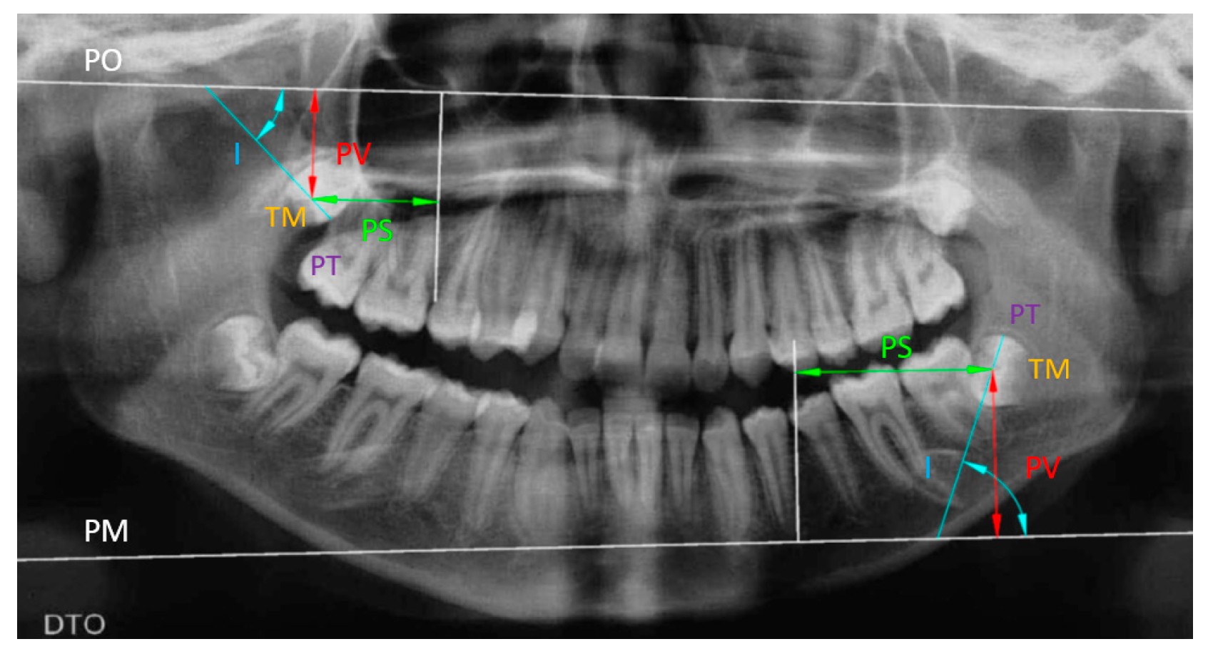
Pre- and post-treatment digital panoramic radiographs were taken for each patient. All radiographs were taken using the same machine (Gendex Orthoralix 9200 DDE, Gendex Dental Systems, Des Plains, IL, USA). Radiographic analysis was performed using Auto CAD 2021 for Window. Landmarks and planes used in Tavano method [
11] were determined on each panoramic radiograph (
Figure 1). An intra-examiner reproducibility study was undertaken on eight panoramic radiographs by repeating the tracing after two weeks by the same operator. The results confirmed an excellent agreement between the two trials for all the variables (≥95%).
The inclination, the sagittal, and the vertical positions of the third molars were evaluated for each radiograph. Descriptive statistics and a mixed model repeated-measures ANOVA were used to determine the measurement differences between the two time points in each group at a significance level of 5%.
3. Results
The only statistically significant difference between the two groups was observed in the vertical position of the lower left third molar, which was more proximal to the Menton plane by −2.24 mm in the FFRD group compared to the controls at
p = 0.010 (
Table 1). However, all the other extracted measurements were similar in both groups (
p > 0.05).
4. Discussion
The mandibular third molar is the most prevalent impacted tooth, followed by the maxillary third molar. The third molars’ size and shape dimorphism, variability in position, root formation, duration of calcification, lack of required space and altered eruption path make their eruption one of the most unpredictable events in the evolution of human dentition [
8].
It has been demonstrated that FFRD produces relatively more dentoalveolar effects, a combination of mesialization of the lower molars and distalization of the upper molars, which substantially contribute to class II molar relationship correction. The literature reported an increase in the mandibular retromolar area due to the effect of FFRD [
10,
12]. On the other hand, there is a controversy concerning the influence of FFRD on the position of the maxillary third molar. On one side, Heinrichs et al. [
13], confirmed the existence of a significant distalization of the maxillary third molars due to the use of FFRD, while on the other side, Jones et al. [
14], observed a significant mesialization effect. In our FFRD group, there was a reduction in the angle of inclination of both the upper and lower third molars. This reduced inclination value was not significantly different compared to controls. This result might reflect the non-significant effect of FFRD on the inclination or the third molar, or it might be a false-negative finding due to using a convenience sample with no power calculation. According to our literature search, this study was the first to assess the effect of non-extraction treatment of class II malocclusion with FFRD on the position of the maxillary and mandibular third molars combined. Using the present findings to conduct another study based on a power calculation is recommended.
Within the limitation of this study, we could conclude that orthodontic treatment of class II malocclusion using FFRD device does not seem to influence the eruption path of third molars; accordingly, the probability of the eruption of third molars is multifactorial and does not rely only on orthodontic treatment with FFDR.
Author Contributions
Principal investigator, undertaking the research study to fulfil the requirements for the degree of specialist in orthodontics, methodology, original draft preparation, J.G.; supervision, conceptualization, writing up, P.M.P.; statistical analysis, J.B.; writing up, editing, I.B. All authors have read and agreed to the published version of the manuscript.
Funding
This research received no external funding.
Institutional Review Board Statement
The study was conducted in accordance with the Declaration of Helsinki and approved by the Institutional Review Board of Egas Moniz School of Health and Science, (protocol code 975, 27 May 2021).
Informed Consent Statement
Informed consent was obtained from all subjects involved in the study.
Data Availability Statement
Research data are available upon request.
Conflicts of Interest
The authors declare no conflict of interest.
References
- Franchi, L.; Alvetro, L.; Giuntini, V.; Masucci, C.; Defraia, E.; Baccetti, T. Effectiveness of comprehensive fixed appliance treament used with the forsus fatigue resistant device in class II patients. Angle Orthod. 2011, 81, 678–683. [Google Scholar] [CrossRef] [PubMed]
- McNamara, J.A. Components of Class II malocclusion in children 8–10 years of age. Angle Orthod. 1981, 51, 177–202. [Google Scholar] [PubMed]
- Tendulkar, P.M.; Pradhan, T. Effects of fixed twin-block and forsus fatigue resistant device on mandibular third molar angulation–A comparative study. Ind. J. Health Sci. Biomed. Res. KLEU 2021, 14, 340–347. [Google Scholar] [CrossRef]
- Ritto, A.K.; Ferreira, A.P. Fixed functional appliances- a classification. Funct. Orthod. 2000, 17, 12–30. [Google Scholar] [PubMed]
- Vogt, W. The Forsus Fatigue Resistant Device. J. Clin. Orthod. 2006, 40, 368–377. [Google Scholar]
- Padhye, M.N.; Dabir, A.V.; Girotra, C.S.; Pandhi, V.H. Pattern of mandibular third molar impaction in the Indian population: A retrospective clinico-radiographic survey. Oral Surg. Oral Med. Oral Pathol. Oral Radiol. Endod 2013, 116, e161–e166. [Google Scholar] [CrossRef] [PubMed]
- Elsey, M.J.; Rock, W.P. Influence of orthodontic treatment on development of third molars. Br. J. Oral Maxillofac. Surg. 2000, 38, 350–353. [Google Scholar] [CrossRef]
- Hatem, M.; Bugaighis, I.; Taher, E.M. Pattern of third molar impaction in Libyan population: A retrospective radiographic study. Saudi J. Dent. Res. 2016, 7, 7–12. [Google Scholar] [CrossRef][Green Version]
- Carter, K.; Worthington, S. Predictors of third molar impaction: A systematic review and meta-analysis. J. Dent. Res. 2015, 95, 1. [Google Scholar] [CrossRef] [PubMed]
- Belma, I.; Zühre, Z.; Akarslan, K. Effects of Angle class II correction with the forsus fatigue resistant device on mandibular third molars-A retrospective study. J. Orofac. Orthop. 2021, 82, 403–412. [Google Scholar]
- Tavano, O.; Ursi, W.; Almeida, R.; Henriques, J. Determinação de linhas de referências para medições angulares em radiografias ortopantomográficas. Odontol. Atual 1989, 16, 22–55. [Google Scholar]
- Sakuno, A. Tomographic evaluation of dentoskeletal changes due to the treatment of class II malocclusion with Forsus appliance. J. Oral Biol. Craniofac. Res. 2019, 9, 277–279. [Google Scholar] [CrossRef] [PubMed]
- Heinrichs, D.A.; Shammaa, I.; Martin, C.; Razmus, T.; Gunel, E.; Ngan, P. Treatment effects of fixed intermaxillary device to correction of class II malocclusion in growing patients. Prog. Orthod. 2014, 15, 145–151. [Google Scholar] [CrossRef] [PubMed][Green Version]
- Jones, G.; Buschang, P.H.; Kim, K.B.; Oliver, D.R. Class II non-extraction patients treated with the forsus fatigue resistant device versus intermaxillary elastics. Angle Orthod. 2008, 78, 332–338. [Google Scholar] [CrossRef] [PubMed]
| Disclaimer/Publisher’s Note: The statements, opinions and data contained in all publications are solely those of the individual author(s) and contributor(s) and not of MDPI and/or the editor(s). MDPI and/or the editor(s) disclaim responsibility for any injury to people or property resulting from any ideas, methods, instructions or products referred to in the content. |
© 2023 by the authors. Licensee MDPI, Basel, Switzerland. This article is an open access article distributed under the terms and conditions of the Creative Commons Attribution (CC BY) license (https://creativecommons.org/licenses/by/4.0/).


