Proceeding Paper

Microcrystals and Microfibers of Cellulose from Acrocomia aculeata (Arecaceae) Characterization †

by
Shirley Duarte
1,*,
Magna Monteiro
2,
Porfirio Andrés Campuzano
1,
Natalia Giménez
1 and
María Cristina Penayo
1,*
1
Faculty of Chemistry, National University of Asunción, San Lorenzo 1055, Paraguay
2
Polytechnic School, National University of Asuncion, Mcal. Estigarribia km 11, Asuncion 1209, Paraguay
*
Authors to whom correspondence should be addressed.
Presented at the 1st International Conference of the Red CYTED ENVABIO100 “Obtaining 100% Natural Biodegradable Films for the Food Industry”, San Lorenzo, Paraguay, 14–16 November 2022.
Biol. Life Sci. Forum 2023, 28(1), 8; https://doi.org/10.3390/blsf2023028008
Published: 9 November 2023

Abstract

:
In the context of the so-called lignocellulose bio-refinery, the coconut shell (S) and pulp (P) of Acrocomia aculeata (Arecaceae) are interesting agro-industrial wastes that can be used as feedstock for the production of high value-added products. The aim of this work was to evaluate these lignocellulosic residues S and P, to obtain the microcrystal (MCC) and microfiber (MFC) of cellulose, and to characterize them to propose possible applications. First, cellulose content in the raw materials was determined, being 39.69% and 45.42% for both (S and P)) respectively, respectively. Then, the purification of residues was carried out via alkaline and bleaching treatments. Next, in order to obtain MCC and MFC from the purified cellulose, a chemical treatment with HCl (for MCC) and a mechanical treatment with a blender (for MFC) were performed. The size and morphology were observed via MEB, and properties were characterized using Fourier transform infrared spectroscopy (FTIR), X-ray diffraction (XRD), and differential thermogravimetric analysis (DTG).

1. Introduction

The South American palm species Acrocomia aculeata (Arecaceae), commonly known as mbocayá, macaw, macauba, or just coconut palm, has attracted the attention of researchers in recent years, mainly for its great potential as a sustainable oil crop [1,2,3]. In Paraguay, the A. aculeata fruit (coconut) has been processed since 1940 [4] for oil extraction. From the process, the shell (S), pulp (P), endocarp, and almond expeller remain as waste, which can be used to obtain new products with greater added value [5,6].
Lignocellulosic wastes as S and P from coconut fruit represent a renewable cellulose source, the primary raw material for nano and micro celluloses (NCs and MCs) [7]. Cellulose derivatives with desirable properties for different applications are a current topic of study in the scientific community. Although NC is a material with exceptional properties, the high consumption of mineral acids during its extraction process is, in most cases, still a disadvantage.
Microcrystal (MCC) and microfiber cellulose (MFC) have gained increasing interest as reinforcing polymeric materials due to their availability, relatively low cost, and high mechanical resistance [8,9,10]. There are also numerous industrial applications for these fibers, which exploit their chemical functionality (reactivity) for crosslinking, their ability to retain water, and their hydrogen bonding capability [11].
MCC and MFC compositions must be studied for each source because characteristics such as crystallinity, thermal stability, and final chemical composition are dependent on their origin [12]. This information is essential to explore MFC and MCC uses as reinforcing agents in developing polymeric biodegradable compounds, e.g., packaging applications with improved properties.
This work describes the characteristics of two lignocellulosic agroindustrial wastes from coconut fruit (S and P), their main components, and the methods used to obtain MCC and MFC from them. The main characteristics of the MCC and MFCs, considering their possible application as reinforcing agents in films for food packaging, are also presented.

2. Materials and Methods

2.1. Raw Materials

Coconut shell (S) and pulp (P) were provided as agroindustrial waste by Industrial Aceitera S.A. paraguayan company. They were washed to remove dirt and other impurities. Then, they were dried in an oven at 105 ± 5 °C for 3 h. Finally, sample particles between 0.85 and 2 mm were obtained using a mill IKA M20, as NREL/TP51042620 establishes them.

2.2. Compositional Analysis

The chemical composition of the constituents (S and P) was determined: organic extractive (TAPPI 204 cm-97), holocellulose, and hemicelluloses based on the standard ASTM D1104 [13,14], soluble and insoluble lignin (NREL/TP510-42618).

2.3. Cellulose Preparation

Cellulose purification was carried out via two chemical treatments: (i) alkaline treatment with 5% NaOH (w/v) solution; (ii) bleaching process, using 1.5% NaClO2 solution (v/v) with acetic acid (pH 4–5), both treatments for 120 min at 80 °C, 600 rpm, and a solid/solution ratio of 1:20. The yield was calculated based on the dry basis weight of each constituent.

Microfibrillated and Microcrystal Cellulose Preparation

MFC was prepared by blending the bleached cellulose in a high rotation brand blender (model BL 767) at 25,000 rpm at different times (5, 10, 20 min), maintaining a ratio of 1% (p/v) (fiber/solution) in a volume of 500 mL of solution. At the end of the corresponding time of the mechanical treatment, the solution was filtered, and the MFCs obtained were dried in an oven at 45 °C for 18 h.
For the MCC, the bleached cellulose was subjected to different times of acid hydrolysis (15, 30, and 60 min) with 2.5 N HCl at a constant temperature of 85 ± 2 °C and a fiber/solution ratio (1:20), with a constant stirring. The reaction was stopped with an ice bath and adding NaOH. It was filtered through a fritted glass filter and washed with distilled water until pH neutral. Subsequently, it was dried in an oven at 45 ± 5 °C for around 18 h.

2.4. Characterization of MCC and MFC

2.4.1. Fourier Transform Infrared Spectroscopy (FTIR)

The presence of functional groups was studied using a Thermo Fisher (Nicolet iS5, Thermo Fisher, Waltham, MA, USA) FT/IR (Fourier transform infrared spectrometer). For the FTIR scan, KBr pellets containing 2 mg of dry sample with 200 mg of KBr powder were prepared. Spectra in the range of 400–4000 cm−1 was obtained with a resolution of 4 cm−1, and the signal was accumulated from 32 scans [15,16].

2.4.2. X-ray Diffraction (XRD)

X-ray diffractometer was measured using the X′Pert3 Powder via Cu-Kα radiation at 45 kV and 40 mA in an angular range of 10° to 40°/2θ with a step size of 0.0170/2θ and a count time of 50.1650 s in each step.
The crystallinity index (CrI) was measured using Segal’s Equation on the diffraction peak height intensity, as indicated in Equation (1):
% C r I = I 002 I a m I 002
where I 002 is the maximum intensity of the diffraction peak, taken at 2θ between 22° and 23° for cellulose I, and I a m is the intensity of the amorphous diffraction peak taken at 2θ between 18° and 19° for cellulose I.

3. Results and Discussion

3.1. Compositional Analysis

The raw materials (S and P) were analyzed to determine the cellulose content (Table 1).
A higher cellulose content (45.4%) is observed in the pulp (P), but the value obtained for the shell (S) is also important (39.6%). Comparing the lignocellulosic composition of P of the coconut fruit with the non-woody and woody raw materials commonly used to obtain cellulose, such as pinewood (37–43%), eucalyptus wood (41–50%), bamboo (43%), and bagasse (42–55%), similar and even higher cellulose contents are observed. These values open the expectations for the use of these lignocellulosic wastes from coconut fruits to obtain cellulose and, after a purification process, obtain MCC and MFC from them.

3.2. Cellulose Preparation and Purification

Table 2 shows the results of the yields after the alkaline and bleached treatments. The lower yield of alkaline treatment may be due to the presence of oil in both raw materials (S and P), mainly in the shell (S), as seen in the content of extractives in Table 1 regarding the bleaching treatment, the yield is quite promising for industrial scaling.
Figure 1 shows the DTG traces for S and P before and after alkaline and bleaching treatments.
The purification of cellulose was verified using DTG as the functional groups and characteristic peaks of hemicellulose (around 250 °C) and lignin (a small shoulder above 390 °C) are not seen, leaving only the characteristic cellulose peak (around 340 °C), intensified in both bleached samples. In addition, the effectiveness of alkali and bleaching treatments of the coconut residues was analyzed using FTIR spectroscopy by the decrease in or disappearance of peaks characteristics of hemicelluloses and lignin.

3.3. Characterization of MCC and MFC

Fourier Transform Infrared Spectroscopy (FTIR)

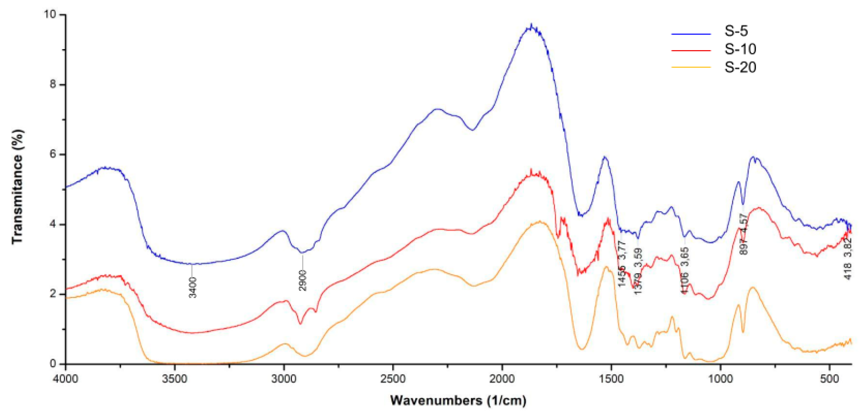
Figure 2 shows the FTIR analyses performed on the MFC of the S at different times of mechanical treatment.
Absorption bands close to 3400, 2900, 1430, 1370, 1160, and 897 cm−1 were associated with cellulose type I [17,18]. The peak at 2900 cm−1 was assigned to the CH stretching of the cellulose. The peak at 1640 cm−1 was due to the vibration of the adsorbed water molecules and also to the carbonyl groups, which may indicate the presence of hemicellulose [19,20]. The absorption peak at 1430 cm−1 was related to the crystalline band, which refers to the symmetric stretching of CH2. The peak at 897 cm−1 was attributed to asymmetric stretching of the out-of-plane ring in cellulose due to β-glucoside bonding. The peak at 1640 cm−1 was associated with the H–O–H stretching vibration of carbohydrate-absorbed water. This indicates the presence of hemicellulose in MFCs, which favors fibrillation during mechanical treatment [20].
Other figures from the FTIR analysis for the MFC of the P and the MCC of the P and S show the same characteristic peaks of cellulose, indicating that purified MCs have been obtained using different treatments.
The crystallinity indices obtained using Equation (1) for the MCC are presented in Table 3.
The peaks identified for the MCC samples correspond to Type I cellulose, and crystallinity index (%CrI) increased as the chemical treatment time increased, as observed in Table 3, for the S60 sample, with the 60 min of treatment. The increase in the crystalline index could be explained by the total or partial elimination of the hemicellulose and lignin structures, which reduced the amorphous region [21].
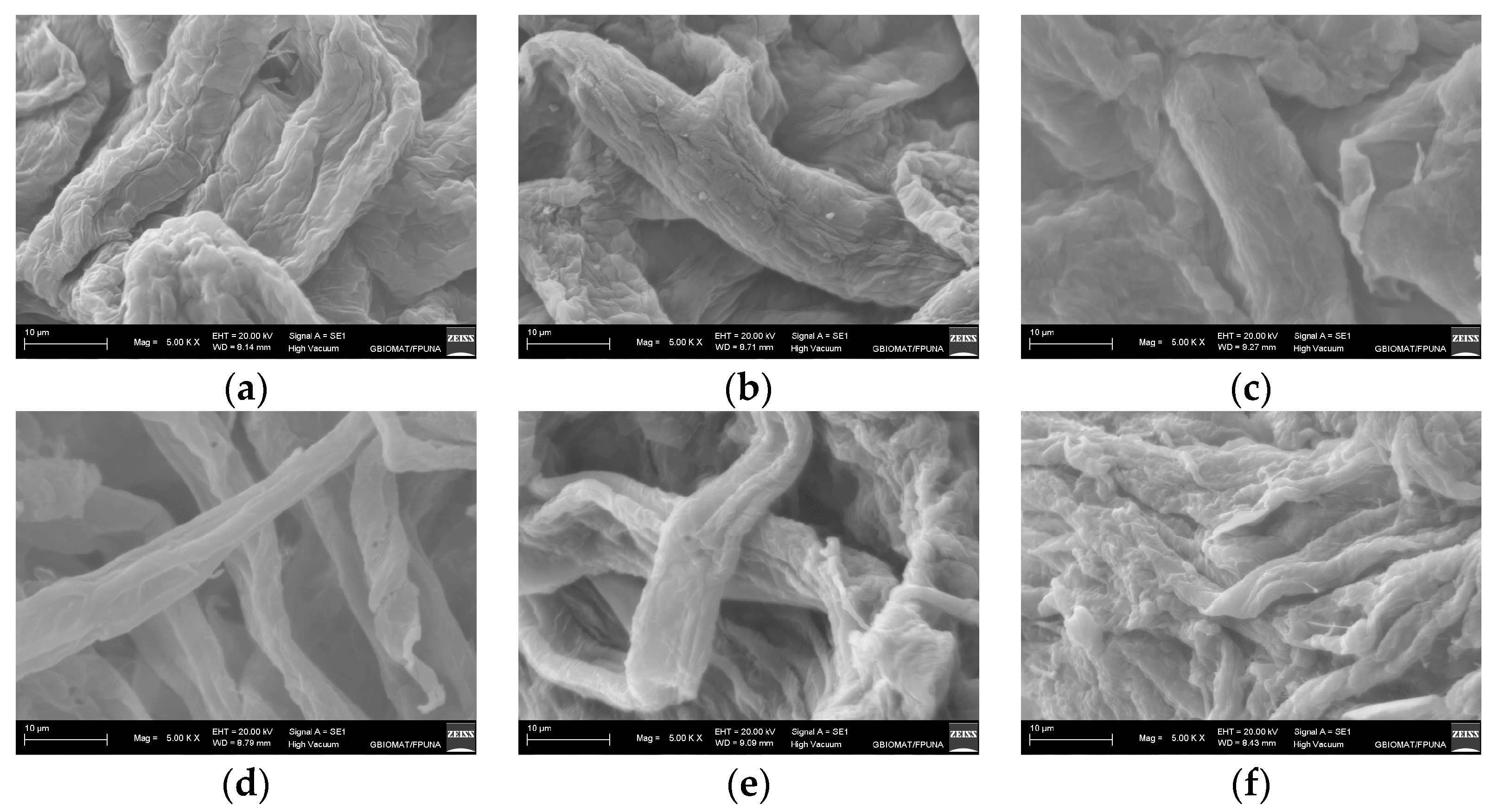
Regarding SEM analysis (Figure 3, a decrease in the diameter was verified for the MCFs as the mechanical treatment increased, being the best condition after 20 min treatment. The estimated diameter, after 20 min of treatment, was between 1 and 3 microns for the pulp (P) and 6 and 8 microns for the shell (S). In addition, a smaller size of the microfibers is evident in the MFCs obtained from the pulp compared to those obtained from the shell.

4. Conclusions

Coconut fruit wastes were valued by obtaining microcrystals (MCC) with high crystallinity index and cellulose microfibers (MFC) with high fibrillation. The purification treatments of the extracted cellulose were effective according to the characterizations carried out after the alkaline treatment and bleaching, verified via FTIR and TGA analysis.
In addition, high yields of purified cellulose for shell (S) and pulp (P) were obtained. Given their richness in cellulose and their (MCC) and (MFC) characteristics, S and P have the potential to be used as reinforcement for food packaging on the way to obtaining environmentally friendly materials.

Author Contributions

Conceptualization, S.D.; methodology, M.C.P., M.M. and S.D.; software, M.C.P.; validation, S.D. and M.M.; formal analysis, M.M. and S.D.; investigation, N.G. and P.A.C.; resources, S.D. and M.C.P.; data curation, M.C.P.; writing—original draft preparation, M.C.P. and S.D.; writing—review and editing, S.D. and M.M.; visualization, S.D.; supervision, S.D.; project administration S.D.; funding acquisition, S.D. All authors have read and agreed to the published version of the manuscript.

Funding

This research was funded by CONACYT under grant Code Project PINV18-514 and the Red ENVABIO100-CYTED (Ref. 121RT0108).

Institutional Review Board Statement

Not applicable.

Informed Consent Statement

Not applicable.

Data Availability Statement

Not applicable.

Acknowledgments

The authors thank Magna Monteiro (GBIOMAT, FP-UNA, Py) for assistance with SEM, FTIR, and XRD analysis. The authors gratefully acknowledge the Red Cyted ENVABIO100 121RT0108 for their financial support. S. Duarte would like to thank PRONII (CONACYT, PY).

Conflicts of Interest

The authors declare no conflict of interest.

References

  1. Evaristo, A.B.; Grossi, J.A.S.; Carneiro, A.d.C.O.; Pimentel, L.D.; Motoike, S.Y.; Kuki, K.N. Actual and putative potentials of macauba palm as feedstock for solid biofuel production from residues. Biomass-Bioenergy 2016, 85, 18–24. [Google Scholar] [CrossRef]
  2. Reis, S.B.; Mercadante-Simões, M.O.; Ribeiro, L.M. Pericarp development in the macaw palm Acrocomia aculeata (Arecaceae). Rodriguésia 2012, 63, 541–549. [Google Scholar] [CrossRef]
  3. Plath, M.; Moser, C.; Bailis, R.; Brandt, P.; Hirsch, H.; Klein, A.-M.; Walmsley, D.; von Wehrden, H. A novel bioenergy feedstock in Latin America? Cultivation potential of Acrocomia aculeata under current and future climate conditions. Biomass-Bioenergy 2016, 91, 186–195. [Google Scholar] [CrossRef]
  4. Ciconini, G.; Favaro, S.; Roscoe, R.; Miranda, C.; Tapeti, C.; Miyahira, M.; Bearari, L.; Galvani, F.; Borsato, A.; Colnago, L.; et al. Biometry and oil contents of Acrocomia aculeata fruits from the Cerrados and Pantanal biomes in Mato Grosso do Sul, Brazil. Ind. Crop. Prod. 2013, 45, 208–214. [Google Scholar] [CrossRef]
  5. Ovelar, R.L.; Ortellado, J.; Echauri, C.; Aguero, J.; Galeano, M. Residuos de “acrocomia aculeata” como fuente de biomasa: Una revisión sistemática. Extensionismo Innov. Y Transf. Tecnol. 2019, 5, 326–330. [Google Scholar] [CrossRef]
  6. Duarte, S.; Lv, P.; Almeida, G.; Rolón, J.C.; Perre, P. Alteration of physico-chemical characteristics of coconut endocarp—Acrocomia aculeata—By isothermal pyrolysis in the range 250–550 °C. J. Anal. Appl. Pyrolysis 2017, 126, 88–98. [Google Scholar] [CrossRef]
  7. Oviedo Ch, A.; Vinueza, G.J. Lignocellulosic waste and its uses, a review. infoANALÍTICA 2020, 8 (Extra 1), 133–147. [Google Scholar]
  8. Hasan, M.; Lai, T.K.; Gopakumar, D.A.; Jawaid, M.; Owolabi, F.A.T.; Mistar, E.M.; Alfatah, T.; Noriman, N.Z.; Haafiz, M.K.M.; Khalil, H.P.S.A. Micro Crystalline Bamboo Cellulose Based Seaweed Biodegradable Composite Films for Sustainable Packaging Material. J. Polym. Environ. 2019, 27, 1602–1612. [Google Scholar] [CrossRef]
  9. Huang, X.; Xie, F.; Xiong, X. Surface-modified microcrystalline cellulose for reinforcement of chitosan film. Carbohydr. Polym. 2018, 201, 367–373. [Google Scholar] [CrossRef] [PubMed]
  10. Li, C.; Luo, J.; Qin, Z.; Chen, H.; Gao, Q.; Li, J. Mechanical and thermal properties of microcrystalline cellulose-reinforced soy protein isolate–gelatin eco-friendly films. RSC Adv. 2015, 5, 56518–56525. [Google Scholar] [CrossRef]
  11. Wypych, G. Fillers—Origin, Chemical Composition, Properties, And Morphology. In Handbook of Fillers; ChemTec Publishing, Science Direct: Toronto, ON, Canada, 2016; pp. 13–266. [Google Scholar]
  12. Ventura-Cruz, S.; Tecante, A. Nanocellulose and microcrystalline cellulose from agricultural waste: Review on isolation and application as reinforcement in polymeric matrices. Food Hydrocoll. 2021, 118, 106771. [Google Scholar] [CrossRef]
  13. Álvarez, A.; Cachero, S.; González-Sánchez, C.; Montejo-Bernardo, J.; Pizarro, C.; Bueno, J.L. Novel method for holocellulose analysis of non-woody biomass wastes. Carbohydr. Polym. 2018, 189, 250–256. [Google Scholar] [CrossRef] [PubMed]
  14. Browning, B.L. Methods of Wood Chemistry Vol I & II; John Wiley & Sons: New York, NY, USA, 1967. [Google Scholar]
  15. Bhawna, S.; El Barbary, H.; Barakat, M. Chemical isolation and characterization of different cellulose nanofibers from cotton stalks. Carbohydr. Polym. 2015, 134, 581–589. [Google Scholar]
  16. Moharram, M.A.; Mahmoud, O.M. FTIR spectroscopic study of the effect of microwave heating on the transformation of cellulose I into cellulose II during mercerization. J. Appl. Polym. Sci. 2008, 107, 30–36. [Google Scholar] [CrossRef]
  17. Brahim, M.M.; El-Zawawy, W.K.; Jüttke, Y.; Koschella, A.; Heinze, T. Cellulose and microcrystalline cellulose from rice straw and banana plant waste: Preparation and characterization. Cellulose 2013, 20, 2403–2416. [Google Scholar] [CrossRef]
  18. Razali, N.; Hossain, M.S.; Taiwo, O.A.; Ibrahim, M. Influence of Acid Hydrolysis Reaction Time on the Isolation of Cellulose Nanowhiskers from Oil Palm Empty Fruit Bunch Microcrystalline Cellulose. BioResources 2017, 12, 6773–6788. [Google Scholar] [CrossRef]
  19. Adel, A.M.; El-Gendy, A.A.; Diab, M.A.; Abou-Zeid, R.E.; El-Zawawy, W.K.; Dufresne, A. Microfibrillated cellulose from agricultural residues. Part I: Papermaking application. Ind. Crop. Prod. 2016, 93, 161–174. [Google Scholar] [CrossRef]
  20. Vora, R.; Shah, Y. Extraction, characterization of micro crystalline cellulose obtained from corn husk using different acid alkali treatment methods. Indo Am. J. Pharm. Sci. 2017, 4, 2399–2408. [Google Scholar]
  21. Martinez-Pavetti, M.B.; Medina, L.; Espínola, M.; Monteiro, M. Study on two eco-friendly surface treatments on Luffa cylindrica for development of reinforcement and processing materials. J. Mater. Res. Technol. 2021, 14, 420–2427. [Google Scholar] [CrossRef]
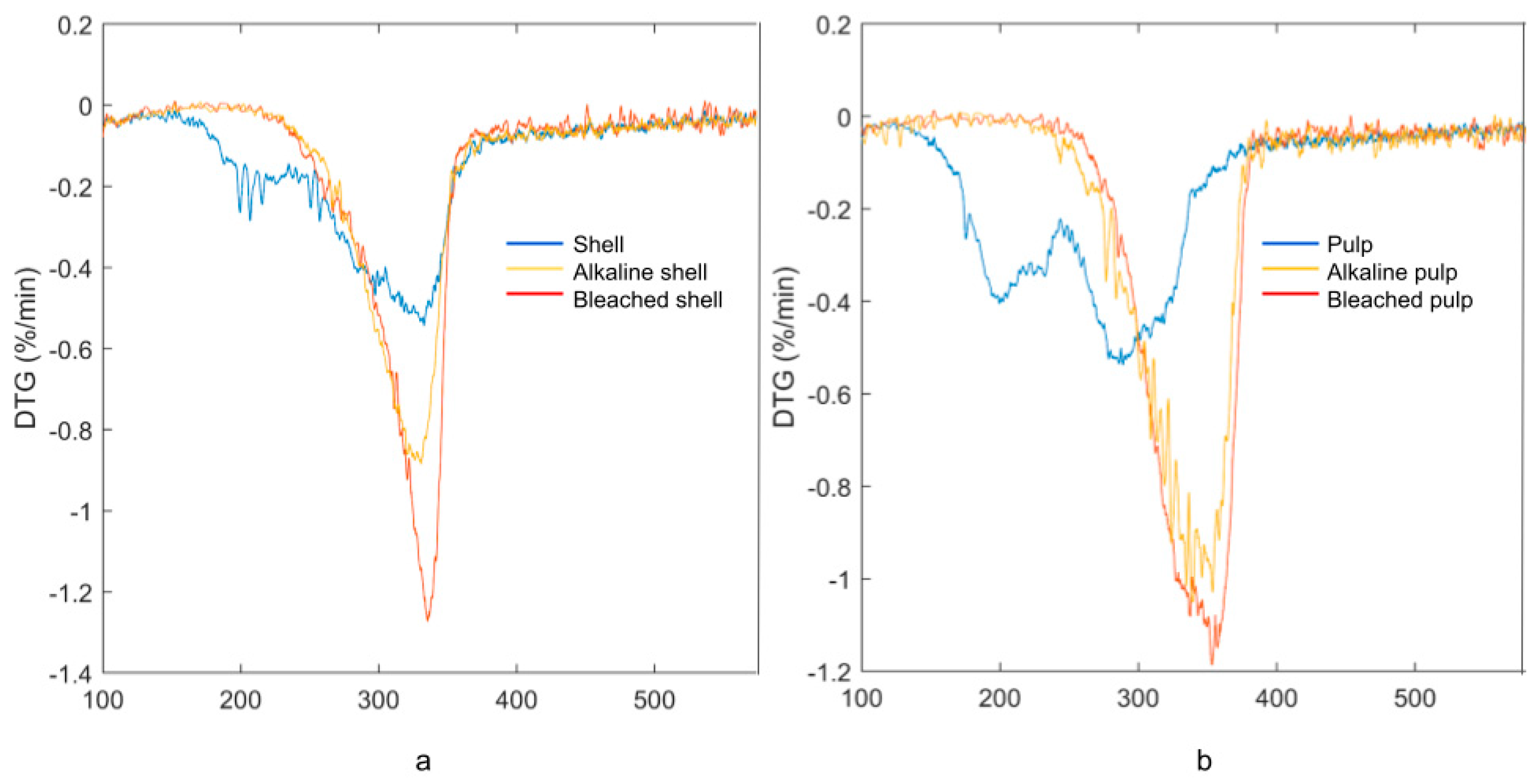
Figure 1. (a) DTG of S and (b) DTG of P before and after the alkaline and bleaching treatments.
Blsf 28 00008 g001
Figure 2. FTIR of the MFC of coconut shell (S) obtained at 5 (S-5), 10 (S-10), and 20 min (S-20).
Blsf 28 00008 g002
Figure 3. SEM micrographs of MFC from coconut fruit shell (ac) and pulp (df) obtained after 5 min (a,d), 10 min (b,e), and 20 min (c,f) of mechanical treatment.
Blsf 28 00008 g003
Table 1. Composition of S and P from coconut fruit.
CompositionShell (%w/w)Pulp (%w/w)
Extractives6.2717.13
Cellulose39.6545.42
Hemicellulose19.2215.89
Lignin30.8017.90
Ash4.063.66
Table 2. Alkali and bleached treatment yields.
Time (min)Pulp (%w/w)Shell (% w/w)
Alkaline treatment24020.5230.88
Bleaching treatment12045.5658.31
Table 3. Crystallinity index for MCC and MFCs of coconut fruit.
Sample%CrISample%CrI
MCCS15′42.42P15′46.40
S30′68.19P30′64.36
S60′69.74P60′67.03
Disclaimer/Publisher’s Note: The statements, opinions and data contained in all publications are solely those of the individual author(s) and contributor(s) and not of MDPI and/or the editor(s). MDPI and/or the editor(s) disclaim responsibility for any injury to people or property resulting from any ideas, methods, instructions or products referred to in the content.