Proceeding Paper

Optimization of Enzymatic Extraction of Melanoidins from Bread by-Products: Bioactivity and Microbiota Modulation †

Department of Biotechnology and Food Science, Faculty of Sciences, Universidad de Burgos, Plaza Misael Bañuelos, 09001 Burgos, Spain
*
Author to whom correspondence should be addressed.
Presented at the 4th International Electronic Conference on Foods, 15–30 October 2023; Available online: https://foods2023.sciforum.net/.
Biol. Life Sci. Forum 2023, 26(1), 25; https://doi.org/10.3390/Foods2023-15102
Published: 14 October 2023
(This article belongs to the Proceedings of The 4th International Electronic Conference on Foods)

Abstract

:
The bakery industry generates substantial food waste, leading to global environment pollution and economic losses. However, these by-products contain valuable bioactive compounds, such as melanoidins, which are brown pigmented compounds with significant antioxidant, antimicrobial and anti-inflammatory properties, making them ideal functional ingredient. This study aimed to evaluate the optimal enzymatic extraction of melanoidins from bread crust using different proteolytic enzymes: pronase E, serine-endo-protease (SP), metalloenzyme protease (MP) and endoprotease papain enzyme (EP). While the yield of melanoidins from MP and EP was lower than that of pronase E, SP yield was significantly higher. No cytotoxicity of bioaccessible, gastrointestinal and colonic fractions was observed in the cell lines Caco-2, HUVEC and SHSY5Y. The antioxidant properties and ability to stabilize the hydroxyl radical were characterized in bioaccessible fractions of the four melanoidins extracts. Moreover, melanoidins positively influenced the intestinal microbiota by increasing beneficial bacteria like Bifidobacterium and Lactobacillus, while reducing non-beneficial bacteria such as Bacteroides spp. and Clostridium leptum, with particularly significant results for SP and EP enzyme extracts. In conclusion, SP and EP enzymes appear to be viable alternatives to pronase E for obtaining melanoidin extracts as functional ingredients. These enzymes offer potential cost advantages and contribute to the sustainable utilization of bakery by-product melanoidins in various food applications. Implementing these alternatives could help to reduce food waste, environmental pollution and economic losses in the bakery industry while promoting the use of valuable bioactive compounds.

1. Introduction

Currently, the bakery industry generates a significant amount of waste, contributing to both environmental pollution and economic losses [1]. Among the bakery waste products, bread crust is an important by-product of the bakery industry that can be revalued by extracting bioactive compounds such as melanoidins, thus reducing environmental contaminants and adding value. Melanoidins are the end products of the Maillard reaction (MRPs) that occur during the heat treatment of food, such as baking. This reaction also contributes to the improvement in the sensory properties of foods such as taste, texture, smell and color [2]. Melanoidins are polymeric, heterogeneous, nitrogenous and brown macromolecules. Their complex structure is not fully defined due to their structural complexity, and they even present great difficulty in extracting, isolating, purifying and identifying them. For bakery products, one commonly used method to extract melanoidins is by digestion with the proteolytic enzyme pronase E. This enzyme is responsible for breaking the gluten network where compounds such as melanoidins are trapped [3]. But there are also other more economical enzymes that are capable of releasing melanoidins such as serine-endo-protease (SP), metalloenzyme protease (MP) and endoprotease enzyme (EP). In this study, we evaluated whether the use of more economical enzymes influences the healthy properties of melanoidins in terms of bioactivity, measured as antioxidant capacity, their cytotoxicity and their ability to modulate the intestinal microbiota in order to propose an alternative extraction method to obtain melanoidins more economically and efficiently.

2. Materials and Methods

2.1. Extraction, Isolation, Digestion and Colonic Fermentation of Melanoidins

Bakery bread were obtained from a local supermarket. Crust samples were ground in a mill and sieved to a particle size <1 mm in a wire mesh sieve (CISA, Barcelona, Spain) and melanoidins were extracted according to Gonzalez-Mateo et al. [4], using four different proteolytic enzymes: pronase E, serine-endo-protease (SP), metalloenzyme protease (MP) and endoprotease enzyme (EP). The soluble fraction was subjected to ultrafiltration using a “dead-end” system using a flat membrane of polyethersulfone 10 kDa (Trisep Flat Sheet Membrane UF10, Sterlitech Corporation, Washington WA, USA).
Then, the gastrointestinal digestion of the samples was continued following the method described by Minekus et al. [5], obtaining the four bioaccessible fractions of melanoidins which were lyophilized. Subsequently, an in vitro colonic fermentation from the residue of the lyophilized gastrointestinal fraction (GI) was carried out. The samples were fermented by the intestinal microbiota from rat fecal samples in a sterile anaerobic environment. They were centrifuged and freeze-dried to obtain the fermented fraction (F) of melanoidins.

2.2. Microbiota Analysis

Bacterial genomic DNA from the lyophilized residue of colonic digestion was isolated using the QIAamp DNA Mini Kit according to the manufacturer’s instructions. Then, qPCR was used to amplify five groups of bacteria in colonic fermentation samples, all bacteria, Bacteroides spp., Lactobacilli spp., Bifidobacterium spp. and Clostridium leptum. The specific 16S rDNA primers and PCR conditions are those described by Gerardi et al. [6]. The results were obtained using a calibration curve and expressed as the number of log copies/ng of DNA.

2.3. Determination of Total Antioxidant Capacity (TAC)

TAC was evaluated using the Q-ABTS assay. Briefly, the ABTS radical (2,20-Azino-Bis-3-Ethylbenzothiazoline-6-Sulfonic acid) was incubated with bioaccessible fractions of melanoidins (10 mg/mL) for 5 min and then the spectrophotometric absorbance of the samples was measured at 734 nm. The results were expressed as the percentage of inhibition with respect to the control without the sample.

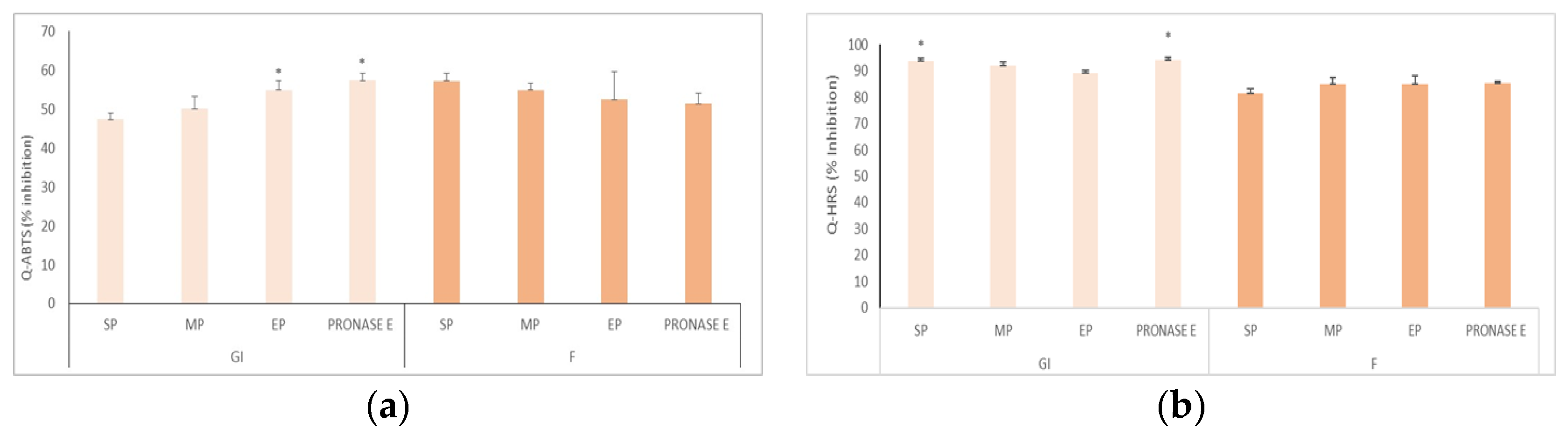
2.4. Determination of Radical Scavenger Activity (Q-HRS)

In the determination of radical scavenger activity, the procedure described by Del Pino et al. [7] was followed. Briefly, 5 mg of the melanoidins samples was weighed and mixed with 100 mM deoxyribose, 10 mM Ascorbic acid, 1 mM H2O2, 10 mM FeCl3, and 10 mM EDTA in Buffer 24 mM sodium phosphate with 15 mM NaCl (pH 7.4). Control samples (samples and buffer) and the oxidized control (without samples) were also prepared. The samples were incubated at 37 °C for 60 min with continuous stirring. After that, 28% (w/v) TCA and 1% (w/v) TBA were added and were heated at 100 °C for 15 min. The absorbance was recorded at 532 nm and the results are expressed as oxidation inhibition percentages with respect to the oxidized control.

2.5. Determination of Metal Chelating Capacity

To measure the chelating activity of bioaccessible fractions of melanoidins, the method described by Yesiloglu [8] was followed. Melanoidin fractions were added to a 0.2 mM FeCl2 solution, 5 mM ferrozine and milli-Q water and incubated at room temperature for 10 min. Absorbance was measured spectrophotometrically at 562 nm in triplicate. The results are expressed as the percentage of inhibition of the formation of the ferrozine-Fe+2 complex: Chelating effect of the metal (%) = (1 − (abs sample/Abs control) × 100.

2.6. Cell Culture and Exposure Conditions

In this study, different cell lines were used, human colon adenocarcinoma (Caco-2), Human Umbilical Vein Endothelial Cells (HUVEC) and human neuroblastoma cell line (SHSY5Y), and they were cultured at 37 °C and 5% CO2 in their specific culture medium.

2.7. MTT Assay

To determine cell viability in the 3 cell lines, 10,000 cells per well were seeded in 96-well plates for 24 h, and then the cells were incubated with the bioaccessible fractions of mela-noidins (GI, F) at a concentration of 150 μg/mL for 24 h. Then, the MTT reagent (5 mg/mL) was added for 2 h. The medium was removed and resuspended in 100 μL DMSO and finally the absorbance was measured at 570 nm. The results were expressed as a percentage of cell viability with respect to the control (cells without treatment).

2.8. Statistical Analysis

All data are expressed as the mean ± standard deviation (SD) of at least three independent measurements for each replicate sample. Statistical analysis was performed using Statgraphics® Centurion 19 (Statpoint Technologies, Inc.,Warranton, VA, USA). Analysis of variance (ANOVA) using Fisher’s Least Significant Difference was performed to detect significant differences between the data (p < 0.05). In addition, the Student’s t-test was applied in the comparison between two samples. The value of p < 0.05 was applied for all analyses.

3. Results and Discussion

In the present study, we evaluated the yield and bioactivity of melanoidins extracted from bread crust using three different proteolytic enzymes (SP, MP and EP) and compared the results with those obtained using the enzyme pronase E. Additionally, we determined the effect of digested fractions (gastrointestinal and colonic fermentation) on the viability of three different cell lines and measured the effect on intestinal microbiota in vitro.
First, we evaluated the yield of melanoidins in the digestion fractions, gastrointestinal (GI) and colonic fermentation (F), obtained from bread crust by extraction with different proteolytic enzymes. The highest yield of GI fractions obtained with the enzyme SP (9.5 g/100 g crust bread) had higher values than that obtained with pronase E enzyme (6.5 g/100 g/crust bread). The yields of the MP and EP enzyme were significantly lower.
Regarding the antioxidant capacity of melanoidins, it is well-known that melanoidin fractions isolated from a wide variety of foods such as bread have beneficial effects (antioxidant, anti-inflammatory and antimicrobial) [9]. Furthermore, other authors have highlighted the significant role of melanoidins in the prevention of oxidative damage due to their ability to chelate metals and eliminate free radicals [10,11]. The antioxidant capacity, measured as Q-ABTS, and the ability to scavenge hydroxyl radicals are shown in Figure 1. The results indicate that all GI samples exhibited high antioxidant capacity (Figure 1a), where the fraction obtained with the enzyme EP showed values similar to the reference enzyme pronase E. The F fractions showed a similar antioxidant capacity to GI fractions and no significant differences were observed among the different samples. The hydroxyl radical is the most toxic oxygen radical known, which can react with almost any molecule in biological systems. In this context, is important to evaluate the hydroxyl scavenger activity of bioaccessible fractions. The results (Figure 1b) showed that all bioaccessible fractions (GI and F) of all extracts of melanoidins (SP, MP, EP and pronase E) have the capacity to scavenge hydroxyl radicals. The highest levels of hydroxyl inhibition were observed for the melanoidins extract obtained with the SP enzyme with values similar to those obtained with the pronase E enzyme.
Although bread crust melanoidins extracted using pronase E have been demonstrated to be non-cytotoxic in cell models such as Caco-2 and HUVEC [12], it remains important to assess whether the GI and F fractions obtained from melanoidins extracted with the other enzymes can alter these properties and release healthy or harmful compounds. Therefore, it is of great relevance to examine their cytotoxicity. As the intestinal epithelium is the first barrier exposed to the compounds under study, the Caco-2 model, commonly used in nutritional studies, was used. The HUVEC model was also employed because endothelial cells are highly sensitive to oxidative changes that can lead to cardiovascular alterations, which may be influenced by the presence of melanoidins [13]. In addition, in this study, we evaluated the effect of the melanoidins digestion fraction on a neuronal model (SHSY5Y). Investigating the effects of these bioactive compounds on neurodegenerative disorders is of great interest to evaluate their therapeutic potential, which is why the differentiated neuronal cell line was used [14].
The viability percentages in the three types of cell lines, Caco-2 model, HUVEC and SHSY5Y, are shown in Table 1. It is noteworthy that the melanoidin digestion fractions GI and F did not show cytotoxicity at concentrations of 50 μg/mL and 100 μg/mL with values of viability higher than 80%. These results are not yet definitive since we must contrast the results with the bioavailable fractions of melanoidins in these three models. However, summarizing these preliminary results, it becomes evident that the melanoidins compounds obtained from bread crust after extraction with enzymes SP, MP and EP did not show cytotoxicity at the tested concentrations in the cell models used.
On the other hand, the biological activity of melanoidins has been studied by other authors, who evaluated the effect of melanoidins on the intestinal microbiota after the in vitro colonic fermentation [15,16]. This study also aimed to evaluate the effect of the different extracts of melanoidins, obtained with the different enzymes, on the intestinal microbiota. The levels of all bacteria, Bacteroides spp., Lactobacillus spp., Bifidobacterium and Clostridium leptum were determined and compared to a control (Figure 2). The results showed that beneficial bacteria such as Lactobacillus and Bifidobacterium increased significantly compared to the control. And non-beneficial bacteria like Bacteroides spp. and Clostridium leptum declined. Therefore, melanoidins have a role in the growth and increase in beneficial microbiota.

4. Conclusions

In conclusion, our study highlights the use of cost-effective proteolytic enzymes for the extraction of melanoidins from bread crust, with potential applications as a functional ingredient. These melanoidins exhibit antioxidant activity, probiotic properties and could be considered safe as they not exhibit cytotoxic effects in the studied cell models. Future research should aim to elucidate the mechanism underlying these effects.

Author Contributions

V.T.: Investigation, Formal Analysis, Methodology, Writing; G.G.: Methodology, Investigation, Supervision, Writing—Review; M.C.-S.: Resources, Investigation, Writing—Review; P.M.: Funding Acquisition, Project Administration, Writing—Review and Editing; G.S.: Conceptualization, Resources, and Writing—Review. All authors have read and agreed to the published version of the manuscript.

Funding

This research was funded by the Autonomous Government of Castilla y Leon and Feder (JCyL/FEDER), grant number BU243P18.

Institutional Review Board Statement

Not applicable.

Informed Consent Statement

Not applicable.

Data Availability Statement

Data from the present study are available upon request from the corresponding author.

Conflicts of Interest

The authors declare no conflict of interest.

References

  1. Verni, M.; Minisci, A.; Convertino, S.; Nionelli, L.; Rizzello, C.G. Wasted Bread as Substrate for the Cultivation of Starters for the Food Industry. Front. Microbiol. 2020, 11, 293. [Google Scholar] [CrossRef]
  2. Pastoriza, S.; Rufián-Henares, J.A. Contribution of melanoidins to the antioxidant capacity of the Spanish diet. Food Chem. 2014, 164, 438–445. [Google Scholar] [CrossRef] [PubMed]
  3. Roncero-Ramos, I.; Delgado-Andrade, C.; Haro, A. Effects of dietary bread crust Maillard reaction products on calcium and bone metabolism in rats. Amino Acids 2013, 44, 1409–1418. [Google Scholar] [CrossRef] [PubMed]
  4. González-Mateo, S.; González-Sanjosé, M.L.; Muñiz, P. Presence of Maillard products in Spanish muffins and evaluation of colour and antioxidant potential. Food Chem. Toxicol. 2009, 47, 2798–2805. [Google Scholar] [CrossRef] [PubMed]
  5. Minekus, M.; Alminger, M.; Alvito, P.; Ballance, S.; Bohn, T.; Bourlieu, C.; Brodkorb, A. A standardised static in vitro digestion method suitable for food—An international consensus. Food Funct. 2014, 5, 1113–1124. [Google Scholar] [CrossRef] [PubMed]
  6. Gerardi, G.; Cavia-Saiz, M.; Rivero-Pérez, M.D.; González-SanJosé, M.L.; Muñiz, P. Modulation of Akt-p38-MAPK/Nrf2/SIRT1 and NF-κB pathways by wine pomace product in hyperglycemic endothelial cell line. J. Funct. Foods 2019, 58, 255–265. [Google Scholar] [CrossRef]
  7. Del Pino-Garcia, R.; Garcia-Lomillo, J.; Rivero-Perez, M.D.; Gonzalez San Jose, M.L.; Muniz, P. Adaptation and validation of quick, easy, new, cheap, and reproducible (QUENCHER) antioxidant capacity assays in model products obtained from residual wine pomace. J. Agric. Food Chem. 2015, 63, 6922–6931. [Google Scholar] [CrossRef] [PubMed]
  8. Yesiloglu, Y.; Aydin, H.; Kilic, I. In Vitro Antioxidant Activity of Various Extracts of Ginger (Zingiber officinale L.) Seed. Asian J. Chem. 2013, 25, 3573–3578. [Google Scholar] [CrossRef]
  9. Celik, E.E.; Rubio, J.M.A.; Andersen, M.L.; Gokmen, V. Interactions of coffee and bread crust melanoidins with hydroxycinnamic and hydroxybenzoic acids in aqueous radical environment. Food Res. Int. 2018, 108, 286–294. [Google Scholar] [CrossRef] [PubMed]
  10. Mesías, M.; Delgado-Andrade, C. Melanoidins as a potential functional food ingredient. Curr. Opin. Food Sci. 2017, 14, 37–42. [Google Scholar] [CrossRef]
  11. Wang, H.; Qian, H.; Yao, W. Melanoidins produced by the Maillard reaction: Structure and biological activity. Food Chem. 2011, 128, 573–584. [Google Scholar] [CrossRef]
  12. Diaz-Morales, N.; Cavia-Saiz, M.; Salazar-Mardones, G.; Rivero-Perez, M.D.; Muñiz, P. Cytotoxicity study of bakery product melanoidins on intestinal and endothelial cell lines. Food Chem. 2021, 343, 128405. [Google Scholar] [CrossRef] [PubMed]
  13. Cossu, A.; Posadino, A.M.; Giordo, R.; Emanueli, C.; Sanguinetti, A.M.; Piscopo, A.; Pintus, G. Apricot melanoidins prevent oxidative endothelial cell death by counteracting mitochondrial oxidation and membrane depolarization. PLoS ONE 2012, 7, e48817. [Google Scholar] [CrossRef] [PubMed]
  14. Temiño, V.; Porras, D.; Salazar-Mardones, G.; Gerardi, G.; Muñiz, P.; Cavia-Sáiz, M. Melanoidins mitigate oxidative distress induced by CoCl2 in neuronal cells (SH-SYY5Y). Free. Radic. Biol. Med. 2023, 201, 63. [Google Scholar] [CrossRef]
  15. Borrelli, R.C.; Fogliano, V. Bread crust melanoidins as potential prebiotic ingredients. Mol. Nutr. Food Res. 2005, 49, 673–678. [Google Scholar] [CrossRef] [PubMed]
  16. Perez-Burillo, S.; Rajakaruna, S.; Pastoriza, S.; Paliy, O.; Angel Rufian-Henares, J. Bioactivity of food melanoidins is mediated by gut microbiota. Food Chem. 2020, 316, 126309. [Google Scholar] [CrossRef] [PubMed]
Figure 1. (a) Antioxidant capacity (Q-ABTS) and (b) hydroxyl radical scavenger (Q-HRS) activity of gastrointestinal (GI) and colonic fermentation (F) fractions of melanoidins. Asterisk (*) indicates significant differences between the values.
Blsf 26 00025 g001
Figure 2. Microbiota composition in cecal samples treated with melanoidins. The results are shown as mean ± standard deviation (n = 3); the statistic was represented by performing a simple ANOVA in each bacterial strain comparing the control without melanoidins with bioaccessible samples. Significant differences (p < 0.05) among the different samples are expressed in Roman letters.
Blsf 26 00025 g002
Table 1. Cell viability of Caco-2 cell treated with bioaccessible melanoidins (GI and F) isolated from bread crust.
ACACO-2
% ViabilityNTSPMPEPPronase E
Fractions 50 μg/mL100 μg/mL50 μg/mL100 μg/mL50 μg/mL100 μg/mL50 μg/mL100 μg/mL
GI100 ± 3.59102 ± 9.2693.41 ± 6.6496.46 ± 5.0198.73 ± 4.3794.14 ± 4.0795.47 ± 4.9199.30 ± 3.6197.88 ± 2.63
F100 ± 9.1195.86 ± 3.9896.95 ± 2.6792.02 ± 2.6694.09 ± 6.9486.12 ± 5.6584.46 ± 6.57 *93.99 ± 2.4392.74 ± 4.27
BHUVEC
% ViabilityNTSPMPEPPronase E
Fractions 50 μg/mL100 μg/mL50 μg/mL100 μg/mL50 μg/mL100 μg/mL50 μg/mL100 μg/mL
GI100 ± 6.5891.94 ± 8.7394.52 ± 9.84106 ± 0.9892.86 ± 11.3982.12 ± 5.26 *75.42 ± 1.28 *91.77 ± 9.5481.45 ± 10.53
F100 ± 6.5890.83 ± 3.4383.41± 5.73 105.85 ± 5.01105.32 ± 3.6185.95 ± 10.8888.72 ± 10.6197.67 ± 9.29 95.24 ± 1.24
CSHSY5Y
% ViabilityNTSPMPEPPronase E
Fractions 50 μg/mL100 μg/mL50 μg/mL100 μg/mL50 μg/mL100 μg/mL50 μg/mL100 μg/mL
GI100 ± 9.5588.5 ± 5.0791.60 ± 6.15 *91.78 ± 7.2585.05 ± 8.093.87 ± 1.72 98.52 ± 9.06100 ± 1.8895.5 ± 5.27
F100 ± 6.5590.30± 5.4490.29 ± 12.0592.39 ± 9.2589.34 ±14.4393.87 ± 4.8694.49 ± 11.696.28 ±12.01 94.76 ± 9.23
A: % viability in cells Caco-2; B: % viability in cells HUVEC; C: % viability in cells SHSY5Y. Asterisk (*) indicates significant differences between the values.
Disclaimer/Publisher’s Note: The statements, opinions and data contained in all publications are solely those of the individual author(s) and contributor(s) and not of MDPI and/or the editor(s). MDPI and/or the editor(s) disclaim responsibility for any injury to people or property resulting from any ideas, methods, instructions or products referred to in the content.

