Proceeding Paper

Effect of Carbon, Nitrogen and Salt Sources on the Growth of Monascus purpureus in Quinoa (Chenopodium quinoa)-Based Culture Media †

by
Evelyn Quispe-Rivera
1,2,
Franz Tucta-Huillca
1,2,
Ursula Gonzales-Barron
3,4,
Vasco Cadavez
3,4,
Marcial Silva-Jaimes
2 and
Juan Juscamaita Morales
1,*
1
Facultad de Ciencias, Universidad Nacional Agraria La Molina (UNALM), Av. La Molina s/n La Molina, Lima 15024, Peru
2
Facultad de Industrias Alimentarias, Universidad Nacional Agraria La Molina (UNALM), Av. La Molina s/n La Molina, Lima 15024, Peru
3
Centro de Investigação de Montanha (CIMO), Instituto Politécnico de Bragança—Campus de Santa Apolónia, 5300-253 Bragança, Portugal
4
Laboratório para a Sustentabilidade e Tecnologia em Regiões de Montanha, Instituto Politécnico de Bragança—Campus de Santa Apolónia, 5300-253 Bragança, Portugal
*
Author to whom correspondence should be addressed.
Presented at the 3rd International Electronic Conference on Foods: Food, Microbiome, and Health—A Celebration of the 10th Anniversary of Foods’ Impact on Our Wellbeing, 1–15 October 2022; Available online: https://sciforum.net/event/Foods2022.
Biol. Life Sci. Forum 2022, 18(1), 64; https://doi.org/10.3390/Foods2022-12999
Published: 30 September 2022

Abstract

:
Monascus purpureus is produced in Asia and used as a food dye and for medicinal purposes. In the present study, the radial growth of quinoa flour-based culture media was evaluated on the tenth day after cultivation with different sources of carbon (glucose, fructose and molasses), nitrogen (monosodium glutamate, fermented fish and fish hydrolysate) and sodium chloride in two percentages (0.5% and 1%) at different pH (5, 6 and 7). The highest value obtained was 72.59 mm with a radial growth rate of 3.629 mm/day, corresponding to the effect of 0.5% (w/v) sodium chloride at pH 6, and the lowest value was 42.05 mm with a radial growth rate of 2.10 mm/day, due to the effect of 0.5% (w/v) monosodium glutamate at pH 7. From this investigation, it was deduced that different sources of carbon, nitrogen and sodium chloride have effects on the development of M. purpureus and that factors such as pH and supplement concentration did not cause changes in the morphology of the colonies.

1. Introduction

Monascus purpureus also known as “red rice koji”, “ang kak”, “akakoji”, “anka”, and misnamed “red rice yeast”, as it is a filamentous fungus, has been consumed in Asia since 800 BC [1] as a traditional food ingredient, for food coloring and medicinal use. Monascus has also been used to make fermented foods, red soybean cheese, red wine, medicines and to preserve meat [2]. Therefore, there are numerous pigments produced using Monascus with a worldwide economic value as a natural coloring agent, as well as being produced on cheap substrates and being easily extractable with solvents such as water and ethanol. Researchers are trying to replace synthetic food coloring with natural Monascus pigments, as they improve the sensory characteristics of food. In pharmacology and medicine, Monascus pigments have wide uses in the prevention and treatment of numerous human diseases due to their antioxidant, antihypertensive, anti-inflammatory, neuroprotective, antihyperlipidemic, antitumor, antibiosis properties, etc. [3].
On the other hand, quinoa (Chenopodium quinoa Willd) is an herbaceous plant belonging to the Chenopodiaceae family, and was cultivated and consumed 5000 years ago by the populations of the Andean indigenous regions [4]. In recent years, quinoa has been recognized as an alternative crop to cereals due to its excellent nutritional value. It is currently grown in mainly Peru, Bolivia, Ecuador and Chile, from where it is exported, with Peru being the main producer (59.8%), followed by Bolivia (38.8%). This grain gained increasing attention, being widely promoted in 2013 by the Food and Agriculture Organization of the United Nations [5]. Nitrogen and pH affect the culture conditions in the biosynthesis of pigment production [6].
In this research, we propose to evaluate the growth of M. purpureus in quinoa flour-based culture media enriched with different carbon, nitrogen and salt supplements.

2. Materials and Methods

2.1. Inoculum Production

The filamentous fungus M. purpureus CECT 2955 from the Spanish Type Culture Collection (CECT) was used. The instructions for the resuspension of lyophilized cultures from the CECT were followed. After activation, the micro-organism was seeded in Petri dishes with PDA medium (HiMEDIA), incubated at 30 °C for 7 days, and stored at 4 °C for later use. The strain was streak inoculated onto a Petri dish containing QFA: quinoa flour agar medium (quinoa 5% w/v, 1.5% w/v agar, and 100 mL distilled water). It was incubated at 30 °C for 7 days. After this time, the medium invaded by the fungus was liquefied with sterile water at room temperature for 15 s. The homogeneous inoculum was used for the investigation.

2.2. Growth Experiment

2.2.1. Preparation of Culture Media and Inoculation

The culture media proposed for this research were based on quinoa flour agar supplemented at 0.5% and 1% with glucose, fructose, monosodium glutamate and sodium chloride (CDH, India), and molasses, fermented fish and fish hydrolysate (donated by the Environmental Biotechnology Laboratory—Bioremediation UNALM). Each of the two concentrations of each supplement was tested at three pH levels 5, 6 and 7. The culture medium was poured per Petri dish (20 mL). After solidification, a well was made in the center of the culture medium with the help of a 0.5 mm diameter punch where 40 µL of the homogenate strain was inoculated per hole. Petri dishes were incubated at 30 °C for 10 days, and all treatments were performed in triplicate (three replicates).

2.2.2. Growth Rate

To evaluate the radial growth, two perpendicular lines were drawn at the base of each Petri dish. With a digital vernier, the colony diameter was measured after 10 days of incubation, making two measurements per plate. The average of these two measurements per plate was taken into account in each repetition. The radial growth rate was calculated by linear regression of the mean diameter as a function of time in mm per day.

2.3. Statistical Analysis

The results obtained for radial growth (mm) of M. purpureus were analyzed using ANOVA and Statgraphics 19 software (Statpoint Technologies Inc., Warrenton, VA, USA). Means were analyzed by Tukey’s test, considering a significance level of 5% throughout the study. To compare the data, a completely randomized statistical design was used with a 7 × 2 × 3 of the substrate, percentage of substrate and pH, respectively, in a factorial arrangement with three replicates.

3. Results and Discussion

Table 1 shows the results of the analysis of variance for the diameter (mm) of the M. purpureus growth trial. The highest diameter obtained was 72.59 mm, which corresponded to a radial growth rate of 3.629 mm/day, with the treatment of 0.5% (w/v) sodium chloride at pH 6. The lowest diameter 42.05 mm, corresponding to a radial growth rate of 2.10 mm/day, was obtained for the treatment with 0.5% (w/v) monosodium glutamate at pH 7. Tukey’s test was applied, and significant differences (p < 0.05) were obtained only for the supplements.
Table 2 shows that no differences (p > 0.05) were found among the supplements of fish hydrolysate, fermented fish, fructose and molasses. No significant differences were found at the three pH levels (5, 6, 7), nor at the concentrations of 0.5 or 1% (data not shown), so the culture medium at pH 6 was chosen as it is close to the initial pH (5.81) of the culture medium formulation and the concentration of 0.5%, reducing costs and formulation time.
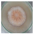
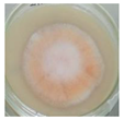
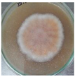
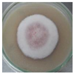
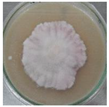
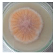
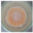
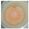
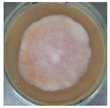
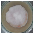
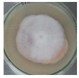
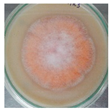
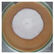
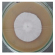
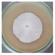
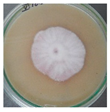
Table 3 shows the mycelial development of M. purpureus on quinoa flour-based culture media enriched with seven supplements. After 240 h of incubation, the greatest radial growth occurred with the sodium chloride supplement at pH 5, 6 and 7 when compared with the other six supplements at their respective pH levels (Figure 1), as well as showing greater pigmentation both on the front of the plate and on the back (images not shown). On the other hand, it is observed that with the glucose and fructose supplements the hyphae were pigmented, which indicated that a stationary phase had been reached where the secondary metabolites were formed in their majority, among them the pigments, in contrast to the white areas on the mycelium, which would indicate that they were mainly forming biomass, such as the media containing molasses, fermented fish, fish hydrolysate and monosodium glutamate supplements at pH 7; it should be noted that the slowest growth and deformed edges of the mycelium occurred with the latter supplement.

4. Conclusions

This research determined that the growth of M. purpureus on quinoa flour agar is affected by the type of supplementation which causes changes in the growth rate and morphology of the fungus. Sodium chloride at a concentration of 0.5% (w/v) and pH 6 showed the highest growth of 72.59 mm with a radial growth rate of 3.629 mm/day. Supplements such as glucose and fructose affected the early pigment production observed in the culture plates. Finally, the lowest growth was produced with a concentration of 0.5% monosodium glutamate at pH 7.

Author Contributions

Conceptualization, E.Q.-R.; M.S.-J. and J.J.M.; methodology, E.Q.-R. and F.T.-H.; software, E.Q.-R.; validation, M.S.-J.; formal analysis, E.Q.-R.; investigation, E.Q.-R. and F.T.-H.; resources, M.S.-J.; data curation, E.Q.-R.; writing—original draft preparation, E.Q.-R.; writing—review and editing, U.G.-B.; visualization, E.Q.-R. and J.J.M.; supervision, M.S.-J., V.C., U.G.-B. and J.J.M.; project administration, M.S.-J.; funding acquisition, M.S.-J. All authors have read and agreed to the published version of the manuscript.

Funding

This research was funded by CONCYTEC-PROCIENCIA under the Basic Research Project 2019-01 [contract 383-2019- FONDECYT].

Institutional Review Board Statement

Not applicable.

Informed Consent Statement

Not applicable.

Data Availability Statement

Summary data available upon request.

Acknowledgments

The authors gratefully acknowledge the financial support obtained from CONCYTEC-PROCIENCIA under the Basic Research Project 2019-01 [contract 383-2019-FONDECYT]. We would also like to thank the Laboratorio de Microbiología de Alimentos UNALM, Laboratorio de Biotecnología Ambiental-Biorremediación UNALM and the Centro de Investigação de Montanha (CIMO). U. Gonzales-Barron would like to thank the FCT for the national funding through the institutional scientific employment program contract.

Conflicts of Interest

The authors declare no conflict of interest.

References

  1. Hong, M.Y.; Seeram, N.P.; Zhang, Y. Heber D. Anticancer effects of Chinese red yeast rice versus monacolin K alone on colon cancer cells. J. Nutr. Biochem. 2008, 19, 448–458. [Google Scholar] [CrossRef] [PubMed]
  2. Akihisa, T.; Tokuda, H.; Yasukawa, K.; Ukiya, M.; Kiyota, A.; Sakamoto, N.; Suzuki, T.; Tanabe, N.; Nishino, H. Azaphilones, Furanoisophthalides, and Amino Acids from the Extracts of Monascus pilosus-Fermented Rice (Red-Mold Rice) and Their Chemopreventive Effects. J. Agric. Food Chem. 2005, 53, 562–565. [Google Scholar] [CrossRef] [PubMed]
  3. Bogsrud, M.P.; Ose, L.; Langslet, G.; Ottestad, I.; Strøm, E.C.; Hagve, T.A.; Retterstøl, K. HypoCol (red yeast rice) lowers plasma cholesterol—A randomized placebo-controlled study. Scand. Cardiovasc. J. 2010, 44, 197–200. [Google Scholar] [CrossRef] [PubMed]
  4. Zhang, X.; Liu, W.; Chen, X.; Cai, J.; Wang, C.; He, W. Effects and mechanism of blue light on Monascus in liquid fermentation. Molecules 2017, 22, 385. [Google Scholar] [CrossRef] [PubMed]
  5. Fabio, A.D.; Parraga, G. Origin, production and utilization of pseudocereals. In Pseudocereals—Chemistry and Technology; Haros, C.M., Schoenlechner, R., Eds.; Wiley & Sons: New York, NY, USA, 2017; pp. 1–27. [Google Scholar]
  6. Patrovsky, M.; Sinovska, K.; Branska, B.; Patakova, P. Effect of initial pH, different nitrogen sources, and cultivation time on the production of yellow or orange Monascus purpureus pigments and the mycotoxin citrinin. Food Sci. Nutr. 2019, 7, 3494–3500. [Google Scholar] [CrossRef] [PubMed] [Green Version]
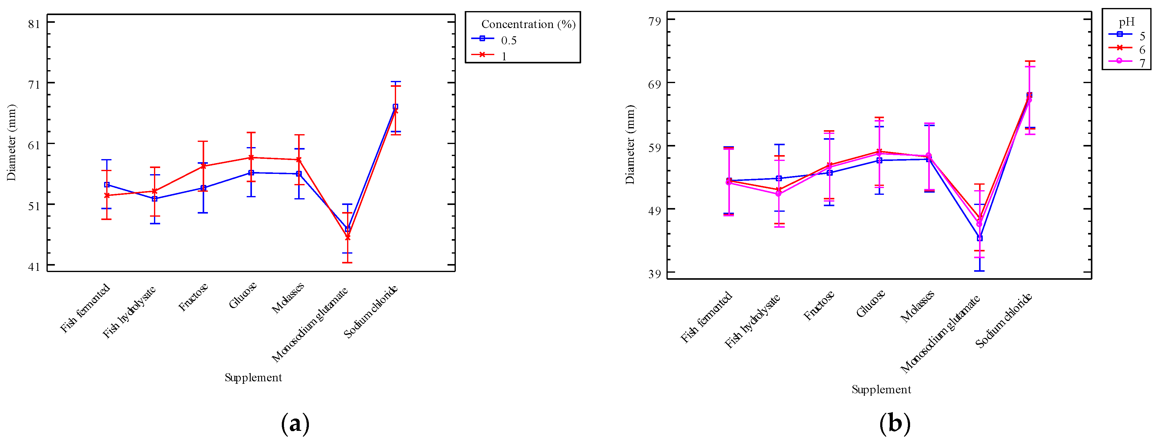
Figure 1. The figure shows (a) the interactions of supplement and substrate concentration in percentages; and (b) the interaction of supplement and pH on the radial growth of the fungus in vitro.
Blsf 18 00064 g001
Table 1. Analysis of variance for diameter (mm) obtained in vitro.
Sources of VariationSum of SquaresDfMean SquareF-Ratiop-Value
Main effects
A: Supplement4227.096704.51528.120.0000
B: Concentration (%)21.10121.0990.840.3611
C: pH9.0724.5330.180.8348
Interactions
AB122.11620.3520.810.5630
AC59.47124.9560.200.9983
BC0.5920.2930.010.9884
Residuals2405.399625.056
Total (Corrected)6844.82125
Table 2. Multiple range tests for the diameter (mm) per supplement used in quinoa flour-based culture media.
SupplementCountLS MeanLS SigmaHomogeneous Groups
Monosodium glutamate1846.1651.1798X
Fish hydrolysate1852.4211.1798X
Fish fermented1853.3421.1798X X
Fructose1855.4441.1798X X
Molasses1857.1471.1798X X
Glucose1857.4821.1798X
Sodium chloride1866.7661.1798X
Count: percentage of substrate (0.5, 1%) × pH (5, 6, 7) × three replicas; LS Mean: least squares mean; LS Sigma: least squares sigma.
Table 3. Mycelial development of M. purpureus in culture media based on quinoa flour supplemented with different sources of carbon, nitrogen and salts at 0.5% (w/v), at 3 pH levels (5, 6 and 7) after 10th days of incubation.
GlucoseFructoseMolassesFermented FishFish HydrolysateMonosodium GlutamateSodium Chloride
5Blsf 18 00064 i001Blsf 18 00064 i002Blsf 18 00064 i003Blsf 18 00064 i004Blsf 18 00064 i005Blsf 18 00064 i006Blsf 18 00064 i007
6Blsf 18 00064 i008Blsf 18 00064 i009Blsf 18 00064 i010Blsf 18 00064 i011Blsf 18 00064 i012Blsf 18 00064 i013Blsf 18 00064 i014
7Blsf 18 00064 i015Blsf 18 00064 i016Blsf 18 00064 i017Blsf 18 00064 i018Blsf 18 00064 i019Blsf 18 00064 i020Blsf 18 00064 i021
Publisher’s Note: MDPI stays neutral with regard to jurisdictional claims in published maps and institutional affiliations.