Article

Unveiling Biocompatibility: Comprehensive Study on Epoxy–Polyetheramine-Based Polymeric Nanogels in CHO-K1 Cell Line

by
Natalia Nascimento Silveira
*,
Heber Eduardo Andrada
,
Julia Mirian Paulino
,
Naiara Cristina da Silva Boaretto
,
Eduardo Ferreira Molina
and
Raquel Alves dos Santos
Department of Exact and Technological Sciences Research, University of Franca, Av. Dr. Armando de Sáles Oliveira, 201, Parque Universitario, Franca 14404-600, SP, Brazil
*
Author to whom correspondence should be addressed.
Future Pharmacol. 2025, 5(3), 54; https://doi.org/10.3390/futurepharmacol5030054
Submission received: 31 July 2025 / Revised: 12 September 2025 / Accepted: 16 September 2025 / Published: 18 September 2025

Abstract

Backgorund/Objectives: Advances in nanotechnology have enabled conventional compounds with low bioavailability to achieve their full therapeutic potential by ensuring targeted tissue delivery. In this context, polymeric nanogels have emerged as a promising option for drug delivery due to their high loading capacity and excellent in vivo stability. Objectives: Given the growing potential of nanogels in drug delivery, their cytotoxicity and genotoxicity must be evaluated to ensure safety in biotechnological applications. This study assessed the genotoxic safety of nanogels synthesized via the reaction of Jeffamine® T-5000 polyoxypropylene triamine (PPO) monomers (Huntsman Chemical, The Woodlands, TX, USA) and poly (ethylene glycol) diglycidyl ether (DPEG) in varying proportions: 1:1 (Nano11), 1:3 (Nano13), and 2:3 (Nano23) PPO/DPEG. Additionally, we determined which of the two components exhibited lower toxicity against the CHO-K1 cell line (Chinese hamster ovary). Methods: To achieve this, short- and long-term cytotoxicity experiments were conducted using the XTT colorimetric assay and clonogenic survival assay, alongside the micronucleus test and comet assay for genotoxicity analysis. Results: The cytotoxicity assays (XTT, clonogenic, and trypan blue) indicated that the nanogels did not exhibit cytotoxic effects at concentrations up to 100 μg/mL, while the genotoxicity assays revealed no evidence of DNA or chromosomal damage at these levels. Conclusions: These findings underscore the safety profile of Jeffamine® T-5000 as an effective carrier, demonstrating its compatibility with DPEG and positioning it as a highly promising and innovative solution for advanced drug delivery systems.

Graphical Abstract

1. Introduction

Factors such as in vivo instability, low bioavailability and solubility, poor absorption in the body, challenges in targeted delivery, suboptimal efficacy, and potential adverse effects of medications pose significant barriers to a drug achieving its full therapeutic potential [1,2].
Nanotechnology offers significant promise for drug delivery by ensuring that therapeutic agents reach their intended target sites while minimizing off-target dispersion to non-specific tissues. Additionally, nano-sized materials can penetrate the extracellular matrix and be engineered to control their clearance rates [3,4,5,6]. This technology facilitates the optimization of targeted drug delivery to specific cells or tissues, enables the transcytosis of drugs across epithelial barriers, supports the intracellular delivery of large macromolecular therapeutics to their sites of action, and allows for real-time monitoring of the in vivo efficacy of these agents [3,4].
Among the wide range of nanocarriers options, hydrogels have emerged as particularly promising in pharmacology. These are three-dimensional, hydrophilic, cross-linked polymeric networks capable of absorbing and retaining large amounts of water or biological fluids without dissolving, thereby maintaining their structural integrity. Their ease of synthesis, tunable porosity, compatibility with biomolecules, biodegradability, and stimulus-responsive properties position them as strong candidates for drug delivery [7,8,9]. Nanogels represent a promising innovation in this domain, capable of adapting to reach target sites and minimizing adverse effects through localized and sustained drug release, thus reducing the need for repeated administrations. They combine characteristics of both hydrogels and nanoparticles, making them ideal for the nanoscale transport of bioactive molecules. Defined as nanoscale cross-linked polymeric networks, nanogels offer high aqueous stability and a large surface-area-to-volume ratio, enabling efficient encapsulation and delivery of therapeutic agents. Their tunable size, biocompatibility, chemical versatility, and responsiveness to physiological stimuli further enhance their potential as smart carriers in drug delivery applications [10,11]. The high loading capacity and in vivo stability of nanogels make them a compelling option for drug delivery. Advances in nanomedicine have enabled the encapsulation of biomolecules, as well as the oral administration of nucleoside analogs, oligonucleotides, or small interfering RNA (siRNA) fragments for treating cancers and viral infections [12,13].
Numerous studies in the literature describe the use of Jeffamines in the construction of nanogels, with the aim of developing controlled drug delivery systems and exploring additional applications [14,15,16,17]. However, there is a lack of in-depth analysis regarding the cytotoxic and genotoxic properties of these formulations, particularly when combined with diepoxy polyethylene glycol (DPEG). The reaction between Jeffamine® and DPEG was selected due to the straightforward interaction between the amine groups and the epoxide structures, yielding a nanogel that is simple to synthesize. Notably, this reaction is not directly pH-dependent and is catalyzed by water [18], which serves as a unique, sustainable, and cost-effective solvent [17].
When introducing a new drug delivery system, ensuring its biocompatibility, particularly in terms of toxicity and genotoxicity, is paramount. This requires establishing an exposure threshold for the compound that poses no toxicological or carcinogenic risks to humans. Genotoxicity assessments are conducted to elucidate the mechanisms of carcinogenicity, and such evaluations have become an integral component of human-exposure risk assessment processes [19]. The objective of this study is to evaluate the cytotoxic and genotoxic effects of polymeric nanogels synthesized from the reaction between Jeffamine® T-5000, a high-molecular-weight triamine with a polypropylene oxide backbone, and a diepoxide with a polyethylene glycol backbone. The aim is to identify safe concentration ranges for future applications, while also analyzing the individual components (monomers) separately to determine which may be responsible for any observed adverse effects.

2. Materials and Methods

2.1. Materials

Poly (ethylene glycol) diglycidyl ether (DPEG; chemical formula: C3H5O2-(C2H4O)n-C3H5O; average molecular weight (Mw) = 500 g/mol; CAS 26403-72-5) and Jeffamine® T-5000 polyoxypropylene triamine (polyetheramine with a polypropylene oxide backbone; Mw = 5000 g/mol) (Huntsman Chemical, The Woodlands, TX, USA) were kindly provided by Huntsman Chemical (The Woodlands, TX, USA). All reagents were used as received, without further purification.

2.2. Synthesis of the Polyetheramine–Epoxide Nanogels (PPO–DPEG)

PPO–DPEG nanogels were synthesized through an aggregation polymerization process, as adapted from established protocols [17,20]. Both monomers—polyoxypropylene triamine (PPO) and poly (ethylene glycol) diglycidyl ether (DPEG)—are water-soluble, facilitating their manipulation in an aqueous environment. To prepare the nanogels, each monomer was dissolved separately in 5 mL of deionized water and maintained under magnetic stirring at 25 °C for 60 min to achieve complete solubilization.
The PPO solution was then added dropwise to the DPEG solution over approximately 20 s, promoting a controlled interaction between the amine groups of PPO and the epoxide groups of DPEG. This gradual addition is crucial to prevent rapid precipitation or uncontrolled gelation, ensuring uniform crosslinking. The resulting 10 mL reaction mixture was stirred for an additional 1 h at room temperature to complete nanogel formation.
The total monomer concentration in the aqueous medium was maintained at 10% (w/v). Nanogels were prepared using three distinct stoichiometric ratios of PPO to DPEG: 1:1 (designated as Nano11), 1:3 (Nano13), and 2:3 (Nano23). The specific masses used were as follows: for the 1:1 ratio, 0.90 g of PPO and 0.10 g of DPEG; for the 1:3 ratio, 0.77 g of PPO and 0.23 g of DPEG; and for the 2:3 ratio, 0.87 g of PPO and 0.13 g of DPEG (summarized in Table 1). The freshly prepared nanogel dispersions exhibited a basic pH of approximately 10, primarily due to unreacted amine groups from PPO. Prior to biological assays, the dispersions were neutralized to a physiological pH of 7.0 by the careful, stepwise addition of 0.1 M HCl.

2.3. Characterization of the Polymeric Nanogels

The hydrodynamic diameter (Dh) and polydispersity index (PDI) of the nanogels were determined by Dynamic Light Scattering (DLS). Zeta potential (ζ), which is indicative of the nanogels’ surface charge, was also measured. All measurements were performed using a Zetasizer Lab Blue system (ZSU3100, Malvern Panalytical, Malvern, UK) equipped with an OBIS solid-state laser source (λ = 633 nm). Zeta potential values were reported in millivolts (mV).
To confirm that nanogel formation resulted exclusively from the addition reaction of the monomers, individual aqueous solutions of Jeffamine® T-5000 and DPEG were also analyzed under identical conditions.

2.4. Transmission Electron Microscopy (TEM)

The morphology of the nanogels was characterized using Transmission Electron Microscopy (TEM) with a JEM-100CXII instrument (JEOL Ltd., Tokyo, Japan) operated at an accelerating voltage of 100 kV. Prior to imaging, nanogel dispersions were diluted 10-fold in deionized water to achieve optimal particle density and minimize aggregation artifacts. For each sample, a drop of the diluted nanogel solution was placed on a copper grid, followed by a drying process at room temperature.

2.5. Cell Cultures

All cell culture procedures were performed under sterile conditions. The CHO-K1 cell line (Chinese hamster ovary) was acquired from the Rio de Janeiro Cell Bank (Rio de Janeiro, Brazil). Cells were routinely cultured at 37 °C in a humidified 5% CO2 atmosphere.
The growth medium consisted of a 1:1 mixture of Ham’s F-10 Medium and Dulbecco’s Modified Eagle Medium (DMEM) (both from Sigma-Aldrich, St. Louis, MO, USA). This base medium was supplemented with 10% (v/v) fetal bovine serum (FBS) (Sigma-Aldrich, St. Louis, MO, USA), 1% (v/v) penicillin/streptomycin solution (100 U/mL penicillin and 100 µg/mL streptomycin) (Sigma-Aldrich, St. Louis, MO, USA), and 10 µg/mL kanamycin sulfate (Sigma-Aldrich, St. Louis, MO, USA).
For routine maintenance and experimental treatments, cells were cultured in 25 cm2 cell culture flasks (TPP, Trasadingen, Switzerland) or multi-well plates (e.g., 96-well or 24-well) as required for specific assays.

2.6. Cell Viability—XTT

Cell viability was assessed using the 2,3-bis-(2-methoxy-4-nitro-5-sulfophenyl)-2H-tetrazolium-5-carboxanilide (XTT) assay, which relies on the reduction of the tetrazolium dye by mitochondrial dehydrogenases in viable cells to form a colored formazan derivative.
For the XTT assay, CHO-K1 cells were seeded at a density of 3 × 10⁴ cells/well in 96-well microplates containing complete growth medium. After 24 h of incubation to allow cell attachment, the cells were treated with various concentrations of DPEG, PPO, Nano11, Nano13, and Nano23, ranging from 10 to 10,000 μg/mL. Untreated cells served as the negative control, while cells treated with 10% (v/v) dimethyl sulfoxide (DMSO) were used as the positive control for cell death.
Colorimetric analysis was performed after 24, 48, and 72 h of treatment. At each time point, the culture medium was removed from the wells, and cells were washed once with phosphate-buffered saline (PBS). Subsequently, the cells were incubated at 37 °C with a XTT labeling mixture, prepared according to the manufacturer’s instructions (Cell Proliferation Kit II; Roche Diagnostics, Basel, Switzerland), in phenol red-free Dulbecco’s Modified Eagle Medium (DMEM). After a 4 h incubation period, the absorbance was measured using a microplate spectrophotometer at a test wavelength of 492 nm and a reference wavelength of 690 nm [21].

2.7. Clonogenic Assay

To evaluate the long-term survival and proliferative capacity of CHO-K1 cells following treatment with the nanogels, a clonogenic survival assay was performed according to the protocol described by Franken et al. (2006) [22].
Briefly, 500 cells per well were seeded into 6-well plates containing 2 mL of complete growth medium (HAM F10 + DMEM supplemented with 10% fetal bovine serum and antibiotics, as detailed in the cell culture section). Cells were allowed to attach for 4 h to ensure uniform plating efficiency. Subsequently, the cells were treated for 24 h with varying concentrations of DPEG, PPO, Nano11, Nano13, or Nano23, ranging from 10 to 1000 μg/mL. The negative control consisted of untreated cells, while the positive control was treated with doxorubicin hydrochloride (0.5 μM), a known cytotoxic agent that induces DNA damage and inhibits colony formation.
After the 24 h treatment period, the medium was aspirated, and the cells were gently washed twice with pre-warmed phosphate-buffered saline (PBS, 1X) to remove residual compounds and prevent carryover effects. Fresh complete growth medium (2 mL per well) was then added, and the plates were returned to the incubator (37 °C, 5% CO2) for colony development. Cultures were monitored daily using an inverted phase-contrast microscope (e.g., at 10× magnification) to assess growth without disturbing the plates.
After approximately 7 days (adjusted based on observed colony size to ensure maturity), when control colonies had reached a sufficient size, the medium was removed, and the plates were washed with 5 mL of pre-warmed PBS (1X, 37 °C) per well. Fixation was achieved by adding a methanol:acetic acid:distilled water solution (1:1:8, v/v/v) at room temperature for 30 min, which preserves cellular morphology and prevents colony disruption. Following fixation, the plates were stained with 5 mL of Giemsa dye diluted in Sorensen buffer (1:20, v/v) at 37 °C for 15–30 min, enhancing visibility of the colonies through nuclear and cytoplasmic contrast.
Colonies were defined as clusters containing more than 50 cells, indicating sustained proliferative potential. These were manually counted using a stereomicroscope at 16x magnification to ensure accuracy and minimize edge effects. The plating efficiency (PE) was calculated for the negative control as the ratio of colonies formed to cells seeded, with the control group normalized to 100% survival. Survival fractions (SF) for treated groups were then determined relative to the negative control using the following equation:
S F = n u m b e r   o f   c o l o n i e s   c o u n t e d   i n   e a c h   t r e a m e n t n u m b e r   o f   c o l o n i e s   o b s e r v e d   i n   t h e   n e g a t i v e   c o n t r o l × 100

2.8. Trypan Blue Exclusion Test

To evaluate cell viability, 3 × 10⁴ cells/well were seeded in 96-well plates in complete medium growth. After 24 h, cells were treated with concentrations ranging from 10 to 10,000 μg/mL of DPEG, PPO, Nano11, Nano13, and Nano23. The negative control did not receive treatment, and the positive control was treated with 10% (v/v) DMSO. The analysis was performed 4 h after the treatment. To do so, cells were washed with 1× PBS and collected using trypsin. A 1:1 proportion of trypan blue and cell suspension was prepared to evaluate the percentage of viable cells using the TC20 Automated Cell Counter.

2.9. Cytokinesis-Blocked Micronucleus Assay (CBMN)

To assess the potential genotoxic effects of the tested compounds, the in vitro micronucleus (MN) test was conducted in accordance with the Organisation for Economic Co-operation and Development (OECD) Test Guideline 487 (TG 487) [23]. The Chinese hamster ovary (CHO-K1) cell line, sourced from the Rio de Janeiro Cell Bank (Rio de Janeiro, Brazil), was employed, and cells were seeded at a density of 1 × 10⁵ cells per 25 cm2 culture flask (TPP, Trasadingen, Switzerland) in complete growth medium, consisting of a 1:1 mixture of Ham’s F-10 and Dulbecco’s Modified Eagle Medium (DMEM; Sigma-Aldrich, St. Louis, MO, USA) supplemented with 10% fetal bovine serum (FBS; Sigma-Aldrich, St. Louis, MO, USA) and antibiotics (penicillin/streptomycin at 10 mL/L and kanamycin sulfate at 10 mg/L; all from Sigma-Aldrich). The flasks were incubated at 37 °C in a humidified atmosphere with 5% CO2 for 24 h to allow cell attachment and entry into the exponential growth phase.
Following the initial 24 h incubation, cells were treated with concentrations of 10 and 100 μg/mL of DPEG, PPO, Nano11, Nano13, and Nano23, along with Cytochalasin B (Sigma-Aldrich, USA), which blocked cytokinesis. The positive control (0.125 µM doxorubicin hydrochloride) was also treated at this time. After a 24 h treatment period, cells were washed, trypsinized, resuspended in a hypotonic solution (0.3% KCl), and then centrifuged. The cell suspension was then resuspended in 3 mL of fixative (methanol: acetic acid, 3:1, v/v) along with a drop of 1% formaldehyde and carefully homogenized. After centrifugation, the supernatant was discarded, and the cell suspension was dropped onto slides that had been previously cleaned and covered with a film of cold distilled water. Slides were stained with a 3% Giemsa solution diluted in Sorensen buffer (0.06 M Na2HPO4 and 0.06 M KH2PO4, pH 6.8) for 7 min. They were then washed with distilled water and air-dried at room temperature.
For the determination of the Nuclear Division Index (NDI), 1000 viable cells with well-preserved cytoplasm were counted per coded slide using a transmitted light microscope (40× objective), noting cells with 1 to 4 nuclei. The NDI was calculated using the following formula:
N D I = M 1 + 2 ( M 2 ) + 3 ( M 3 ) + 4 ( M 4 ) N
where M1 to M4 represent the number of cells with 1, 2, 3, and 4 nuclei, respectively, and N is the total number of viable cells. Additionally, micronucleated cells were counted among 1000 binucleated cells, and the micronucleus frequency was estimated. The criteria for micronucleus identification were based on those described by Fenech (2007) [24].

2.10. Single Cell Gel Electrophoresis Assay

To perform the comet assay, 2 × 10⁵ cells per well were seeded in 24-well plates and subsequently incubated for 24 h in a humidified chamber at 37 °C with 5% CO2. Following this incubation, treatments were applied using concentrations ranging from 10 to 1000 μg/mL of DPEG, PPO, Nano11, Nano13, and Nano23, as determined by prior cytotoxicity analyses. After treatment, the cells were incubated for an additional 4 h and then subjected to trypsinization. Subsequently, 10 μL of the cell suspension was mixed with low-melting-point agarose (0.75%). This mixture was homogenized and deposited onto a microscope slide precoated with normal-melting-point agarose (1.5%). The slides were covered with a coverslip and kept at −20 °C for 5 min to allow solidification of the low-melting-point agarose layer. The coverslip was then removed, and the slides were immersed in lysis solution (2.5 M NaCl, 100 mM Na2EDTA, 10 mM Tris, 1% Triton X-100, and 10% DMSO, pH 10) at 4 °C. After 4 h of lysis, the slides were incubated in alkaline buffer solution (0.3 M NaOH and 1 mM Na2EDTA, pH > 13) for 20 min. Electrophoresis was performed in the same alkaline buffer at 25 V and 300 mA for 25 min. Following electrophoresis, the slides were immersed in neutralization buffer (0.4 M Tris-HCl, pH 7.5) for 15 min and then fixed in absolute ethanol for 5 min [25]. The slides were subsequently stained with the fluorescent dye propidium iodide. Finally, the slides were visualized at 400× magnification using a Zeiss Axio Scope A1 fluorescence microscope (Carl Zeiss AG, Oberkochen, Germany), coupled to a MetaSystems CoolCube 1 camera (MetaSystems, Altlussheim, Germany). For each treatment, two slides were prepared, and 50 nucleoids per slide were analyzed using MetaSystems Comet Imager V2.2 software. Results were tabulated based on the tail intensity of each nucleoid. Each treatment was performed in triplicate.

2.11. Statistical Analysis

The results were analyzed using GraphPad Prism software (version 8.0). All data underwent descriptive statistical analysis to calculate the mean ± standard deviation (SD). The appropriate statistical test was selected based on data distribution and variance, followed by Tukey’s post hoc test [26], with a significance level of p < 0.05 considered as the minimum threshold for determining differences between experimental groups and the negative control.

3. Results

3.1. Characterization

The synthesis of nanogels was conducted according to the procedures outlined in the previous section, utilizing appropriately functionalized polymers. The reaction between Jeffamine® (Figure 1A) and DPEG (Figure 1B) led to the formation of nanogels, as illustrated in the structure shown in Figure 1C. This process produced nanogels with distinct characteristics, arising from the interactions between these two compounds during the reaction.
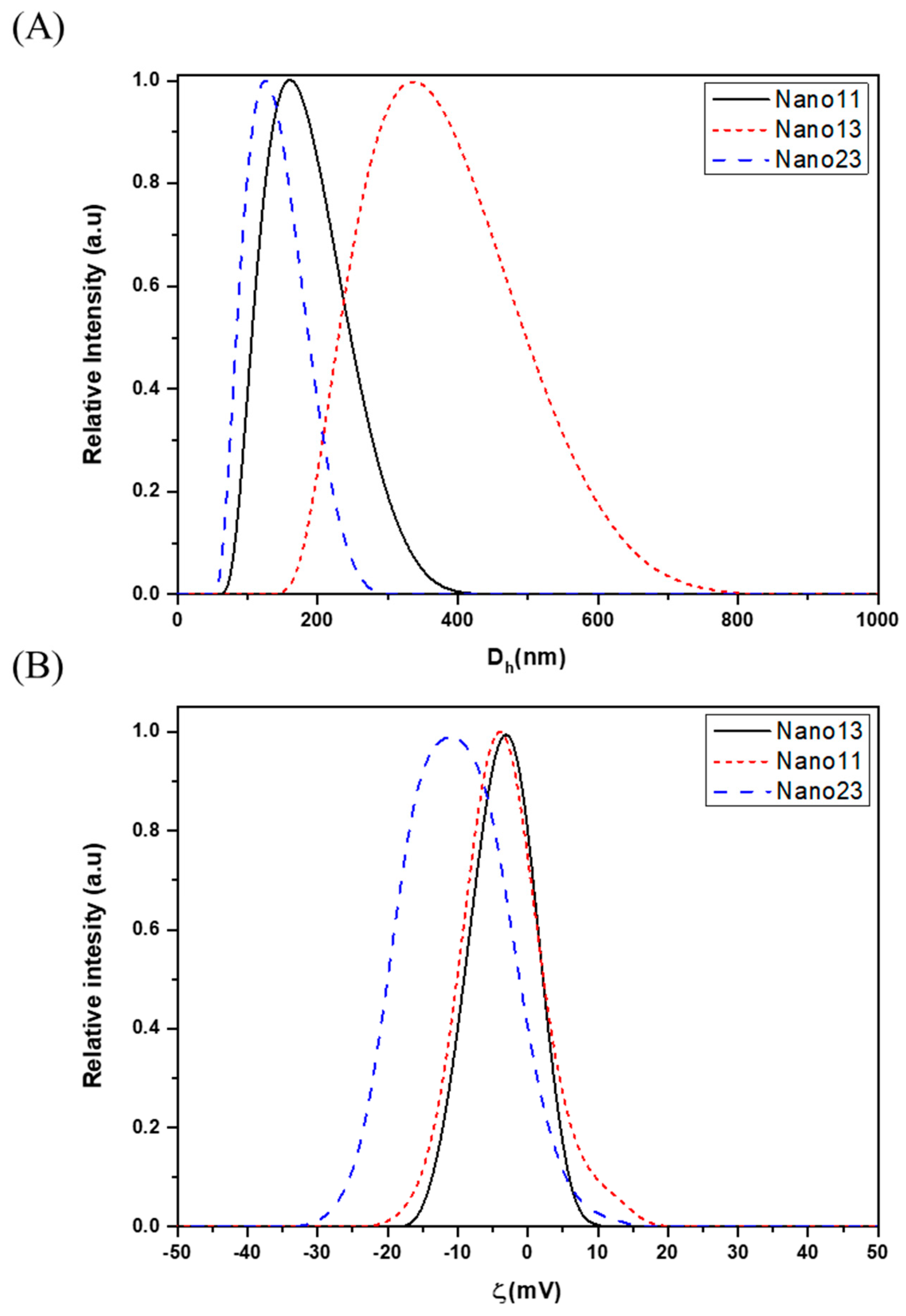
Figure 2 shows a graphical representation of the data obtained via Dynamic Light Scattering (DLS).
The hydrodynamic diameter (Dh) distribution of the synthesized nanogels is presented in Figure 2A. Nano23 and Nano11 exhibit a population with a Dh ranging from 130 to 170 nm. These nanogels consist of smaller gel particles that aggregate to form larger structures. This aggregation effect is more pronounced in Nano13, whose Dh is almost twice that of the others. Figure 2B shows a zeta potential (ζ) graph for the different synthesized nanogels. The ζ values are low, ranging from −3 mV to −13 mV in all cases. As indicated in Figure 2B, these systems exhibit poor stability in solution (for colloidal stability, |ζ| should be ≥±30 mV) and tend to aggregate over time [27,28,29]. The surface charge of the nanogels arises from the presence of both amine and alcohol groups on their surface. This surface charge is closely related to the pH of the medium: at pH 7 or above, the charge is negative, whereas, at pH values below 7, the amine groups become protonated, resulting in a more positive surface charge [17]. Table 2 presents the data for Dh, PDI, and zeta potential (ζ) for the different polymeric nanogels synthesized at pH 7.

3.2. Morphological Analysis of Nanogels by TEM

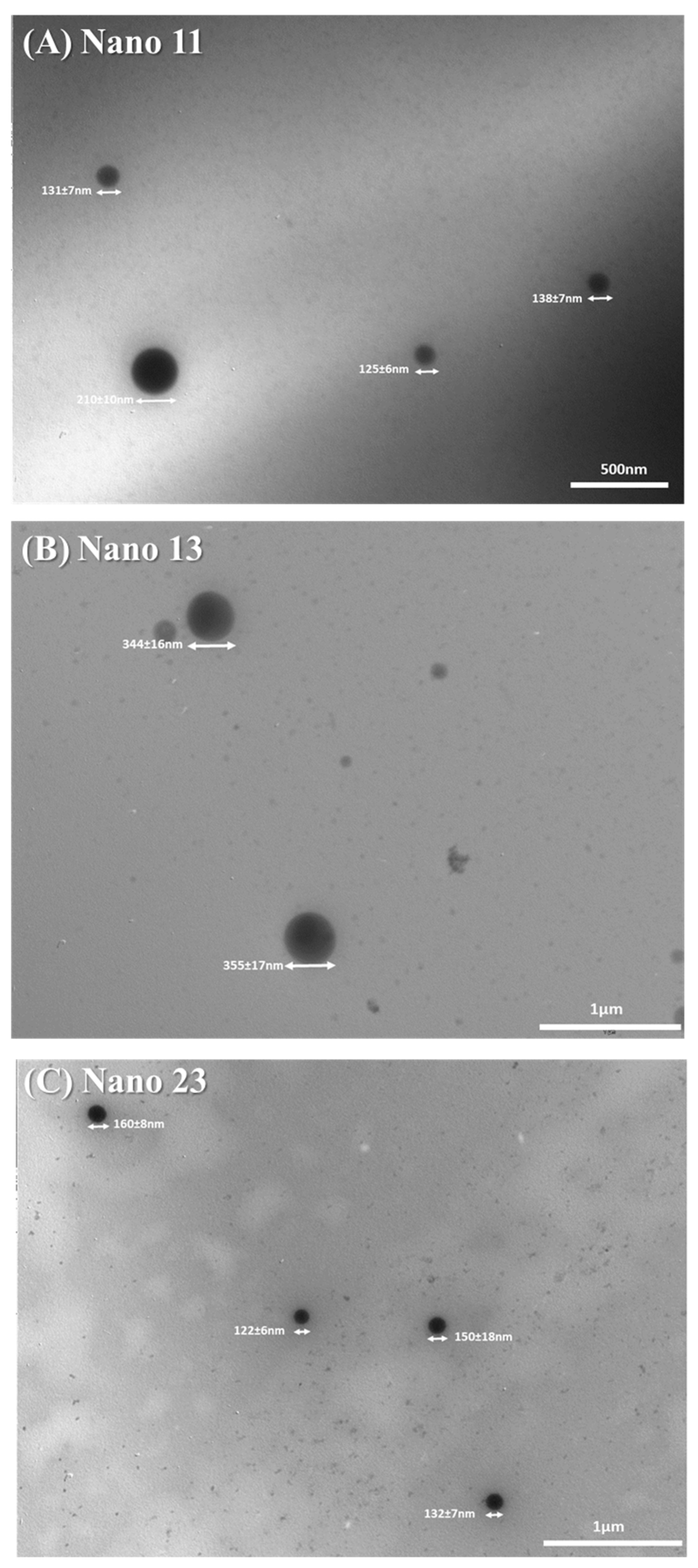
Measurements were performed to obtain data on the morphology and sizes of the synthesized polymeric nanogels. Figure 3 presents micrographs of the synthesized nanogels, along with the observed sizes. A correlation was established between the sizes obtained via TEM and the hydrodynamic diameters measured by DLS. Additionally, the micrographs reveal that these polymeric nanogels consist of aggregates of numerous smaller polymeric particles or nanogels.

3.3. Effect of Nanogels on CHO-K1 Cell Viability (XTT Assay)

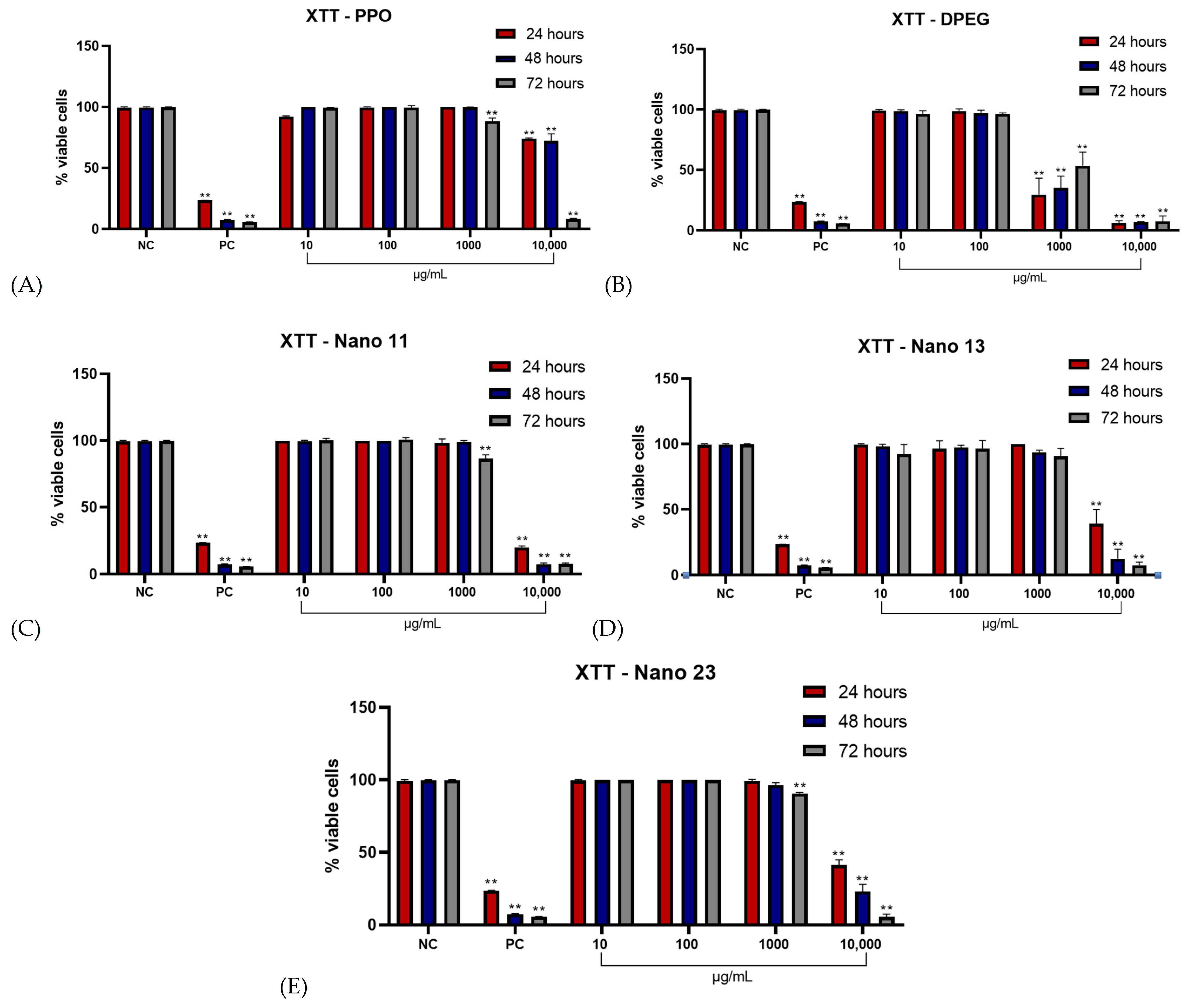
To conduct a thorough analysis of the components and nanogels in this study, cytotoxic assays were first performed to identify subcytotoxic concentrations suitable for accurate genotoxic evaluation. The initial assay employed was the XTT cell viability test, which quantifies the capacity of viable cells to reduce tetrazolium dye through mitochondrial dehydrogenases, yielding a colored formazan derivative [30]. In this assay, the components PPO and DPEG, along with the nanogels Nano11, Nano13, and Nano23, were assessed over treatment periods of 24, 48, and 72 h at concentrations ranging from 10 to 10,000 μg/mL. The results demonstrated that the highest concentration tested induced cytotoxic effects in all samples against the Chinese hamster ovary (CHO-K1) cell line. Furthermore, only the DPEG sample displayed cytotoxicity at 1000 μg/mL across all treatment durations (Figure 4B). With the exception of Nano13, all samples showed statistically significant cytotoxicity at 1000 μg/mL after 72 h of treatment.
When comparing the 1000 μg/mL concentration of the produced nanogels and their constituent reagents, a difference in behavior is observed relative to the DPEG reagent, indicating a statistically significant difference in the percentage of viable cells (Figure 5).

3.4. Long-Term Cell Survival and Proliferative Capacity

The XTT assay was employed as a short-term test to evaluate cell viability. Subsequently, a clonogenic assay was performed to assess the cells’ capacity to form colonies following treatment. The results from the XTT assay allowed us to select concentrations that permitted sufficient cell survival for colony formation. Accordingly, concentrations of 10, 100, and 1000 μg/mL were chosen for further cytotoxicity analysis, as these did not induce a statistically significant decrease in cell viability for most samples. However, the highest concentration tested led to a significant reduction in the survival fraction for all samples, with the epoxy component proving uniquely cytotoxic, preventing colony formation starting at a concentration of 100 μg/mL (Figure 6A).
Similar to the comparative analysis in the prior clonogenic assay at higher concentrations, an evaluation was conducted at 100 μg/mL for the study samples (including the synthesized nanogels and their constituent reagents). As anticipated based on the concentration–response trends observed, the samples demonstrated statistically significant differences in survival fractions relative to DPEG (Figure 7).

3.5. Effect of Nanogels on CHO-K1 Cell Viability (Trypan Blue Exclusion Test)

Prior to advancing to the definitive genotoxicity evaluation via the comet assay, it was essential to identify subcytotoxic concentrations optimized for the assay’s distinct experimental parameters, particularly its shorter 4 h treatment duration. This step was crucial to avoid confounding cytotoxicity-induced artifacts in DNA damage measurements, ensuring that any observed genotoxic effects could be attributed specifically to the test substances rather than overt cell death. Accordingly, the Trypan Blue Exclusion Test—a rapid, dye-based method for assessing cell membrane integrity and viability—was employed across the full range of study concentrations for all samples, including the nanogel formulations (Nano11, Nano13, Nano23) and their constituent reagents (PPO, DPEG, and epoxy).
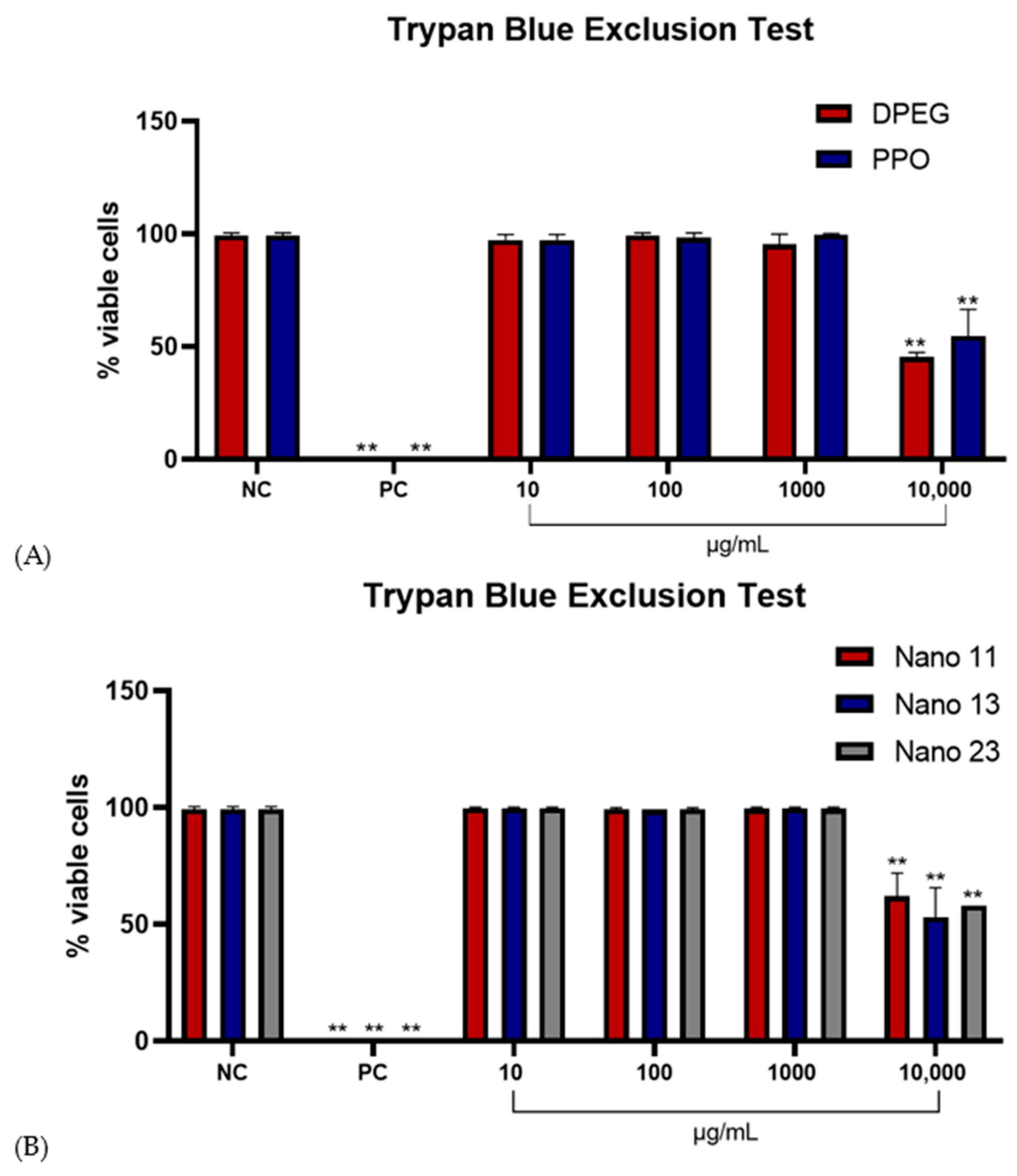
As anticipated based on patterns from preceding assays, only the highest concentration (10,000 μg/mL) elicited a statistically significant reduction in the percentage of viable cells across all samples, indicating membrane compromise and potential necrotic or apoptotic induction (Figure 8). This concentration was therefore excluded from subsequent comet assay protocols to maintain assay sensitivity and specificity. In contrast, lower concentrations (e.g., 10–1000 μg/mL) preserved cell viability above predefined thresholds (typically >70–80% viability for genotoxicity testing), allowing for reliable progression to DNA strand break analysis without baseline cytotoxicity interference.

3.6. Evaluation of Genotoxic Effects by Micronucleus Formation

Short- and long-term cytotoxicity assays were essential for determining subcytotoxic concentrations appropriate for subsequent genotoxicity evaluations. In this study, concentrations of 10 and 100 μg/mL were employed in the micronucleus assay. The results demonstrated that the DPEG sample exhibited genotoxicity at 100 μg/mL. In contrast, none of the other samples induced DNA damage in the CHO-K1 cell line (Table 3). Furthermore, in a comparative analysis, the PPO, Nano 11, Nano 13, and Nano 23 samples displayed statistically significant differences in micronucleus frequency relative to the DPEG sample (Table 4).

3.7. Comet Assay

The comet assay is a method for evaluating the extent of DNA damage induced by various substances prior to the activation of cellular DNA repair mechanisms. This technique is particularly valuable because it enables researchers to assess the genotoxic potential of a substance through analysis of the initial damage inflicted. In this study, the chosen parameter for damage evaluation was comet tail intensity, which quantifies the fragmented DNA migrating from the nucleus under an electrophoretic field [31].
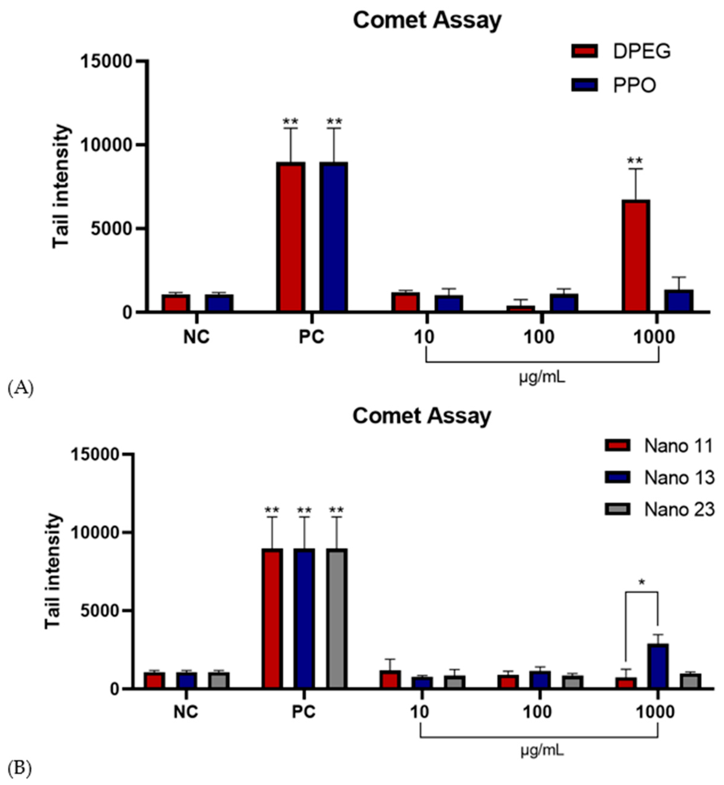
Analysis of this parameter revealed that, except for the 1000 μg/mL concentration of the DPEG sample, none of the other tested samples induced observable DNA damage in the CHO-K1 cell line (Figure 9). These findings suggest that, aside from the high concentration of DPEG, the tested components and nanogels did not elicit significant genotoxic effects under the experimental conditions employed.
Comparison of tail intensities at the 1000 μg/mL concentration across all samples revealed a statistically significant difference between the DPEG sample and the others, indicating greater DNA damage in cells exposed to DPEG (Figure 10).

4. Discussion

Nanogels represent a class of nanomaterials consisting of sub-micron-sized, three-dimensionally cross-linked polymer networks. These structures are composed of hydrogel particulates at the nanoscale and exhibit characteristics of both nanoparticles and hydrogels [32,33]. In drug delivery applications, nanogels emerge as an ideal carrier owing to their high loading capacity, biocompatibility, and stability, which have garnered significant attention in recent years [33]. According to Sharma et al. (2016) [34], nanogels are among the most effective systems for controlled drug release. Key attributes that distinguish them from other drug delivery options include high biocompatibility, ease of administration, versatility in encapsulating both hydrophilic and hydrophobic drugs, and compatibility with a wide array of small molecules [34,35,36,37,38,39,40,41].
Although nanogels represent a relatively novel class of materials in the scientific literature, several researchers have investigated their formulations and potential applications in drug delivery. For instance, He et al. (2015) [42] developed a nanogel based on lipoproteins and sodium carboxymethyl cellulose that successfully delivered doxorubicin (DOX) to cancer cells. Similarly, Gerecke et al. (2017) [43] examined the biocompatibility and characterization of polyglycerol-based nanogels in normal human keratinocyte (NHK) cells. In another study, Ansari et al. (2019) [44] employed zinc gluconate-loaded chitosan nanoparticles to achieve effective antiarthritic effects in rats. A common thread among these investigations is the evaluation of not only drug delivery efficacy but also the safety profile of blank (unloaded) nanogels.
According to Chacko et al. (2012) [45], an ideal nanogel-based drug delivery system should exhibit several key features, including robust encapsulation stability, responsiveness to external stimuli, capabilities for both passive and active targeting, low or negligible toxicity, and straightforward synthesis. The authors further emphasize that the toxicity profile of a nanogel critically determines its viability as a drug delivery vehicle, underscoring the necessity of rigorous toxicity screening for novel candidates. Moreover, the broader literature highlights the importance of evaluating nanogel toxicity. Guidelines from organizations such as the Organisation for Economic Co-operation and Development (OECD, 2023) [23] provide standardized protocols to ensure the safety of materials intended for pharmaceutical applications. Examples of successful nanogel systems are presented in Table 5.
To address the existing gap in comprehensive biocompatibility analysis and the characterization of varying proportions within the PPO–DPEG nanogel system, as previously described by Andrada et al. (2024) [17], this study employed an aggregation polymerization process. This approach aligns with the key features outlined by Chacko et al. (2012) [45], representing a straightforward methodology for nanogel synthesis. Data from Dynamic Light Scattering (DLS) revealed that, unlike Nano13, the synthesized nanogels Nano23 and Nano11 displayed hydrodynamic diameters ranging from 130 to 170 nm, resulting from aggregates of smaller particles. In contrast, the Nano13 nanogel exhibited a more pronounced aggregation effect, with diameters nearly twice as large. Particle shape and size are critical factors in processes that govern drug delivery efficiency; notably, particles exceeding 5 μm in size are typically not internalized by cells [46]. Zeta potential measurements indicated low values (ranging from −3 mV to −13 mV), which suggest limited colloidal stability in solution and a propensity for aggregation over time. However, this aggregation may paradoxically contribute to enhanced stability under certain conditions, as supported by prior studies [17,27,28].
Transmission Electron Microscopy (TEM) confirmed the size and morphology of the previously described nanogels, verifying these characteristics and enabling the observation of aggregates composed of smaller polymeric particles. Ahmad et al. (2022) [47] reported that nanoparticles smaller than 300 nm are generally the least toxic and most biocompatible with various cell lines, which aligns with the properties of the present nanogels as characterized by Dynamic Light Scattering (DLS) and TEM.
The Organisation for Economic Co-operation and Development (OECD, 2023) [23] recommends the micronucleus assay for evaluating the genotoxic potential of chemicals. This assay detects fragments that may arise from acentric chromosomes or whole chromosomes lost during the anaphase stage of cell division, providing a foundation for investigating chromosomal damage in cells exposed to a test substance that subsequently undergoes division. For a comprehensive biocompatibility analysis, cytotoxic assays are essential to identify subcytotoxic concentrations, which can then be used in conjunction with genotoxic assays.
The literature highlights several widely used in vitro assays for cytotoxicity and genotoxicity evaluation, including the MTT assay, trypan blue dye exclusion test, and comet assay [47,48]. In line with OECD guidelines, this study also incorporated micronucleus and clonogenic survival assays. The XTT assay, which assesses cell viability via mitochondrial dehydrogenase activity, revealed that concentrations up to 1000 μg/mL induced cytotoxic effects in most samples across 24, 48, and 72 h treatments, with the exception of DPEG. The clonogenic assay validated the viability of selected concentrations (10 and 100 μg/mL), as they permitted colony formation, rendering them suitable for subsequent genotoxic analysis. The Trypan Blue Exclusion Test identified viable subcytotoxic concentrations for the comet assay under a shorter 4 h treatment, excluding the highest concentration due to a significant decline in cell viability. Notably, most biologically compatible and efficient drug delivery systems exhibit no cytotoxic effects at concentrations below 400 μg/mL, as determined by the XTT assay [6,42,43,44]. This observation is consistent with the XTT results for PPO–DPEG, which showed no cytotoxicity up to 1000 μg/mL.
Pinelli et al. (2020) [15] demonstrated the successful development of a nanogel system based on the reaction between polyethylene glycol (PEG) and Jeffamine®, where cells maintained high viability post-treatment, confirming the system’s in vitro biocompatibility. However, while their study established overall biocompatibility, our research extends this by determining optimal concentrations that preserve biocompatibility across varying proportions tested, offering a more nuanced understanding of concentration-dependent performance. Similarly, Castan et al. (2017) [14] evaluated the cytotoxicity of Jeffamines in isolation. One variant, PPO 2000, which differs in chemical structure and molecular weight from the Jeffamine ® used here, exhibited significant cytotoxicity at a low concentration of 1.95 μM (equivalent to 3.9 μg/mL) under identical experimental conditions (XTT assay and cell line), demonstrating that the chemical structure of the Jeffamines component affects the biocompatibility of the experimental model.
To further elucidate the safety profile of the PPO–DPEG nanogel systems, genotoxicity assessments were conducted in alignment with the cytotoxic findings. As anticipated from the preliminary cytotoxicity analyses, only the epoxy component demonstrated genotoxic effects at a concentration of 100 μg/mL, whereas no other samples induced DNA damage in CHO-K1 cells under the tested conditions. The comet assay, a sensitive method for detecting initial DNA lesions such as single- and double-strand breaks, alkali-labile sites, and incomplete excision repair sites, revealed that, apart from DPEG at the highest tested concentration of 1000 μg/mL, none of the nanogel components or formulations caused significant DNA damage. This suggests that, within the parameters of this study, the PPO–DPEG nanogels and their constituent materials exhibit a generally non-genotoxic profile, which is a critical attribute for biocompatible drug delivery vehicles.
Comparative analysis with the existing literature provides valuable context for these results. For instance, Castan et al. (2017) [14] evaluated the genotoxic potential of various Jeffamines, including PPO 2000, using both micronucleus and comet assays. They reported that PPO 2000 induced detectable DNA damage at a notably low concentration of 0.25 µM (equivalent to 0.5 μg/mL) in the comet assay. However, this damage appeared to be mitigated by cellular DNA repair mechanisms, as it was not evident in the subsequent micronucleus test, which assesses more persistent chromosomal aberrations. In contrast, our genotoxicity assays demonstrated that neither the synthesized PPO–DPEG nanogels nor the specific Jeffamine® T-5000 PPO variant elicited DNA damage under analogous conditions. This discrepancy may stem from differences in molecular weight, chemical structure, or formulation context—highlighting the importance of the cross-linking with DPEG in enhancing biocompatibility. Notably, while the comet assay in our study identified statistically significant differences in DNA damage at the highest concentrations (1000 μg/mL) for nanogels Nano11 and Nano13 compared to lower doses, these effects were not deemed significant relative to the negative control and did not manifest in the micronucleus assay. Such observations align with the repair mechanisms posited by Castan et al. (2017) [14], suggesting that any transient lesions induced by higher nanogel concentrations are efficiently resolved by cellular machinery, thereby preventing long-term genotoxic risks.
Further corroboration comes from Manivong (2022) [6], who investigated chitosan-based nanogels and reported no genotoxic effects at concentrations up to 100 µg/mL across similar assays. These findings mirror our results, where the PPO–DPEG systems showed neither cytotoxic nor genotoxic impacts at comparable levels, reinforcing their high potential as functional carriers for therapeutic agents. As previously detailed by Andrada et al. (2024) [17], the aggregation polymerization process employed here yields nanogels with optimized hydrodynamic diameters (130–170 nm for Nano23 and Nano11 and larger aggregates for Nano13), zeta potentials indicative of moderate stability (−3 to −13 mV), and morphologies confirmed via TEM—all of which contribute to their biocompatibility. Collectively, these genotoxicity data fill a critical gap in the literature by providing proportion-specific insights into PPO–DPEG systems, demonstrating that optimal formulations (e.g., at 10–100 μg/mL) maintain cellular integrity without inducing DNA damage, thus paving the way for safer, more effective drug delivery platforms.
The assays also highlighted the difference in cytotoxicity and genotoxicity of the DPEG sample compared to the other study samples. Although it exhibited higher toxicity compared to Jeffamine® poly (propylene oxide), its presence in the nanogels, even at high proportions, did not increase toxicity. This result validates the efficacy of the nanogel synthesis, as it indicates that, once formed, these systems lose the cytotoxic and genotoxic characteristics of DPEG, evidencing the conversion of the epoxide into the final non-toxic nanogel structure. Although the precise mechanisms underlying the DNA damage observed for free DPEG at 1000 μg/mL remain to be clarified, our findings demonstrate that this effect is not reproduced in the nanogel form. Therefore, the toxicological profile of the final construct should be interpreted independently from its precursors, underscoring the relevance of nanogel formation for safe biomedical applications.
The final balance between hydrophilic and hydrophobic characteristics depends on the relative proportions of Jeffamine® and DPEG used in the synthesis. Overall, considering the properties of DPEG and Jeffamine® [49,50], the nanogels exhibit high hydrophilicity, allowing for water absorption, while also containing internal hydrophobic domains that can interact with hydrophobic molecules. The Jeffamine® T-5000 component contains terminal amine groups that are hydrophilic, along with poly (propylene oxide) (PPO) chains that are hydrophobic. The diepoxide PEG (DPEG) contributes highly hydrophilic poly (ethylene oxide) (PEO) segments, with terminal epoxide groups that react with the amines of Jeffamine® while maintaining hydrophilicity [49,50]. In addition, chemical groups such as –OH confer the ability to establish hydrophilic and polar interactions, which are present in the structure of the nanogels [17,51]. This dual nature, characterized by predominantly hydrophilic domains and some hidden hydrophobic regions, suggests that they are primarily compatible with polar bioactive molecules, while also retaining a limited capacity to interact with non-polar compounds. Their size, mostly ranging from 130 to 170 nm, is suitable for encapsulating small drugs, peptides, and short oligonucleotides. The PEG segments confer biocompatibility, reduce nonspecific interactions with cells and proteins, and help minimize recognition by the immune system, decreasing rapid clearance [52,53].
PEG-based delivery systems have been widely utilized to encapsulate a diverse array of bioactive molecules, including enzymes (such as adenosine deaminase and L-asparaginase), chemotherapeutics (doxorubicin, paclitaxel, irinotecan, and cisplatin), growth factors (e.g., granulocyte colony-stimulating factor [G-CSF] and filgrastim), and therapeutic proteins (e.g., interferons and epoetin beta) [53]. Given the amphiphilic nature and nanoscale dimensions of PPO–DPEG nanogels, it is reasonable to hypothesize that these systems possess comparable encapsulation capabilities, thereby underscoring their potential as versatile carriers for pharmaceutical and cosmetic applications. Moreover, the widespread use of PEG in food products, pharmaceuticals (as an excipient in oral, topical, and injectable formulations), and cosmetics (as a humectant, emulsifier, penetration enhancer, and skin conditioner) highlights its established safety profile and versatility. Additionally, PEGylation strategies in drug delivery—wherein PEG is covalently conjugated to drugs or nanocarriers—have demonstrated enhanced therapeutic efficacy and pharmacokinetics, further supporting the promise of PPO–DPEG nanogels as a safe and effective delivery platform [54,55,56,57].
Jeffamines have also been documented in the literature as effective systems for cosmetic applications. For example, studies by Paderes et al. (2020) [58] have successfully illustrated the cosmetic utility of Jeffamine-based systems, emphasizing how variations in the polymer backbone, molecular weight, and hydrophobic modifications can influence gel properties, viscoelasticity, and compatibility with diverse formulation media.
Collectively, the chemical versatility and amphiphilic characteristics of PPO–DPEG nanogels position them as promising multifunctional delivery systems for both pharmaceutical and cosmetic purposes. Nevertheless, although the current experiments establish a foundational safety profile for these nanogels at the in vitro level, additional investigations are required to verify their broader applicability and long-term safety. In particular, in vivo studies will be crucial for validating the biocompatibility, biodistribution, and therapeutic efficacy of PPO–DPEG nanogels, thereby establishing them as viable candidates for translational applications in biomedical and cosmetic domains.

5. Conclusions

This study holds particular significance as it offers a foundational exploration of PPO–DPEG-based nanogels—a topic that remains underexplored in the existing literature—thereby establishing a valuable starting point for future investigations in this emerging and promising field. The results demonstrate that cells exhibited a concentration-dependent response in cytotoxicity assays. Notably, with the exception of DPEG, which was identified as the primary contributor to cytotoxic and genotoxic effects, the nanogels did not display such effects at concentrations up to 100 µg/mL, and no statistically significant differences were observed among the tested proportions. These findings underscore Jeffamine® T-5000 as a promising carrier when combined with DPEG. Furthermore, the statistical differences between the DPEG sample and the other formulations confirm the occurrence of a chemical reaction between PPO and DPEG, resulting in the formation of a new, non-toxic molecular structure. While this study represents a valuable foundational step in exploring PPO–DPEG-based nanogels, future research should prioritize in vivo safety evaluations and assessments of drug release kinetics. These investigations are crucial for comprehensively validating the translational potential of these nanogels in pharmaceutical and cosmetic applications.

Author Contributions

N.N.S. led the conceptualization and contributed to the methodology, analysis, data curation, and drafting of the manuscript and its review; H.E.A. contributed to the methodology, analysis, data curation, writing, and manuscript review; J.M.P. and N.C.d.S.B. contributed to the methodology; E.F.M. supported the methodology and supervision; and R.A.d.S. oversaw supervision, project administration, and the manuscript review. All authors have read and agreed to the published version of the manuscript.

Funding

This research was funded by FAPESP (grant numbers 2021/06552-1, 2021/14619-9, and 2022/06507-9) and CNPq (grant number 307696/2021-9).

Data Availability Statement

The data presented in this study are available on request from the corresponding author.

Conflicts of Interest

The authors declare no conflicts of interest.

Abbreviations

The following abbreviations are used in this manuscript:
BINBinucleated Cells
CHO-K1Chinese Hamster Ovary Cells
DhHydrodynamic Diameter
DLSDynamic Light Scattering
DMEMDulbecco’s Modified Eagle Medium
DMSODimethyl Sulfoxide
DOXDoxorubicin
DPEGDiepoxy Poly(ethylene glycol)
MNMicronucleus
MONMononucleated Cells
NCNegative Control
NDINuclear Division Index
NHKNormal Human Keratinocytes
OECDOrganisation for Economic Co-operation and Development
PCPositive Control
PDIPolydispersity Index
TEMTransmission Electron Microscopy
TRNTrinucleated Cells

References

  1. Patra, J.K.; Das, G.; Fraceto, L.F.; Campos, E.V.R.; Rodriguez-Torres, M.D.P.; Acosta-Torres, L.S.; Diaz-Torres, L.A.; Grillo, R.; Swamy, M.K.; Sharma, S.; et al. Nano Based Drug Delivery Systems: Recent Developments and Future Prospects. J. Nanobiotechnol. 2018, 16, 71. [Google Scholar] [CrossRef]
  2. Silva, P.; Bonifácio, B.; Ramos, M.; Negri, K.; Maria Bauab, T.; Chorilli, M. Nanotechnology-Based Drug Delivery Systems and Herbal Medicines: A Review. Int. J. Nanomed. 2013, 8, 1–15. [Google Scholar] [CrossRef]
  3. Liong, M.; Lu, J.; Kovochich, M.; Xia, T.; Ruehm, S.G.; Nel, A.E.; Tamanoi, F.; Zink, J.I. Multifunctional Inorganic Nanoparticles for Imaging, Targeting, and Drug Delivery. ACS Nano 2008, 2, 889–896. [Google Scholar] [CrossRef]
  4. Farokhzad, O.C.; Langer, R. Impact of Nanotechnology on Drug Delivery. ACS Nano 2009, 3, 16–20. [Google Scholar] [CrossRef]
  5. Brown, S.; Kumar, S.; Sharma, B. Intra-Articular Targeting of Nanomaterials for the Treatment of Osteoarthritis. Acta Biomater. 2019, 93, 239–257. [Google Scholar] [CrossRef]
  6. Manivong, S.; Garcia Ac, A.; Patten, S.A.; Fernandes, J.C.; Benderdour, M.; Banquy, X.; Roullin, V.G. Chitosan-Based Nanogels: Synthesis and Toxicity Profile for Drug Delivery to Articular Joints. Nanomaterials 2022, 12, 1337. [Google Scholar] [CrossRef]
  7. Hussain AL-Mayahy, M.; Imad Hameed, H. Hydrogels and Nanogels as a Promising Carrier for Drug Delivery. In Hydrogels and Nanogels-Applications in Medicine; Umeyor, C., Uronnachi, E., Kakade, P., Eds.; IntechOpen: London, UK, 2023. [Google Scholar] [CrossRef]
  8. Ludwig, J.D.C.; Grigoletto, D.F.; Renzi, D.F.; Abraham, W.-R.; De Paula, D.; Khalil, N.M. Hydrogels and Nanogels: Effectiveness in Dermal Applications. Beilstein J. Nanotechnol. 2025, 16, 1216–1233. [Google Scholar] [CrossRef]
  9. Delgado-Pujol, E.J.; Martínez, G.; Casado-Jurado, D.; Vázquez, J.; León-Barberena, J.; Rodríguez-Lucena, D.; Torres, Y.; Alcudia, A.; Begines, B. Hydrogels and Nanogels: Pioneering the Future of Advanced Drug Delivery Systems. Pharmaceutics 2025, 17, 215. [Google Scholar] [CrossRef]
  10. Blagojevic, L.; Kamaly, N. Nanogels: A Chemically Versatile Drug Delivery Platform. Nano Today 2025, 61, 102645. [Google Scholar] [CrossRef]
  11. Kubeil, M.; Suzuki, Y.; Casulli, M.A.; Kamal, R.; Hashimoto, T.; Bachmann, M.; Hayashita, T.; Stephan, H. Exploring the Potential of Nanogels: From Drug Carriers to Radiopharmaceutical Agents. Adv. Healthc. Mater. 2024, 13, 2301404. [Google Scholar] [CrossRef]
  12. Vashist, A.; Kaushik, A.; Vashist, A.; Bala, J.; Nikkhah-Moshaie, R.; Sagar, V.; Nair, M. Nanogels as Potential Drug Nanocarriers for CNS Drug Delivery. Drug Discov. Today 2018, 23, 1436–1443. [Google Scholar] [CrossRef]
  13. Soni, K.S.; Desale, S.S.; Bronich, T.K. Nanogels: An Overview of Properties, Biomedical Applications and Obstacles to Clinical Translation. J. Control. Release 2016, 240, 109–126. [Google Scholar] [CrossRef]
  14. Castan, L.; José da Silva, C.; Ferreira Molina, E.; Alves dos Santos, R. Comparative Study of Cytotoxicity and Genotoxicity of Commercial Jeffamines® and Polyethylenimine in CHO-K1 Cells. J. Biomed. Mater. Res. Part B 2018, 106, 742–750. [Google Scholar] [CrossRef]
  15. Pinelli, F.; Pizzetti, F.; Ortolà, Ó.F.; Marchetti, A.; Rossetti, A.; Sacchetti, A.; Rossi, F. Influence of the Core Formulation on Features and Drug Delivery Ability of Carbamate-Based Nanogels. Int. J. Mol. Sci. 2020, 21, 6621. [Google Scholar] [CrossRef]
  16. Van Le, H.; Dulong, V.; Picton, L.; Le Cerf, D. Thermoresponsive Nanogels Based on Polyelectrolyte Complexes between Polycations and Functionalized Hyaluronic Acid. Carbohydr. Polym. 2022, 292, 119711. [Google Scholar] [CrossRef]
  17. Andrada, H.E.; Fico, B.A.; Alves, F.B.; Paulino, J.M.; Silveira, N.N.; Dos Santos, R.A.; Molina, E.F.; José da Silva, C.; Ferreira Molina, E. Multifunctional Polyetheramine—Epoxide Gels and Their Prospective Applications in Health and Agriculture. New J. Chem. 2024, 48, 703–711. [Google Scholar] [CrossRef]
  18. Stropoli, S.J.; Elrod, M.J. Assessing the Potential for the Reactions of Epoxides with Amines on Secondary Organic Aerosol Particles. J. Phys. Chem. A 2015, 119, 10181–10189. [Google Scholar] [CrossRef]
  19. Hobson, D.W. Nanotoxicology: The Toxicology of Nanomaterials and Nanostructures. Int. J. Toxicol. 2016, 35, 3–4. [Google Scholar] [CrossRef]
  20. Alves, F.B.; Andrada, H.E.; Fico, B.A.; Reinaldi, J.S.; Tavares, D.C.; Squarisi, I.S.; Montanha, G.S.; Nuevo, L.G.; De Carvalho, H.W.P.; Pérez, C.A.; et al. Facilitating Seed Iron Uptake through Amine-Epoxide Microgels: A Novel Approach to Enhance Cucumber (Cucumis Sativus) Germination. J. Agric. Food Chem. 2024, 72, 14570–14580. [Google Scholar] [CrossRef]
  21. Scudiero, D.A.; Shoemaker, R.H.; Paull, K.D.; Monks, A.; Tierney, S.; Nofziger, T.H.; Boyd, M.R. Evaluation of a Soluble Tetrazolium/Formazan Assay for Cell Growth and Drug Sensitivity in Culture Using Human and Other Tumor Cell Lines. Cancer Res. 1988, 48, 4827–4833. [Google Scholar]
  22. Franken, N.A.; Rodermond, H.M.; Stap, J.; Haveman, J.; Van Bree, C. Clonogenic Assay of Cells In Vitro. Nat. Protoc. 2006, 1, 2315–2319. [Google Scholar] [CrossRef]
  23. Organisation for Economic Cooperation and Development. Test No. 487: In Vitro Mammalian Cell Micronucleus Test; OECD Publishing: Paris, France, 2023. [Google Scholar] [CrossRef]
  24. Fenech, M. Cytokinesis-Block Micronucleus Cytome Assay. Nat. Protoc. 2007, 2, 1084–1104. [Google Scholar] [CrossRef]
  25. Tice, R.R.; Agurell, E.; Anderson, D.; Burlinson, B.; Hartmann, A.; Kobayashi, H.; Sasaki, Y.F. Single Cell Gel/Comet Assay: Guidelines for In Vitro and In Vivo Genetic Toxicology Testing. Environ. Mol. Mutagen. 2000, 35, 206–221. [Google Scholar] [CrossRef]
  26. Mejía-Jaramillo, A.M.; Gómez-Hoyos, C.; Cañas Gutierrez, A.I.; Correa-Hincapié, N.; Zuluaga Gallego, R.; Triana-Chávez, O. Tackling the Cytotoxicity and Genotoxicity of Cellulose Nanofibers from the Banana Rachis: A New Food Packaging Alternative. Heliyon 2023, 9, e21560. [Google Scholar] [CrossRef]
  27. Smith, M.C.; Crist, R.M.; Clogston, J.D.; McNeil, S.E. Zeta Potential: A Case Study of Cationic, Anionic, and Neutral Liposomes. Anal. Bioanal. Chem. 2017, 409, 5779–5787. [Google Scholar] [CrossRef]
  28. Bhattacharjee, S. DLS and Zeta Potential—What They Are and What They Are Not? J. Control. Release 2016, 235, 337–351. [Google Scholar] [CrossRef]
  29. Bhattacharjee, S. In Relation to the Following Article “DLS and Zeta Potential—What They Are and What They Are Not?”. J. Control. Release 2016, 238, 311–312. [Google Scholar] [CrossRef]
  30. Roehm, N.W.; Rodgers, G.H.; Hatfield, S.M.; Glasebrook, A.L. An Improved Colorimetric Assay for Cell Proliferation and Viability Utilizing the Tetrazolium Salt XTT. J. Immunol. Methods 1991, 142, 257–265. [Google Scholar] [CrossRef]
  31. Rojas, E.; Lopez, M.C.; Valverde, M. Single Cell Gel Electrophoresis Assay: Methodology and Applications. J. Chromatogr. B 1999, 722, 225–254. [Google Scholar] [CrossRef]
  32. Hamidi, M.; Azadi, A.; Rafiei, P. Hydrogel Nanoparticles in Drug Delivery. Adv. Drug Deliv. Rev. 2008, 60, 1638–1649. [Google Scholar] [CrossRef]
  33. Zhang, H.; Zhai, Y.; Wang, J.; Zhai, G. New Progress and Prospects: The Application of Nanogel in Drug Delivery. Mater. Sci. Eng. C 2016, 60, 560–568. [Google Scholar] [CrossRef]
  34. Sharma, A.; Garg, T.; Aman, A.; Panchal, K.; Sharma, R.; Kumar, S.; Markandeywar, T. Nanogel—An Advanced Drug Delivery Tool: Current and Future. Artif. Cells Nanomed. Biotechnol. 2016, 44, 165–177. [Google Scholar] [CrossRef]
  35. Guerrero-Ramírez, L.G.; Nuno-Donlucas, S.M.; Cesteros, L.C.; Katime, I. Smart Copolymeric Nanohydrogels: Synthesis, Characterization and Properties. Mater. Chem. Phys. 2008, 112, 1088–1092. [Google Scholar] [CrossRef]
  36. Oh, J.K.; Drumright, R.; Siegwart, D.J.; Matyjaszewski, K. The Development of Microgels/Nanogels for Drug Delivery Applications. Prog. Polym. Sci. 2008, 33, 448–477. [Google Scholar] [CrossRef]
  37. Samah, N.A.; Williams, N.; Heard, C.M. Nanogel Particulates Located within Diffusion Cell Receptor Phases Following Topical Application Demonstrates Uptake into and Migration across Skin. Int. J. Pharm. 2010, 401, 72–78. [Google Scholar] [CrossRef]
  38. Sawada, S.I.; Sasaki, Y.; Nomura, Y.; Akiyoshi, K. Cyclodextrin-Responsive Nanogel as an Artificial Chaperone for Horseradish Peroxidase. Colloid Polym. Sci. 2011, 289, 685–691. [Google Scholar] [CrossRef]
  39. Garg, T.; Singh, S.; Goyal, A. Stimuli-Sensitive Hydrogels: An Excellent Carrier for Drug and Cell Delivery. Crit. Rev. Ther. Drug Carr. Syst. 2013, 30, 429–464. [Google Scholar] [CrossRef]
  40. Garg, T.; Goyal, A.K. Biomaterial-Based Scaffolds—Current Status and Future Directions. Expert Opin. Drug Deliv. 2014, 11, 767–789. [Google Scholar] [CrossRef]
  41. Soni, S.; Ruhela, R.K.; Medhi, B. Nanomedicine in Central Nervous System (CNS) Disorders: A Present and Future Prospective. Adv. Pharm. Bull. 2016, 6, 319–330. [Google Scholar] [CrossRef]
  42. He, L.; Liang, H.; Lin, L.; Shah, B.R.; Li, Y.; Chen, Y.; Li, B. Green-Step Assembly of Low Density Lipoprotein/Sodium Carboxymethyl Cellulose Nanogels for Facile Loading and pH-Dependent Release of Doxorubicin. Colloids Surf. B 2015, 126, 288–296. [Google Scholar] [CrossRef]
  43. Gerecke, C.; Edlich, A.; Giulbudagian, M.; Schumacher, F.; Zhang, N.; Said, A.; Yealland, G.; Lohan, S.; Neumann, F.; Meinke, M.C.; et al. Biocompatibility and Characterization of Polyglycerol-Based Thermoresponsive Nanogels Designed as Novel Drug-Delivery Systems and Their Intracellular Localization in Keratinocytes. Nanotoxicology 2017, 11, 267–277. [Google Scholar] [CrossRef]
  44. Ansari, M.M.; Ahmad, A.; Mishra, R.K.; Raza, S.S.; Khan, R. Zinc Gluconate-Loaded Chitosan Nanoparticles Reduce Severity of Collagen-Induced Arthritis in Wistar Rats. ACS Biomater. Sci. Eng. 2019, 5, 3380–3397. [Google Scholar] [CrossRef]
  45. Chacko, R.T.; Ventura, J.; Zhuang, J.; Thayumanavan, S. Polymer Nanogels: A Versatile Nanoscopic Drug Delivery Platform. Adv. Drug Deliv. Rev. 2012, 64, 836–851. [Google Scholar] [CrossRef] [PubMed]
  46. Liu, Y.; Tan, J.; Thomas, A.; Ou-Yang, D.; Muzykantov, V.R. The Shape of Things to Come: Importance of Design in Nanotechnology for Drug Delivery. Ther. Deliv. 2012, 3, 181–194. [Google Scholar] [CrossRef]
  47. Ahmad, A.; Imran, M.; Sharma, N. Precision Nanotoxicology in Drug Development: Current Trends and Challenges in Safety and Toxicity Implications of Customized Multifunctional Nanocarriers for Drug-Delivery Applications. Pharmaceutics 2022, 14, 2463. [Google Scholar] [CrossRef]
  48. Gupta, A.; Kumar, S.; Kumar, V. Challenges for Assessing Toxicity of Nanomaterials. In Biochemical Toxicology-Heavy Metals and Nanomaterials; IntechOpen: London, UK, 2020. [Google Scholar] [CrossRef]
  49. Vyas, B.; Pillai, S.A.; Bahadur, P. Influence of Surfactant’s Polar Head Group Charge on the Self-Assembly of Three PEO–PPO–PEO Triblock Copolymers of Widely Varying Hydrophobicity. J. Mol. Liq. 2020, 316, 113858. [Google Scholar] [CrossRef]
  50. Bar, H.; Bianco-Peled, H. Modification of Shellac Coating Using Jeffamine® for Enhanced Mechanical Properties and Stability. Prog. Org. Coat. 2020, 141, 105559. [Google Scholar] [CrossRef]
  51. Pashirova, T.; Afonso, A.; Terekhova, N.; Kamalov, M.; Masson, P.; Souto, E.B. Nanogels for Drug Delivery: Physicochemical Properties, Biological Behavior, and In Vivo Applications; Academic Press: Cambridge, MA, USA, 2023; pp. 95–131. [Google Scholar] [CrossRef]
  52. Zalipsky, S. Chemistry of Polyethylene Glycol Conjugates with Biologically Active Molecules. Adv. Drug Deliv. Rev. 1995, 16, 157–182. [Google Scholar] [CrossRef]
  53. Zalba, S.; Ten Hagen, T.L.M.; Burgui, C.; Garrido, M.J. Stealth Nanoparticles in Oncology: Facing the PEG Dilemma. J. Control. Release 2022, 351, 22–36. [Google Scholar] [CrossRef]
  54. Gaballa, S.; Naguib, Y.; Mady, F.; Khaled, K. Polyethylene Glycol: Properties, Applications, and Challenges. J. Adv. Biomed. Pharm. Sci. 2023, 7, 26–36. [Google Scholar] [CrossRef]
  55. Harris, J.M.; Chess, R.B. Effect of Pegylation on Pharmaceuticals. Nat. Rev. Drug Discov. 2003, 2, 214–221. [Google Scholar] [CrossRef] [PubMed]
  56. Knop, K.; Hoogenboom, R.; Fischer, D.; Schubert, U.S. Poly(Ethylene Glycol) in Drug Delivery: Pros and Cons as Well as Potential Alternatives. Angew. Chem. Int. Ed. 2010, 49, 6288–6308. [Google Scholar] [CrossRef] [PubMed]
  57. Jang, H.-J.; Shin, C.Y.; Kim, K.-B. Safety Evaluation of Polyethylene Glycol (PEG) Compounds for Cosmetic Use. Toxicol. Res. 2015, 31, 105–136. [Google Scholar] [CrossRef]
  58. Paderes, M.C.; James, C.; Jamieson, S.A.; Mai, A.H.; Limon, J.H.; Dolatkhani, M.; Fernandez-Prieto, S.; De Borggraeve, W.M.; Fratini, E. Tuning the Properties of Polyether Alkyl Urea Derivatives as Rheology Modifiers in Cosmetic Solvents. ACS Appl. Polym. Mater. 2020, 2, 2902–2909. [Google Scholar] [CrossRef]
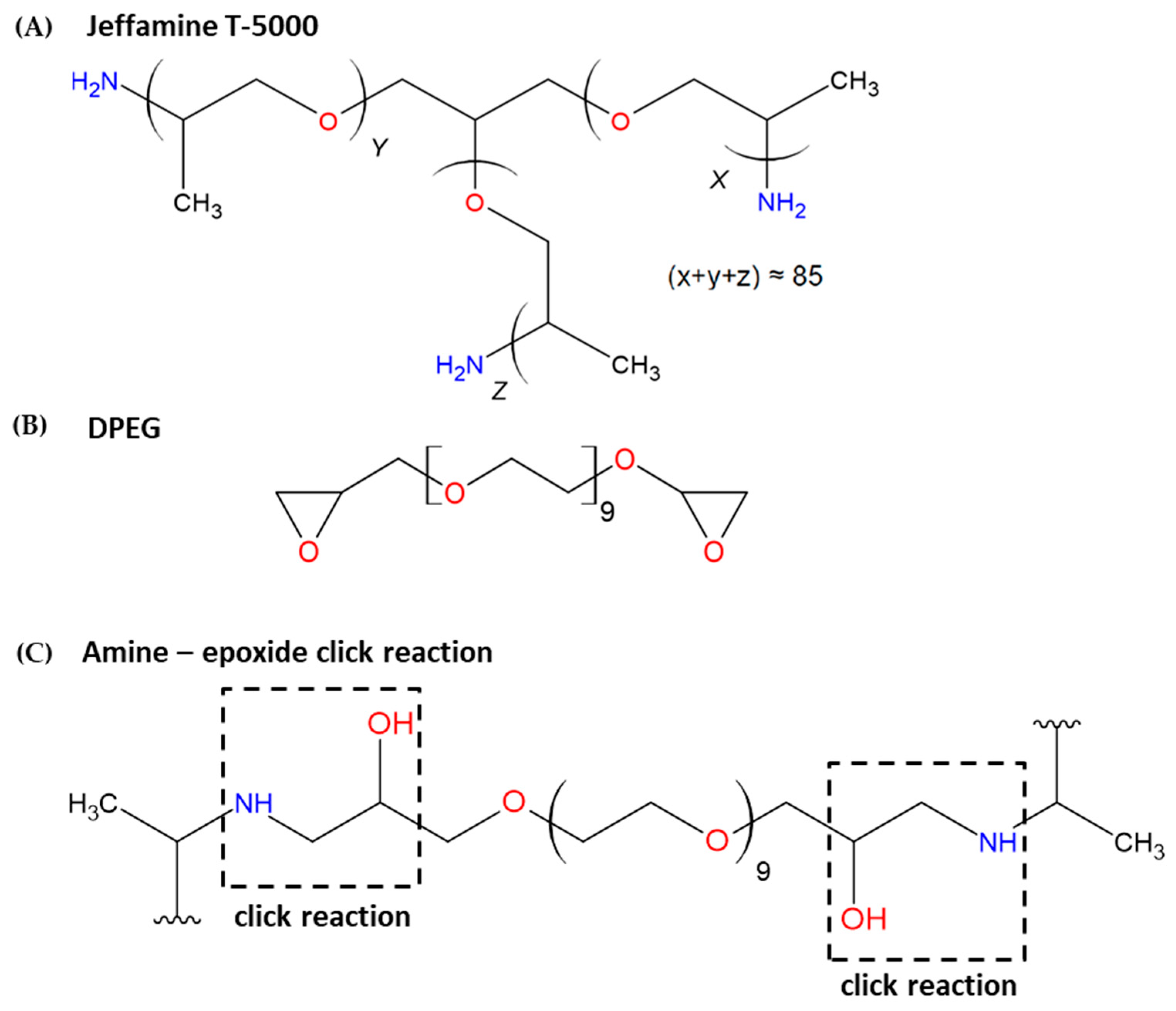
Figure 1. Chemical structures of Jeffamine® T-5000 (PPO) (A), Diepoxy poly (ethylene glycol) (DPEG) (B), and Click reactions between amine T-5000 and epoxide DPEG for the formation of nanogels (C).
Futurepharmacol 05 00054 g001
Figure 2. Hydrodynamic diameter (Dh) distribution graphs obtained by DLS for the synthesized nanogels (A) and surface charge measurements of the different synthesized polymeric nanogels obtained through zeta potential (ζ) measurements (B).
Futurepharmacol 05 00054 g002
Figure 3. Transmission Electron Microscopy (TEM) micrographs of the synthesized nanogels: (A) Nano11, (B) Nano13, and (C) Nano23.
Futurepharmacol 05 00054 g003
Figure 4. Percentage of viable cells detected by the XTT assay after 24, 48, and 72 h of treatment with different concentrations of (A) PPO (Jeffamine® T-5000), (B) DPEG (Poly (ethylene glycol) diglycidyl ether), and the nanogels 11, 13, and 23 ((C), (D) and (E), respectively) in the CHO-K1 cell line. NC: negative control; PC: positive control (10% DMSO). ** p < 0.01 compared to NC.
Futurepharmacol 05 00054 g004
Figure 5. Percentage of viable cells detected by the XTT assay after 24, 48, and 72 h of treatment with a concentration of 1000 μg/mL of DPEG (Poly (ethylene glycol) diglycidyl ether), PPO (Jeffamine® T-5000), and nanogels 11, 13, and 23 in the CHO-K1 cell lineage. ** p < 0.01 compared to DPEG.
Futurepharmacol 05 00054 g005
Figure 6. Percentage of viable cells detected by the clonogenic survival assay after 24 h of treatment with different concentrations of the (A) PPO (Jeffamine® T-5000) and DPEG (Poly (ethylene glycol) diglycidyl ether), and (B) the nanogels Nano11, Nano13, and Nano23 in the CHO-K1 cell line. NC: negative control; PC: positive control (doxorubicin 0.5 μM). ** p < 0.01 compared to NC.
Futurepharmacol 05 00054 g006
Figure 7. Percentage of viable cells detected by the clonogenic survival assay after 24 h of treatment with a concentration of 100 μg/mL of DPEG (Poly (ethylene glycol) diglycidyl ether), PPO (Jeffamine® T-5000), and the nanogels Nano11, Nano13, and Nano23 in the CHO-K1 cell line. * p < 0.05 compared to DPEG.
Futurepharmacol 05 00054 g007
Figure 8. Percentage of viable cells detected by the Trypan Blue Exclusion Test after 4 h of treatment with different concentrations of the (A) PPO (Jeffamine® T-5000), (B) DPEG (Poly (ethylene glycol) diglycidyl ether), and the nanogels 11, 13, and 23 respectively in the CHO-K1 cell line. NC: negative control; PC: positive control (10% DMSO). ** p < 0.01 compared to NC.
Futurepharmacol 05 00054 g008
Figure 9. DNA damage detected by the comet assay in the CHO-K1 cell lineage after 4 h treatment with different concentrations of DPEG (Poly (ethylene glycol) diglycidyl ether), PPO (Jeffamine® T-5000) (A), and the nanogels Nano11, Nano13, and Nano23 (B). CN: negative control; CP: positive control (doxorubicin 0.75 μM). * p < 0.05 compared to CN. ** p < 0.01 compared to CN.
Futurepharmacol 05 00054 g009
Figure 10. DNA damage was detected by the comet assay in the CHO-K1 cell line after 4 h of treatment with 1000 μg/mL of DPEG (Poly (ethylene glycol) diglycidyl ether), PPO (Jeffamine® T-5000), and the nanogels Nano11, Nano13, and Nano23. * p < 0.05 compared to DPEG. ** p < 0.01 compared to DPEG.
Futurepharmacol 05 00054 g010
Table 1. Proportions of reagents in each synthesized nanogel.
PPODPEG
Nano 110.90 g0.10 g
Nano 130.77 g0.23 g
Nano 230.87 g0.23 g
PPO: Jeffamine® T-5000. DPEG: (Poly (ethylene glycol) diglycidyl ether).
Table 2. Hydrodynamic diameter (Dh), polydispersity index (PDI), and zeta potential (ζ) values of the nanogels obtained at pH = 7.
SampleDh (nm)PDIζ (mV)
Nano11170 ± 80.20−3 ± 1
Nano13350 ± 170.07−5 ± 2
Nano23130 ± 60.25−13 ± 5
Table 3. Nuclear division index and cells binucleated with micronucleus and micronucleus frequency obtained by the micronucleus test on the CHO-K1 cell line after a 24 h treatment with different concentrations of DPEG (Poly (ethylene glycol) diglycidyl ether), PPO (Jeffamine® T-5000), and the nanogels Nano11, Nano13, and Nano23.
TreatmentsMicronucleus Test—CHO-K1
NDI/1000 CellsBinucleated Cells with MNMN/1000 Cells
NC1.88 ± 0.011.7 ± 0.171.63 ± 0.21
PC1.16 ± 0.04 **14 ± 0.28 **12.5 ± 0.32 **
PPO 101.86 ± 0.0062.30 ± 0.452.10 ± 0.30
PPO 1001.82 ± 0.012.53 ± 0.312.33 ± 0.27
DPEG 101.73 ± 0.0052.13 ± 0.031.93 ± 0.12
DPEG 1001.72 ± 0.035.86 ± 0.61 **5.4 ± 0.58 **
Nano11 101.90 ± 0.052.20 ± 0.452.10 ± 0.50
Nano11 1001.81 ± 0.062.86 ± 1.412.56 ± 1.16
Nano13 101.83 ± 0.031.63 ± 0.141.46 ± 0.88
Nano13 1001.78 ± 0.042.23 ± 0.632.13 ± 0.53
Nano23 101.89 ± 0.011.30 ± 0.201.2 ± 0.15
Nano23 1001.77 ± 0.031.53 ± 0.171.43 ± 0.17
NDI: nuclear division index. MN: micronucleus. NC: negative control; PC: positive control (doxorubicin 0.5 μM). ** p < 0.01 compared to NC. Concentration in 10 and 100 μg/mL.
Table 4. Binucleated cells with micronuclei and micronucleus frequency obtained from the micronucleus test on the CHO-K1 cell line after 24 h of treatment in a concentration of 100 μg/mL of DPEG (Poly (ethylene glycol) diglycidyl ether), PPO (Jeffamine® T-5000), and the nanogels Nano11, Nano13, and Nano23.
TreatmentsMicronucleus Test—CHO-K1
Binucleated Cells with MNMN/1000 Cells
PPO 1002.53 ± 0.31 *2.33 ± 0.27 *
DPEG 1005.86 ± 0.615.40 ± 0.58
Nano11 1002.86 ± 1.412.56 ± 1.16 *
Nano13 1002.23 ± 0.63 *2.13 ± 0.53 *
Nano23 1001.53 ± 0.17 *1.43 ± 0.17 **
MN: micronucleus. * p < 0.05 compared to DPEG. ** p < 0.01 compared to DPEG. Concentration of 100 μg/mL.
Table 5. Compilation of nanogel studies.
NanogelExperimental ModelAssaysResultsReference
Lipoprotein/carboxymethylcellulose sodium nanogelsHeLa and HepG2 cell lines (in vitro)MTT assayNo damage at the highest tested concentration (8 μg/mL)[42]
Thermoresponsive polyglycerol-based nanogels (tNG_dPG_tP and tNG_dPG_pNIPAM)NHK cell line (in vitro)MTT assay, comet assay, and Carboxy-H2DCFDA testNo damage at the highest tested concentration (500 μg/mL)[43]
Chitosan-based nanogelsrTERT-BJ and L-929
cell lines (in vitro)
MTT assayNo damage at the highest tested concentration (400 μg/mL)[45]
PEG-PEI, PE-PEI, and Plu-PEI nanogelsMicroglia culture (in vitro)MTS and LDH assaysNo damage at the highest tested concentration (0.05 w/v)[15]
Chitosan-based nanogelsChondrocytes, synoviocytes, and osteoblasts (in vitro)
Zebrafish (in vivo)
MTS, LDH, nitric oxide quantification, DNA drag test, and zebrafish embryo assayDid not affect cell proliferation or nitric oxide production in vitro but exhibited moderate genotoxicity in a dose-dependent manner, without inducing embryotoxicity[6]
Polyetheramine–epoxide-based nanogelsGM07492-A (in vitro)XTT assayNo damage up to a concentration of 1000 μg/mL[17]
Disclaimer/Publisher’s Note: The statements, opinions and data contained in all publications are solely those of the individual author(s) and contributor(s) and not of MDPI and/or the editor(s). MDPI and/or the editor(s) disclaim responsibility for any injury to people or property resulting from any ideas, methods, instructions or products referred to in the content.