Review

Oxygen-Generating Metal Peroxide Particles for Cancer Therapy, Diagnosis, and Theranostics

by
Adnan Memić
1,* and
Turdimuhammad Abdullah
2,3
1
Center of Nanotechnology, King Abdulaziz University, Jeddah 21589, Saudi Arabia
2
İzel Kimya Research and Development Center, Dilovası 41455, Turkey
3
Department of Metallurgical & Meterials Engineering, Kocaeli University, İzmit 41001, Turkey
*
Author to whom correspondence should be addressed.
Future Pharmacol. 2025, 5(3), 41; https://doi.org/10.3390/futurepharmacol5030041
Submission received: 31 May 2025 / Revised: 1 July 2025 / Accepted: 11 July 2025 / Published: 30 July 2025

Abstract

Theranostic materials, which combine therapeutic and diagnostic capabilities, represent a promising advancement in cancer treatment by improving both the precision and personalization of therapies. Recently, metal peroxides (MePOs) have attracted significant interest from researchers for their potential use in both cancer diagnosis and therapy. This review provides an overview of recent developments in the application of MePOs for innovative cancer treatment strategies. The unique properties of MePOs, such as oxygen generation, are highlighted for their potential to improve therapeutic outcomes, especially in hypoxic tumor microenvironments. Initially, methods for MePO synthesis are briefly discussed, including hydrolyzation–precipitation, reversed-phase microemulsion, and sonochemical techniques, emphasizing the role of surfactants in regulating the particle size and enhancing bioactivity. Next, we discuss the main therapeutic approaches where MePOs have shown promise. These applications include chemotherapy, photodynamic therapy (PDT), immunotherapy, and radiation therapy. Overall, we focus on integrating MePOs into theranostic platforms to enhance cancer treatment and enable diagnostic imaging for improved clinical outcomes. Finally, we discuss potential future research directions that could lead to clinical translation and the development of advanced medicines.

Graphical Abstract

1. Introduction

Materials that have both therapeutic and diagnostic imaging modalities are referred to as theranostic, blending the two words together (i.e., therapeutic and diagnostic), a term originally coined by Funkhouser in 2002 [1,2]. However, the origins of the concept can be traced to 1946 in nuclear medicine’s use of radio-iodine pharmaceuticals for thyroid cancers [3]. This unique approach allows for the simultaneous delivery of an imaging agent and a therapeutic one, significantly aiding in the therapy of some diseases [4,5]. For example, in the treatment of cancer, it is essential to initially carry out disease staging, which is often performed with the aid of imaging before starting therapy [6,7]. Nevertheless, separate materials are often used for imaging and therapy, which may lead to differences in biodistribution. This means that the imaging and therapeutic agents may not accumulate in the same areas at the same time, reducing treatment effectiveness.
A more effective approach would be to integrate both imaging and therapeutic functions into a single theranostic platform or material. This approach ensures a consistent biodistribution, thereby improving the precision of the treatment and minimizing side effects [8,9]. Ultimately, using theranostic nanomaterials, it might be possible to obtain real-time feedback and/or the long-term monitoring of cancer progression with the goal of tuning the therapy more quickly and precisely [10,11]. Thus, shifting cancer treatment to include theranostic approaches using nanomaterials could propel more personalized and effective treatments with improved outcomes for a more diverse range of patients [5,12].
One of the more promising aspects of using theranostically based nanomaterials is their potential for minimizing unwanted side effects by being precisely targeted to diseased tissues [12,13]. Larger particles are usually limited in their penetration of tumors and are generally localized to tumor tissue due to a phenomenon known as the enhanced permeability and retention (EPR) effect [14,15]. The EPR effect is a result of the poor lymphatic drainage typical of tumors and the often irregular, enlarged, and leaky vasculature surrounding the tumor tissue, which enables these particles to escape from the bloodstream [15].
Targeted nanomaterials can also benefit from their reduced size by not being easily filtered out by the kidneys, allowing them to remain in the bloodstream longer [16]. Furthermore, altering nanomaterial surface properties and engineering them with targeting ligands and cloaking agents, such as poly (ethylene glycol) (PEG), can significantly improve tumor targeting and reduce undesired systemic toxicity [17,18]. In addition, achieving a higher concentration of nanomaterials in tumor tissue could also improve the contrast in imaging applications [19]. The high surface-to-area ratio relative to the volume of targeted nanomaterials enhances their capacity to simultaneously carry large loads of both therapeutic and imaging agents [12,18].
A variety of theranostic nanomaterials, including gold nanoparticles, carbon dots, graphene, mesoporous silica, and metal peroxides (MePOs), have been investigated for their potential applications in cancer treatment [20]. Among them, the latter has attracted significant interest due to their multifunctional nature [21,22,23]. One of the main advantages of MePOs is that they can generate oxygen, reactive oxygen species (ROS), and/or hydrogen peroxide [22,23]. These unique features of MePOs could be important for improving therapeutic outcomes and preventing disease progression [22,24,25]. For example, calcium peroxides have been extensively investigated in cancer treatment applications [22,24]. Calcium peroxide (CPO) is particularly attractive since calcium is the most abundant metal element in the human body and can have a better biocompatibility profile versus other peroxides [26,27]. Similarly, CPO can release Ca ions and generate hydrogen peroxide under acidic conditions usually found in the tumor microenvironment (TME) [26,28].
Alternatively, under more neutral pH environments, CPO can release oxygen that could be augmented by another MePO (i.e., manganese peroxide, MnO2, MPO) [28,29,30]. The addition of MPO can significantly improve the release of oxygen from CPO particles [28]. Furthermore, MPO is also unique as it has been used as a contrast agent in magnetic resonance imaging (MRI) [31,32]. Similarly to CPO in the acidic environment, MPO can release Mn(II) ions that have five unpaired electrons, which can increase the likelihood of the presence of Mn(II) paramagnetic centers when in contact with water molecules (i.e., providing the improved MRI contrast) [32,33]. Therefore, the combination of these MePOs can be used in a single platform, providing a theranostic modality for both cancer treatment and imaging. Moreover, when dealing with the nanoparticles of MePOs, the possibility arises for the additional conjugation of drugs and therapeutics on the surface of these materials, thereby extending their applications [34]. Similarly, it is possible to surface modify or add polymer coatings that prevent the premature degradation of the MePO, such as CPO [35,36]. As such, there have been several key aspects in which MePOs have been used in cancer treatment and imaging. In this review, we focus on four main therapeutic approaches: chemotherapy, photodynamic therapy (PDT), immunotherapy, and radiation therapy.

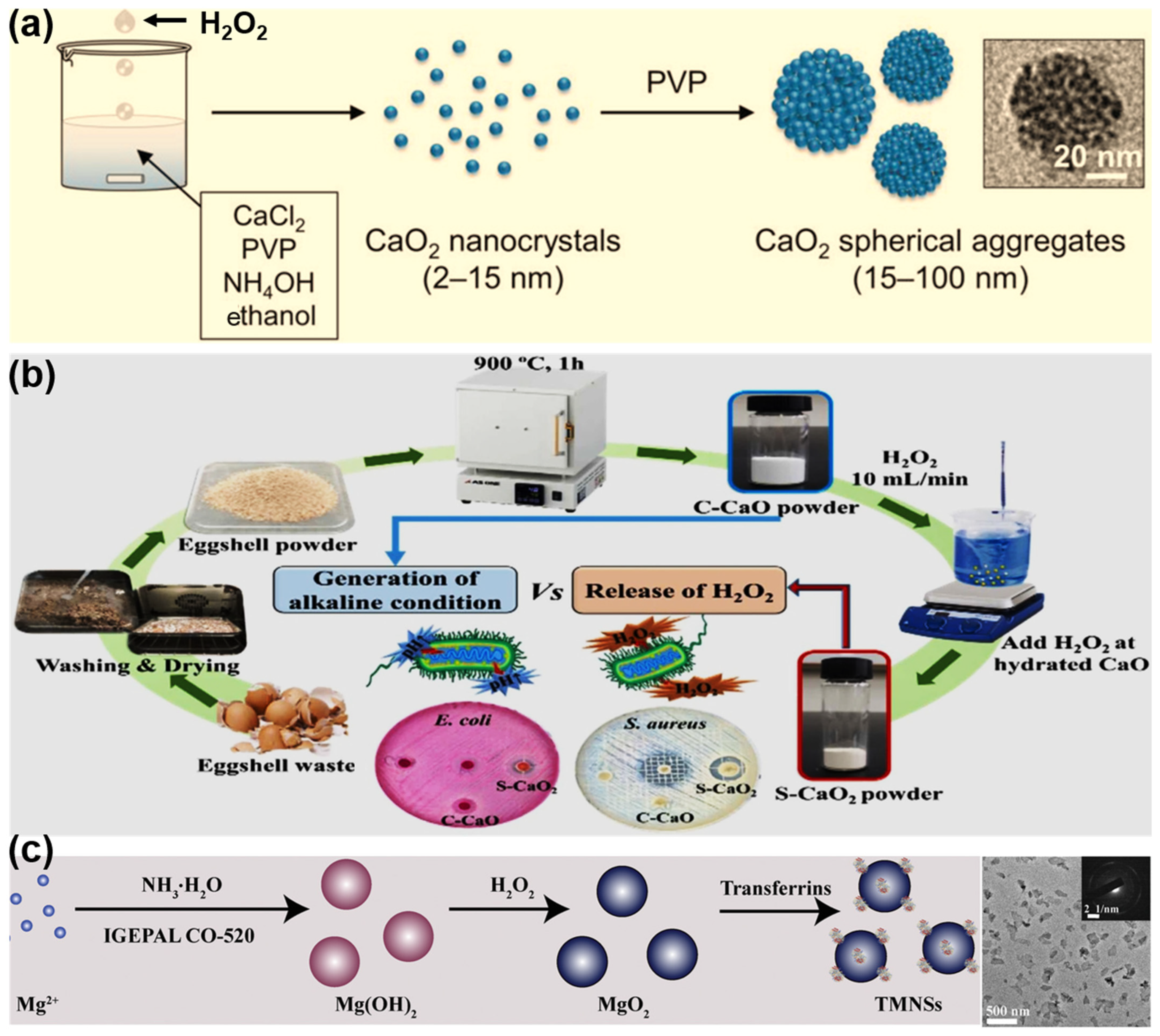
2. Synthesis of MePO Particles

A variety of techniques have been developed for the synthesis of MePO micro- or nanoparticles (Table 1 and Figure 1), including hydrolyzation–precipitation [37,38,39], reversed-phase microemulsion [36,40], and sonochemical methods [41,42]. In the majority of cases, water-soluble polymers, such as PEG [29,43], polyvinyl pyrrolidone (PVP) [39,41], and hyaluronic acid (HA) [44], have been employed as surfactants and/or stabilizers to regulate the particle size and improve the dispersibility of the MePO particles [35]. Furthermore, the introduction of these polymers also enhances the stability and bioactivity of the MePO as well as its suitability for biomedical applications [35,45].
The hydrolyzation–precipitation method is the most prevalent technique for the synthesis of MePO particles, offering a number of advantages, including simplicity, mild conditions, low costs, and size adjustability [35,46]. The process typically involves the use of metal salts as precursors, which are reacted with hydrogen peroxide in an alkaline solution to form insoluble MePO nanoparticles. By employing this approach, Shen et al. [39] successfully synthesized CaO2 nanoparticles and nanocrystals. They used ethanol as a solvent to minimize the hydrolysis of CaO2. The addition of H2O2 to an ethanol solution containing CaCl2 and PVP resulted in the formation of primary nanocrystals with a diameter of 2–15 nm. The resulting CaO2 nanoparticles exhibited antibacterial activity, as evidenced by the release of H2O2 and O2 upon contact with water. Zhang et al. [38] synthesized BaO2 using a similar method but incorporated organic ligands in order to control the growth and morphology of the resulting material. The use of coordinating ligands enabled the synthesis of nanosized particles, while modifications of BaO2 with biodegradable agents such as L-glutamic acid reduced the toxicity of Ba2+ and enabled tumor-targeted X-ray therapy applications. In several studies, metal oxides were employed directly in the synthesis process, rather than their corresponding salts. For instance, Heo et al. [37] transformed eggshell waste into calcium oxide (CaO) through calcination at 900 °C, followed by the reaction of the calcined CaO with water, which resulted in the generation of a calcium hydroxide (Ca(OH)2) solution. Subsequently, a gradual addition of H2O2 was employed to synthesize CaO2 powder. However, the resulting CaO2 exhibited an irregular particle size and shape, which may be attributed to the absence of surfactants during the synthesis process.
Another promising technique for the preparation of MePO particles is the reversed-phase microemulsion method [35,47]. This method is advantageous for several reasons, including its suitability for the production of small, well-dispersed, and impurity-free particles; its precise control over the nanoparticle size and morphology; and the simplicity of its implementation. The method employs the use of a nanoscale microreactor environment, which is formed by the microemulsion system. As an illustration, Tang et al. [40] synthesized MgO2 nanosheets through the combination of cyclohexane and CO-520 with a MgCl2 solution, followed by an ammonium hydroxide injection to facilitate the formation of Mg(OH)2. Subsequently, hydrogen peroxide was added to regulate the progression of the reaction, while the addition of ethanol acted to disassemble the microemulsion structure, allowing for the formation of MgO2 nanosheets (Figure 1c). Furthermore, this method allows for the direct incorporation of chemotherapeutic pharmaceuticals into nanoparticles, thereby enabling the nanoparticle synthesis and drug loading to occur concurrently. For example, He et al. [36] successfully fabricated CaO2 nanoparticles, simultaneously incorporating the chemotherapeutic drug cisplatin and coating the nanoparticles with a negatively charged phospholipid layer using this method.
Recently, sonochemistry has emerged as a prominent approach for synthesizing nanoparticles, including those of MePOs [48,49,50]. By employing a high-intensity ultrasound to initiate, enhance, or control chemical reactions, this method has enabled the formation of uniform nanoparticles with unique properties [51]. Pirouzmand et al. [42] employed a sonochemical method to fabricate ZnO2 nanoparticles. This involved dissolving a zinc salt (ZnSO4) in an aqueous NaOH solution (pH 8), followed by a H2O2 reaction, and finally subjecting the solution to ultrasound sonication. Despite the formation of monodisperse nanoparticles, aggregation was observed. In another study conducted by Alipoor et al. [41], the use of PVP during the ZnO2 preparation process by the same method resulted in the absence of aggregation. This highlights the crucial role of stabilizers/surfactants during nanoparticle synthesis. Additionally, several novel approaches, including pulsed laser ablation in liquid [52,53], Leidenfrost dynamic chemistry [54], and gas diffusion [30], have also been used to synthesize MO2 nanoparticles.

3. Application of MePO Particles in Cancer Diagnosis and Therapy

3.1. Chemotherapy

One of the hallmarks of cancer is hypoxia, or low oxygen levels, in the tissue surrounding the tumor [55]. Through the action of several proteins involved or upregulated in hypoxia, the tumor microenvironment (TME) becomes increasingly resistant to chemotherapy, which leads to the efflux of the drugs from the cancer cells they are intended to kill [55,56]. Therefore, increasing the oxygen supply to the TME may inhibit some of these effects and improve chemotherapeutic outcomes [32,57]. In addition, MePOs, such as CPO, might not only increase oxygen but also increase other ROS in the TME that could be proapoptotic in the presence of chemotherapies. For example, Cheng et al. [58] produced magnetic CPO nanoparticles to generate oxygen and enhance the efficacy of the FDA-approved chemotherapeutic agent doxorubicin in the hypoxic microenvironment of triple-negative breast cancer (TNBC). The synthesized nanoparticles reduced the HIF-1α expression, inhibited autophagy, induced apoptosis, and significantly improved the tumor suppression in TNBC when combined with doxorubicin, both in vitro and in an orthotopic mouse model. Specifically, the HIF-1α expression decreased 6-fold (i.e., from 2.5 to 0.4) with the introduction of 20 ppm CaO2 nanoparticles, and the tumor weight decreased by over 90% with the combination treatment (CaO2-MNPs + DOX) compared to the treatment with DOX alone.
As mentioned above, chemotherapeutic formulations might benefit from dual MPO and CPO combinations. In such cases, the presence of MPO could improve chemotherapeutic treatments by catalyzing and enhancing the oxygen release from the CPO degradation that initially yields hydrogen peroxide [59,60]. For example, recently, researchers generated MPO on the surface of CPO particles, which significantly enhanced oxygen production [61]. The total dissolved oxygen level in the sample reached 8 mg/L after 3 h, whereas CPO alone generated only 0.5 mg/L of dissolved oxygen within the same time frame. The increased oxygen levels via down-regulating hypoxia-dependent pathways improved the chemotherapeutic agent retention and TME penetration. The water-responsive formulation provided a controlled doxorubicin release in the lipase-rich TME that degraded the solid lipid monostearin used to coat the CPO/MPO nanocomposite. In addition, in such cases it is possible to perform imagining using MPO as an additional imaging modality. However, CPO could also be used in a catalyst-independent manner. For example, the degradation of CPO in the presence of water leads to the formation of Ca(OH)2 [62]. This in turn yields an alkaline microenvironment that supports the degradation of H2O2, allowing CPO to directly generate O2 in a catalyst-free manner [24]. Previous groups have utilized this catalyst-free approach in enhancing hypoxic hepatocellular carcinoma or breast cancer chemotherapy [63].

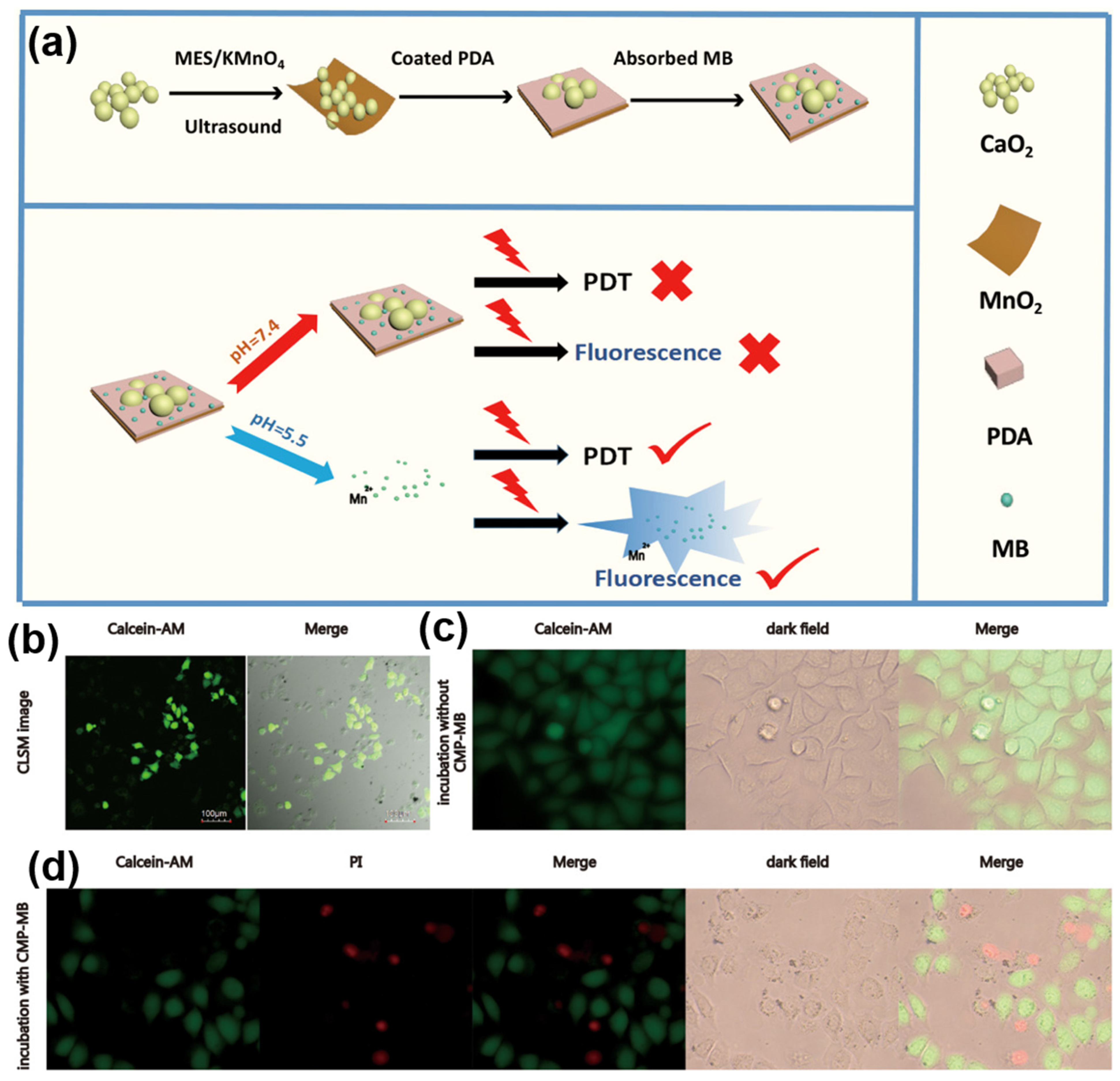
3.2. Photodynamic Therapy

Photodynamic therapy (PDT) is a minimally invasive treatment modality intended to selectively destroy diseased cells and tissues (usually cancer) by using a combination of a photosensitizer, a light source, and molecular oxygen [64,65]. For example, once the photosensitizer is accumulated in the cancerous tissue, it can be activated using a specific wavelength of light, which in turn can harness that energy and convert the oxygen present in the TME into ROS that destroy the surrounding cancerous cells [65,66]. Although PDT has a low therapy resistance and side effects, the presence of hypoxia in the TME usually presents a challenge for effective PDT treatments, especially in solid tumors [66]. Therefore, to improve oxygenation in the TME, researchers have turned to MePOs as part of the treatment approach. For example, several research groups have turned to CPO and other oxygen-generating peroxides using different coating strategies to tailor the release of oxygen species in the TME to modulate PDT with varying success [24,67]. Some used pH-sensitive polymer coatings, others turned to photosensitive polymer shells, and others, on the other hand, used lipids of liposomes to deliver CPO to the TME in order to improve the PDT efficacy [68,69]. More recently, researchers have used MPO to improve PDT, in which MPO acts as an enzyme that catalyzes the hydrogen peroxide decomposition reaction [70,71]. The TME of solid tumors with increased oxygen can be significant, with markedly improved therapeutic outcomes [70,72]. In addition, MPO can be used as a contrast agent, providing a dual functionality and an opportunity to follow, in real time, the therapeutic effects of PDT [31,73].
However, as in the previous case with chemotherapy, the best outcomes are achieved when combining CPO and MPO into a single platform [29,74,75]. For example, Shi et al. [74] developed a nanosystem comprising a MPO nanosheet coated on CPO nanoparticles. This combination had the unique property of a methylene blue emission suppression that was incorporated on the CPO/MPO platform. In turn, this nanosystem provided switch-controlled (i.e., ON/OFF) fluorescent cell imaging and tumor inhibition capabilities that also provided oxygen for an improved PDT (Figure 2). Specifically, the system exhibited minimal cytotoxicity without laser irradiation. However, when light was irradiated, the tumor cell viability decreased remarkably, dropping to 28% at a concentration of 100 μg/mL of CPO/MPO nanosheets. In addition to using MPO to control fluorescent imaging, MPO can be directly used in the MRI T1 modality as a contrast agent. Therefore, the combination of MPO and CPO enhances the efficacy of PDT-mediated treatments, while allowing for the detailed imaging of tumors [29]. In addition to CPO and MPO, iridium peroxide (IrOx) has generated significant interest in PDT due to its distinctive optical and chemical properties [76,77]. Ma et al. [76] designed a nanoplatform that responds to both acidic conditions and excess glutathione (GSH). This platform is composed of a multi-caged IrOx coated by a metal–polyphenol network (MPN) composed of Fe3+ and tannic acid (TA). The research demonstrated that the MPN shell can be degraded under the acidic TME and intracellular excess of GSH. This results in the release of Fe3+ and the exposure of the IrOx core. As a result, the efficient dual-pathway chemodynamic therapy (CDT) is facilitated. The nanoplatform has been shown to effectively mitigate the attenuation of CDT by consuming excess GSH within the tumor. The multi-caged structure of IrOx is advantageous for implementing photothermal therapy (PTT) in coordination with CDT, further enhancing the therapeutic efficacy of tumors. Finally, the team demonstrated the remarkable capabilities of IrOx@MPN in terms of its outstanding computed tomography (CT) and magnetic resonance imaging (MRI) (T1/T2) multimodal imaging. These capabilities are instrumental in facilitating an early diagnosis and timely treatment, which is crucial for effective patient care and management.

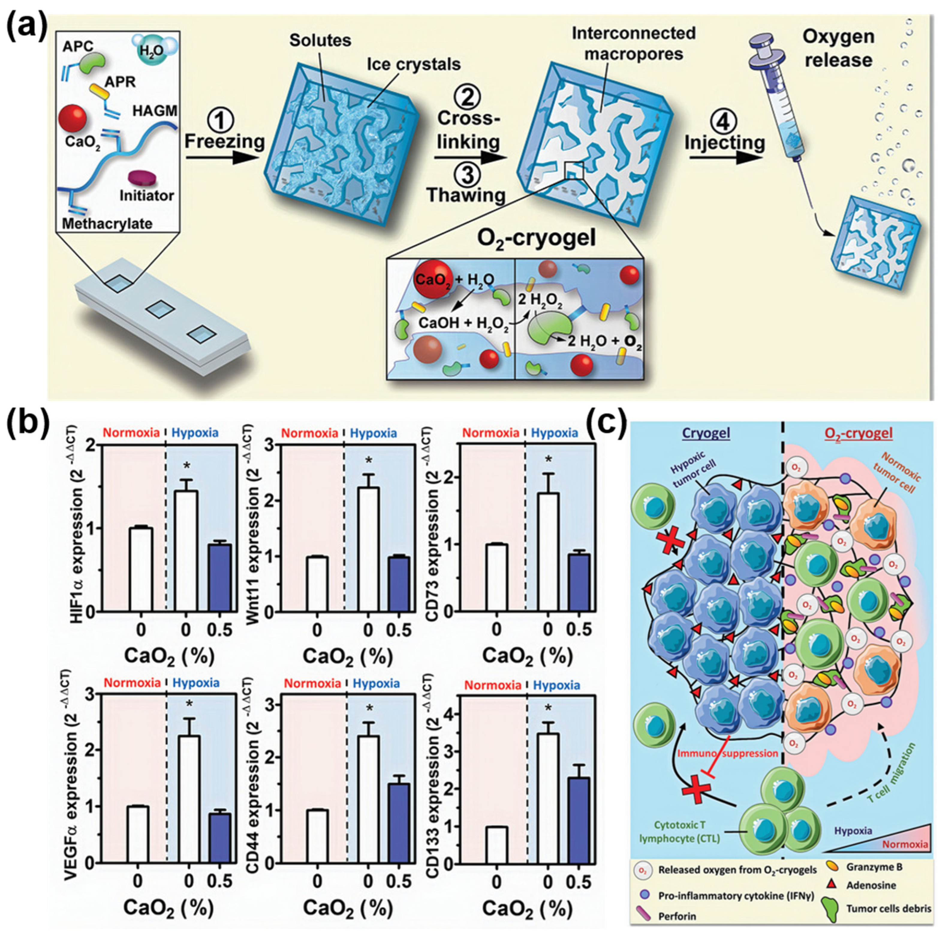
3.3. Immunotherapy and Radiation Therapy

A new area of research that has found applications for oxygen-generating particles is immunotherapy [22,78]. The goal of immunotherapy is to improve the body’s natural immune response to recognize and attack cancerous cells more effectively, leading to disease regression [79,80]. The hypoxic microenvironment of solid tumors can create a barrier to antitumor immune responses by inducing immunosuppressive conditions that inhibit cytotoxic T lymphocyte activity [22,81]. These effects are primarily driven by hypoxia-inducible factors (HIFs) and A2A adenosine receptors that lead to suppressing the antitumor activity of T cells and natural killer cells via altered cytokine and chemokine secretion [82]. Although the use of hyperoxic breathing has shown success in overcoming the immunosuppression of cancer hypoxia, this approach could significantly benefit from local oxygen generation by micro- and nanoparticles [83].
One example of such an approach is by Colombani et al. [22], where they developed a syringe-injectable oxygen-generating scaffold (i.e., cryogels) by incorporating CPO particles into the polymer walls (Figure 3). To improve the oxygenation of the TME and to mitigate the possible cytotoxicity from hydrogen peroxide, a CPO degradation byproduct, the researchers also conjugated a catalase enzyme into the polymeric scaffolds to improve the CPO conversion. They showed that the incorporated CPO particles were homogeneously distributed within the cryogels, providing a predictable, controlled, and sustained oxygen release under both normoxic and hypoxic conditions with minimal hydrogen peroxide-derived toxicity due to the catalase enzyme presence. This approach was able to reverse the TME hypoxia while simultaneously down-regulating the hypoxia-induced protein expression and increasing proinflammatory cytokines that lead to lower immunosuppression. Ultimately, the researchers demonstrate the preservation of the T cell function and antitumor activity using these oxygen-generating cryogels in what would otherwise be a highly immunosuppressive TME of an advanced-stage murine melanoma model. Nevertheless, the authors emphasized the necessity for more extensive toxicological and long-term safety evaluations before preclinical trials. Furthermore, human immune responses differ significantly from those in murine models, particularly with regard to T cell infiltration levels and the sensitivity to reactive oxygen species. These differences could impact the efficacy of O2-cryogels when translating these findings into clinical settings with cancer patients.
Similarly, CPO has been used to improve the immune response by inducing enhanced antigen-presenting cell (APC) maturation via ROS generation in draining lymph nodes [84,85]. For example, Su et al. [85] developed a nanovaccine that included CPO particles. These nanovaccines were able to induce ROS generation in endo/lysosomes of APCs found in the draining lymph nodes, at which the nanovaccines were targeted. The stimulation and lipid peroxidation in APCs improved their maturation and enhanced the antigen presentation to T cells, which generated a more robust immune response.
Furthermore, MePOs have demonstrated significant radiosensitization effects, enhancing the efficacy of radiotherapy by generating reactive oxygen species (ROS), alleviating tumor hypoxia, and boosting antitumor immunity [86,87,88]. Salah et al. [88] demonstrated the anti-cancer effects of titanium peroxide nanoparticles (TiOxNPs) combined with ionizing radiation on radioresistant cancer stem cells (CSCs) in both in vitro and in vivo models. TiOxNPs have shown a synergistic effect with radiation on pancreatic spheres’ enriched CSCs by decreasing self-renewal regulatory factors and CSC surface markers. In addition, the combination treatment effectively suppressed the epithelial–mesenchymal transition, migration, and invasion properties in primary and aggressive pancreatic cancer cells. In another study, Nakamaya et al. [87] the compared radiotherapeutic efficacy of TiOxNPs with their polyacrylic acid-modified counterparts (PAA-TiOxNPs). They found that PAA-TiOxNPs generate considerably higher levels of hydroxyl radicals and H2O2 upon X-ray irradiation compared to TiO2 alone. In both in vitro assays with MIA PaCa-2 pancreatic cancer cells and in vivo xenograft models, PAA-TiOxNPs significantly increased the DNA damage and suppressed the tumor growth when combined with radiation, without an observable systemic toxicity over a 43-day period. These findings indicate that PAA-TiOxNPs are effective radiosensitizers that are safe and enhance the efficacy of radiotherapeutic treatments. They achieve this by increasing the production of ROS over time and improving the response of tumors to treatment.

4. Conclusions and Future Direction

Oxygen-generating particles, and specifically MePOs, present a promising platform for both improving cancer treatment outcomes and also providing the potential for diagnostic imaging. It is clear that MePOs have been applied in several cancer treatment strategies, ranging from chemotherapy and PDT to more recent and developing fields like immunotherapy and radiation therapy. Furthermore, it is clear that MePOs’ functions stretch well beyond just oxygen generation and can include other mechanisms through which they derive treatment benefits. However, several challenges still exist.
One of the first challenges that should be addressed, to achieve clinically relevant results and translation, is the engineering of MePO stability. Most of the MePOs are likely to react with water and potentially decompose prematurely, leading to a loss of efficacy [24]. This is especially important for biological processes and responses that require days or weeks to achieve their full potential [24,68]. It might be possible to improve MePO stability and mitigate toxicity by surface modifications or coating by various biomolecules [36,68,89]. For example, multi-anchor phosphonic-PEG, dextran/PEG bilayers, and zwitterionic polymers form protective, hydrophilic shells that stabilize metal peroxide nanoparticles. Biomimetic coatings—such as lipids or cell-membrane-derived vesicles, as well as protein/peptide grafts—enhance biocompatibility, immune evasion, and controlled oxygen release kinetics, thereby reducing toxicity in biomedical environments [90,91]. However, to date, long-term stability while preserving a low toxicity and controlled prolonged oxygen release, which might be particularly beneficial for immunotherapy-based approaches, still remains challenging [22]. Some of the challenges stem from the inorganic nature of MePOs that tend to aggregate and precipitate in aqueous environments. The tendency to agglomerate not only lowers the accessible surface area but also can lead to potential side effects, including toxicity in normal tissues and a proportional decrease in the oxygen release potential. Therefore, biocompatibility in general is another important aspect to take into consideration [92]. As MePOs like CPO decompose, they generate calcium and hydroxyl ions as well as hydrogen peroxide, which in some applications might be beneficial but in other settings might derive unnecessary toxicity for normal tissues. Biomolecular coatings can improve biocompatibility, but close attention needs to be given to the formulation so that the controlled release profile of the oxygen species is preserved [35]. Ultimately, more comprehensive and long-term toxicity studies of MePOs need to be performed, not only in vitro but also in in vivo settings, to ensure the absence of systemic toxicity.
Other challenges that exist are related to the way MePOs are synthesized and developing methods that provide tunable control of the MePO particle synthesis. For example, having the ability to precisely control the size and shape of MePO particles on scales that would support commercial production is still lacking. Having optimized synthetic protocols that provide predictable MePO physicochemical properties would address a major obstacle to the clinical translation of such therapies [35,39]. Finally, biomedical applications of MePO nanoparticles encounter stringent regulatory challenges due to their altered physicochemical properties at the nanoscale. The regulatory standards established by the FDA (Montgomery County, MD, USA) and the EPA (Washington, DC, USA) are applicable to bulk chemicals, rather than to nanoscale forms. This discrepancy results in the following consequences: unclear size thresholds; an inconsistent jurisdiction over nanomaterials; redundant reviews (e.g., under TSCA, FDA, FIFRA); and burdensome, case-by-case data requirements that strain agency resources and slow innovation.
To conclude, MePOs and their properties, including oxygen generation, hold great potential in both the treatment and diagnosis of cancer, which have been applied in several different settings. The promising antitumor effects of MePOs could be further improved and lead to the clinical translation and development of advanced medicines guided by filling the necessary research gaps and addressing the existing challenges.

Author Contributions

The manuscript was written and edited with contributions from A.M. and T.A. All authors have read and agreed to the published version of the manuscript.

Funding

Deanship of Scientific Research (DSR) at King Abdulaziz University, Jeddah under grant no. (IPP: 824-903-2025).

Acknowledgments

This project was funded by the Deanship of Scientific Research (DSR) at King Abdulaziz University, Jeddah under grant no. (IPP: 824-903-2025). The authors, therefore, acknowledge with thanks DSR for technical and financial support.

Conflicts of Interest

T.A. is employed in Izel Kimya A.S. Izel Kimya had no role in the design of the study; in the collection, analyses, or interpretation of data; in the writing of the manuscript, or in the decision to publish the results. The authors declare no conflicts of interest.

References

  1. Moorthy, H.; Govindaraju, T. Dendrimer Architectonics to Treat Cancer and Neurodegenerative Diseases with Implications in Theranostics and Personalized Medicine. ACS Appl. Bio Mater. 2021, 4, 1115–1139. [Google Scholar] [CrossRef] [PubMed]
  2. Kevadiya, B.D.; Ottemann, B.M.; Thomas, M.B.; Mukadam, I.; Nigam, S.; McMillan, J.E.; Gorantla, S.; Bronich, T.K.; Edagwa, B.; Gendelman, H.E. Neurotheranostics as Personalized Medicines. Adv. Drug Deliv. Rev. 2019, 148, 252–289. [Google Scholar] [CrossRef] [PubMed]
  3. Filippi, L.; Chiaravalloti, A.; Schillaci, O.; Cianni, R.; Bagni, O. Theranostic Approaches in Nuclear Medicine: Current Status and Future Prospects. Expert Rev. Med Devices 2020, 17, 331–343. [Google Scholar] [CrossRef] [PubMed]
  4. Altinbasak, I.; Alp, Y.; Sanyal, R.; Sanyal, A. Theranostic Nanogels: Multifunctional Agents for Simultaneous Therapeutic Delivery and Diagnostic Imaging. Nanoscale 2024, 16, 14033–14056. [Google Scholar] [CrossRef] [PubMed]
  5. Bauri, S.; Tripathi, S.; Choudhury, A.M.; Mandal, S.S.; Raj, H.; Maiti, P. Nanomaterials as Theranostic Agents for Cancer Therapy. ACS Appl. Nano Mater. 2023, 6, 21462–21495. [Google Scholar] [CrossRef]
  6. Fitzgerald, R.C.; Antoniou, A.C.; Fruk, L.; Rosenfeld, N. The Future of Early Cancer Detection. Nat. Med. 2022, 28, 666–677. [Google Scholar] [CrossRef] [PubMed]
  7. Connal, S.; Cameron, J.M.; Sala, A.; Brennan, P.M.; Palmer, D.S.; Palmer, J.D.; Perlow, H.; Baker, M.J. Liquid Biopsies: The Future of Cancer Early Detection. J. Transl. Med. 2023, 21, 118. [Google Scholar] [CrossRef] [PubMed]
  8. van de Looij, S.M.; Hebels, E.R.; Viola, M.; Hembury, M.; Oliveira, S.; Vermonden, T. Gold Nanoclusters: Imaging, Therapy, and Theranostic Roles in Biomedical Applications. Bioconjugate Chem. 2021, 33, 4–23. [Google Scholar] [CrossRef] [PubMed]
  9. Dammes, N.; Peer, D. Monoclonal Antibody-Based Molecular Imaging Strategies and Theranostic Opportunities. Theranostics 2020, 10, 938–955. [Google Scholar] [CrossRef] [PubMed]
  10. Panda, S.; Hajra, S.; Kaushik, A.; Rubahn, H.G.; Mishra, Y.K.; Kim, H.J. Smart Nanomaterials as the Foundation of a Combination Approach for Efficient Cancer Theranostics. Mater. Today Chem. 2022, 26, 101182. [Google Scholar] [CrossRef]
  11. Su, T.; Zhao, F.; Ying, Y.; Li, W.; Li, J.; Zheng, J.; Qiao, L.; Che, S.; Yu, J. Self-Monitoring Theranostic Nanomaterials: Emerging Visual Agents for Real-Time Monitoring of Tumor Treatment Processes. Small Methods 2023, 8, e2301470. [Google Scholar] [CrossRef] [PubMed]
  12. Lim, E.K.; Kim, T.; Paik, S.; Haam, S.; Huh, Y.M.; Lee, K. Nanomaterials for Theranostics: Recent Advances and Future Challenges. Chem. Rev. 2015, 115, 587–775. [Google Scholar] [CrossRef] [PubMed]
  13. Wong, X.Y.; Sena-Torralba, A.; Álvarez-Diduk, R.; Muthoosamy, K.; Merkoçi, A. Nanomaterials for Nanotheranostics: Tuning Their Properties According to Disease Needs. ACS Nano 2020, 14, 2585–2627. [Google Scholar] [CrossRef] [PubMed]
  14. Onzi, G.; Guterres, S.S.; Pohlmann, A.R.; Frank, L.A. Passive Targeting and the Enhanced Permeability and Retention (EPR) Effect. In The ADME Encyclopedia; Springer: Cham, Switzerland, 2021. [Google Scholar]
  15. Kalyane, D.; Raval, N.; Maheshwari, R.; Tambe, V.; Kalia, K.; Tekade, R.K. Employment of Enhanced Permeability and Retention Effect (EPR): Nanoparticle-Based Precision Tools for Targeting of Therapeutic and Diagnostic Agent in Cancer. Mater. Sci. Eng. C 2019, 98, 1252–1276. [Google Scholar] [CrossRef] [PubMed]
  16. Xu, M.; Qi, Y.; Liu, G.; Song, Y.; Jiang, X.; Du, B. Size-Dependent In Vivo Transport of Nanoparticles: Implications for Delivery, Targeting, and Clearance. ACS Nano 2023, 17, 20825–20849. [Google Scholar] [CrossRef] [PubMed]
  17. Cao, J.; Huang, D.; Peppas, N.A. Advanced Engineered Nanoparticulate Platforms to Address Key Biological Barriers for Delivering Chemotherapeutic Agents to Target Sites. Adv. Drug Deliv. Rev. 2020, 167, 170–188. [Google Scholar] [CrossRef] [PubMed]
  18. Lin, G.; Zhang, M. Ligand Chemistry in Antitumor Theranostic Nanoparticles. Acc. Chem. Res. 2023, 56, 1578–1590. [Google Scholar] [CrossRef] [PubMed]
  19. Siddique, S.; Chow, J.C.L. Application of Nanomaterials in Biomedical Imaging and Cancer Therapy. Nanomaterials 2020, 10, 1700. [Google Scholar] [CrossRef] [PubMed]
  20. Kashyap, B.K.; Singh, V.V.; Solanki, M.K.; Kumar, A.; Ruokolainen, J.; Kesari, K.K. Smart nanomaterials in cancer theranostics: Challenges and opportunities. ACS Omega 2023, 8, 14290–14320. [Google Scholar] [CrossRef] [PubMed]
  21. Mohammed, A.; Saeed, A.; Elshaer, A.; Melaibari, A.A.; Memić, A.; Hassanin, H.; Essa, K. Fabrication and Characterization of Oxygen-Generating Polylactic Acid/Calcium Peroxide Composite Filaments for Bone Scaffolds. Pharmaceuticals 2023, 16, 627. [Google Scholar] [CrossRef] [PubMed]
  22. Colombani, T.; Eggermont, L.J.; Hatfield, S.M.; Rogers, Z.J.; Rezaeeyazdi, M.; Memic, A.; Sitkovsky, M.V.; Bencherif, S.A. Oxygen-Generating Cryogels Restore T Cell Mediated Cytotoxicity in Hypoxic Tumors. Adv. Funct. Mater. 2021, 31, 2102234. [Google Scholar] [CrossRef] [PubMed]
  23. Abdullah, T.; Gauthaman, K.; Hammad, A.H.; Joshi Navare, K.; Alshahrie, A.A.; Bencherif, S.A.; Tamayol, A.; Memic, A. Oxygen-Releasing Antibacterial Nanofibrous Scaffolds for Tissue Engineering Applications. Polymers 2020, 12, 1233. [Google Scholar] [CrossRef] [PubMed]
  24. He, C.; Zhang, S.; Liu, X.; Wang, J.; Huang, Y.; Zhang, A.; Zhang, X. CaO2 Nanomedicines: A Review of Their Emerging Roles in Cancer Therapy. Nanotechnology 2023, 34, 482002. [Google Scholar] [CrossRef] [PubMed]
  25. Mohammed, A.H.; Kovacev, N.; Elshaer, A.; Melaibari, A.A.; Iqbal, J.; Hassanin, H.; Essa, K.; Memić, A. Preparation of Polylactic Acid/Calcium Peroxide Composite Filaments for Fused Deposition Modelling. Polymers 2023, 15, 2229. [Google Scholar] [CrossRef] [PubMed]
  26. Huang, Y.; Fu, Z.; Wang, H.; Liu, Z.; Gao, M.; Luo, Y.; Zhang, M.; Wang, J.; Ni, D. Calcium Peroxide-Based Hydrogels Enable Biphasic Release of Hydrogen Peroxide for Infected Wound Healing. Adv. Sci. 2024, 11, 2404813. [Google Scholar] [CrossRef] [PubMed]
  27. Zoroddu, M.A.; Aaseth, J.; Crisponi, G.; Medici, S.; Peana, M.; Nurchi, V.M. The Essential Metals for Humans: A Brief Overview. J. Inorg. Biochem. 2019, 195, 120–129. [Google Scholar] [CrossRef] [PubMed]
  28. Khorshidi, S.; Younesi, S.; Karkhaneh, A. Peroxide Mediated Oxygen Delivery in Cancer Therapy. Colloids Surf. B Biointerfaces 2022, 219, 112832. [Google Scholar] [CrossRef] [PubMed]
  29. Hu, Y.; Wang, X.; Zhao, P.; Wang, H.; Gu, W.; Ye, L. Nanozyme-Catalyzed Oxygen Release from Calcium Peroxide Nanoparticles for Accelerated Hypoxia Relief and Image-Guided Super-Efficient Photodynamic Therapy. Biomater. Sci. 2020, 8, 2931–2938. [Google Scholar] [CrossRef] [PubMed]
  30. Wang, J.; Fang, L.; Li, P.; Ma, L.; Na, W.; Cheng, C.; Gu, Y.; Deng, D. Inorganic Nanozyme with Combined Self-Oxygenation/Degradable Capabilities for Sensitized Cancer Immunochemotherapy. Nano-Micro Lett. 2019, 11, 74. [Google Scholar] [CrossRef] [PubMed]
  31. Cai, X.; Zhu, Q.; Zeng, Y.; Zeng, Q.; Chen, X.; Zhan, Y. Manganese Oxide Nanoparticles as Mri Contrast Agents in Tumor Multimodal Imaging and Therapy. Int. J. Nanomed. 2019, 14, 8321–8344. [Google Scholar] [CrossRef] [PubMed]
  32. Xu, X.; Duan, J.; Liu, Y.; Kuang, Y.; Duan, J.; Liao, T.; Xu, Z.; Jiang, B.; Li, C. Multi-Stimuli Responsive Hollow MnO2-Based Drug Delivery System for Magnetic Resonance Imaging and Combined Chemo-Chemodynamic Cancer Therapy. Acta Biomater. 2021, 126, 445–462. [Google Scholar] [CrossRef] [PubMed]
  33. Fan, W.; Bu, W.; Shen, B.; He, Q.; Cui, Z.; Liu, Y.; Zheng, X.; Zhao, K.; Shi, J. Intelligent MnO2 Nanosheets Anchored with Upconversion Nanoprobes for Concurrent PH-/H2O2-Responsive UCL Imaging and Oxygen-Elevated Synergetic Therapy. Adv. Mater. 2015, 27, 4155–4161. [Google Scholar] [CrossRef] [PubMed]
  34. Wu, D.; Zhu, Z.Q.; Tang, H.X.; Shi, Z.E.; Kang, J.; Liu, Q.; Qi, J. Efficacy-Shaping Nanomedicine by Loading Calcium Peroxide into Tumor Microenvironment-Responsive Nanoparticles for the Antitumor Therapy of Prostate Cancer. Theranostics 2020, 10, 9808–9829. [Google Scholar] [CrossRef] [PubMed]
  35. He, J.; Fu, L.H.; Qi, C.; Lin, J.; Huang, P. Metal Peroxides for Cancer Treatment. Bioact. Mater. 2021, 6, 2698–2710. [Google Scholar] [CrossRef] [PubMed]
  36. He, C.; Zhang, X.; Yan, R.; Zhao, P.; Chen, Y.; Li, M.; Chen, C.; Fan, T.; Lu, Y.; Wang, C.; et al. Enhancement of Cisplatin Efficacy by Lipid-CaO2 Nanocarrier-Mediated Comprehensive Modulation of the Tumor Microenvironment. Biomater. Sci. 2019, 7, 4260–4272. [Google Scholar] [CrossRef] [PubMed]
  37. Heo, W.; Shin, H.; Ansari, J.R.; Park, K.; Seo, J. Preparation and Properties of Calcium Oxide and Calcium Peroxide from Eggshell Waste for Enhanced Antimicrobial Activity. Mater. Today Commun. 2024, 41, 110531. [Google Scholar] [CrossRef]
  38. Zhang, M.; Shen, B.; Song, R.; Wang, H.; Lv, B.; Meng, X.; Liu, Y.; Liu, Y.; Zheng, X.; Su, W.; et al. Radiation-Assisted Metal Ion Interference Tumor Therapy by Barium Peroxide-Based Nanoparticles. Mater. Horiz. 2019, 6, 1034–1040. [Google Scholar] [CrossRef]
  39. Shen, S.; Mamat, M.; Zhang, S.; Cao, J.; Hood, Z.D.; Figueroa-Cosme, L.; Xia, Y. Synthesis of CaO2 Nanocrystals and Their Spherical Aggregates with Uniform Sizes for Use as a Biodegradable Bacteriostatic Agent. Small 2019, 15, e1902118. [Google Scholar] [CrossRef] [PubMed]
  40. Tang, Z.-M.; Liu, Y.-Y.; Ni, D.-L.; Zhou, J.-J.; Zhang, M.; Zhao, P.-R.; Lv, B.; Wang, H.; Jin, D.-Y.; Bu, W.-B. Biodegradable Nanoprodrugs: “Delivering” ROS to Cancer Cells for Molecular Dynamic Therapy. Adv. Mater. 2020, 32, e1904011. [Google Scholar] [CrossRef] [PubMed]
  41. Alipoor, M.; Meshkini, A.; Sistanipour, E. Self-Sustained H2O2 and O2 Generation by Calcium Carbonate/Zinc Peroxide Nanocomposite, Enhancing Osteosarcoma Cell Differentiation and Antibacterial Activity. Surf. Interfaces 2025, 56, 105575. [Google Scholar] [CrossRef]
  42. Pirouzmand, M.; Sani, P.S.; Ghasemi, Z.; Azizi, S. Citric Acid-Crosslinked β-Cyclodextrin Supported Zinc Peroxide as a Biocompatible H2O2 Scavenger. J. Biol. Inorg. Chem. 2020, 25, 411–417. [Google Scholar] [CrossRef] [PubMed]
  43. Dong, S.; Chen, Y.; Yu, L.; Lin, K.; Wang, X. Magnetic Hyperthermia–Synergistic H2O2 Self-Sufficient Catalytic Suppression of Osteosarcoma with Enhanced Bone-Regeneration Bioactivity by 3D-Printing Composite Scaffolds. Adv. Funct. Mater. 2020, 30, 1907071. [Google Scholar] [CrossRef]
  44. Han, Y.; Ouyang, J.; Li, Y.; Wang, F.; Jiang, J.H. Engineering H2O2 Self-Supplying Nanotheranostic Platform for Targeted and Imaging-Guided Chemodynamic Therapy. ACS Appl. Mater. Interfaces 2020, 12, 288–297. [Google Scholar] [CrossRef] [PubMed]
  45. Zhang, M.; Song, R.; Liu, Y.; Yi, Z.; Meng, X.; Zhang, J.; Tang, Z.; Yao, Z.; Liu, Y.; Liu, X.; et al. Calcium-Overload-Mediated Tumor Therapy by Calcium Peroxide Nanoparticles. Chem 2019, 5, 2171–2182. [Google Scholar] [CrossRef]
  46. Mbugua, S.N. Targeting Tumor Microenvironment by Metal Peroxide Nanoparticles in Cancer Therapy. Bioinorg. Chem. Appl. 2022, 2022, 5041399. [Google Scholar] [CrossRef] [PubMed]
  47. Cárdenas-Arenas, A.; Bailón-García, E.; Lozano-Castelló, D.; Da Costa, P.; Bueno-López, A. Stable NiO–CeO2 Nanoparticles with Improved Carbon Resistance for Methane Dry Reforming. J. Rare Earths 2022, 40, 57–62. [Google Scholar] [CrossRef]
  48. Gedanken, A.; Perelshtein, I.; Perkas, N. Power Ultrasound for the Production of Nanomaterials. In Power Ultrasonics: Applications of High-Intensity Ultrasound, 2nd ed.; Elsevier Ltd.: London, UK, 2023. [Google Scholar]
  49. Nobre, F.X.; Mendes, O.C.; da Silva, A.P.J.; Junior, J.L.S.; do Nascimento, M.V.B.; Pessoa Junior, W.A.G.; Manzato, L.; Brandim, A.S.; Matos, J.M.E.; Brito, W.R.; et al. Fast and Efficient Green Synthesis of CaWO4 NPs Using Eggshells as a Biogenic Calcium Source: Structure, Optical Property, and Morphology. J. Photochem. Photobiol. A Chem. 2023, 439, 114589. [Google Scholar] [CrossRef]
  50. Ghosh, S.; Sarkar, B.; Kaushik, A.; Mostafavi, E. Nanobiotechnological Prospects of Probiotic Microflora: Synthesis, Mechanism, and Applications. Sci. Total Environ. 2022, 838, 156212. [Google Scholar] [CrossRef] [PubMed]
  51. Hinman, J.J.; Suslick, K.S. Nanostructured Materials Synthesis Using Ultrasound. Top. Curr. Chem. 2017, 375, 12. [Google Scholar] [CrossRef] [PubMed]
  52. Perdana, M.Y.; Hassan, M.; Ramelan, A.H.; Gondal, M.A. Synthesis and Characterizations of Zinc Peroxide by Pulsed Laser Ablation in Liquid (PLAL) and Zinc Oxide Nanoparticles by Simple and Low-Temperature Heating Treatment. J. Phys. Conf. Ser. 2023, 2556, 012004. [Google Scholar] [CrossRef]
  53. Yang, D.; Gondal, M.A.; Yamani, Z.H.; Baig, U.; Qiao, X.; Liu, G.; Xu, Q.; Xiang, D.; Mao, J.; Shen, K. 532 Nm Nanosecond Pulse Laser Triggered Synthesis of ZnO2 Nanoparticles via a Fast Ablation Technique in Liquid and Their Photocatalytic Performance. Mater. Sci. Semicond. Process. 2017, 57, 124–131. [Google Scholar] [CrossRef]
  54. Elbahri, M.; Abdelaziz, R.; Disci-Zayed, D.; Homaeigohar, S.; Sosna, J.; Adam, D.; Kienle, L.; Dankwort, T.; Abdelaziz, M. Underwater Leidenfrost Nanochemistry for Creation of Size-Tailored Zinc Peroxide Cancer Nanotherapeutics. Nat. Commun. 2017, 8, 15319. [Google Scholar] [CrossRef] [PubMed]
  55. Li, X.; Wu, Y.; Zhang, R.; Bai, W.; Ye, T.; Wang, S. Oxygen-Based Nanocarriers to Modulate Tumor Hypoxia for Ameliorated Anti-Tumor Therapy: Fabrications, Properties, and Future Directions. Front. Mol. Biosci. 2021, 8, 683519. [Google Scholar] [CrossRef] [PubMed]
  56. Ge, X.; Pan, M.H.; Wang, L.; Li, W.; Jiang, C.; He, J.; Abouzid, K.; Liu, L.Z.; Shi, Z.; Jiang, B.H. Hypoxia-Mediated Mitochondria Apoptosis Inhibition Induces Temozolomide Treatment Resistance through MiR-26a/Bad/Bax Axis. Cell Death Dis. 2018, 9, 1128. [Google Scholar] [CrossRef] [PubMed]
  57. Newland, B.; Baeger, M.; Eigel, D.; Newland, H.; Werner, C. Oxygen-Producing Gellan Gum Hydrogels for Dual Delivery of Either Oxygen or Peroxide with Doxorubicin. ACS Biomater. Sci. Eng. 2017, 3, 787–792. [Google Scholar] [CrossRef] [PubMed]
  58. Cheng, F.Y.; Chan, C.H.; Wang, B.J.; Yeh, Y.L.; Wang, Y.J.; Chiu, H.W. The Oxygen-Generating Calcium Peroxide-Modified Magnetic Nanoparticles Attenuate Hypoxia-Induced Chemoresistance in Triple-Negative Breast Cancer. Cancers 2021, 13, 606. [Google Scholar] [CrossRef] [PubMed]
  59. Hsieh, T.E.; Lin, S.J.; Chen, L.C.; Chen, C.C.; Lai, P.L.; Huang, C.C. Optimizing an Injectable Composite Oxygen-Generating System for Relieving Tissue Hypoxia. Front. Bioeng. Biotechnol. 2020, 8, 511. [Google Scholar] [CrossRef] [PubMed]
  60. dos Santos, D.M.; Dias, L.M.; Surur, A.K.; de Moraes, D.A.; Pavarina, A.C.; Fontana, C.R.; Correa, D.S. Electrospun Composite Bead-on-String Nanofibers Containing CaO2 Nanoparticles and MnO2 Nanosheets as Oxygen-Release Systems for Biomedical Applications. ACS Appl. Nano Mater. 2022, 5, 14425–14436. [Google Scholar] [CrossRef]
  61. Zhang, X.; He, C.; Sun, Y.; Liu, X.; Chen, Y.; Chen, C.; Yan, R.; Fan, T.; Yang, T.; Lu, Y.; et al. A Smart O2-Generating Nanocarrier Optimizes Drug Transportation Comprehensively for Chemotherapy Improving. Acta Pharm. Sin. B 2021, 11, 3608–3621. [Google Scholar] [CrossRef] [PubMed]
  62. Wang, H.; Zhao, Y.; Li, T.; Chen, Z.; Wang, Y.; Qin, C. Properties of Calcium Peroxide for Release of Hydrogen Peroxide and Oxygen: A Kinetics Study. Chem. Eng. J. 2016, 303, 450–457. [Google Scholar] [CrossRef]
  63. He, C.; Zhang, X.; Chen, C.; Liu, X.; Chen, Y.; Yan, R.; Fan, T.; Gai, Y.; Lee, R.J.; Ma, X.; et al. A Solid Lipid Coated Calcium Peroxide Nanocarrier Enables Combined Cancer Chemo/Chemodynamic Therapy with O2/H2O2 Self-Sufficiency. Acta Biomater. 2021, 122, 354–364. [Google Scholar] [CrossRef] [PubMed]
  64. Niculescu, A.G.; Grumezescu, A.M. Photodynamic Therapy—An up-to-Date Review. Appl. Sci. 2021, 11, 3626. [Google Scholar] [CrossRef]
  65. Sobhani, N.; Samadani, A.A. Implications of Photodynamic Cancer Therapy: An Overview of PDT Mechanisms Basically and Practically. J. Egypt. Natl. Canc. Inst. 2021, 33, 34. [Google Scholar] [CrossRef] [PubMed]
  66. Zhao, W.; Wang, L.; Zhang, M.; Liu, Z.; Wu, C.; Pan, X.; Huang, Z.; Lu, C.; Quan, G. Photodynamic Therapy for Cancer: Mechanisms, Photosensitizers, Nanocarriers, and Clinical Studies. MedComm 2024, 5, e603. [Google Scholar] [CrossRef] [PubMed]
  67. Yu, Q.; Huang, T.; Liu, C.; Zhao, M.; Xie, M.; Li, G.; Liu, S.; Huang, W.; Zhao, Q. Oxygen Self-Sufficient NIR-Activatable Liposomes for Tumor Hypoxia Regulation and Photodynamic Therapy. Chem. Sci. 2019, 10, 9091–9098. [Google Scholar] [CrossRef] [PubMed]
  68. Sheng, Y.; Nesbitt, H.; Callan, B.; Taylor, M.A.; Love, M.; McHale, A.P.; Callan, J.F. Oxygen Generating Nanoparticles for Improved Photodynamic Therapy of Hypoxic Tumours. J. Control. Release 2017, 264, 333–340. [Google Scholar] [CrossRef] [PubMed]
  69. Liu, S.; Lai, H.; Xing, F.; Xiao, P. Polymer-Coated Calcium Peroxide Nanoparticles as an Oxygen Self-Supplying Platform for Enhanced Photodynamic Therapy. Eur. Polym. J. 2022, 177, 111458. [Google Scholar] [CrossRef]
  70. Zhu, X.; Liu, Y.; Yuan, G.; Guo, X.; Cen, J.; Gong, Y.; Liu, J.; Gang, Y. In Situ Fabrication of MS@MnO2hybrid as Nanozymes for Enhancing ROS-Mediated Breast Cancer Therapy. Nanoscale 2020, 12, 22317–22329. [Google Scholar] [CrossRef] [PubMed]
  71. Yang, G.; Ji, J.; Liu, Z. Multifunctional MnO2 Nanoparticles for Tumor Microenvironment Modulation and Cancer Therapy. Wiley Interdiscip. Rev. Nanomed. Nanobiotechnol. 2021, 13, e1720. [Google Scholar] [CrossRef] [PubMed]
  72. Tian, X.T.; Cao, P.P.; Zhang, H.; Li, Y.H.; Yin, X.B. GSH-Activated MRI-Guided Enhanced Photodynamic- and Chemo-Combination Therapy with a MnO2-Coated Porphyrin Metal Organic Framework. Chem. Commun. 2019, 55, 6241–6244. [Google Scholar] [CrossRef] [PubMed]
  73. Zeng, W.; Zhang, H.; Deng, Y.; Jiang, A.; Bao, X.; Guo, M.; Li, Z.; Wu, M.; Ji, X.; Zeng, X.; et al. Dual-Response Oxygen-Generating MnO2 Nanoparticles with Polydopamine Modification for Combined Photothermal-Photodynamic Therapy. Chem. Eng. J. 2020, 389, 124494. [Google Scholar] [CrossRef]
  74. Ji, C.; Lu, Z.; Xu, Y.; Shen, B.; Yu, S.; Shi, D. Self-Production of Oxygen System CaO2/MnO2@PDA-MB for the Photodynamic Therapy Research and Switch-Control Tumor Cell Imaging. J. Biomed. Mater. Res. Part B Appl. Biomater. 2018, 106, 2544–2552. [Google Scholar] [CrossRef] [PubMed]
  75. Gulzar, A.; He, F.; Gulzar, A.; Kuang, Y.; Zhang, F.; Gai, S.; Yang, P.; Wang, C. In Situ Oxygenating and 808 Nm Light-Sensitized Nanocomposite for Multimodal Imaging and Mitochondria-Assisted Cancer Therapy. J. Mater. Chem. B 2021, 9, 131–146. [Google Scholar] [CrossRef] [PubMed]
  76. Ma, R.; Zhang, P.; Chen, X.; Zhang, M.; Han, Q.; Yuan, Q. Dual-Responsive Nanoplatform for Integrated Cancer Diagnosis and Therapy: Unleashing the Power of Tumor Microenvironment. Front. Chem. 2024, 12, 1475131. [Google Scholar] [CrossRef] [PubMed]
  77. Zhang, Q.; Chen, D.; Liu, X.; Deng, Z.; Li, J.; Zhu, S.; Ma, B.; Liu, R.; Zhu, H. High Photocytotoxicity Iridium (III) Complex Photosensitizer for Photodynamic Therapy Induces Antitumor Effect Through GPX4-Dependent Ferroptosis. Small 2025, 21, 2403165. [Google Scholar] [CrossRef] [PubMed]
  78. Jiang, Q.; Qiao, B.; Lin, X.; Cao, J.; Zhang, N.; Guo, H.; Liu, W.; Zhu, L.; Xie, X.; Wan, L.; et al. A Hydrogen Peroxide Economizer for On-Demand Oxygen Production-Assisted Robust Sonodynamic Immunotherapy. Theranostics 2022, 12, 59–75. [Google Scholar] [CrossRef] [PubMed]
  79. Pilard, C.; Ancion, M.; Delvenne, P.; Jerusalem, G.; Hubert, P.; Herfs, M. Cancer Immunotherapy: It’s Time to Better Predict Patients’ Response. Br. J. Cancer 2021, 125, 927–938. [Google Scholar] [CrossRef] [PubMed]
  80. Sahu, M.; Suryawanshi, H. Immunotherapy: The Future of Cancer Treatment. J. Oral Maxillofac. Pathol. 2021, 25, 371. [Google Scholar] [CrossRef] [PubMed]
  81. Chen, Z.; Han, F.; Du, Y.; Shi, H.; Zhou, W. Hypoxic Microenvironment in Cancer: Molecular Mechanisms and Therapeutic Interventions. Signal Transduct. Target. Ther. 2023, 8, 70. [Google Scholar] [CrossRef] [PubMed]
  82. Figarella, K.; Kim, J.; Ruan, W.; Mills, T.; Eltzschig, H.K.; Yuan, X. Hypoxia-Adenosine Axis as Therapeutic Targets for Acute Respiratory Distress Syndrome. Front. Immunol. 2024, 15, 1328565. [Google Scholar] [CrossRef] [PubMed]
  83. Wang, H.; Guo, Y.; Wang, C.; Jiang, X.; Liu, H.; Yuan, A.; Yan, J.; Hu, Y.; Wu, J. Light-Controlled Oxygen Production and Collection for Sustainable Photodynamic Therapy in Tumor Hypoxia. Biomaterials 2021, 269, 120621. [Google Scholar] [CrossRef] [PubMed]
  84. Chen, Z.; Liu, Y.; Yu, Y.; Yang, S.; Feng, J.; Zhu, Y.; Huang, W.; Qin, B.; Guan, X.; He, Z.; et al. Micro-to-Nano Oncolytic Microbial System Shifts from Tumor Killing to Tumor Draining Lymph Nodes Remolding for Enhanced Immunotherapy. Adv. Mater. 2024, 36, e2306488. [Google Scholar] [CrossRef] [PubMed]
  85. Su, R.; Chong, G.; Dong, H.; Gu, J.; Zang, J.; He, R.; Sun, J.; Zhang, T.; Zhao, Y.; Zheng, X.; et al. Nanovaccine Biomineralization for Cancer Immunotherapy: A NADPH Oxidase-Inspired Strategy for Improving Antigen Cross-Presentation via Lipid Peroxidation. Biomaterials 2021, 277, 121089. [Google Scholar] [CrossRef] [PubMed]
  86. Hassan, M.; Nakayama, M.; Salah, M.; Akasaka, H.; Kubota, H.; Nakahana, M.; Tagawa, T.; Morita, K.; Nakaoka, A.; Ishihara, T.; et al. A Comparative Assessment of Mechanisms and Effectiveness of Radiosensitization by Titanium Peroxide and Gold Nanoparticles. Nanomaterials 2020, 10, 1125. [Google Scholar] [CrossRef] [PubMed]
  87. Nakayama, M.; Sasaki, R.; Ogino, C.; Tanaka, T.; Morita, K.; Umetsu, M.; Ohara, S.; Tan, Z.; Nishimura, Y.; Akasaka, H.; et al. Titanium Peroxide Nanoparticles Enhanced Cytotoxic Effects of X-Ray Irradiation against Pancreatic Cancer Model through Reactive Oxygen Species Generation in Vitro and in Vivo. Radiat. Oncol. 2016, 11, 91. [Google Scholar] [CrossRef] [PubMed]
  88. Salah, M.; Akasaka, H.; Shimizu, Y.; Morita, K.; Nishimura, Y.; Kubota, H.; Kawaguchi, H.; Sogawa, T.; Mukumoto, N.; Ogino, C.; et al. Reactive Oxygen Species-Inducing Titanium Peroxide Nanoparticles as Promising Radiosensitizers for Eliminating Pancreatic Cancer Stem Cells. J. Exp. Clin. Cancer Res. 2022, 41, 146. [Google Scholar] [CrossRef] [PubMed]
  89. Ren, S.Z.; Zhu, X.H.; Wang, B.; Liu, M.; Li, S.K.; Yang, Y.S.; An, H.; Zhu, H.L. A Versatile Nanoplatform Based on Multivariate Porphyrinic Metal-Organic Frameworks for Catalytic Cascade-Enhanced Photodynamic Therapy. J. Mater. Chem. B 2021, 9, 4678–4689. [Google Scholar] [CrossRef] [PubMed]
  90. Hu, H.; Yu, L.; Qian, X.; Chen, Y.; Chen, B.; Li, Y. Chemoreactive Nanotherapeutics by Metal Peroxide Based Nanomedicine. Adv. Sci. 2021, 8, 2000494. [Google Scholar] [CrossRef] [PubMed]
  91. Yu, M.; Huang, S.; Yu, K.J.; Clyne, A.M. Dextran and Polymer Polyethylene Glycol (PEG) Coating Reduce Both 5 and 30 Nm Iron Oxide Nanoparticle Cytotoxicity in 2D and 3D Cell Culture. Int. J. Mol. Sci. 2012, 13, 5554–5570. [Google Scholar] [CrossRef] [PubMed]
  92. Liu, Q.; Kim, Y.J.; Im, G.B.; Zhu, J.; Wu, Y.; Liu, Y.; Bhang, S.H. Inorganic Nanoparticles Applied as Functional Therapeutics. Adv. Funct. Mater. 2021, 31, 2008171. [Google Scholar] [CrossRef]
Figure 1. Different methods for the synthesis of MePOs. (a) A schematic illustration of the CaO2 nanoparticle preparation procedure by the hydrolyzation–precipitation method (adapted with permission from [39], Wiley, 2019). (b) A diagram of the preparation process of CaO2 nanoparticles using bio-waste eggshells by calcination followed by the hydrolyzation–precipitation method (adapted with permission from [37], Elsevier, 2024). (c) A schematic illustration of the synthesis of MgO2 nanosheets by the reverse-phase microemulsion method (adapted with permission from [40], Wiley, 2019).
Futurepharmacol 05 00041 g001
Figure 2. CaO2/MnO2@PDA-MB oxygen generation system for photodynamic therapy and tumor cell imaging. (a) Illustration of CPO/MPO nanosheet synthesis and performance for PDT and ON/OFF control tumor cell imaging. (b) LSCM image of Hela cells incubated with CMP-MB under irradiation; green fluorescence is derived from calcein AM after exposure to irradiation for 5 min. Micrographs of Hela cells stained with AM (green, live cells) and PI (red, dead cells) and incubated without (c) and with (d) CMP-MB. (Adapted with permission from [74], Wiley, 2018).
Futurepharmacol 05 00041 g002
Figure 3. Injectable oxygen-generating scaffold cryogels for immunotherapy. (a) A schematic illustration of the capacity of the cryogels to reverse the immunosuppressive TME and restore the impaired effector T cell function. (b) The expression levels of HIF1α, Wnt11, CD73, VEGFα, CD44, and CD133 in B16-F10 cells after 24 h of incubation in cryogels and O2-cryogels containing 0.5% CaO2 under normoxic and hypoxic conditions are presented. * p < 0.05. (c) A schematic depicting the mechanism of action of an O2-cryogel: (i) the induction of local oxygenation in an oxygen-deprived solid tumor, (ii) the reversal of hypoxia-driven immunosuppression, and (iii) the restoration of the tumoricidal function of cytotoxic T cells, which are fatal to tumor cells (adapted with permission from [22], Wiley, 2021).
Futurepharmacol 05 00041 g003
Table 1. A comparative summary of metal peroxide particle synthesis methods.
MethodMechanismAdvantagesDisadvantages
Hydrolyzation–PrecipitationMetal ions form hydroxo–peroxo complexes → nucleation → growth and precipitation.Simple, low cost, low temperature, scalability, good component homogeneityLimited control over size/morphology; wide size distribution; possible incomplete precipitation; biocompatibility requires surface functionalization
Reversed-Phase MicroemulsionNanodroplets act as microreactors—controlled nucleation/growth inside micelles.Produces narrow size distribution, controlled morphology, versatile shapes (core–shell, nanowires)Surfactant residues may lead to cytotoxicity and are difficult to remove; complex formulation; scale-up challenging due to emulsion stability
SonochemicalAcoustic cavitation produces radicals → peroxo complexes → rapid nucleation inside bubbles.Green (no toxic reagents), quick, mild conditions, yields clean, small (<30 nm), porous particles, easy core–shell formation, moderate scalabilityRequires specialized ultrasound equipment; potential agglomeration if not controlled
Disclaimer/Publisher’s Note: The statements, opinions and data contained in all publications are solely those of the individual author(s) and contributor(s) and not of MDPI and/or the editor(s). MDPI and/or the editor(s) disclaim responsibility for any injury to people or property resulting from any ideas, methods, instructions or products referred to in the content.

