Article

An In Vitro Diacetylcurcumin Study for Periodontitis: A New Approach to Controlling Subgingival Biofilms

by
Valdo Antonio Aires da Silva
1,
Bruno Bueno-Silva
1,2,
Luciene Cristina Figueiredo
1,
Tatiane Tiemi Macedo
1,
Lucas Daylor Aguiar da Silva
1,
Helio Chagas Chaves de Oliveira Junior
1,
Carlos Roberto Polaquini
3,
Luís Octávio Regasini
3 and
Janaina de Cássia Orlandi Sardi
1,*
1
Dental Research Division, Guarulhos University, Guarulhos 07023-070, SP, Brazil
2
Department of Biosciences, Piracicaba Dental School, University of Campinas, Piracicaba 13414-903, SP, Brazil
3
Department of Chemistry and Environmental Sciences, Júlio de Mesquita Filho University, São Jose do Rio Preto 15054-000, SP, Brazil
*
Author to whom correspondence should be addressed.
Future Pharmacol. 2025, 5(2), 19; https://doi.org/10.3390/futurepharmacol5020019
Submission received: 19 December 2024 / Revised: 27 March 2025 / Accepted: 8 April 2025 / Published: 25 April 2025
(This article belongs to the Special Issue Feature Papers in Future Pharmacology 2024)

Abstract

:
Background: Periodontal disease (PD) is a chronic inflammatory condition associated with dysbiotic biofilm, leading to the destruction of bone and periodontal ligament. Scaling and root planing (SRP) is the gold-standard treatment for PD, but some patients may not respond adequately, necessitating adjunctive therapies. This study investigated the antimicrobial activity of diacetylcurcumin (DAC), a modified curcumin, against multispecies subgingival biofilm associated with periodontitis. Methods: The biofilm, containing 40 bacterial species, was cultured for seven days in the Calgary apparatus. Treatments with DAC (200 μg/mL), 0.12% chlorhexidine (CHX), and a vehicle (control) were applied twice daily for 1 min, starting on the third day. On the seventh day, biofilms were analyzed for metabolic activity (MA) and bacterial counts via DNA-DNA hybridization. DAC toxicity was tested on Galleria mellonella larvae. Results: DAC reduced biofilm metabolic activity by 51%, while CHX achieved 88% reduction compared to the vehicle (p < 0.05). DAC also significantly decreased counts of key periodontal pathogens, including P. gingivalis, T. forsythia, P. intermedia, and A. actinomycetemcomitans (p < 0.05). At the tested concentration, DAC showed no toxicity in larvae. Conclusions: These findings suggest that DAC effectively reduces biofilm activity and periodontal pathogen counts, presenting a promising adjunctive therapy for PD.

1. Introduction

Periodontal disease is a chronic, multifactorial inflammatory condition commonly associated with the presence of dysbiotic biofilm, leading to the progressive destruction of bone and periodontal ligament. Periodontopathogenic bacteria trigger exacerbated immune responses, resulting in the release of cytokines, matrix metalloproteinases, and prostaglandins, promoting the destruction of periodontal tissues and eventual tooth loss. In addition to compromising oral health, periodontal disease is associated with systemic conditions such as cardiovascular diseases, diabetes, and rheumatoid arthritis [1,2,3].
The subgingival biofilm, the main etiological factor of periodontal disease, is a complex community composed of various bacterial species. Initially, bacteria associated with periodontitis were classified into five complexes based on their relationship with health and disease [4]. The species most associated with diseased sites include Porphyromonas gingivalis, Tanerella forsythia, and Treponema denticola.
Recent studies suggest that other species, such as Filifactor alocis, may be periodontal pathogens, indicating that the biofilm related to periodontal disease is more diverse than previously thought [5,6]. P. gingivalis, along with T. forsythia, Streptococcus gordonii, and other oral bacteria, significantly contributes to the dysbiosis of the subgingival biofilm, leading to the development of periodontitis [7].
Effective biofilm control is essential in periodontal treatment. Although scaling and root planing (SRP) is an effective method for reducing bacterial load, there are limitations in deep sites. Hence, local antimicrobial agents, such as gels and nanoparticles, have been explored as adjuncts. Natural products, such as curcumin, have gained prominence due to their antimicrobial, anti-inflammatory, and antioxidant properties, as well as their biocompatibility. Derived from the rhizome of Curcuma longa, curcumin is applied in various health fields, including dentistry, being widely used in topical forms, such as gels [8,9].
Curcumin modulates inflammation by inhibiting enzymes like cyclooxygenase-2 and lipoxygenase, in addition to regulating the production of pro-inflammatory cytokines [10]. These properties help reduce inflammatory edema and promote re-epithelialization. However, limitations such as low water solubility and bioavailability compromise its therapeutic efficacy [11]. To overcome these challenges, synthetic analogs like diacetylcurcumin (DAC) have been developed. In DAC, phenolic hydroxyl groups are replaced with acetyl groups, resulting in greater antimicrobial potency against Streptococcus mutans, methicillin-sensitive Staphylococcus aureus (MSSA), and methicillin-resistant Staphylococcus aureus (MRSA) compared to curcumin. Furthermore, its efficacy against MRSA biofilms is comparable to vancomycin, with low toxicity in human cells [12,13,14].
The local administration of curcumin and DAC offers significant advantages. These compounds can achieve high concentrations directly in the subgingival site, minimizing side effects associated with systemic administration. Curcumin-based gels have shown efficacy as adjuncts in the non-surgical treatment of periodontitis, promoting both anti-inflammatory and antimicrobial control, particularly in deep periodontal pockets. However, the lack of well-established protocols underscores the need for more clinical studies to validate long-term benefits [10].
The search for new therapies for periodontitis, including the use of curcumin derivatives such as DAC, offers promising alternatives, not only for their broad-spectrum antimicrobial action, but also for their potential to modulate the inflammatory response. These advances represent a more targeted and biocompatible approach in periodontal treatment, reducing the disease’s impact on oral quality of life and the risk of systemic complications.

2. Materials and Methods

2.1. Synthesis of DAC

DAC was synthesized through the acetylation of curcumin, using acetic anhydride in pyridine at 100 °C for 96 h. The crude product was purified using a silica gel column, and its structure was confirmed by 1H and 13C NMR [11]. For microbiological assays, DAC was diluted following the protocol described by Sardi and collaborators (2017) [12].
Futurepharmacol 05 00019 i001
Structure of curcumin and diacetylcurcumin.
Physico-chemical data for DAC. Yellow solid. Yield: 91%. 1H NMR (CDCl3; 400 MHz) δH (mult.; J in Hz): 2.35 (s; 4′-OCOCH3); 3.90 (s; 3′-OCH3); 5.88 (s; H-4); 6.59 (d; J = 16.0 Hz; H-2); 7.09 (d; J = 8.0 Hz; H-5′); 7.15 (d; J = 2.0; H-2′); 7.18 (dd; J = 2.0 e 8.0 Hz; H-6′); 7.64 (d; J = 16.0 Hz; H-1). 13C NMR (CDCl3; 100 MHz) δC: 20.7 (4′-OCOCH3); 55.9 (3′-OCH3); 101.8 (C-4); 111.0 (C-2′); 121.1 (C-6′) 123.3 (C-5′); 124.3 (C-2); 134.0 (C-1′); 140.0 (C-1); 141.3 (C-4′) 151.4 (C-3′); 168.8 (4′-OCOCH3); 183.1 (C-3).

2.2. Multispecies Subgingival Biofilm Model

For this study, 40 bacterial species were used, as described in Table 1.
The microorganisms were cultured on tryptic soy agar supplemented with 5% sheep blood under anaerobic conditions (85% nitrogen, 10% carbon dioxide, and 5% hydrogen). The multispecies subgingival biofilm model was developed using the Calgary Biofilm Device (CBD), as described by Soares et al. [15]. On the third day, the culture medium was replaced and daily treatments began, performed twice a day (early morning and late afternoon) for 1 min each until the sixth day of biofilm formation to mimic the use of mouthwash. The treatment was conducted using a concentration of 200 µg/mL. In addition to the negative control (treated with the vehicle), 0.12% chlorhexidine was used as a positive control. On the seventh day, biofilms were collected for the analysis of metabolic activity and DNA-DNA hybridization [15]. The experiment was conducted in triplicate in two independent experiments.

2.3. Metabolic Activity of Biofilm

The metabolic activity of the biofilm was evaluated by measuring the percentage reduction in 2,3,5-triphenyltetrazolium chloride (TTC) through spectrophotometry. TTC is commonly used to differentiate metabolically active cells from inactive ones. This colorless compound is enzymatically reduced by dehydrogenases in live bacterial cells to produce red formazan (1,3,5-triphenyl, TPHP). The resulting color change is quantified spectrophotometrically and serves as an indirect indicator of biofilm metabolic activity. For the assay, biofilm-coated pins were washed twice with a washing solution to remove non-adherent cells and then transferred to microplates containing 190 µL of fresh BHI medium supplemented with 1% hemin and 10 µL of 0.1% TTC solution. The plates were incubated under anaerobic conditions at 37 °C for 6–8 h. Following incubation, the reduction in TTC was measured at 485 nm using a BioTek Epoch Microplate Spectrophotometer (Agilent Technologies, Santa Clara, CA, USA). The resulting data provided an indirect assessment of bacterial metabolic activity [15].

2.4. DNA-DNA Hybridization (Checkerboard Technique)

Seven-day biofilms, adhered to the ‘pegs’ from the Calgary Biofilm Device, were washed twice with PBS (Phosphate-Buffered Saline—Sigma-Aldrich, St. Louis, MO, USA) and transferred to Eppendorf tubes containing 150 μL of TE buffer. After adding 100 μL of 0.5 M NaOH, the tubes were boiled for 10 min and then neutralized with 0.8 mL of 5 M ammonium acetate. The extracted DNA was analyzed for 40 bacterial species using DNA-DNA hybridization. DNA samples were applied to nylon membranes using a Minislot device (Immunetics), fixed, and hybridized with digoxigenin-labeled probes specific to subgingival bacteria in a Miniblotter 45. Probe detection was performed using a digoxigenin-specific antibody conjugated with alkaline phosphatase, and the signals were revealed with AttoPhos substrate and analyzed using the Typhoon Trio Plus system (GE Healthcare Life Sciences, Chicago, IL, USA). Signals detected by the Typhoon Trio scanner (GE Healthcare Life Sciences, Chicago, IL, USA). were converted to absolute counts based on the standards present on the same membrane. Two lanes in each run contained standards with 10⁵ or 10⁶ cells of each species. The signals obtained with the Typhoon Trio were converted into absolute counts by comparison with the standards on the same membrane. A lack of signals was recorded as zero. Finally, the values from the treated samples were compared to those from the ‘pegs’ in the positive and negative control groups [15].

2.5. Determination of Acute Toxic Potential in an Alternative In Vivo Model

This assay, based on the method described by Megaw et al. [16], evaluated the acute toxic effects of DAC. For the experiment, groups of 10 larvae weighing 0.2–0.3 g, with no signs of melanization, were used. Each larva received a 10 μL injection of DAC into the hemocoel via the last left proleg using a Hamilton syringe (Hamilton Inc., Benvallis, NV, USA). The larvae were incubated at 37 °C in the dark, and survival was monitored at specific intervals over 72 h. Larvae unresponsive to touch and showing extensive melanization were considered dead.

2.6. Statistical Analysis
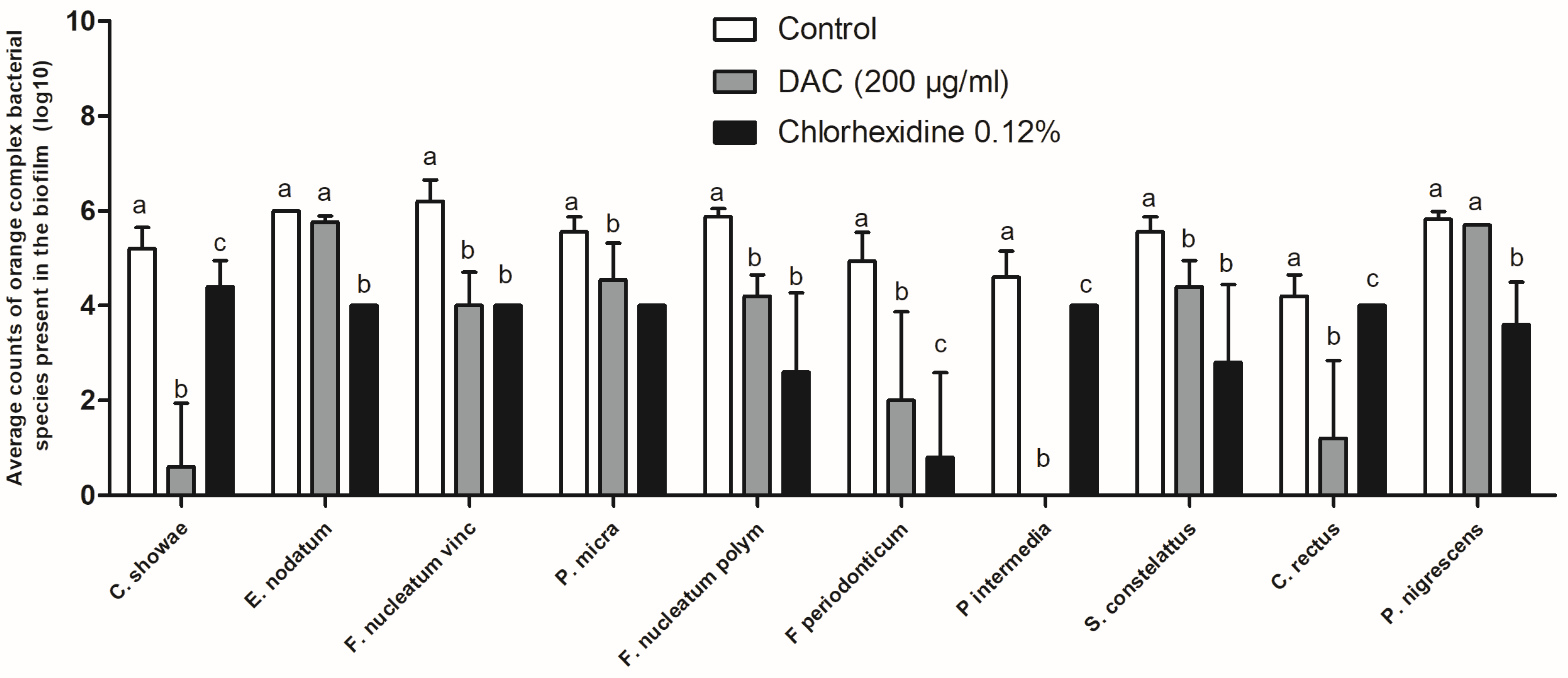
The assays were conducted in duplicates across three independent experiments. Biofilm assay data were analyzed using the Kruskal–Wallis test with Dunn’s post hoc analysis, applying a significance level of 5%. For the Galleria mellonella model, survival curves were generated in GraphPad Prism 5.0 using the log-rank (Mantel–Cox) test, and survival differences were assessed through the log-rank test.

3. Results

Antibiofilm Activity of DAC

Diacetylcurcumin (DAC), at a concentration of 200 µg/mL, was evaluated in a biofilm comprising 40 bacterial species. Figure 1 shows the metabolic activity of the biofilm after treatments. Treatment with DAC reduced the biofilm’s metabolic activity by 51%, while chlorhexidine at 0.12% showed a reduction of 88% compared to the untreated control (p < 0.05). These results highlight the potential of DAC as a promising alternative, although less potent than chlorhexidine, for antimicrobial interventions targeting multispecies subgingival biofilms.
Figure 2 shows the total biofilm counts obtained using the checkerboard technique for a biofilm formed over 7 days, with treatment starting on the 3rd day of formation. Treatments were performed twice daily, each lasting 1 min, using DAC (200 µg/mL) and chlorhexidine at 0.12%. Treatment with DAC reduced total biofilm counts by approximately 55% compared to the vehicle-treated group (p < 0.05). In contrast, treatment with chlorhexidine resulted in a reduction of approximately 80% in total biofilm counts, which was statistically different from the vehicle-treated control (p < 0.05) but not statistically different from the DAC treatment (p > 0.05).
Figure 3 shows the mean counts of each bacterial species from the experiments, with biofilms treated from the third day of formation, twice a day for 1 min each, with DAC (200 μg/mL) and 0.12% chlorhexidine. Treatment with chlorhexidine reduced the counts of 36 distinct species (p < 0.05), while DAC (200 μg/mL) reduced the counts of 27 species. Although DAC did not achieve the same level of total biofilm reduction as chlorhexidine, it significantly decreased key bacteria associated with periodontitis, notably Actinomyces israelli, Selenomonas noxia, Prevotella melaninogenica, Fusobacterium, Prevotella intermedia, Porphyromonas gingivalis, and Tanerella forsythia.
Figure 4 illustrates the effect of DAC specifically on bacteria from Socransky’s red complex.
Figure 4 demonstrates that DAC exhibited significant activity against complex red bacteria, with no statistical differences compared to 0.12% chlorhexidine, the gold standard in dentistry.
DAC demonstrated remarkable efficacy in inhibiting the growth of eight out of ten strains from the orange complex, including C. showae, F. nucleatum vincentii, P. micra, F. nucleatum polymorphum, F. periodonticum, P. intermedia, S. constellatus, and C. rectus, with complete inhibition of P. intermedia (Figure 5).
In Figure 6, it is possible to observe that DAC at the concentration tested in the biofilm did not present acute toxicity when injected into G. mellonella larvae. Studies carried out by Sardi et al., 2017, and Sanches et al., 2019 [12,13], also did not find DAC toxicity.

4. Discussion

The development of complex multispecies subgingival biofilm models, composed of up to 40 bacterial species, presents a promising approach to study periodontitis, one of the most prevalent diseases in the oral cavity. However, faithfully replicating in vivo conditions in in vitro models remains a significant challenge. Despite significant advances in studies on single-species biofilms, the complex interactions present in multispecies biofilms still require a deeper understanding to optimize strategies for the prevention, diagnosis, and treatment of periodontal infections, as well as to determine prognosis [17].
Socransky’s complex is a fundamental classification for understanding bacterial dynamics in the oral cavity, especially in the context of periodontal diseases. In this model, bacteria are grouped into complexes based on their association with the progression of periodontitis. The red complex, consisting of Porphyromonas gingivalis, Tannerella forsythia, and Treponema denticola, is particularly relevant as these species are strongly associated with the more advanced stages of periodontal disease. Additionally, the orange complex, which includes pathogens such as Prevotella intermedia, also plays a crucial role in periodontal pathogenesis, often found in synergy with bacteria from the red complex. This classification helps clarify how different species interact within the subgingival biofilm and contribute to the inflammation and tissue destruction characteristic of periodontitis [18].
Curcumin, a natural compound widely recognized for its pharmacological activities, including antioxidant, anti-inflammatory, antimicrobial, and anticancer properties, has been extensively investigated for combating oral pathogens. Previous studies have demonstrated its effectiveness against various bacterial species associated with periodontitis, such as Streptococcus mutans, Lactobacillus casei, Actinomyces viscosus, Porphyromonas gingivalis, and Fusobacterium nucleatum [12]. Li et al. [19] reported that curcumin significantly inhibited the biofilm formation and metabolism of S. mutans, while Izui et al. [20] highlighted its ability to reduce the virulence factors of P. gingivalis, such as the proteases kgp and rgp. However, curcumin’s low bioavailability, due to its hydrophobic nature and rapid hepatic metabolism, limits its therapeutic applicability [21,22].
The search for curcumin analogs with higher bioactivity and bioavailability has been a promising strategy, and among these alternatives, diacetylcurcumin (DAC), a synthetic derivative of curcumin, stands out. Studies have shown that DAC exhibits higher antimicrobial activity compared to curcumin, being effective against planktonic cells of S. mutans, methicillin-sensitive Staphylococcus aureus (MSSA), and methicillin-resistant Staphylococcus aureus (MRSA) [12]. Additionally, DAC demonstrated efficacy comparable to vancomycin against MSSA and MRSA biofilms, highlighting its potential as a therapeutic agent against oral infections [12,13]. In a recent study conducted by our group, DAC also proved effective against Enterococcus faecalis biofilms, serving as a promising intracanal medication [23].
In this study conducted by our group, we demonstrated for the first time the antimicrobial activity of DAC against periodontopathogenic biofilms. DAC was effective in significantly reducing the bacterial proportions in multispecies biofilms, particularly affecting species from the red and orange complexes of Socransky, such as A. israelli, S. noxia, P. melaninogenica, and P. intermedia, some of which were completely eliminated. Other species, such as P. gingivalis, T. forsythia, and Fusobacterium periodonticum, also showed substantial reductions [24]. This antimicrobial effect highlights the potential of DAC not only in reducing oral pathogens, but also in modulating the interactions within the biofilm, which is crucial for the treatment of periodontitis.
In particular, Porphyromonas gingivalis and Tannerella forsythia, members of the red complex, are primary pathogens in periodontitis and have significant systemic implications, being associated with cardiovascular diseases, diabetes, and Alzheimer’s disease due to their ability to invade tissues and induce systemic inflammation [24,25]. Reducing these species in the biofilm with the use of DAC could have a significant impact on reducing the bacterial load and mitigating the systemic effects associated with these oral infections.
Recent studies support the efficacy of curcumin and its derivatives in combating periodontal pathogens. Hr et al. [26] showed that nanocurcumin inhibited P. gingivalis, T. forsythia, Aggregatibacter actinomycetemcomitans, and P. intermedia at low concentrations, while Guru [27] reported that the use of curcumin in patients with chronic periodontitis improved clinical parameters, reducing the bacterial load of P. gingivalis, T. forsythia, and A. actinomycetemcomitans. These studies reinforce the relevance of curcumin-derived compounds as alternative or complementary treatments to conventional approaches, such as the use of chlorhexidine gel. Additionally, DAC eliminated A. israelii, a bacterium belonging to the Actinomyces sp. group, associated with both aggressive generalized and chronic periodontitis, and found in shallow and deep periodontal pockets [28,29]. This finding underscores the importance of eliminating this bacterium for periodontitis control.
Another important finding was the reduction in A. actinomycetemcomitans, previously considered the main causative agent of localized aggressive periodontitis. It is now recognized that this bacterium is part of a pathogenic consortium that suppresses the host’s initial response and promotes the overgrowth of other pathogens [30]. Therefore, the reduction in A. actinomycetemcomitans by DAC has significant therapeutic implications.
Additionally, toxicity studies using Galleria mellonella indicate that DAC has low toxicity, offering ethical and economic advantages, as this invertebrate species is commonly used to evaluate antimicrobial and therapeutic agents [31]. Galleria mellonella, a wax moth larva, has gained prominence as an alternative model in biomedical research due to its simplicity, low cost, and ease of maintenance, providing a viable option to replace vertebrate animal models. Its immune response, while distinct from humans, shares fundamental characteristics with mammalian immune responses, such as the activation of hemocytes and the production of cytokines, allowing the evaluation of the toxicity and antimicrobial efficacy of compounds. Furthermore, Galleria mellonella has a short life cycle, enabling rapid experimentation, with the added benefit of reducing the need for vertebrate animal testing, aligning with ethical guidelines for biomedical research. The use of this model has proven effective in studies assessing the efficacy of various therapeutic compounds, including antibiotics and natural substances, providing a preliminary analysis of toxicological profiles and antimicrobial properties before more complex model testing. In conclusion, diacetylcurcumin has shown great potential in controlling periodontopathogenic biofilms, effectively targeting bacterial complexes associated with periodontitis. The observed antimicrobial efficacy, combined with its low toxicity, positions DAC as a promising alternative for the treatment of periodontitis and other oral infections, standing out in the landscape of therapies based on modified natural compounds [32,33].

5. Conclusions

Based on the findings of this study, it can be concluded that treatment with DAC at 200 µg/mL reduced the metabolic activity of the multispecies subgingival biofilm by 49%, as well as the counts of P. gingivalis, T. forsythia, F. periodonticum, F. nucleatum, P. intermedia, A. israelli, S. noxia, P. melaninogenica, C. showae, and C. rectus. Furthermore, at the tested concentrations, no toxicity was observed. These results suggest that DAC has a positive effect on the formation of periodontopathogenic biofilms. Further studies should focus on its immunomodulatory activity and long-term toxicity in other models.

Author Contributions

Conceptualization, B.B.-S., L.C.F. and J.d.C.O.S.; Formal analysis, B.B.-S., L.C.F. and J.d.C.O.S.; Funding acquisition, L.C.F. and J.d.C.O.S.; Investigation, B.B.-S., L.C.F., L.O.R., C.R.P. and J.d.C.O.S.; Methodology, V.A.A.d.S., T.T.M., L.D.A.d.S., H.C.C.d.O.J. and J.d.C.O.S.; Project administration, J.d.C.O.S.; Resources, L.O.R., C.R.P. and J.d.C.O.S.; Supervision, B.B.-S., L.C.F. and J.d.C.O.S.; Validation, V.A.A.d.S., T.T.M., L.D.A.d.S., H.C.C.d.O.J. and J.d.C.O.S.; Visualization J.d.C.O.S.; Writing—original draft V.A.A.d.S., B.B.-S., L.C.F., T.T.M., L.D.A.d.S., H.C.C.d.O.J. and J.d.C.O.S.; Writing—review & editing B.B.-S., L.C.F., C.R.P., L.O.R. and J.d.C.O.S. All authors have read and agreed to the published version of the manuscript.

Funding

This research was funded by Coordination for the Improvement of Higher Education Personnel (CAPES).

Informed Consent Statement

Not applicable.

Data Availability Statement

The original contributions presented in this study are included in the article. Further inquiries can be directed to the corresponding authors.

Conflicts of Interest

The authors declare no conflicts of interest.

References

  1. Sharma, A.; Raman, A.; Pradeep, A.R. Role of 1% alendronate gel as adjunct to mechanical therapy in the treatment of chronic periodontitis among smokers. J. Appl. Oral Sci. 2017, 25, 243–249. [Google Scholar] [CrossRef] [PubMed]
  2. Tonetti, M.S.; Greenwell, H.; Kornman, K.S. Staging and grading of periodontitis: Framework and proposal of a new classification and case definition. J. Periodontol. 2018, 89, 1475. [Google Scholar] [CrossRef] [PubMed]
  3. Janakiram, C.; Dye, B.A. A public health approach for prevention of periodontal disease. Periodontol 2000 2020, 84, 202–214. [Google Scholar] [CrossRef] [PubMed]
  4. Socransky, S.S.; Haffajee, A.D.; Dzink, J.L.; Hillman, J.D. Associations between microbial species in subgingival plaque samples. Oral Microbiol. Immunol. 1988, 3, 1–7. [Google Scholar] [CrossRef]
  5. Aja, E.; Mangar, M.; Fletcher, H.M.; Mishra, A. Filifactor alocis: Recent Insights and Advances. J. Dent. Res. 2021, 100, 790–797. [Google Scholar] [CrossRef]
  6. Mishra, A.; Dou, Y.; Wang, C.; Fletcher, H.M. Filifactor alocis enhances survival of Porphyromonas gingivalis W83 in response to H2O2-induced stress. Mol. Oral Microbiol. 2024, 39, 12–26. [Google Scholar] [CrossRef]
  7. Hajishengallis, G. Periodontitis: From microbial immune subversion to systemic inflammation. Nat. Rev. Immunol. 2015, 15, 30–44. [Google Scholar] [CrossRef]
  8. Kumbar, V.M.; Peram, M.R.; Kugaji, M.S.; Shah, T.; Patil, S.P.; Muddapur, U.M.; Bhat, K.G. Effect of curcumin on growth, biofilm formation and virulence factor gene expression of Porphyromonas gingivalis. Odontology 2021, 109, 18–28. [Google Scholar] [CrossRef]
  9. Anuradha, B.R.; Bai, Y.D.; Sailaja, S.; Sudhakar, J.; Priyanka, M.; Deepika, V. Evaluation of Anti-Inflammatory Effects of Curcumin Gel as an Adjunct to Scaling and Root Planing: A Clinical Study. J. Int. Oral Health 2015, 7, 90–93. [Google Scholar]
  10. Peng, Y.; Ao, M.; Dong, B.; Jiang, Y.; Yu, L.; Chen, Z.; Hu, C.; Xu, R. Anti-Inflammatory Effects of Curcumin in the Inflammatory Diseases: Status, Limitations and Countermeasures. Drug Des. Dev. Ther. 2021, 15, 4503–4525. [Google Scholar] [CrossRef]
  11. Polaquini, C.R.; Marques, B.C.; Ayusso, G.M.; Morão, L.G.; Sardi, J.C.O.; Campos, D.L.; Silva, I.C.; Cavalca, L.B.; Scheffers, D.J.; Rosalen, P.L.; et al. Antibacterial activity of a new monocarbonyl analog of curcumin MAC 4 is associated with divisome disruption. Bioorg Chem. 2021, 109, 104668. [Google Scholar] [CrossRef] [PubMed]
  12. Sardi, J.C.O.; Polaquini, C.R.; Freires, I.A.; Galvão, L.C.C.; Lazarini, J.G.; Torrezan, G.S.; Regasini, L.O.; Rosalen, P.L. Antibacterial activity of diacetylcurcumin against Staphylococcus aureus results in decreased biofilm and cellular adhesion. J. Med. Microbiol. 2017, 66, 816–824. [Google Scholar] [CrossRef] [PubMed]
  13. Sanches, C.V.G.; Sardi, J.C.O.; Terada, R.S.S.; Lazarini, J.G.; Freires, I.A.; Polaquini, C.R.; Torrezan, G.S.; Regasini, L.O.; Fujimaki, M.; Rosalen, P.L. Diacetylcurcumin: A new photosensitizer for antimicrobial photodynamic therapy in Streptococcus mutans biofilms. Biofouling 2019, 35, 340–349. [Google Scholar] [CrossRef] [PubMed]
  14. Polaquini, C.R.; Torrezan, G.S.; Santos, V.R.; Nazaré, A.C.; Campos, D.L.; Almeida, L.A.; Silva, I.C.; Ferreira, H.; Pavan, F.R.; Duque, C.; et al. Antibacterial and Antitubercular Activities of Cinnamylideneacetophenones. Molecules 2017, 22, 1685. [Google Scholar] [CrossRef]
  15. Soares, G.M.; Teles, F.; Starr, J.R.; Feres, M.; Patel, M.; Martin, L.; Teles, R. Effects of azithromycin, metronidazole, amoxicillin, and metronidazole plus amoxicillin on an in vitro polymicrobial subgingival biofilm model. Antimicrob. Agents Chemother. 2015, 59, 2791–2798. [Google Scholar] [CrossRef]
  16. Megaw, J.; Thompson, T.P.; Lafferty, R.A.; Gilmore, B.F. Galleria mellonella as a novel in vivo model for assessment of the toxicity of 1-alkyl-3-methylimidazolium chloride ionic liquids. Chemosphere 2015, 139, 197–201. [Google Scholar] [CrossRef]
  17. Miranda, S.L.F.; Damaceno, J.T.; Faveri, M.; Figueiredo, L.C.; Soares, G.M.S.; Feres, M.; Bueno-Silva, B. In Vitro Antimicrobial Effect of Cetylpyridinium Chloride on Complex Multispecies Subgingival Biofilm. Braz. Dent. J. 2020, 31, 103–108. [Google Scholar] [CrossRef]
  18. Socransky, S.S.; Haffajee, A.D.; Cugini, M.A.; Smith, C.; Kent, R.L., Jr. Microbial complexes in subgingival plaque. J. Clin. Periodontol. 1998, 25, 134–144. [Google Scholar] [CrossRef]
  19. Li, B.; Li, X.; Lin, H.; Zhou, Y. Curcumin as a Promising Antibacterial Agent: Effects on Metabolism and Biofilm Formation in S. mutans. Biomed. Res. Int. 2018, 2018, 4508709. [Google Scholar]
  20. Izui, S.; Sekine, S.; Maeda, K.; Kuboniwa, M.; Takada, A.; Amano, A.; Nagata, H. Antibacterial Activity of Curcumin Against Periodontopathic Bacteria. J. Periodontol. 2016, 87, 83–90. [Google Scholar] [CrossRef]
  21. Wang, Y.J.; Pan, M.H.; Cheng, A.L.; Lin, L.I.; Ho, Y.S.; Hsieh, C.Y.; Lin, J.K. Stability of curcumin in buffer solutions and characterization of its degradation products. J. Pharm. Biomed. Anal. 1997, 15, 1867–1876. [Google Scholar] [CrossRef] [PubMed]
  22. Kharat, M.; Du, Z.; Zhang, G.; McClements, D.J. Physical and Chemical Stability of Curcumin in Aqueous Solutions and Emulsions: Impact of pH, Temperature, and Molecular Environment. J. Agric. Food Chem. 2017, 65, 1525–1532. [Google Scholar] [CrossRef]
  23. Souza, V.; Polaquini, C.R.; de Moraes, G.R.; Oliveira Braga, A.R.; da Silva, P.V.; da Silva, D.R.; Ribeiro Lima, F.R.; Regasini, L.O.; Cássia Orlandi Sardi, J. Diacetylcurcumin: A novel strategy against Enterococcus faecalis biofilm in root canal disinfection. Future Microbiol. 2024, 19, 647–654. [Google Scholar] [CrossRef] [PubMed]
  24. Bregaint, S.; Boyer, E.; Fong, S.B.; Meuric, V.; Bonnaure-Mallet, M.; Jolivet-Gougeon, A. Porphyromonas gingivalis outside the oral cavity. Odontology 2022, 110, 1–19. [Google Scholar] [CrossRef] [PubMed]
  25. Olsen, I.; Taubman, M.A.; Singhrao, S.K. Porphyromonas gingivalis suppresses adaptive immunity in periodontitis, atherosclerosis, and Alzheimer’s disease. J. Oral Microbiol. 2016, 8, 33029. [Google Scholar] [CrossRef]
  26. Hr, V.; Daniel, R.A.; Prabhu, A.P.S.; Basavaraju, S. Susceptibility of periodontal pathogens to a novel target-specific drug delivery system containing self-nanoemulsifying curcumin: An in vitro study. J. Adv. Periodontol. Implant. Dent. 2023, 15, 67–73. [Google Scholar] [CrossRef]
  27. Guru, S.R.; Reddy, K.A.; Rao, R.J.; Padmanabhan, S.; Guru, R.; Srinivasa, T.S. Comparative evaluation of 2% turmeric extract with nanocarrier and 1% chlorhexidine gel as an adjunct to scaling and root planing in patients with chronic periodontitis: A pilot randomized controlled clinical trial. J. Indian Soc. Periodontol. 2020, 24, 244–252. [Google Scholar] [CrossRef]
  28. Heller, D.; Silva-Boghossian, C.M.; do Souto, R.M.; Colombo, A.P. Subgingival microbial profiles of generalized aggressive and chronic periodontal diseases. Arch. Oral Biol. 2012, 57, 973–980. [Google Scholar] [CrossRef]
  29. Vielkind, P.; Jentsch, H.; Eschrich, K.; Rodloff, A.C.; Stingu, C.S. Prevalence of Actinomyces spp. in patients with chronic periodontitis. Int. J. Med. Microbiol. 2015, 305, 682–688. [Google Scholar] [CrossRef]
  30. Fine, D.H.; Patil, A.G.; Velusamy, S.K. Aggregatibacter actinomycetemcomitans (Aa) Under the Radar: Myths and Misunderstandings of Aa and Its Role in Aggressive Periodontitis. Front. Immunol. 2019, 10, 728. [Google Scholar] [CrossRef]
  31. Ménard, G.; Rouillon, A.; Cattoir, V.; Donnio, P.Y. Galleria mellonella as a Suitable Model of Bacterial Infection: Past, Present and Future. Front. Cell Infect. Microbiol. 2021, 11, 782733. [Google Scholar] [CrossRef] [PubMed]
  32. Tsai, C.J.; Loh, J.M.; Proft, T. Galleria mellonella infection models for the study of bacterial diseases and for antimicrobial drug testing. Virulence 2016, 7, 214–229. [Google Scholar] [CrossRef] [PubMed]
  33. Santos, T.A.; Scorzoni, L.; Santos, A.C.; Junqueira, J.C.; Anbinder, A.L. Galleria mellonella as an experimental model for studying periodontopathogens. J. Indian Soc. Periodontol. 2020, 24, 593–596. [Google Scholar] [PubMed]
Figure 1. Effect of two daily treatments, each lasting 1 min, in control (C), with DAC at 200 μg/mL and 0.12% chlorhexidine (CHR) on the metabolic activity of biofilms formed over 7 days (n = 6—3 independent experiments). Different letters (a, b, or c) indicate statistically significant differences between groups, determined using the Kruskal–Wallis test followed by Dunn’s post hoc test (p < 0.05).
Futurepharmacol 05 00019 g001
Figure 2. Mean and standard deviation of total biofilm counts treated with the negative vehicle control (control), DAC 200 µg/mL, and 0.12% chlorhexidine (CHX), analyzed using the DNA-DNA checkerboard hybridization technique. Different letters (a or b) indicate a statistically significant difference, determined by the Kruskal–Wallis test followed by Dunn’s post hoc test (p < 0.05).
Futurepharmacol 05 00019 g002
Figure 3. Average counts of each bacterial species present in the biofilm, treated from the third day twice a day for 1 min each with DAC (200 µg/mL) and chlorhexidine 0.12%.
Futurepharmacol 05 00019 g003
Figure 4. Mean counts of bacterial species from the red complex present in the biofilm, treated from the third day of formation twice a day for 1 min each with DAC (200 µg/mL) and 0.12% chlorhexidine. Statistical analysis was performed using the Kruskal–Wallis test followed by Dunn’s post hoc test (p < 0.05). All groups were compared to each other (n = 6—3 distinct experiments). Different letters (a, b) indicate a statistically significant difference, determined by the Kruskal–Wallis test followed by Dunn’s post hoc test (p < 0.05).
Futurepharmacol 05 00019 g004
Figure 5. Average counts of bacterial species of the orange complex present in the biofilm treated from the third day, 2/x per day for 1 min each with DAC (200 µg/mL) and chlorhexidine 0.12%. Statistical analysis was performed using the Kruskal–Wallis test followed by Dunn’s post hoc test (p < 0.05). All groups were compared with each other, and different letters (a, b or c) indicate statistical difference (n = 6—3 different experiments).
Futurepharmacol 05 00019 g005
Figure 6. Percentage survival over time of G. mellonella larvae injected with DAC at concentrations of 200, 100 and 50 μg/mL, respectively, which correspond to their effective anti-biofilm concentrations (200 μg/mL) (p > 0.05, log-rank test).
Futurepharmacol 05 00019 g006
Table 1. Bacteria used in the formation of multispecies biofilm.
BacteriaATCCBacteriaATCC
Actinomyces naeslundiiATCC12104Actinomyces orisATCC43146
Actinomyces gerencseriaeATCC23840Actinomyces israeliiATCC12102
Veillonella parvulaATCC10790Actinomyces odontolyticusATCC17929
Streptococcus sanguinisATCC10556Streptococcus oralisATCC35037
Streptococcus intermediusATCC27335Streptococcus gordoniiATCC10558
Streptococcus mitisATCC49456Aggregatibacter actinomycetemcomitansATCC29523
Capnocytophaga ochraceaATCC33596Capnocytophaga gingivalisATCC33624
Eikenella corrodensATCC23834Capnocytophaga sputigenaATCC33612
Streptococcus constellatusATCC27823Eubacterium nodatumATCC33099
Fusobacterium nucleatum vincentiiATCC49256Parvimonas micraATCC33270
Fusobacterium nucleatum polymorphumATCC10953Fusobacterium nucleatum nucleatumATCC25586
Campylobacter showaeATCC51146Capnocytophaga sputigenaATCC33612
Campylobacter gracilisATCC33236Leptotrichia buccalisATCC14201
Campylobacter rectusATCC33238Fusobacterium periodonticumATCC33693
Prevotella intermediaATCC25611Porphyromonas gingivalisATCC33277
Tannerella forsythiaATCC43037Prevotella melaninogenicaATCC25845
Prevotella nigrescensATCC33563Eubacterium saburreumATCC33271
Streptococcus anginosusATCC33397Selenomonas noxiaATCC43541
Neisseria mucosaATCC19696Propionibacterium acnesATCC11827
Gemella morbillorumATCC27824Streptococcus mutansATCC25175
Disclaimer/Publisher’s Note: The statements, opinions and data contained in all publications are solely those of the individual author(s) and contributor(s) and not of MDPI and/or the editor(s). MDPI and/or the editor(s) disclaim responsibility for any injury to people or property resulting from any ideas, methods, instructions or products referred to in the content.

