Review

Anticancer Efficacy of Decursin: A Comprehensive Review with Mechanistic Insights

by
Tanzila Akter Eity
1,2,
Md. Shimul Bhuia
2,3,
Raihan Chowdhury
2,3,
Md. Arman Ali
3,
Mst Muslima Khatun
2,3,
Salehin Sheikh
2,3,
Md. Sakib Al Hasan
2,3,
Rubel Hasan
2,3,
Ivo Cavalcante Pita Neto
4,
Isaac Moura Araújo
5,
Henrique D. M. Coutinho
5,* and
Muhammad Torequl Islam
2,3,6,*
1
Department of Biotechnology and Genetic Engineering, Gopalganj Science and Technology University, Gopalganj 8100, Bangladesh
2
BioLuster Research Center Ltd., Gopalganj 8100, Bangladesh
3
Department of Pharmacy, Gopalganj Science and Technology University, Gopalganj 8100, Bangladesh
4
CECAPE College, Av. Padre Cícero 3917, São José, Juazeiro do Norte 63024-015, CE, Brazil
5
Department of Biological Chemistry, Regional University of Cariri, Crato 63105-000, CE, Brazil
6
Pharmacy Discipline, Khulna University, Khulna 9208, Bangladesh
*
Authors to whom correspondence should be addressed.
Future Pharmacol. 2025, 5(2), 17; https://doi.org/10.3390/futurepharmacol5020017
Submission received: 17 February 2025 / Revised: 19 March 2025 / Accepted: 24 March 2025 / Published: 10 April 2025
(This article belongs to the Special Issue Feature Papers in Future Pharmacology 2025)

Abstract

:
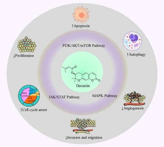
Introduction: Decursin is a pyranocoumarin natural phytochemical found in the Angelica gigas Nakai herb, which shows various therapeutic properties and beneficial effects against various diseases. Objective: The aim of this study was to find the anticancer potential of decursin and its molecular mechanisms involved with different anticancer effects. Methodology: All of the relevant data concerning this compound and cancer were collected using different scientific search engines, including PubMed, Scopus, Springer Link, Wiley Online, Web of Science, Scifinder, ScienceDirect, and Google Scholar. Results: This study found that decursin shows anticancer properties through various mechanisms, such as apoptosis, cell cycle arrest, inhibition of cell proliferation, autophagy, inhibition of angiogenesis, cytotoxicity, and the inhibition of invasion and migration against a number of cancers, including breast, bladder, lung, colon, skin, ovarian, prostate, pancreatic, and bone cancers. This study also discovered that decursin has the ability to affect several signaling pathways in the molecular anticancer mechanisms, such as the PI3K/AKT/mTOR, JAK/STAT, and MAPK signaling pathways. Findings also revealed that decursin expresses poor oral bioavailability. Conclusions: Based on the data analysis from this literature-based study, decursin has properties to be considered as a potential candidate in the treatment of cancer. However, more clinical research is suggested to establish proper efficacy, safety, and human dosage.

Graphical Abstract

1. Introduction

Cancer encompasses numerous medical conditions characterized by the unregulated division of cells and the ability to infiltrate surrounding tissues, which can metastasize to different body regions via the circulatory and lymphatic systems, which also aid in flushing harmful substances [1,2]. Cancer is a remarkable global health burden because it is the second major cause of mortality in numerous countries, after heart disease [3]. The majority of cancer, about 70% of the burden, is shouldered by developing countries [4]. Significant advancements have been established over the past few years, but the incidence of cancer is growing due to increasing global populations, in addition to factors that increase risk, such as smoking, being overweight, and eating habits [5]. In 2020, there were approximately 19.3 million new instances of cancer in the world (excluding nonmelanoma skin cancer), resulting in nearly 10.0 million deaths (excluding nonmelanoma skin cancer). It is estimated that by 2030, there are going to be approximately 26 million new cases of cancer and 17 million fatalities from cancer annually, and by 2040, there will be a total of 28.4 million cancer cases worldwide [5,6]. This rise will be more significant in transitioning countries [6]. In Bangladesh, in 2022, the number of new cancer cases was 167,256, and the number of deaths due to cancer was 116,598 [7].
The cancer with the greatest percentage of causes that can be related to these potential risks, including cervical, lung, and esophageal cancer. The most significant risk factors for these malignant tumors encompassed sexual transmission of HPV that results in infections that persist for the long term with oncogenic viruses, alcohol consumption, smoking, and inadequate consumption of fruits and vegetables [8]. Additional instances encompass several substances, including those now employed within research laboratories, such as ethidium bromide, a profoundly carcinogenic chemical [9]. Researchers started looking at the potential more than a century ago that viruses and other infectious substances could be the origin of malignant tumors [10]. An investigation was conducted to examine the correlation between the standard of life in cancer patients and many factors, including medication, pain, early identification, psychological distress, organ transplantation, the length of illnesses, and attendants [11,12]. In addition, cancer can overlap in patients with other significant disorders, such as Parkinson’s disease, Down’s syndrome, obesity, schizophrenia, allergy-related conditions, Alzheimer’s disease, and multiple sclerosis [13]. Tumor microcirculation, pH level in the tissues, oxygen and nutrition delivery, and bioenergetic condition can all have a significant impact on how effectively malignant tumors cope with treatment such as chemotherapy, traditional radiation therapy, and other endoscopic procedures for treatment [14,15,16]. The growing prevalence of prognostic mutations in various types of tumors demands the application of comprehensive, multiplied, and extremely accurate sequencing equipment in regular medical treatment [17,18].
Though intense protocols of concurrent chemotherapy and radiation therapy can result in more efficacy, they also give rise to significant adverse consequences, including mucositis, xerostomia, dysphagia, a reduction in weight, neutropenia, and sepsis [19,20]. There is still a group of individuals whose tumors do not react to stringent chemoradiotherapy, and it is crucial to determine specific and consistent molecular indicators of tumor susceptibility to chemoradiotherapy [21]. Therefore, there is a critical requirement for innovative and targeted therapeutic solutions to treat various types of human cancers [22,23].
The molecules obtained from natural substances, such as alkaloids, terpenoids, organosulfur derivatives, and polyphenols, have numerous possible configurations and can effectively demonstrate cancer chemopreventive and chemotherapeutic properties [22,24,25]. Stilbenes, phenolic acids, and flavonoids are the predominant organic polyphenols, with flavonoids accounting for approximately two-thirds of all polyphenolic synthetic substances, which are exceptionally common in many plant-based products, such as fruits and vegetables, and are commonly ingested by people as an ordinary part of their daily diet [26,27,28]. Investigation of natural products (NPs) through the previous three decades indicates that almost 40% of the medicinal medications that received approval from the US Food and Drug Administration (FDA) were NPs, or artificial compounds mimicking NPs [29]. Decursin and its derivatives are also a type of synthetic compound that exhibit not only anticancer properties but also anti-inflammatory capabilities [30].
Decursin (2,2-dimethyl-8-oxo-3,4-dihydropyrano 3-methylbut-2-enoate) (Figure 1) is a pyranocoumarin phytochemical collected from the roots of the Angelica gigas Nakai herb, which is found in several Asian nations, such as Korea, China, and Japan [30,31,32]. Stopping tumor growth can be effectively achieved by inhibiting the abnormal growth of the cell cycle in cancer cells [33]. Decursin caused a halt in the cell cycle at the G1 phase [34]. Decursin demonstrated viability as a therapy strategy against prostate cancer cells (RC-58T/h/SA#4) by activating the jnkjnk-dependent mechanism [35]. Research findings indicate that decursin exhibited anti-inflammatory activities on skin keratinocytes, antifibrotic activities on hepatocytes, and antioxidant properties on renal epithelial cells [36,37,38,39]. The compound also has several other therapeutic activities, including antiviral, antidiabetic [40], angiogenesis [33], and effects on neurological diseases, such as Parkinson’s disease, depression, and Alzheimer’s disease [41]. The study investigates the techniques by which decursin exerts its anticancer effects, including the underlying molecular pathways, cellular processes, and interconnections. Furthermore, we highlighted the botanical origins and physiochemical features.

2. Materials and Methods

2.1. Literature Search Stratagem

The data were collected (up to date 2024) by searching electronic databases such as PubMed, ScienceDirect, Springer Link, Scopus, Wiley Online, Web of Science, ResearchGate and Google Scholar with the terms “Decursin”, then paired with “Cancer”, “Tumor”, “Pathophysiology of cancer”, “Anticancer activity”, “Antiproliferation activity”, “Apoptotic effect”, “Oxidative stress”, “Protective effect”, “Cytotoxic activity”, “Genotoxic activity“, “Carcinogenesis“, “Anti-angiogenic effect”, “Antitumor activity”, “Human cancer”, “Biological activities”, “Biological evaluation”, “Chemical features”, “Pharmacokinetics”, “Biopharmaceutics”, “Medicinal use”, “Pharmacology”, “Pharmacological effects”, “Pharmacological activities”, “In vivo studies”, or “In vitro studies”. No language restrictions were imposed. The studies were thoroughly assessed, with information on the sources, dose, concentration, test system, hypothesized anticancer effect mechanism, and overall conclusion provided. The following are the criteria for inclusion and exclusion:

2.2. Inclusion and Exclusion Criteria

Inclusion criteria: (a) studies performed in different laboratory animals, humans, and their derived tissues or cells; (b) studies of the anticancer activities of decursin; (c) studies with decursin in combination with other molecules; (d) studies with or without hypothesized mechanisms of action; (e) studies with the physical and chemical characteristics of decursin; (f) studies with the biopharmaceutical profiles of decursin or its preparations; (g) studies with the toxicological profile of decursin; (h) studies of clinical investigation of decursin; (i) studies of the anticancer properties of decursin investigated up to date 2024.
Exclusion criteria: (i) studies exhibited duplicate data and/or titles and abstracts that did not meet the inclusion criteria; (ii) decursin, in conjunction with other studies, sheds light on the current issue; (iii) papers written in languages other than English; (iv) studies do not have complete written content accessible; (v) case reports, letters, editorials, and commentaries.

3. Anticancer Activity of Decursin: Mechanistic Analysis

3.1. Cytotoxicity

Destruction of cancer cells by anticancer drugs is one of the prominent approaches in cancer therapy [42]. As cancer cells themselves are harmful to the body because of the abnormally high rate of divisibility that leads to the growth and expansion of tumors. So, it is a prime concern to target and kill those cancerous cells in an efficient way with the drugs used in cancer treatment. In this regard, researchers initially examine the toxic effects of chemical compounds or biological plant extracts using in vitro and in vivo methods. After achieving promising cytotoxic effects, investigators usually proceed to the next stage of study in anticancer drug development [1,43]. Therefore, phytochemical compounds showing significant cytotoxic effects might have the capability to be studied for further anticancer drug discovery.
To explore the cytotoxic activity of decursin on bladder and colon cancer, Kim and his team found that decursin exerted significant cytotoxic effects on 253J and HCT11 cells [38]. Similar results were demonstrated in another study on U266, MM.1S, and ARH77 cell lines and on mice where increased cytotoxicity was experienced upon treatment with decursin (Table 1) [44,45,46]. In addition, there is an investigation on pancreatic cancer cells (PANC-1 and MIA PaCa-2), which aimed at exploring decursin’s anticancer effect, and the results support the cytotoxic effect of that compound. The study evidently shows that decursin at the concentrations of 20–60 µM exhibited an anticancer effect through increasing cell cytotoxicity [47]. Further, an in vitro study on doxorubicin-resistant NCI/ADR-RES ovarian cancer cells stated that decursin exhibited cell cytotoxicity with an IC50 value of 23 μg/mL. Published data also suggest that decursin could induce apoptotic cell death of doxorubicin-resistant ovarian cancer cells through blocking P-glycoprotein expression [48]. Consequently, decursin may be presented as a potential novel candidate for anticancer treatment due to its cytotoxic capabilities.

3.2. Cell Cycle Arrest

Signaling pathways, which regulate the termination of preceding processes before progressing to the next stage of the eukaryotic cell cycle, are known as cell cycle checkpoints [65,66]. The irregularities of the cell cycle are responsible for the abnormal expansion of cancer cells. Additionally, the lack of proper control over cell cycle checkpoints leads to the proliferation of cancer [67]. Usually, cell cycle arrest happens at the G1/S or G2/M boundaries. Even in the presence of cellular damage, the cell enters S phase or mitosis when the regulation of checkpoint arrest is disrupted. Genetic instability could arise from this, increasing the possibility of cancer developing and spreading. However, the inability of malignant cells to effectively halt the cell cycle can be strategically used for therapeutic purposes [68,69].
The strong capability of decursin to arrest the cell cycle checkpoint at G1/S is demonstrated in different studies. From a study by Kim and his colleagues, it was revealed that decursin could induce cell cycle arrest by decreasing the level of cyclin D1 in breast cancer cells (MDA-MB-231, MDA-MB-453, MDAMB-157, MCF-7, and MCF-10A) at 80 µM concentrations [49]. Another study found that decursin also had the potential to halt the cell cycle of colorectal cancer cells (CT-26) at a concentration of 10–20 µM through lowering the matrix metalloproteinase (MMP)-2 and MMP-9 expression as well as ERK and c-Jun N-terminal kinase (JNK) phosphorylation, which can block DNA synthesis [51]. A further study carried out by Kim and his co-workers found that decursin could induce cell cycle arrest in bladder and colon cancer cells (355J, HCT116, respectively) via lowering the expression of JNK, ERK, and MAP kinase expressions at 50–100 µM concentrations [38]. There is another study by Kim and his colleagues that revealed that decursin had a high effect on cell cycle arrest in gastric cancer cells (SNU216, NCI-N87) via decreasing CDK 4/6, LC3-II expression levels [39]. Similarly, decursin had an impact on pancreatic cancer cells (PANC-1 and MIA PaCa-2). It induces cell cycle arrest via downregulating cyclin D1, CDK4, and MMP-2 [47]. Decursin could also halt the cell cycle in prostate and lung cancer cells (DU145, PC-3, and LNCaP) at a concentration of 25–100 µM [34,57]. A study of Joo and his co-workers demonstrated that decursin could block cell cycle at the G0/G1 checkpoint in head and neck cancer cells (HNSCC) [60]. Li and his team found that decursin could cause cell cycle arrest in skin cancer cells (HepG2) at 5–80 µM concentrations [63]. By inducing cell cycle arrest, decursin worked as a treatment to diagnose different cancer cells via halting their growth.

3.3. Apoptotic Cell Death

Apoptosis, also known as a particular kind of genetically programmed cell death, which contributes to the systematic and effective elimination of dysfunctional cells [70], such as those caused by damage to the genome or occurring during the course of expansion, can be initiated by endogenous signals, such as genotoxic stress, as well as exogenous indications, for example, the interaction between ligands and receptors found on the cell surface [7,71].
Currently, the majority of the essential factors in the regulation of cell death have been discovered and are able to be specifically targeted by medical treatments [72]. Apoptotic mechanisms are promising targets for medicinal treatment where the regulation of Bcl-2 expression is the most sophisticated step in the development of drugs compared to other methods based on apoptosis [73,74]. Apoptosis is triggered by the sequential activation of caspase pathways through two different but convergent pathways referred to as the intrinsic mechanisms and extrinsic mechanisms [75,76]. Intrinsic mechanisms, also called mitochondrial pathways, are primarily regulated by the Bcl-2 peptide groups and initiated through DNA-damaging agents, overload of Ca2+, oncogene activation, oxidants, and microtubule-targeted drugs, and deficiency of growth factor [77]. The external route of apoptosis is activated by external signals that connect to apoptotic receptors, such as tumor necrosis factor receptors (TNF-R), Fas receptors, TNF-related death-inducing ligand proteins (TRAIL-R), and tumor necrosis factor receptors (TNF-R), which are seen on the outer layer of numerous cells [27].
Several preclinical studies found that the phytochemical has potent apoptotic effects against various cancer cells. Decursin (50−100 µM) induced apoptosis was correlated with a downregulation in the expression of the anti-apoptotic protein Bcl-2 and a boost in activation of pro-apoptotic factors such as cytochrome c, caspase 3, and Bax in both types of cells (235J cells and HCT116 cells) associated with bladder and colon cancer, respectively [78]. Another study found the impact of decursin (10, 30, 60, and 90 μmol/L−1) on the upregulation of programmed cell death in colorectal cancer cells (HT29 and HCT116) by targeting the phosphatidylinositol 3-kinase (PI3K) or serine-threonine kinase (Akt) pathway [52]. Different studies revealed that decursin could upregulate apoptosis in skin cancer cells (U266, MM.1S, and ARH77) at concentrations of 40–160 µM by triggering caspase-3, -8, and -9, which are linked with the decrease in the expression of cyclin D1, Bcl-2, survivin, Bcl-xL, and the vascular endothelial growth factor (VEGF) as well as in B16F10 cells via enhancing p38 phosphorylation and the synthesis of Bax, while reducing the phosphorylation of extrinsic signaling-regulated kinase (ERK) and the translation of Bcl-2 [45,53]. Recent research by Oh et al. (2019) investigated the activation of decursin-induced apoptosis in glioblastoma cancer cells (U87 and glioblastoma) at concentrations ranging between 10 and 200 µM [55]. Decursin also induced programmed cell death in prostate cancer cells (DU145, and PC-3) and pancreatic cancer cells (PANC-1 and MIA PaCa-2) through increasing the expression of caspase-3 and the breakdown of poly (ADP-ribose) polymerase [47]. Decursin had great efficiency in suppressing the expansion and inducing the demise of DU145 cells and prostate cancer PC-3 and LNCaP cells [34]. In a combination therapy, decursin also expressed notable apoptotic effects. The study revealed that decursin and doxorubicin could remarkably increase apoptosis via the mTOR and STAT3-mediated signaling cascade in numerous myeloma cells (U266, RPMI8226, and MM.1S) [53].

3.4. Inhibition of Cancer Cell Proliferation

Cell proliferation is a fundamental process among living organisms that is necessary for general development and physiological processes. Cell proliferation involves regulated cell growth and cell division rates that result in an increase in the number and size of cells [79,80]. There are many physiological regulations to ensure systematic tissue–organ development and replenishment of impairment [81]. But in a cancerous cell, those regulations are broken down. Moreover, significant changes are observed in the expression or activity of proteins that are associated with cell proliferation or cell cycle regulation. In addition to this, crucial pathways like MAPK, PI3K, and mTOR signaling pathways, which play pivotal roles in the regulation of cellular proliferation and growth, are often disrupted by various types of cancer [52]. Apart from this, signal transduction and transcription activation (STAT3) plays a vital role in the regulation of biological processes involved in the onset of malignant transformation, especially the beginning of cancerous cell growth [82]. Hence, downregulation of these pathways may be a potential objective in anticancer drug development studies.
Several studies revealed the antiproliferative effect of decursin while using various cancer cell lines. Such an investigation was conducted by Kim and this colleague, where decursin effectively prevented proliferation of breast cancer cells (MDA-MB-231, MDA-MB-453, MDAMB-157, MCF-7, and MCF-10A cell lines) through regulation of the expression of NF-κB and cyclin D1 levels with a concentration ranging from 1 to 80 µM [50]. Another study also showed that decursin decreased breast cancer cell proliferation via halting the G1 phase of the cell cycle [49]. In bladder and colon cancer cells, an antiproliferative effect of decursin (50–100 µM) was also observed, which was mediated by increasing apoptosis factors Bcl-2, caspase-3, and Bax, and lowering CDK2, CDK4, Bcl-2, Cyclin D1, and Cyclin E in 53J and HCT116 cell lines [38]. Similar effects were exerted by decursin during an investigation carried out on skin cancer cell lines [44,45]. The downregulation of PI3K and AKT by decursin was reported in cancer-related research while explaining the decursin-induced antiproliferative effects. The investigation used colorectal cancer cell lines such as CT-26, HT29, and HCT116, where the compound decursin was applied at a concentration ranging from 10 µM to 90 µM [51,52]. Likewise, other studies on common cancers such as gastric, prostate, and lung cancers also confirmed that decursin could prevent cell growth and proliferation [56,60,83]. Highly phosphorylated STAT3 is associated with tumorigenesis in some cancers, such as in some breast, prostate, and cervical cancer cells [84]. Cyclin A is found to activate two different cyclin-dependent kinases (CDKs) and play an important role in both the S phase and mitosis. Importantly, as a key cell cycle regulator, elevated expression of cyclin A is found in a variety of cancers [65,85]. Another member of the cyclin family, Cyclin E, a part of the cell cycle machinery, is crucial for progression through the G1 phase of the cell cycle. It also interacts and activates cyclin-dependent kinase 2 (CDK2) that results in DNA replication [86].
Significantly, it has been shown that decursin prevented cell proliferation of head and neck cancer (HNSCC cell line) in a concentration ranging from 50 µM to 100 µM through decreasing phosphorylation of STAT3, Cyclin A, Cyclin E, and CDK2, as well as enhancing G0/G1 cell cycle arresting [60]. In addition, data on ovarian cancer cells reported that decursin (5–50 µg/mL) suppressed cancer cell proliferation through increasing caspase-3, -8, and -9, cleaved PARP levels [48].

3.5. Inhibition of Invasion and Migration

Migration and invasion of cancerous cells inside the body’s healthy organs is one of the prime attributes of cancer [70]. The condition of cancer patients usually worsens when secondary malignant tumors emerge from other cancer cells. This spreading of life-threatening cancerous cells occurs due to their capacity to migrate from the affected organ to other normal tissue by using the blood and lymphatic systems as the main sources of migratory medium [87]. Various studies have been carried out to explain the migratory mechanism of cancer cells. There are two mechanisms suggested by prominent research: one is collective cell migration, and the other is individual cell migration [88]. Both of these mechanisms enable cancer cells to invade surrounding tissue by overcoming the defense employed by the extracellular matrix (ECM). To overcome the ECM-presented defense, cancer cells require some changes on their cell surface, including the recruitment and activation of several MMPs. This activation of MMPs results in the accommodation of cancer cells by restricting the ECM and enabling them to move forward in the migratory journey [82,89]. Additionally, there are some pathways, including STAT3 and AKT/GSK 3β/Snail signaling pathways, that play a remarkable role in the migration and invasion process towards metastasis [89]. Researchers who are currently undertaking efforts on anticancer drug design usually focus on these pathways during their anticancer drug discovery and development. It might be a worthy strategy to downregulate those pathways and MMPs in the study of anticancer drug development.
Many pieces of the literature have established evidence regarding the ability of decursin to resist cancerous cell migration and invasion. Such a study was conducted by Kim with his investigation team and revealed that decursin (1–50 µM) could inhibit the invasion of breast cancer cells (MCF-7) by interfering with TPA-induced MMP-9 [49]. It is reported that MMP-2 and MMP-9 largely contribute to the angiogenesis and progression of cancer cells, including gastric carcinoma [90]. When examining the anticancer effects of decursin, the investigation revealed that decursin at concentrations of 10–20 µM exerted anti-invasive properties under treatment on CT-26 colon carcinoma cells by decreasing MMP-2 and MMP-9 levels [51]. Kweon and his team conducted a study in which pancreatic ductal adenocarcinoma cells (PANC-1 and MIA PaCa-2) treated with decursin from a concentration ranging from 20 µM to 60 µM showed that the level of MMP-9 fell significantly [47].
Homotypic and heterotypic cell-cell adhesion is mediated by a calcium-dependent single-chain transmembrane glycoprotein called N-cadherin. This N-cadherin expression promotes cancer metastasis and invasion [91]. On the contrary, a decrease in E-cadherin level is positively associated with cancer cell invasion [92]. So, increasing E-cadherin and conversely decreasing N-cadherin levels by decursin is another mechanism to prevent invasion and metastasis. This mechanism is reported in a study using colorectal cancer cell lines (HT29 and HCT116) where decursin (10–90 μmol/L) caused an upregulation in E-cadherin expression as well as a decrease in N-cadherin expression.
The literature shows that abnormal stimulation of the PI3K-Akt pathway may elevated cell invasiveness and facilitate particularly in prostate cancer progression [93]. Decursin interfered with EMT-related gene expression and led to a decrease in the AKT level and barred cancer invasion [52]. An investigation on the assessment of the anti-invasive effect of decursin on U266, MM.1S, and ARH77 cell lines (skin cancer) asserted that the compound (10 µM) decreased AKT levels significantly [44]. A similar study revealed that there was a decrease in STAT3 to prevent distant organ metastasis. The experiment was conducted on skin cancer cells (U266, MM.1S, and ARH77) at concentrations of 40–160 µM [53]. Additionally, results from Joo and his colleague’s investigation indicated the reduction of phosphorylation of STAT3 and supported the inhibitory effect of decursin (50–100 µM) on the invasion and migration of head and neck cancer cells (HNSCC cell line) [60].

3.6. Inhibition of Angiogenesis

The creation of new capillary blood vessels, known as angiogenesis, mostly takes place throughout human growth and reproduction, but faulty regulation of angiogenesis is also a basic process observed in a number of pathologic disorders, such as cancer [94]. The Food and Drug Administration in the USA recently granted approval for angiogenesis inhibitors for the treatment of cancer [94].
Various antiangiogenic agents, together with extracellular matrix components and cell types, work together to influence the type, site, and magnitude of the angiogenic reaction [95]. Vascular endothelial growth factor-A (VEGF-A) plays a significant role in endothelial cell activities related to angiogenesis. VEGFA/VEGFR2 signal transduction networks, the primary ligand–receptor interaction in the VEGF system, trigger endothelial cell proliferation, migration, survival, and the development of new blood vessels in angiogenesis [96].
There are several studies that revealed the capability of decursin in the inhibition of angiogenesis in several cancer cells to treat cancer. Decursin could inhibit VEGF-induced angiogenesis in skin cancer cells (U266, MM.1S, and ARH77) at 40–160 µM concentrations [53]. Another study of Lee and his co-workers found that decursin (isolated by ethanol extraction of Angelica gigas) could block angiogenesis in prostate cancer cells (DU145 and PC-3) and lung cancer cells (LLC) by reducing VEGF expression and microvessel density. IHC staining for CD34-positive vascular endothelial cells to detect new microvessels was conducted in this study and found reduced microvessel density in the DU-145 tumors in nude mice, which indicated the anti-angiogenesis properties of decursin [55]. A combination study of Son and his colleagues revealed that decursin blocked VEGF-induced angiogenesis by decreasing the activation of ERK and JNK in HUVECs and had strong in vivo anti-angiogenic activity along with the beneficial effects of oral dosing in different cancer cells. A total of 1 µM decursin completely halted VEGF-induced angiogenesis by inhibiting tube formation and cell migration. In the Chorioallantoic membrane (CAM) assay, an avascular zone (P4 mm diameter) was examined, which indicated the inhibition of angiogenesis [62]. Another study also found that decursin could inhibit VEGF-induced angiogenesis in vitro, including cell migration, proliferation, and tube formation of human umbilical vein endothelial cells through the inhibition of phosphorylation of VEGFR-2, extracellular signal-regulated kinases, and c-Jun N-terminal kinase mitogen-activated protein kinases. In mice, it inhibited neovessel formation in the chick chorioallantoic membrane and tumor development (jung et al., 2009). Significant advancements have been achieved with the development of treatments that target tumor angiogenesis within the past 15 years.

3.7. Autophagy

Autophagy is a biological process that breaks down and removes damaged or unnecessary proteins and organelles [97]. Furthermore, PI3-binding proteins, PI3-phosphatases, and Rab proteins play an important part in autophagy [98]. There are several pathways associated with autophagy in cancer cells, such as multiple crucial autophagic mediators (e.g., Beclin-1, UVRAG, Bcl-2, Class III and I PI3K, mTOR, and p53), which also play an important role [97]. Autophagy activation can act as a suppressor of tumors by breaking down defective cells and other physiological parts [99]. In addition, applying small molecule inhibitors to target specific protein kinases, which play a role in regulating autophagy could be a viable strategy in cancer treatment [97].
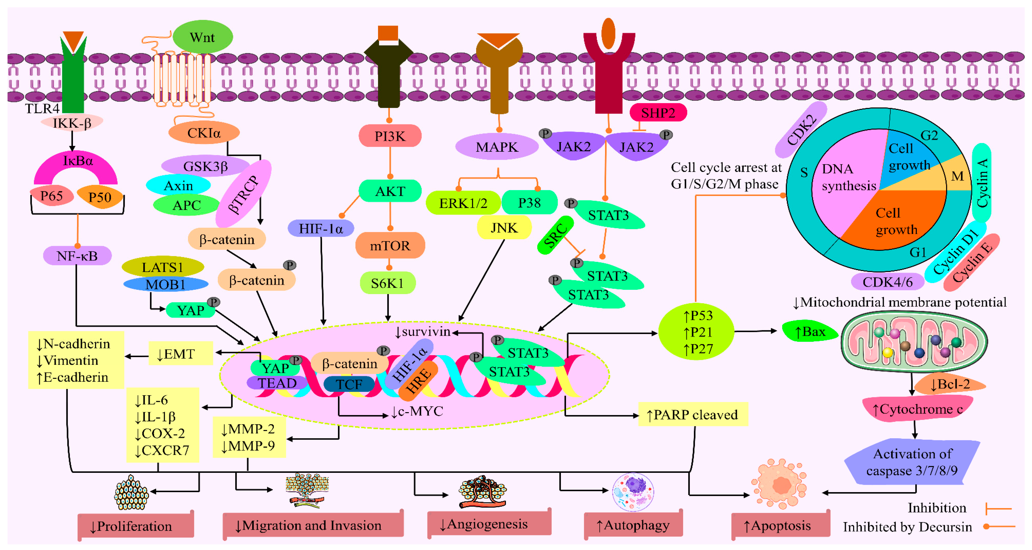
Decursin triggered autophagy in gastric cancer cells (SNU216 and NCI-N87) via lowering the development of spheroids and patient-derived gastric organoids. It also upregulated the translation of CTSC and autophagy-related proteins, including the ATG9A trafficking system, ULK1/2 kinase core complex, ATG12, the autophagy-specific class III PI3K complex, and the LC3 ubiquitin-like conjugation systems. Moreover, decursin also increased SQSTM1 accumulation, which indicated inhibition of the autophagic flux. The expression level of SQSTM1 is an important indicator of autophagic flux. Decursin-mediated inhibition of autophagic flux was observed in gastric, colon, cervical, breast, and lung cancer cells, which were identified by treatment with decursin and bafilomycin A, a well-known inhibitor of autophagy [39,100]. The study also revealed that decursin could act as a potential therapeutic target that blocks cell growth and autophagy at the same time. Figure 2 depicts the possible anticancer mechanisms of decursin.

4. Pharmacokinetics

Pharmacokinetics (PK) is the study of how medicines move through the body once they are administered. It involves the quantitative analysis of the physiological processes of absorption, distribution, metabolism, and excretion. (ADME) [101]. Estimating human PK and dose are fundamental elements of risk prevention methods in the field of drug discovery [102,103]. Because undesirable PK and toxicity are major factors contributing to the failure of drug development [104,105]. In addition, PK studies could explain the relationship between drug exposure and clinical response, which are crucial in optimizing therapeutic results while reducing unwanted effects [106]. Pharmacokinetic factors are also used to understand the drug’s action, including clearance, apparent volume of distribution, elimination half-life, and bioavailability [107]. Therapeutic efficiency studies in animal models for anticancer medicines are possible prior to or after PK profiling studies [108,109]. Due to the high rate of non-responders and the high cost of cancer therapy, it is necessary to explore novel strategies to enhance both clinical efficacy and affordability; PK modeling can be utilized to optimize both outcomes.
Decursin (C19H20O5) is obtained as a white to beige powder with a molecular mass of 328.35 g and a density of 1.2 ± 0.1 g/cm3 [110,111]. Pharmacokinetic studies performed on both animal and human models found that the appropriate dosage of decursin, in terms of its physiological effects, was within the nanomolar range. The plasma concentration of decursin reached a mean peak concentration (Cmax) of 43.7 ng/mL in rats following a single oral administration of decursin at a dose of 50 mg/kg. After being administered orally, it showed high permeability through the blood-brain barrier in rats [112,113]. Decursin (122.12 μg/mL) exhibited >100 μg/mL kinetic solubility [114]. The elimination half-life (t1/2) of decursin was 3.03 h, and it takes 0.44 h to attain the maximum plasma concentration (Tmax) [57]. An in silico pharmacokinetic study revealed decursin was able to pass through the skin and be absorbed by the human gut when taken orally [115].
Decursin had a markedly limited ability to be absorbed orally due to its extensive hepatic first-pass metabolism. Decursin had no direct relationship to the bioactivity of Angelica gigas; it may act as a type of natural prodrug of decursinol. Decursin was extensively metabolized to decursinol (an ester of angelic acid or 3-methyl-2-pentenoic acid), which was also pharmacologically active [116]. In addition, the pharmacological efficacy of decursin is limited due to its low water solubility (Muralikrishnan et al., 2024). Further investigations on various drug delivery approaches to improve decursin’s oral bioavailability and solubility may significantly increase its potential as a natural lead molecule for a range of therapeutic purposes. Studies demonstrated that the conversion of decursin to decursinol primarily occurred in the liver in mice. In addition, the metabolism of decursin by human liver S9 fraction is slower than in mouse and rat liver S9 fraction [112,117]. In a study of decursin (125 or 250 mg for four weeks) in Sprague-Dawley rats, researchers found that there were no histological change anomalies in the spleen, kidney, heart, or cerebrum, which proved that decursin was non-toxic [118]. Another investigation found that rats of both sexes had 50% lethal dose (LD50) values greater than 2000 mg/kg after the treatment with decursin (2 mg/kg). The findings indicated that dosages up to 2000 mg/kg are safe [119].

Extraction and Isolation of Decursin

The biosynthetic pathway of decursin follows the coumarin biosynthesis route, which includes phenylalanine, cinnamic acid, umbelliferone, decursinol, and decursin. It can also be synthesized by the mevalonate or methyl-D-erythritol phosphate (MEP) pathway in which D-[1–13C] glucose is used as a precursor molecule [119].
Decursin can be obtained from the Angelica gigas plant through various extracted approaches. Some other natural sources of decursin include Notopterygium incisum Ting ex H. T. Chang, Angelica dahurica, Angelica glauca Edgew, and Angelica czernaevia [120]. In a study conducted by Hwang and his team, they isolated decursin from the methanolic extract of A. gigas, where 100 g of air-dried A. gigas was submerged in methanol, and the concentrated extract (17 g) was suspended in water and then extracted with n-butanol (n-BuOH), ethylacetate (EtOAc), and di-chloromethane (MC). In this study, 448 mg of decursin was obtained and purified by silica column chromatography [121]. Li and his team isolated and purified decursin from ethanol extracts of the root of Angelica gigas Nakai by silica column chromatography, which have been shown to exhibit antitumor activities [122]. Kim and his co-workers isolated decursin from the ethylacetate fraction of Angelica gigas Nakai using silica gel column chromatography and tested purity (98%) by HPLC [53].

5. Conclusions and Future Direction

Cancer is a dynamic disease and results in a huge number of deaths each year worldwide. In recent years, NPs have played a vital role in the therapeutic treatment of cancer as they are used in drug discovery through different processes. They have played a vital role in advancing therapeutic development for cancer and infectious disorders. Decursin, a pyranocoumarin phytochemical, has shown various anticancer effects in different in vivo and in vitro studies. In this review, it has been manifested that decursin showed a potential anticancer effect against numerous types of cancers, including breast, bladder, colon, skin, ovarian, prostate, lung, pancreatic, gastric, head, neck, glioblastoma, blood, and bone marrow cancers, etc., via various mechanisms like apoptosis, cytotoxicity, cell cycle arrest, inhibition of invasion and migration, inhibition of angiogenesis, autophagy, anti-proliferative effect, etc. This study found various signaling pathways modified by decursin, including modification of the PI3K/AKT/mTOR signaling route, interference with the JAK/STAT signaling system, suppression of the MAPK signaling pathway, and modulation of the MMP signaling pathway, which gives an anticancer effect. This signaling capability indicates that decursin has a greater potential for controlling cancers. The pharmacokinetics investigation demonstrated that oral bioavailability and water solubility of decursin were very low, and it is extremely absorbed in the intestine. So, this review suggests some directions for further research, such as structural modification of the active side chain at the C-7 position of decursin and investigation of alternative novel drug delivery approaches, like nanoformulations, to enhance their anticancer potential. In addition, decursin is able to inhibit tumor growth by targeting the CXCR7 receptor, but the mechanism is unknown, and there is no such research on it. Therefore, this review suggests further investigations to find out the potential of decursin as a CXCR7-targeting anticancer agent. Though there is no human clinical evidence on the use of decursin in cancer treatment, this review suggests that research should be conducted on extensive clinical investigations in the future to figure out the workable anticancer effects of decursin and its effectiveness for long-term treatment of malignant disease.

Author Contributions

Conceptualization, T.A.E. and M.S.B.; methodology, M.S.B. and S.S.; data curation, M.S.A.H. and R.H., writing—original draft preparation, T.A.E., M.S.B., M.M.K., M.S.A.H. I.M.A. and M.A.A.; writing—review and editing, M.S.B., H.D.M.C., I.C.P.N. and R.C.; mechanism draw, R.C.; supervision, M.T.I.; project administration, M.T.I. All authors have read and agreed to the published version of the manuscript.

Funding

This research received no external funding.

Institutional Review Board Statement

Not applicable.

Informed Consent Statement

Not applicable.

Data Availability Statement

Not applicable.

Conflicts of Interest

The authors Md. Sakib Al Hasan, Rubel Hasan and Muhammad Torequl Islam received a speaker honorarium from Bioluster Research Center; The authors Tanzila Akter Eity, Md. Shimul Bhuia and Raihan Chowdhury received products from the Bioluster Research Center for their research works; The authors Mst Muslima Khatun and Salehin Sheikh has been involved as a consultant for Bioluster Research Center. The authors declare that the research was conducted in the absence of any commercial or financial relationships that could be identified as a potential conflict of interest. The company had no role in the design of the study; in the collection, analysis, or interpretation of data; in the writing of the manuscript; or in the decision to publish the results.

Abbreviations

The following abbreviations are used in this manuscript:
ACCAcetyl-CoA carboxylase
APCAdenomatous polyposis coli
BaxBCL2-associated X protein
CDKCyclin-dependent kinase
CDK2Cyclin-dependent kinase 2
c-MYCCellular Myc
COXCyclooxygenase
CTRCatenin response transcription
EGFREpidermal growth factor receptor
EKRExtracellular signal-regulated kinases
EMTEpithelial–mesenchymal transition
ERK1/2Extracellular signal-regulated kinase 1/2
FASFatty acid synthase
HIF-1αHypoxia-inducible factor 1-alpha
IKK-βInhibitor of nuclear factor kappa-B kinase subunit beta
ILInterleukin
JAKJanus-activated kinase
JNKc-Jun N-terminal kinase
LATS1Large tumor suppressor kinase 1
LLCLewis lung cancer
LPSLipopolysaccharide
MAPMitogen-activated protein
MAPKMitogen-activated protein kinase
MMPMatrix metalloproteinase
mTORMammalian target of rapamycin
NF-κBNuclear factor kappa-light-chain-enhancer of activated B-cells
PI3KPhosphoinositide 3-kinase
PPARPeroxisome proliferator-activated receptor
SHP2Src homology 2 domain-containing phosphatase 2
STATSignal transduction and transcription activation
STAT3Signal transducer and activator of transcription 3
TCFT-cell factor
YAPYes-associated protein
TEADTranscriptional enhancer factor domain

References

  1. Bhuia, M.S.; Chowdhury, R.; Sonia, F.A.; Biswas, S.; Ferdous, J.; El-Nashar, H.A.S.; El-Shazly, M.; Islam, M.T. Efficacy of Rotundic Acid and Its Derivatives as Promising Natural Anticancer Triterpenoids: A Literature-based Study. Chem. Biodivers. 2024, 21, e202301492. [Google Scholar] [CrossRef] [PubMed]
  2. Brown, J.S.; Amend, S.R.; Austin, R.H.; Gatenby, R.A.; Hammarlund, E.U.; Pienta, K.J. Updating the Definition of Cancer. Mol. Cancer Res. 2023, 21, 1142–1147. [Google Scholar] [CrossRef]
  3. Ma, D.; Zou, B.; Cai, G.; Hu, X.; Liu, J.O. Total Synthesis of the Cyclodepsipeptide Apratoxin A and Its Analogues and Assessment of Their Biological Activities. Chem. Eur. J. 2006, 12, 7615–7626. [Google Scholar] [CrossRef]
  4. Bray, F.; Ferlay, J.; Soerjomataram, I.; Siegel, R.L.; Torre, L.A.; Jemal, A. Global Cancer Statistics 2018: GLOBOCAN Estimates of Incidence and Mortality Worldwide for 36 Cancers in 185 Countries. CA. Cancer J. Clin. 2018, 68, 394–424. [Google Scholar] [CrossRef]
  5. Katzke, V.A.; Kaaks, R.; Kühn, T.J. Lifestyle and cancer risk. Can. J. 2015, 21, 104–110. [Google Scholar] [CrossRef]
  6. Sung, H.; Ferlay, J.; Siegel, R.L.; Laversanne, M.; Soerjomataram, I.; Jemal, A.; Bray, F. Global Cancer Statistics 2020: GLOBOCAN Estimates of Incidence and Mortality Worldwide for 36 Cancers in 185 Countries. CA. Cancer J. Clin. 2021, 71, 209–249. [Google Scholar] [CrossRef] [PubMed]
  7. Wasim Bin, H.; Mohammad Al-Masum, M. State of Cancer Care in Bangladesh. The Daily Star. Available online: https://www.thedailystar.net/supplements/world-cancer-day-2022/news/state-cancer-care-bangladesh-2953926 (accessed on 20 January 2024).
  8. Danaei, G.; Vander Hoorn, S.; Lopez, A.D.; Murray, C.J.L.; Ezzati, M. Causes of Cancer in the World: Comparative Risk Assessment of Nine Behavioural and Environmental Risk Factors. Lancet 2005, 366, 1784–1793. [Google Scholar] [CrossRef] [PubMed]
  9. Gherghi, I.C.; Girousi, S.T.; Voulgaropoulos, A.; Tzimou-Tsitouridou, R. Interaction of the Mutagen Ethidium Bromide with DNA, Using a Carbon Paste Electrode and a Hanging Mercury Drop Electrode. Anal. Chim. Acta 2004, 505, 135–144. [Google Scholar] [CrossRef]
  10. Trichopoulos, D.; Li, F.P.; Hunter, D.J. What Causes Cancer? Sci. Am. 1996, 275, 80–87. [Google Scholar] [CrossRef]
  11. Isikhan, V.; Güner, P.; Kömürcü, S.; Özet, A.; Arpaci, F.; Öztürk, B. The Relationship between Disease Features and Quality of Life in Patients with Cancer—I. Cancer Nurs. 2001, 24, 490–495. [Google Scholar] [CrossRef]
  12. Islam, M.T.; Sarkar, C.; Hossain, R.; Bhuia, M.S.; Mardare, I.; Kulbayeva, M.; Ydyrys, A.; Calina, D.; Habtemariam, S.; Kieliszek, M. Therapeutic Strategies for Rheumatic Diseases and Disorders: Targeting Redox Imbalance and Oxidative Stress. Biomed. Pharmacother. 2023, 164, 114900. [Google Scholar] [CrossRef]
  13. Tabarés-Seisdedos, R.; Dumont, N.; Baudot, A.; Valderas, J.M.; Climent, J.; Valencia, A.; Crespo-Facorro, B.; Vieta, E.; Gómez-Beneyto, M.; Martínez, S. No Paradox, No Progress: Inverse Cancer Comorbidity in People with Other Complex Diseases. Lancet Oncol. 2011, 12, 604–608. [Google Scholar] [CrossRef] [PubMed]
  14. Sutherland, R.M. Cell and Environment Interactions in Tumor Microregions: The Multicell Spheroid Model. Science 1988, 240, 177–184. [Google Scholar] [CrossRef] [PubMed]
  15. Vaupel, P. Physiological Mechanisms of Treatment Resistance. In The Impact of Tumor Biology on Cancer Treatment and Multidisciplinary Strategies; Springer: Berlin/Heidelberg, Germany, 2009; pp. 273–290. [Google Scholar]
  16. Vaupel, P.; Kallinowski, F.; Okunieff, P. Blood Flow, Oxygen and Nutrient Supply, and Metabolic Microenvironment of Human Tumors: A Review. Cancer Res. 1989, 49, 6449–6465. [Google Scholar] [PubMed]
  17. Hanahan, D.; Weinberg, R.A. Hallmarks of Cancer: The next Generation. Cell 2011, 144, 646–674. [Google Scholar] [CrossRef]
  18. Jayson, G.C.; Kohn, E.C.; Kitchener, H.C.; Ledermann, J.A. Ovarian Cancer. Lancet 2014, 384, 1376–1388. [Google Scholar] [CrossRef]
  19. Lee, J.H.; Machtay, M.; Unger, L.D.; Weinstein, G.S.; Weber, R.S.; Chalian, A.A.; Rosenthal, D.I. Prophylactic Gastrostomy Tubes in Patients Undergoing Intensive Irradiation for Cancer of the Head and Neck. Arch. Otolaryngol. Neck Surg. 1998, 124, 871–875. [Google Scholar] [CrossRef]
  20. Sourati, A.; Ameri, A.; Malekzadeh, M. Acute Side Effects of Radiation Therapy: A Guide to Management, 1st ed.; Springer Cham: Berlin/Heidelberg, Germany, 2017; p. 227. [Google Scholar]
  21. Hanna, E.; Alexiou, M.; Morgan, J.; Badley, J.; Maddox, A.M.; Penagaricano, J.; Fan, C.-Y.; Breau, R.; Suen, J. Intensive Chemoradiotherapy as a Primary Treatment for Organ Preservation in Patients with Advanced Cancer of the Head and Neck: Efficacy, Toxic Effects, and Limitations. Arch. Otolaryngol. Neck Surg. 2004, 130, 861–867. [Google Scholar] [CrossRef]
  22. Beg, S.; Rahman, M.; Jain, A.; Saini, S.; Midoux, P.; Pichon, C.; Ahmad, F.J.; Akhter, S. Nanoporous Metal Organic Frameworks as Hybrid Polymer–Metal Composites for Drug Delivery and Biomedical Applications. Drug Discov. Today 2017, 22, 625–637. [Google Scholar] [CrossRef]
  23. Hale, K.J. Monosaccharides: Use in the Asymmetric Synthesis of Natural Products. In Second Supplements to the 2nd Edition of Rodd’s Chemistry of Carbon Compounds; Elsevier: Amsterdam, The Netherlands, 1991; pp. 315–435. [Google Scholar]
  24. Agarwal, A.; DeNunzio, N.J.; Ahuja, D.; Hirsch, A.E. Beyond the Standard Curriculum: A Review of Available Opportunities for Medical Students to Prepare for a Career in Radiation Oncology. Int. J. Radiat. Oncol. Biol. Phys. 2014, 88, 39–44. [Google Scholar] [CrossRef]
  25. Ali Khan, M.; El-Kersh, D.M.; Islam, M.S.; Ara Khan, S.; Kamli, H.; Sarkar, C.; Bhuia, M.S.; Islam, T.; Chandra Shill, M.; Gobe, G.C. Mikania Micrantha Kunth: An Ethnopharmacological Treasure Trove of Therapeutic Potential. Chem. Biodivers. 2023, 20, e202300392. [Google Scholar] [CrossRef] [PubMed]
  26. Gupta, A.P.; Pandotra, P.; Kushwaha, M.; Khan, S.; Sharma, R.; Gupta, S. Alkaloids: A Source of Anticancer Agents from Nature. Stud. Nat. Prod. Chem. 2015, 46, 341–445. [Google Scholar]
  27. Kashyap, D.; Garg, V.K.; Goel, N. Intrinsic and Extrinsic Pathways of Apoptosis: Role in Cancer Development and Prognosis. Adv. Protein Chem. Struct. Biol. 2021, 125, 73–120. [Google Scholar]
  28. Chandra Shill, M.; El-Nashar, H.A.S.; Prova Mollick, P.; Nath Acharyya, R.; Afrin, S.; Hossain, H.; Halder, S.; Torequl Islam, M.; Bhuia, M.S.; Reza, H.M. Longevity Spinach (Gynura Procumbens) Ameliorated Oxidative Stress and Inflammatory Mediators in Cisplatin-induced Organ Dysfunction in Rats: Comprehensive in Vivo and in Silico Studies. Chem. Biodivers. 2024, 21, e202301719. [Google Scholar] [CrossRef]
  29. Newman, D.J.; Cragg, G.M. Natural Products as Sources of New Drugs over the 30 Years from 1981 to 2010. J. Nat. Prod. 2012, 75, 311–335. [Google Scholar] [CrossRef] [PubMed]
  30. Lee, S.; Lee, J.H.; Kim, E.; Kim, W.; Suk, K.; Kim, J.; Song, G.Y.; Lee, W. A Novel Derivative of Decursin, CSL-32, Blocks Migration and Production of Inflammatory Mediators and Modulates PI3K and NF-κB Activities in HT1080 Cells. Cell Biol. Int. 2012, 36, 683–688. [Google Scholar] [CrossRef]
  31. Scholten, D.J.; Canals, M.; Maussang, D.; Roumen, L.; Smit, M.J.; Wijtmans, M.; De Graaf, C.; Vischer, H.F.; Leurs, R. Pharmacological Modulation of Chemokine Receptor Function. Br. J. Pharmacol. 2012, 165, 1617–1643. [Google Scholar] [CrossRef]
  32. Shehzad, A.; Parveen, S.; Qureshi, M.; Subhan, F.; Lee, Y.S. Decursin and Decursinol Angelate: Molecular Mechanism and Therapeutic Potential in Inflammatory Diseases. Inflamm. Res. 2018, 67, 209–218. [Google Scholar] [CrossRef]
  33. Graña, X.; Reddy, E.P. Cell Cycle Control in Mammalian Cells: Role of Cyclins, Cyclin Dependent Kinases (CDKs), Growth Suppressor Genes and Cyclin-Dependent Kinase Inhibitors (CKIs). Oncogene 1995, 11, 211–220. [Google Scholar]
  34. Yim, D.; Singh, R.P.; Agarwal, C.; Lee, S.; Chi, H.; Agarwal, R. A Novel Anticancer Agent, Decursin, Induces G1 Arrest and Apoptosis in Human Prostate Carcinoma Cells. Cancer Res. 2005, 65, 1035–1044. [Google Scholar] [CrossRef]
  35. Choi, S.-R.; Lee, J.-H.; Kim, J.-Y.; Park, K.-W.; Jeong, I.-Y.; Shim, K.-H.; Lee, M.-K.; Seo, K.-I. Decursin from Angelica Gigas Nakai Induces Apoptosis in RC-58T/h/SA# 4 Primary Human Prostate Cancer Cells via a Mitochondria-Related Caspase Pathway. Food Chem. Toxicol. 2011, 49, 2517–2523. [Google Scholar] [PubMed]
  36. Choi, Y.J.; Kim, D.H.; Kim, S.J.; Kim, J.; Jeong, S.-I.; Chung, C.H.; Yu, K.-Y.; Kim, S.-Y. Decursin Attenuates Hepatic Fibrogenesis through Interrupting TGF-Beta-Mediated NAD (P) H Oxidase Activation and Smad Signaling in Vivo and in Vitro. Life Sci. 2014, 108, 94–103. [Google Scholar] [CrossRef] [PubMed]
  37. Han, J.; Jin, W.; Ho, N.A.; Hong, J.; Kim, Y.J.; Shin, Y.; Lee, H.; Suh, J.-W. Decursin and Decursinol Angelate Improve Wound Healing by Upregulating Transcription of Genes Encoding Extracellular Matrix Remodeling Proteins, Inflammatory Cytokines, and Growth Factors in Human Keratinocytes. Biochem. Biophys. Res. Commun. 2018, 499, 979–984. [Google Scholar] [CrossRef]
  38. Kim, J.H.; Jeong, S.-J.; Kwon, H.-Y.; Park, S.Y.; Lee, H.-J.; Lee, H.-J.; Lieske, J.C.; Kim, S.-H. Decursin Prevents Cisplatin-Induced Apoptosis via the Enhancement of Antioxidant Enzymes in Human Renal Epithelial Cells. Biol. Pharm. Bull. 2010, 33, 1279–1284. [Google Scholar] [CrossRef]
  39. Kim, W.-J.; Lee, S.-J.; Choi, Y.D.; Moon, S.-K. Decursin Inhibits Growth of Human Bladder and Colon Cancer Cells via Apoptosis, G1-Phase Cell Cycle Arrest and Extracellular Signal-Regulated Kinase Activation. Int. J. Mol. Med. 2010, 25, 635–641. [Google Scholar]
  40. Taechowisan, T.; Chanaphat, S.; Ruensamran, W.; Phutdhawong, W.S. Antibacterial Activity of Decursin from Streptomyces Sp. GMT-8; an Endophyte in Zingiber Officinale Rosc. J. Appl. Pharm. Sci. 2013, 3, 74–78. [Google Scholar]
  41. Sharifi-Rad, M.; Lankatillake, C.; Dias, D.A.; Docea, A.O.; Mahomoodally, M.F.; Lobine, D.; Chazot, P.L.; Kurt, B.; Boyunegmez Tumer, T.; Catarina Moreira, A. Impact of Natural Compounds on Neurodegenerative Disorders: From Preclinical to Pharmacotherapeutics. J. Clin. Med. 2020, 9, 1061. [Google Scholar] [CrossRef]
  42. Chari, R.V.J. Targeted Cancer Therapy: Conferring Specificity to Cytotoxic Drugs. Acc. Chem. Res. 2008, 41, 98–107. [Google Scholar] [CrossRef]
  43. Setiawati, A.; Candrasari, D.S.; Setyajati, F.D.E.; Prasetyo, V.K.; Setyaningsih, D.; Hartini, Y.S. Anticancer Drug Screening of Natural Products: In Vitro: Cytotoxicity Assays, Techniques, and Challenges. Asian Pac. J. Trop. Biomed. 2022, 12, 279–289. [Google Scholar] [CrossRef]
  44. Ahmed, M.B.; Islam, S.U.; Lee, Y.S. Decursin Negatively Regulates LPS-Induced Upregulation of the TLR4 and JNK Signaling Stimulated by the Expression of PRP4 in Vitro. Animal Cells Syst. 2020, 24, 44–52. [Google Scholar] [CrossRef]
  45. Kim, B.S.; Seo, H.; Kim, H.-J.; Bae, S.M.; Son, H.-N.; Lee, Y.J.; Ryu, S.; Park, R.-W.; Nam, J.-O. Decursin from Angelica Gigas Nakai Inhibits B16F10 Melanoma Growth through Induction of Apoptosis. J. Med. Food 2015, 18, 1121–1127. [Google Scholar] [CrossRef] [PubMed]
  46. Ki, D.; Go, S.H.; Song, Y.; Lee, D.K.; Park, J.R. Decursin Induces G1 Cell Cycle Arrest and Apoptosis through Reactive Oxygen Species-Mediated Endoplasmic Reticulum Stress in Human Colorectal Cancer Cells in In Vitro and Xenograft Models. Int. J. Mol. Sci. 2024, 25, 9939. [Google Scholar]
  47. Kweon, B.; Han, Y.-H.; Kee, J.-Y.; Mun, J.-G.; Jeon, H.D.; Yoon, D.H.; Choi, B.-M.; Hong, S.-H. Effect of Angelica Gigas Nakai Ethanol Extract and Decursin on Human Pancreatic Cancer Cells. Molecules 2020, 25, 2028. [Google Scholar] [CrossRef]
  48. Choi, H.S.; Cho, S.; Kim, M.K.; Kim, M.S.; Moon, S.H.; Kim, I.H.; Ko, S. Decursin in Angelica Gigas Nakai (AGN) Enhances Doxorubicin Chemosensitivity in NCI/ADR-RES Ovarian Cancer Cells via Inhibition of P-glycoprotein Expression. Phyther. Res. 2016, 30, 2020–2026. [Google Scholar] [CrossRef]
  49. Kim, J.-M.; Noh, E.-M.; Kim, M.-S.; Hwang, J.-K.; Hwang, H.-Y.; Ryu, D.-G.; Kim, H.-J.; Yu, H.-N.; You, Y.-O.; Kim, J.-S. Decursin Prevents TPA-Induced Invasion through Suppression of PKCα/P38/NF-ΚB-Dependent MMP-9 Expression in MCF-7 Human Breast Carcinoma Cells. Int. J. Oncol. 2014, 44, 1607–1613. [Google Scholar] [CrossRef]
  50. Kim, J.; Jung, J.H.; Kim, S.; Jeong, S. Decursin Exerts Anti-cancer Activity in MDA-MB-231 Breast Cancer Cells via Inhibition of the Pin1 Activity and Enhancement of the Pin1/P53 Association. Phyther. Res. 2014, 28, 238–244. [Google Scholar] [CrossRef] [PubMed]
  51. Son, S.H.; Park, K.; Park, S.K.; Kim, Y.C.; Kim, Y.S.; Lee, S.; Chung, W. Decursin and Decursinol from Angelica Gigas Inhibit the Lung Metastasis of Murine Colon Carcinoma. Phyther. Res. 2011, 25, 959–964. [Google Scholar] [CrossRef] [PubMed]
  52. Yang, Y.; Hu, Y.-E.; Zhao, M.-Y.; Jiang, Y.-F.; Fu, X.; You, F.-M. Decursin Affects Proliferation, Apoptosis, and Migration of Colorectal Cancer Cells through PI3K/Akt Signaling Pathway. Zhongguo Zhong yao za zhi= Zhongguo Zhongyao Zazhi = China J. Chinese Mater. Medica 2023, 48, 2334–2342. [Google Scholar]
  53. Kim, H.J.; Kim, S.M.; Park, K.R.; Jang, H.J.; Na, Y.S.; Ahn, K.S.; Kim, S.H.; Ahn, K.S. Decursin chemosensitizes human multiple myeloma cells through inhibition of STAT3 signaling pathway. Cancer Lett. 2011, 301, 29–37. [Google Scholar] [CrossRef]
  54. Ge, Y.; Yoon, S.-H.; Jang, H.; Jeong, J.-H.; Lee, Y.-M. Decursin promotes HIF-1α proteasomal degradation and immune responses in hypoxic tumour microenvironment. Phytomedicine 2020, 78, 153318. [Google Scholar] [CrossRef]
  55. Oh, S.T.; Lee, S.; Hua, C.; Koo, B.-S.; Pak, S.C.; Kim, D.-I.; Jeon, S.; Shin, B.A. Decursin Induces Apoptosis in Glioblastoma Cells, but Not in Glial Cells via a Mitochondria-Related Caspase Pathway. Korean J. Physiol. Pharmacol. 2019, 23, 29–35. [Google Scholar] [CrossRef]
  56. Lee, H.J.; Lee, H.J.; Lee, E.O.; Lee, J.H.; Lee, K.S.; Kim, K.H.; Kim, S.-H.; Lü, J. In Vivo Anti-Cancer Activity of Korean Angelica Gigas and Its Major Pyranocoumarin Decursin. Am. J. Chin. Med. 2009, 37, 127–142. [Google Scholar] [CrossRef] [PubMed]
  57. Bhat, T.A.; Dheeraj, A.; Nambiar, D.K.; Singh, S.P.; Yim, D.S.; Singh, R.P. Decursin Inhibits EGFR-ERK1/2 Signaling Axis in Advanced Human Prostate Carcinoma Cells. Prostate 2023, 83, 534–546. [Google Scholar] [CrossRef]
  58. Song, G.-Y.; Lee, J.-H.; Cho, M.; Park, B.-S.; Kim, D.-E.; Oh, S.J. Decursin suppresses human androgen-independent PC3 prostate cancer cell proliferation by promoting the degradation of β-catenin. Mol. Pharmacol. 2007, 72, 1599–1606. [Google Scholar] [CrossRef] [PubMed]
  59. Kim, S.; Kim, J.-E.; Kim, N.; Joo, M.; Lee, M.-W.; Jeon, H.J.; Ryu, H.; Song, I.-C.; Song, G.-Y.; Lee, H.J.J. Decursin inhibits tumor growth, migration, and invasion in gastric cancer by down-regulating CXCR7 expression. Am. J. Cancer Res. 2019, 9, 2007. [Google Scholar]
  60. Joo, M.; Heo, J.B.; Kim, S.; Kim, N.; Jeon, H.J.; An, Y.; Song, G.-Y.; Kim, J.-M.; Lee, H.J. Decursin Inhibits Tumor Progression in Head and Neck Squamous Cell Carcinoma by Downregulating CXCR7 Expression in Vitro. Oncol. Rep. 2022, 47, 1–11. [Google Scholar]
  61. Kim, H.H.; Ahn, K.S.; Han, H.; Choung, S.Y.; Choi, S.-Y.; Kim, I.-H.J. Decursin and PDBu: Two PKC activators distinctively acting in the megakaryocytic differentiation of K562 human erythroleukemia cells. Leuk. Res. 2005, 29, 1407–1413. [Google Scholar] [CrossRef]
  62. Son, S.H.; Kim, M.-J.; Chung, W.-Y.; Son, J.-A.; Kim, Y.S.; Kim, Y.-C.; Kang, S.S.; Lee, S.-K.; Park, K.-K. Decursin and Decursinol Inhibit VEGF-Induced Angiogenesis by Blocking the Activation of Extracellular Signal-Regulated Kinase and c-Jun N-Terminal Kinase. Cancer Lett. 2009, 280, 86–92. [Google Scholar] [CrossRef]
  63. Li, J.; Wang, H.; Wang, L.; Tan, R.; Zhu, M.; Zhong, X.; Zhang, Y.; Chen, B.; Wang, L. Decursin Inhibits the Growth of HepG2 Hepatocellular Carcinoma Cells via Hippo/YAP Signaling Pathway. Phyther. Res. 2018, 32, 2456–2465. [Google Scholar] [CrossRef]
  64. Ahn, Q.; Jeong, S.-J.; Lee, H.-J.; Kwon, H.-Y.; Han, I.; Kim, H.S.; Lee, H.-J.; Lee, E.-O.; Ahn, K.S.; Jung, M.-H.J.C.l. Inhibition of cyclooxygenase-2-dependent survivin mediates decursin-induced apoptosis in human KBM-5 myeloid leukemia cells. Cancer Lett. 2010, 298, 212–221. [Google Scholar] [CrossRef]
  65. Yam, C.H.; Fung, T.K.; Poon, R.Y.C. Cyclin A in Cell Cycle Control and Cancer. Cell. Mol. Life Sci. CMLS 2002, 59, 1317–1326. [Google Scholar] [CrossRef]
  66. Matthews, H.K.; Bertoli, C.; de Bruin, R.A. Cell cycle control in cancer. Nat. Rev. Mol. Cell Biol. 2022, 23, 74–88. [Google Scholar] [CrossRef] [PubMed]
  67. Williams, G.H.; Stoeber, K. The Cell Cycle and Cancer. J. Pathol. 2012, 226, 352–364. [Google Scholar] [CrossRef] [PubMed]
  68. Bhuia, M.S.; Chowdhury, R.; Sonia, F.A.; Kamli, H.; Shaikh, A.; El-Nashar, H.A.S.; El-Shazly, M.; Islam, M.T. Anticancer Potential of the Plant-Derived Saponin Gracillin: A Comprehensive Review of Mechanistic Approaches. Chem. Biodivers. 2023, 20, e202300847. [Google Scholar] [CrossRef] [PubMed]
  69. Shapiro, G.I.; Harper, J.W. Anticancer Drug Targets: Cell Cycle and Checkpoint Control. J. Clin. Investig. 1999, 104, 1645–1653. [Google Scholar] [CrossRef]
  70. Bhuia, M.S.; Wilairatana, P.; Ferdous, J.; Chowdhury, R.; Bappi, M.H.; Rahman, M.A.; Mubarak, M.S.; Islam, M.T. Hirsutine, an Emerging Natural Product with Promising Therapeutic Benefits: A Systematic Review. Molecules 2023, 28, 6141. [Google Scholar] [CrossRef]
  71. Bhuia, M.S.; Chowdhury, R.; Ara, I.; Mamun, M.; Rouf, R.; Khan, M.A.; Uddin, S.J.; Shakil, M.A.K.; Habtemariam, S.; Ferdous, J. Bioactivities of Morroniside: A Comprehensive Review of Pharmacological Properties and Molecular Mechanisms. Fitoterapia 2024, 175, 105896. [Google Scholar] [CrossRef]
  72. Fischer, U.; Schulze-Osthoff, K. Apoptosis-Based Therapies and Drug Targets. Cell Death Differ. 2005, 12, 942–961. [Google Scholar] [CrossRef]
  73. Los, M.; Burek, C.J.; Stroh, C.; Benedyk, K.; Hug, H.; Mackiewicz, A. Anticancer Drugs of Tomorrow: Apoptotic Pathways as Targets for Drug Design. Drug Discov. Today 2003, 8, 67–77. [Google Scholar] [CrossRef]
  74. Kvansakul, M.; Hinds, M.G.J.A. The Bcl-2 family: Structures, interactions and targets for drug discovery. Apoptosis 2015, 20, 136–150. [Google Scholar] [CrossRef]
  75. Shi, Y. Mechanical Aspects of Apoptosome Assembly. Curr. Opin. Cell Biol. 2006, 18, 677–684. [Google Scholar] [CrossRef]
  76. Thushara, R.; Hemshekhar, M.; Santhosh, M.; Devaraja, S.; Kemparaju, K.; Girish, K.J. Differential action of phytochemicals on platelet apoptosis: A biological overview. Curr. Med. Chem. 2013, 20, 1018–1027. [Google Scholar] [PubMed]
  77. Moyer, A.; Tanaka, K.; Cheng, E.H. Apoptosis in Cancer Biology and Therapy. Annu. Rev. Pathol. 2025, 20, 303–328. [Google Scholar] [CrossRef] [PubMed]
  78. Kim, S.; Lee, S.-I.; Kim, N.; Joo, M.; Lee, K.-H.; Lee, M.-W.; Jeon, H.J.; Ryu, H.; Kim, J.-M.; Sul, J.-Y. Decursin Inhibits Cell Growth and Autophagic Flux in Gastric Cancer via Suppression of Cathepsin C. Am. J. Cancer Res. 2021, 11, 1304. [Google Scholar] [PubMed]
  79. Bhuia, M.S.; Wilairatana, P.; Chowdhury, R.; Rakib, A.I.; Kamli, H.; Shaikh, A.; Coutinho, H.D.M.; Islam, M.T. Anticancer Potentials of the Lignan Magnolin: A Systematic Review. Molecules 2023, 28, 3671. [Google Scholar] [CrossRef]
  80. Zhu, J.; Thompson, C.B. Metabolic regulation of cell growth and proliferation. Mol. Cell Biol. 2019, 20, 436–450. [Google Scholar] [CrossRef]
  81. Duronio, R.J.; Xiong, Y. Signaling Pathways That Control Cell Proliferation. Cold Spring Harb. Perspect. Biol. 2013, 5, a008904. [Google Scholar] [CrossRef]
  82. Gu, Y.; Mohammad, I.S.; Liu, Z. Overview of the STAT-3 Signaling Pathway in Cancer and the Development of Specific Inhibitors. Oncol. Lett. 2020, 19. [Google Scholar] [CrossRef]
  83. Bhat, P.; Kriel, J.; Shubha Priya, B.; Basappa; Shivananju, N.S.; Loos, B. Modulating autophagy in cancer therapy: Advancements and challenges for cancer cell death sensitization. Biochem. Pharmacol. 2018, 147, 170–182. [Google Scholar] [CrossRef]
  84. Page, C.; Huang, M.; Jin, X.; Cho, K.; Lilja, J.; Reynolds, R.K.; Lin, J. Elevated Phosphorylation of AKT and Stat3 in Prostate, Breast, and Cervical Cancer Cells. Int. J. Oncol. 2000, 17, 23–31. [Google Scholar] [CrossRef]
  85. Bendris, N.; Lemmers, B.; Blanchard, J.M. Cell cycle, cytoskeleton dynamics and beyond: The many functions of cyclins and CDK inhibitors. Cell Cycle 2015, 14, 1786–1798. [Google Scholar] [CrossRef] [PubMed]
  86. Mazumder, S.; DuPree, E.L.; Almasan, A. A Dual Role of Cyclin E in Cell Proliferation and Apotosis May Provide a Target for Cancer Therapy. Curr. Cancer Drug Targets 2004, 4, 65–75. [Google Scholar] [CrossRef]
  87. Krakhmal, N.V.; Zavyalova, M.V.; Denisov, E.V.; Vtorushin, S.V.; Perelmuter, V.M. Cancer Invasion: Patterns and Mechanisms. Acta Naturae (aнглoязычнaя версия) 2015, 7, 17–28. [Google Scholar] [CrossRef]
  88. Apoptosis Lintz, M.; Muñoz, A.; Reinhart-King, C.A. The Mechanics of Single Cell and Collective Migration of Tumor Cells. J. Biomech. Eng. 2017, 139, 21005. [Google Scholar]
  89. Qi, X.; Sun, L.; Wan, J.; Xu, R.; He, S.; Zhu, X. Tensin4 Promotes Invasion and Migration of Gastric Cancer Cells via Regulating AKT/GSK-3β/Snail Signaling Pathway. Pathol. Pract. 2020, 216, 153001. [Google Scholar] [CrossRef]
  90. Zheng, H.; Takahashi, H.; Murai, Y.; Cui, Z.; Nomoto, K.; Niwa, H.; Tsuneyama, K.; Takano, Y. Expressions of MMP-2, MMP-9 and VEGF Are Closely Linked to Growth, Invasion, Metastasis and Angiogenesis of Gastric Carcinoma. Anticancer. Res. 2006, 26, 3579–3583. [Google Scholar] [PubMed]
  91. Cao, Z.-Q.; Wang, Z.; Leng, P. Aberrant N-Cadherin Expression in Cancer. Biomed. Pharmacother. 2019, 118, 109320. [Google Scholar] [CrossRef]
  92. Suciu, C.; Cîmpean, A.M.; Muresan, A.M.; Izvernariu, D.; Raica, M. E-Cadherin Expression in Invasive Breast Cancer. Rom. J. Morphol. Embryol. 2008, 49, 517–523. [Google Scholar]
  93. Shukla, S.; MacLennan, G.T.; Hartman, D.J.; Fu, P.; Resnick, M.I.; Gupta, S. Activation of PI3K-Akt Signaling Pathway Promotes Prostate Cancer Cell Invasion. Int. J. Cancer 2007, 121, 1424–1432. [Google Scholar] [CrossRef]
  94. Zhao, Y.; Adjei, A.A. Targeting Angiogenesis in Cancer Therapy: Moving beyond Vascular Endothelial Growth Factor. Oncologist 2015, 20, 660–673. [Google Scholar] [CrossRef]
  95. Kieran, M.W.; Kalluri, R.; Cho, Y.-J. The VEGF Pathway in Cancer and Disease: Responses, Resistance, and the Path Forward. Cold Spring Harb. Perspect. Med. 2012, 2, a006593. [Google Scholar] [CrossRef] [PubMed]
  96. Abhinand, C.S.; Raju, R.; Soumya, S.J.; Arya, P.S.; Sudhakaran, P.R. VEGF-A/VEGFR2 Signaling Network in Endothelial Cells Relevant to Angiogenesis. J. Cell Commun. Signal. 2016, 10, 347–354. [Google Scholar] [CrossRef] [PubMed]
  97. Liu, B.; Cheng, Y.; Liu, Q.; Bao, J.; Yang, J.-M. Autophagic Pathways as New Targets for Cancer Drug Development. Acta Pharmacol. Sin. 2010, 31, 1154–1164. [Google Scholar] [CrossRef]
  98. Tanida, I.; Ueno, T.; Kominami, E. LC3 and Autophagy. Autophagosome Phagosome 2008, 445, 77–88. [Google Scholar] [CrossRef]
  99. Carew, J.S.; Kelly, K.R.; Nawrocki, S.T. Autophagy as a Target for Cancer Therapy: New Developments. Cancer Manag. Res. 2012, 4, 357–365. [Google Scholar] [CrossRef] [PubMed]
  100. Li, X.; He, S.; Ma, B. Autophagy and Autophagy-Related Proteins in Cancer. Mol. Cancer 2020, 19, 12. [Google Scholar] [CrossRef]
  101. Sharma, P.; Patel, N.; Prasad, B.; Varma, M.V.S. Pharmacokinetics: Theory and Application in Drug Discovery and Development. In Drug Discovery and Development; Poduri, R., Ed.; Springer: Singapore, 2021; pp. 297–355. [Google Scholar] [CrossRef]
  102. Lucas, A.J.; Sproston, J.L.; Barton, P.; Riley, R.J. Estimating Human ADME Properties, Pharmacokinetic Parameters and Likely Clinical Dose in Drug Discovery. Expert. Opin. Drug Discov. 2019, 14, 1313–1327. [Google Scholar] [CrossRef]
  103. Hasan, R.; Alshammari, A.; Albekairi, N.A.; Bhuia, M.S.; Afroz, M.; Chowdhury, R.; Khan, M.A.; Ansari, S.A.; Ansari, I.A.; Mubarak, M.S. Antiemetic Activity of Abietic Acid Possibly through the 5HT3 and Muscarinic Receptors Interaction Pathways. Sci. Rep. 2024, 14, 6642. [Google Scholar] [CrossRef]
  104. Ferdous, J.; Bhuia, M.S.; Chowdhury, R.; Rakib, A.I.; Aktar, M.A.; Al Hasan, M.S.; Melo Coutinho, H.D.; Islam, M.T. Pharmacological Activities of Plant-derived Fraxin with Molecular Mechanisms: A Comprehensive Review. Chem. Biodivers. 2024, 21, e202301615. [Google Scholar] [CrossRef]
  105. Wu, F.; Zhou, Y.; Li, L.; Shen, X.; Chen, G.; Wang, X.; Liang, X.; Tan, M.; Huang, Z. Computational Approaches in Preclinical Studies on Drug Discovery and Development. Front. Chem. 2020, 8, 726. [Google Scholar] [CrossRef]
  106. Luo, Z.; Yu, G.; Han, X.; Yang, T.; Ji, Y.; Huang, H.; Wang, G.; Liu, Y.; Sun, W. Prediction of the Pharmacokinetics and Pharmacodynamics of Topiroxostat in Humans by Integrating the Physiologically Based Pharmacokinetic Model with the Drug-Target Residence Time Model. Biomed. Pharmacother. 2020, 121, 109660. [Google Scholar] [CrossRef]
  107. Chowdhury, R.; Bhuia, M.S.; Rakib, A.I.; Hasan, R.; Coutinho, H.D.M.; Araújo, I.M.; de Menezes, I.R.A.; Islam, M.T. Assessment of Quercetin Antiemetic Properties: In Vivo and in Silico Investigations on Receptor Binding Affinity and Synergistic Effects. Plants 2023, 12, 4189. [Google Scholar] [CrossRef]
  108. Gallo, J.M.; Vicini, P.; Orlansky, A.; Li, S.; Zhou, F.; Ma, J.; Pulfer, S.; Bookman, M.A.; Guo, P. Pharmacokinetic Model-Predicted Anticancer Drug Concentrations in Human Tumors. Clin. Cancer Res. 2004, 10, 8048–8058. [Google Scholar] [CrossRef] [PubMed]
  109. Talmadge, J.E.; Singh, R.K.; Fidler, I.J.; Raz, A. Murine models to evaluate novel and conventional therapeutic strategies for cancer. Am. J. Pathol. 2007, 170, 793–804. [Google Scholar] [CrossRef] [PubMed]
  110. Mahat, B.; Chae, J.; Baek, I.; Song, G.; Song, J.; Cho, S.; Kwon, K. Physicochemical Characterization and Toxicity of Decursin and Their Derivatives from Angelica Gigas. Biol. Pharm. Bull. 2012, 35, 1084–1090. [Google Scholar] [CrossRef]
  111. Park, Y.; Park, P.S.; Jeong, D.H.; Sim, S.; Kim, N.; Park, H.; Jeon, K.S.; Um, Y.; Kim, M.-J. The Characteristics of the Growth and the Active Compounds of Angelica Gigas Nakai in Cultivation Sites. Plants 2020, 9, 823. [Google Scholar] [CrossRef] [PubMed]
  112. Li, L.; Zhang, J.; Xing, C.; Kim, S.-H.; Lü, J. Single Oral Dose Pharmacokinetics of Decursin, Decursinol Angelate, and Decursinol in Rats. Planta Med. 2013, 79, 275–280. [Google Scholar] [CrossRef]
  113. Mahat, B.; Chae, J.; Baek, I.; Song, G.; Song, J.; Ma, J.; Kwon, K. Biopharmaceutical Characterization of Decursin and Their Derivatives for Drug Discovery. Drug Dev. Ind. Pharm. 2013, 39, 1523–1530. [Google Scholar] [CrossRef]
  114. Kim, S.-J.; Ko, S.-M.; Choi, E.-J.; Ham, S.-H.; Kwon, Y.-D.; Lee, Y.-B.; Cho, H.-Y. Simultaneous Determination of Decursin, Decursinol Angelate, Nodakenin, and Decursinol of Angelica Gigas Nakai in Human Plasma by UHPLC-MS/MS: Application to Pharmacokinetic Study. Molecules 2018, 23, 1019. [Google Scholar] [CrossRef]
  115. Choi, H.; Yoon, J.-H.; Youn, K.; Jun, M. Decursin Prevents Melanogenesis by Suppressing MITF Expression through the Regulation of PKA/CREB, MAPKs, and PI3K/Akt/GSK-3β Cascades. Biomed. Pharmacother. 2022, 147, 112651. [Google Scholar] [CrossRef]
  116. Park, H.S.; Kim, B.; Oh, J.-H.; Kim, Y.C.; Lee, Y.-J. First-Pass Metabolism of Decursin, a Bioactive Compound of Angelica Gigas, in Rats. Planta Med. 2012, 78, 909–913. [Google Scholar] [CrossRef]
  117. Zhang, J.; Li, L.; Hale, T.W.; Chee, W.; Xing, C.; Jiang, C.; Lü, J. Single Oral Dose Pharmacokinetics of Decursin and Decursinol Angelate in Healthy Adult Men and Women. PLoS ONE 2015, 10, e0114992. [Google Scholar] [CrossRef] [PubMed]
  118. Kim, K.-M.; Kim, T.-H.; Park, Y.-J.; Kim, I.-H.; Kang, J.-S. Evaluation of the genotoxicity of decursin and decursinol angelate produced by Angelica gigas Nakai. J. Mol. Cell Toxicol. 2009, 5, 83–87. [Google Scholar]
  119. Muralikrishnan, A.; Sekar, M.; Kumarasamy, V.; Gan, S.H.; Ravi, S.; Subramaniyan, V.; Wong, L.S.; Wu, Y.S.; Khattulanuar, F.S.; Mat Rani, N.N.I. Chemistry, Pharmacology and Therapeutic Potential of Decursin: A Promising Natural Lead for New Drug Discovery and Development. Dru Des. Dev. Therap. 2024, 18, 3741–3763. [Google Scholar] [CrossRef] [PubMed]
  120. Chu, Y.; Yuan, Q.; Jiang, H.; Wu, L.; Xie, Y.; Zhang, X.; Li, L. A comprehensive review of the anticancer effects of decursin. Front. Pharmacol. 2024, 15, 1303412. [Google Scholar] [CrossRef]
  121. Hwang, J.T.; Kim, S.H.; Hur, H.J.; Kim, H.J.; Park, J.H.; Sung, M.J.; Yang, H.J.; Ryu, S.Y.; Kim, Y.S.; Cha, M.R.; et al. Decursin, an active compound isolated from Angelica gigas, inhibits fat accumulation, reduces adipocytokine secretion and improves glucose tolerance in mice fed a high-fat diet. Phytotherap Res. 2012, 26, 633–638. [Google Scholar] [CrossRef]
  122. Li, L.; Zhang, J.; Shaik, A.A.; Zhang, Y.; Wang, L.; Xing, C.; Kim, S.H.; Lü, J. Quantitative determination of decursin, decursinol angelate, and decursinol in mouse plasma and tumor tissue using liquid-liquid extraction and HPLC. Planta Med. 2012, 78, 252–259. [Google Scholar] [CrossRef]
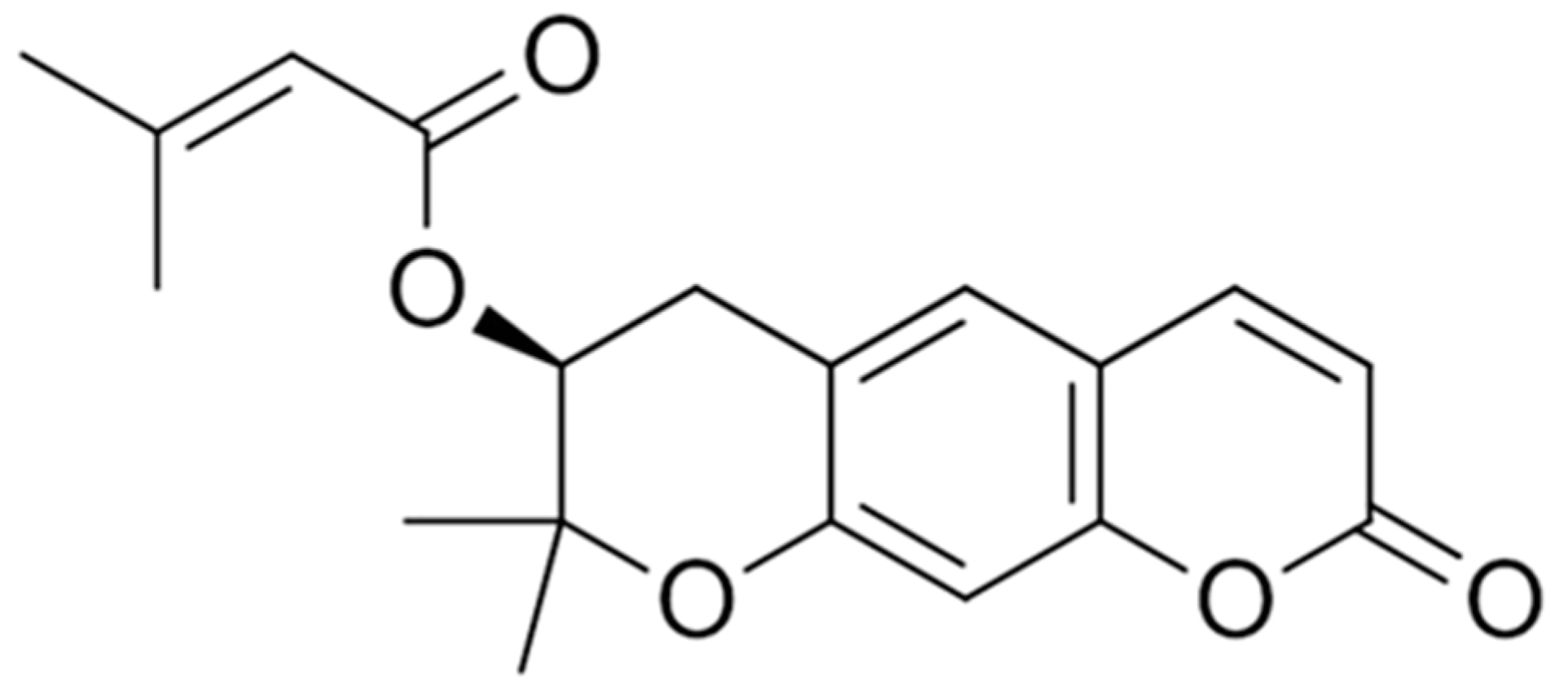
Figure 1. Chemical structure of decursin.
Futurepharmacol 05 00017 g001
Figure 2. Possible anticancer mechanism of decursin against cancer. Decursin actively binds and inactivates tyrosine kinase receptor, which blocks the PI3K/AKT/mTOR pathway and induce apoptosis. It also inactivates the ability of ligands to bind with the cytokine receptor which inhibits the JAK/STAT pathway and blocks the transcription of gene responsible for growth hormones. As a result, tumor cell growth reduces and apoptosis increases. Decursin increases the production of IKKβ, which then inhibits the release of NF-κB to induce the growth-stimulating gene transcription. Decursin activates the wnt signaling pathway. It also increases p52, p51, and p27 expression to block cell cycle at G1/S/G2/M phase and produce Bax to reduce Bcl-2 expression, which results in apoptosis. By all these mechanisms, decursin suppress tumor cell proliferation, migration, invasion, and angiogenesis by lowering c-MYC expression. Decursin induces autophagy and apoptosis in cancer cells. [PI3K: phosphoinositide 3-kinase; MAPK: mitogen-activated protein kinase; STAT3: signal transducer and activator of transcription 3; mTOR: mammalian target of rapamycin; NF-κB: nuclear factor kappa-light-chain-enhancer of activated B-cells; LATS1: large tumor suppressor kinase 1; APC: adenomatous polyposis coli; HIF-1α: hypoxia-inducible factor 1-alpha; ERK1/2: extracellular signal-regulated kinase 1/2; JAK: Janus kinase; IKK-β: inhibitor of nuclear factor kappa-B kinase subunit beta; Bax: BCL2-associated X protein; IL: interleukin; COX: cyclooxygenase; MMP: matrix metalloproteinase; EMT: epithelial–mesenchymal transition; TCF: T-cell factor; YAP: yes-associated protein; TEAD: transcriptional enhancer factor domain; SHP2: Src homology 2 domain-containing phosphatase 2; CDK2: cyclin-dependent kinase 2.].
Futurepharmacol 05 00017 g002
Table 1. Anticancer activity of decursin against different cancers observed in the literature reports.
Cancer TypeTest SystemDose/
Concentration/(R/A)
IC50Results/Possible MechanismReferences
Breast cancerMCF-7 cells, in vitro1–50 µM-↓ TPA-induce MMP-9, cell invasion, ↓ NF-κB, PKCα, MAPK[49]
MDA-MB-231, MDA-MB-453, MDAMB-157, MCF-7, and MCF-10A cells, in vitro80 µM-↑ G1 arrest, cell cycle arrest, p53 protein,
↓ Cyclin D1 level, protein expression, Pin1
[50]
Bladder and colon cancerHuman urinary bladder cancer 235J cells and colon cancer HCT116 cells, in vitro50 and 100 µM-↑ Cytotoxicity, apoptosis, cytochrome c, caspase -3 and Bax, protein levels of p21waf1, cytoplasmic DNA-histone complex, ERK,
↓ Bcl-2, cyclin D1, cyclin E, CDK-2,4, cell cycle, cell growth, proliferation
[38]
Colorectal cancerCT-26 colon carcinoma cells, in vitro10–20 µM-↓ Proliferation, invasion, MMP-2 and MMP-9, formation of tumor nodules, ERK, JNK, growth of cancer cells, ↑ lung weight[51]
HT29 and HCT116, in vitro10–90 μmol/L-↑ Apoptosis, Bax, expression of E-cadherin, p53, EMT
↓ Colony number, cell proliferation, Bcl-2, wound healing, expression of N-cadherin, vimentin, expression of PI3K, Akt
[52]
Skin cancerU266, MM.1S, and ARH77 cells,
in vitro
40–160µM-↑ Apoptosis, caspase-3, -8, and -9, tumor growth, cytotoxicity, cleavage of procaspase-8, procaspase-9
↓ Cyclin D1, bcl-2, Bcl-xL, survivin, VEGF, STAT3, JAK2, interleukin-6, angiogenesis
[53]
B16F10 and NIH-3T3 cells, in vitro20–100 µM-↑ Apoptosis, cytotoxicity, Bax, phosphorylation of p38, caspase-3,
↓ Proliferation, ERK, Bcl-2, tumor growth, tumor weight
[45]
In vivo tumor and histological assay in male C57BL/6J mice, n = 610 mg/kg (i.p.)
(B16-F10) cells, in vitro studies10 µM-↓ IL-6, IL-1β, TLR4, and NF-κB, Akt, JNK, ERK, and p-ERK[44]
Lung cancerHEK-293 cells, A549 cancer cells, in vitro studies10–50 µM-↑ Apoptosis, oxygen-dependant hydroxylation and ubiquitination,
↓ HIF-1, HIF-1α and PD-L1, mRNA expression, proliferation, invasion
[54]
C57BL/6 mice, in vivo studies (n = 5)10 mg/kg (i.p.) ↓ Tumor growth
Glioblastoma cancerU87 cells, glioblastoma cells, in vitro10–200 µM49.01 µM↑ Apoptosis, apoptotic bodies, phosphorylated JNK, p38, caspase-3, -7, and -9, sub-G1 DNA population, PARP-1
↓ Bcl-2, CDK-4 cyclin D1
[55]
Ovarian cancerOvarian cancer cells, in vitro studies5–50 µg/mL23 μg/mL,
8 μg/mL
↑ Caspase-3, -8 and -9, cleaved PARP level, apoptosis
↓ Proliferation of NCI/ADR-RES, P-glycoprotein expression
[48]
Prostate cancerHuman DU145, PC-3 prostate cancer and LLC cell lines, in vivo30–100 mg/kg (i.p and p.o.)-↑ Apoptosis
↓ Tumor volume, cell proliferation, angiogenesis
[56]
Mouse LLC allograft tumor and human PC-3 and DU145 xenograft models in E right flank of C57BL/6 mice, in vivo (n = 5–16)
Pancreatic cancerPancreatic ductal adenocarcinoma cells (PANC-1 and MIA PaCa-2), in vitro20–60 µM-↑ Cytotoxicity, G0/G1 phase arrest, apoptosis, caspase-3, poly (ADP-ribose) polymerase (PARP) cleavage
↓ Colony formation, cyclin D1, CDK4, p38 phosphorylation, Proliferation, MMP-2, MMP-9
[47]
Prostate and lung cancerDU145 and 22Rv1 cells, in vitro studies25–100 µM-↑ Cell cycle arrest, apoptosis, p107 and p130, growth inhibition, cell death,
↓ CDK-2 and CDK-4, EGFR, ERK1/2, cell proliferation, number of surviving colonies, E2F-3, E2F-4 and E2F-5, growth of androgen, EGF ligand
[57]
PC-3 cells, in vitro50–200 µM-↓ Expression of cyclin D1 and c-myc, beta-catenin, growth of PC3, Wnt/β-catenin pathway[58]
DU145, PC-3, and LNCaP cells,
in vitro
25–100 µmol/L-↑ Binding of CDK inhibitor (CDKI) with CDK, apoptosis, caspase-3, -9, cell death, a Strong G1 Arrest, protein levels of Cip1/p21, Kip1/p27 Levels, ↓ CDK2,4,6, cyclin D1, and cyclin E, Cell growth,[34]
Gastric cancerIn vitro and vivo studies human gastric cancer cell lines-SNU484 and SNU21620–40 µM-↓ Cell growth, migration, invasion, survival, cell proliferation, expression of CXCR7, Bcl-2, c-Myc expression[59]
Head and neck cancerHNSCC cell line, in vitro studies50–100 µM-↑ Stress fiber formation, G0/G1 cell cycle arrest,
↓ CXCR7 expression, cell proliferation, migration, invasion, cyclin A, cyclin E, and CDK2, cell growth, S phase and G2/M phase, cell motility, STAT-3 phosphorylation, c-MYC expression
[60]
Blood and bone marrow cancerK562 cells, in vitro50 µM-↓ Bleb formation, megakaryocytic differentiation, translocation of PKCα and βII, binding of PDBu to PKC, PDBu,[61]
-HUVEC and mice, in vitro1–20 µM-↓ Proliferation, migration, capillary-tube formation, microvessel formation, P-ERK, p-JNK, angiogenesis[62]
HepG2 cells
In vitro studies
5–80 µM-↑ LATS1 and βTRCP, apoptosis ↓ Growth of HepG2 cells, cell proliferation, cell cycle[63]
Leukemialeukemic KBM-5 cells, in vitro studies10–80 µM-↑ Apoptosis, ↓ COX-2, surviving.[64]
Gastric cancerSNU216, NCI-N87 cells, in vitro--↑ Cell cycle arrest, LC3-II levels, DNA synthesis ↓ Cell growth, autophagic flux, the expression of lysosomal protein cathepsin C (CTSC), cell proliferation, E2F3, growth of spheroids, patient-derived gastric organoids, autophagy, CDK4/6,[39]
(↑): upregulation/increase/stimulation, (↓): downregulation/decrease/inhibition, LLC: Lewis lung cancer, LPS: lipopolysaccharide, CDK: cyclin-dependent kinase, MMP: matrix metalloproteinase, EKR: extracellular signal-regulated kinases, JNK: c-Jun N-terminal kinase, MAP: mitogen-activated protein, STAT: signal transduction and transcription activation, c-MYC: cellular Myc, EGFR: epidermal growth factor receptor, PPAR: peroxisome proliferator-activated receptor, FAS: fatty acid synthase, ACC: acetyl-CoA carboxylase, CTR: catenin response transcription, JAK: Janus-activated kinase, EMT: epithelial–mesenchymal transition.
Disclaimer/Publisher’s Note: The statements, opinions and data contained in all publications are solely those of the individual author(s) and contributor(s) and not of MDPI and/or the editor(s). MDPI and/or the editor(s) disclaim responsibility for any injury to people or property resulting from any ideas, methods, instructions or products referred to in the content.