Review

An Overview of Sargassum Seaweed as Natural Anticancer Therapy

by
Kelly Johanna Muñoz-Losada
,
Manuela Gallego-Villada
and
Miguel Angel Puertas-Mejía
*
Grupo de Investigación en Compuestos Funcionales, Facultad de Ciencias Exactas y Naturales, Universidad de Antioquia, UdeA, Calle 70 No. 52-21, Medellín 050010, Colombia
*
Author to whom correspondence should be addressed.
Future Pharmacol. 2025, 5(1), 5; https://doi.org/10.3390/futurepharmacol5010005
Submission received: 11 December 2024 / Revised: 10 January 2025 / Accepted: 16 January 2025 / Published: 20 January 2025
(This article belongs to the Special Issue Feature Papers in Future Pharmacology 2024)

Abstract

:
Algae have great therapeutic value and have attracted a great deal of attention due to the abundance of bioactive compounds they contain, which may be the key to fighting diseases of various origins, such as skin cancer, breast cancer, or osteosarcoma. In this regard, global trends indicate that cancer is likely to become the leading cause of death and the main obstacle to increased life expectancy in the 21st century, which is related to multiple factors, including the various effects of climate change, which will continue to cause afflictions to human health. Then, excess exposure to ultraviolet radiation (UVR) causes damage to DNA, proteins, enzymes, and various cellular structures and leads to the development of cancer, premature aging of the skin (wrinkles, dryness, dilation of blood vessels, and loss of collagen and elastin), or alterations of the immune system. In addition, multidrug resistance (MDR) is characterized by the overexpression of efflux pumps, such as P-glycoprotein or P-gp, that expel chemotherapeutic drugs out of the cancer cell being the main obstacle to their efficacy. Some molecules inhibit efflux pumps when co-administered with antineoplastic agents, such as glycolipids. Mycosporin-like amino acids and glycolipids isolated from Sargassum have shown an important role as potential anticancer agents. The results show that glycolipids and mycosporin-like amino acids present in brown algae of the genus Sargassum exhibit cytotoxic effects on different types of cancer, such as breast cancer, leukemia, and osteosarcoma, which is a key criterion to be considered as a natural anti-cancer strategy; but, more in-depth in vitro studies are needed to represent them at the in vivo level, as well as their validation in preclinical assays.

Graphical Abstract

1. Introduction

Cancer is one of the pathologies that appears among the main causes of death worldwide according to the World Health Organization (WHO); indeed, it is the first or second cause of premature death (i.e., at ages 30–69 years) in 134 of 183 countries. The World Cancer Report 2020 projects the global burden of cancer to exceed 27 million new cancer cases per year by 2040, representing a 50% increase compared to the estimated 18.1 million cancer cases reported in 2018. Forecasts indicated that lung, liver, colorectal, and breast cancers will experience a notable increase. Although cancer in particular is declining in higher-income countries, this progress is lacking in lower-income countries. For example, in the period from 2016 to 2020, the cancer mortality rate per 100,000 population in Germany declined by 10%, while in Colombia the decline was 3.5% [1,2].
Cancer is a specific type of neoplasm (uncontrolled growth of abnormal cells or tissues in the body) that is characterized by the ability to spread to other tissues, which is called metastasis. The transformation of normal cells into cancer cells is mainly caused by environmental agents, various types of radiation, physical inactivity, dietary factors, obesity, smoking, alcoholism, heredity factors, and infectious agents such as parasites, viruses, and bacteria, which cause mutations in the DNA so that the cells continue their uncontrolled growth and spread. This process is known as carcinogenesis, and the following four phases are recognized [3]:
  • Induction or initiation: DNA mutations appear that turn healthy cells into cancer cells: uncontrolled division, capacity for local invasion, and distant spread.
  • Cancer ‘in situ’: increase in the number of cancer cells in the organ or tissue in which it originates, producing the primary tumor.
  • Local invasion: extension of the primary tumor to neighboring structures, invading them and giving rise to the first symptoms.
  • Distant invasion or metastasis: the cancerous cells enter the bloodstream and spread to other organs, giving rise to secondary tumors.
On the other hand, those that do not spread are called benign neoplasms, and can therefore be removed surgically. In both cases, the mass formed by the neoplasia is called a tumor [4]. There are more than 100 types of cancer, and they can be grouped into the following categories:
  • Carcinoma: this is the most common cancer of epithelial origin and is capable of affecting organs or secretory glands. There are two subtypes of carcinoma, adenocarcinoma and squamous cell carcinoma.
  • Sarcoma: cancer that forms in connective and supportive tissues such as bone tissue and soft tissues, including muscle, adipose tissue, lymphatic vessels, blood vessels, tendons, and fibrous tissue.
  • Myeloma: cancer that starts in plasma cells (a type of white blood cell) in the bone marrow, forming tumors in many bones and preventing the production of healthy blood cells.
  • Leukemia: cancer derived from blood-forming bone marrow tissue or its precursors. This type of cancer does not form solid tumors; instead, abnormal white blood cells (leukemic cells) accumulate in the blood, displacing normal blood cells in the blood.
  • Lymphoma: cancer of the lymphatic system that specifically affects B-cells, which unlike leukemia produces solid tumors that involve lymph nodes or other organs in the body.
Therefore, the most common way to combat the uncontrolled growth of cancer cells is through the application of conventional treatments such as surgery, radiotherapy, and chemotherapy or more selective treatments such as hormone therapy. Even with advances in cancer therapy, chemotherapy remains one of the most important treatments, involving the use of chemical drugs that suppress and kill cancer cells, mostly in a non-specific manner [5]. The drugs implemented in chemotherapy could then be classified in different ways according to their chemical nature/source, molecular target, mechanism of action, or efficacy in model systems against various types of malignancies. However, it is possible to assemble chemotherapy drugs into alkylating agents, antimetabolites, antitumor antibiotics, topoisomerase inhibitors, mitosis inhibitors, and corticosteroids [6]. Among the most commonly used chemotherapeutic agents is Daunorubicin, whose mechanism of action is based on its intercalation in DNA, inhibiting the synthesis of nucleic acids, as it hinders the progress of the enzyme topoisomerase II, which is responsible for the unwinding of DNA in the transcription process [7]. Vincristine acts at the metaphase stage of the cell cycle by binding to tubulin, inhibiting the assembly of microtubules, which are necessary for chromosome separation during anaphase. Inhibition of these structures results in cell death [8], and 5-fluorouracil acts by inhibiting essential biosynthetic processes or by incorporating them into macromolecules, such as DNA and RNA, and inhibiting their normal function [9].
However, cancer cells have generated resistance to the drugs used in chemotherapy, creating an obstacle to the treatment of neoplasms. This limits the effectiveness of therapy and compromises the choice of chemotherapy in cases of recurrence. Tumors may be intrinsically resistant before or during treatment, so understanding the mechanism of chemical resistance is an important factor in developing an appropriate approach to cancer therapy [10].
Seaweeds are an important source of biologically active substances and play a crucial role in ecology and economy. Among these valuable algae, the genus Sargassum, belonging to the class Phaeophyceae (brown algae) in the order Fucales [11], is well known for its presence in warm, tropical waters around the world, often forming vast floating expanses known as Sargassum [12]. Today, these floating masses have increased considerably along the coasts of the Caribbean, Africa, and the Gulf of Mexico, a phenomenon that has unfavorable economic (loss of tourism), environmental (threat to the survival of coastal areas and fish kills), and sanitary (beach pollution) consequences [13]. Nevertheless, the scientific community, searching for sustainable solutions, has explored various applications of Sargassum (Figure 1). Its use as a source of biogas and bioenergy [14,15], fertilizer [16], and its incorporation into building materials [17] are some of them, without leaving behind the growth of Sargassum research as a source of sulfated compounds, phenolics, fucoidans, and polysaccharides with antitumor, antioxidant, immunological, hypoglycemic, anticoagulant, anti-aging, bone growth-promoting, antiviral, and antibacterial activities [18,19,20].
From an environmental point of view, the cultivation and exploitation of seaweed represent an environmentally friendly activity, as it does not generate waste or effluents; it also increases local biodiversity by serving as a substrate and refuge for numerous species of fish and invertebrates and diversifies traditional productive activities, reducing the fishing of certain endangered species such as sea turtles, queen conch, and lobster. On the other hand, when the amount of algae is not high, they do not generate any damage to the ecosystem they inhabit, but when they accumulate in coastal areas, they generate environmental, economic, and social problems. For example, rotting algae release hydrogen sulfide, turning this biomass into a highly toxic source. Therefore, excess algae in coastal areas not only endangers the local economy and ecosystems but also represents a high health risk.
Although the exploitation of macroalgae is mainly focused on the production of biomass for human consumption and the production of hydrocolloids, in recent years this rich source of bio-compounds has attracted the attention of pharmaceutical, food, and cosmetic companies, among others, for the development of new drugs and products of natural origin [25]. This is largely because they have demonstrated a wide range of biological activities such as antiviral, antibiotic, cytotoxic, antimutagenic, anticancer, antitumor, fungicidal, anti-inflammatory, etc. Therefore, the purpose of this review is, in addition to advancing chemical and biological knowledge of marine biodiversity, to establish the medicinal, economic, and social potential of Sargassum species that proliferate as contaminating biomass on the Caribbean coast as a natural strategy for the treatment of cancer.

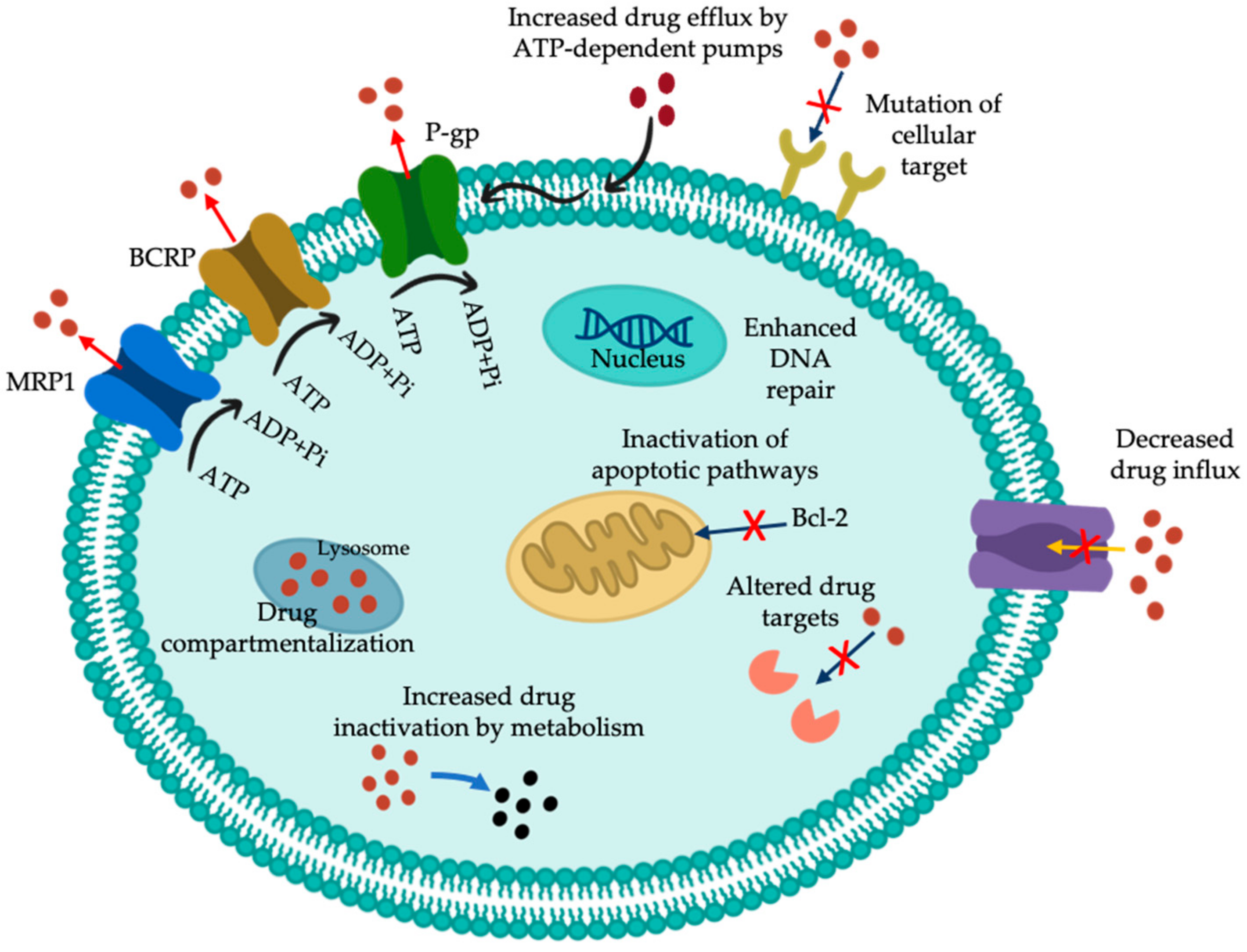
2. Multidrug-Resistance (MDR) Activity

Cancer resistance can be classified into two categories: innate and acquired. Innate drug resistance occurs before treatment, while acquired resistance develops after initial therapy [26]. In some patients, prolonged exposure to a single chemotherapeutic agent may contribute to the development of cross-resistance to other drugs, a phenomenon referred to as multidrug resistance (MDR) [27]. The underlying mechanisms for the development of resistance to anticancer drugs are complicated and involve altered apoptosis pathways leading to cell survival, changes in the specific targets of each drug, increased drug efflux (efflux pumps) or decreased entry, hypoxia-mediated chemoresistance, altered enzymatic behavior leading to anti-cancer drug activation, and DNA repair, among others (Figure 2).
One prevalent mechanism in cancer is the MDR phenotype, which is characterized by overexpression of a class of efflux pumps called energy-dependent binding cassette (ABC) transporters. Efflux pumps are transmembrane proteins capable of expelling any xenobiotic compounds, such as anticancer drugs, from inside the cell to the extracellular milieu, reducing its accumulation and thus preventing tumor cell death [28]. There are approximately 49 types of ABC transporters in humans, distributed in subfamilies from ABCA to ABCG, of which 15 have been identified as clinically active transporters involved in drug resistance [29]. Among them, the transporters ABCB1, drug resistance protein or P-glycoprotein (P-gp); ABCC1, multidrug resistance-associated protein (MRP); and ABCG2, breast cancer resistance protein (BCRP) are the most representative [30]. Despite the role of the transporters in normal physiology, overexpression of these transporters in tumor cells leads to resistance to various cytotoxic agents in cancer chemotherapy [31].
ABCB1 inhibitors are classified into four generations according to their potency, selectivity, and drug-drug interaction capacity. The first generation includes calcium channel blockers (e.g., verapamil); immunosuppressants (e.g., cyclosporin A); antihypertensives (e.g., reserpine); antiarrhythmics (e.g., quinidine); and anti-estrogens (e.g., tamoxifen and toremifene) [32]. Many of these drugs are themselves substrates of P-gp and therefore act in competition for output with other P-gp substrates (competitive inhibition) [33]. However, the main limitations of the clinical use of these inhibitors are related to undesirable immunosuppressive and cardiovascular effects. Therefore, second and third-generation inhibitors, which more specifically modulate ABCB1, were developed to improve the toxicity profile of the first-generation inhibitors. These are analogs of the first-generation compounds that underwent structural modifications to reduce their primary therapeutic activity and increase efficacy and potency. These include the non-immunosuppressive analogs of cyclosporin A, valspodar (PSC-833); the R-enantiomer of verapamil, dexverapamil; and other compounds such as biricodar (VX-710), timcodar (VX-853), and dofequidar (MS-209) [34]. Finally, the fourth generation focuses on isolated natural product inhibitors and their derivatives, surfactants and lipids, peptidomimetics, and agents that combine transport inhibition with other beneficial biological activity [35]. The P-glycoprotein (Pg-p), also known as ABCB1 was the first ABC transporter identified, composed of 1280 amino acids and located on chromosome 7p21 [36]. Since then, Pg-p has been extensively investigated and experimentally evaluated to understand its structure, composition, and mechanism of action and thus find the most effective way to inhibit or modulate its activity.

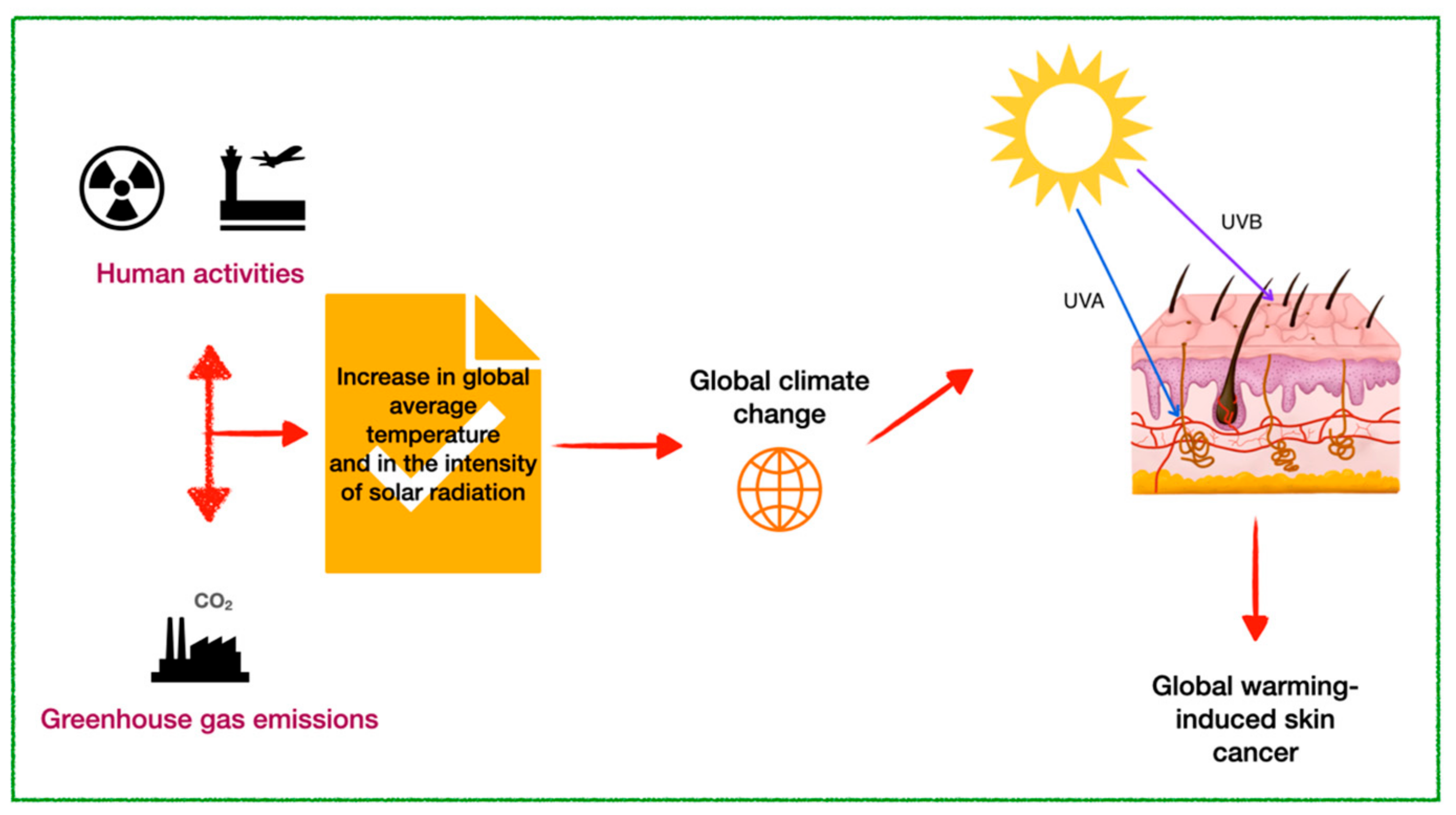
3. Skin Cancer Linked to Global Warming

In recent decades, the concentration of greenhouse gases in the atmosphere has increased considerably, especially gases such as carbon dioxide, methane, nitrous oxide, and gases derived from industrial processes, including hydrofluorocarbons, sulfur hexafluoride, and perfluorocarbons [37]. The presence of these gases accelerates climate change, affecting the ozone layer and heat retention in the atmosphere, with the consequent increase in the planet’s average temperature and the intensity of solar radiation (Figure 3) [38,39,40].
Ultraviolet radiation (UVR) from the sun affects and is affected by global climate change, and whatever affects sunlight affects UVR. The decrease in stratospheric ozone allows more harmful UVB rays (a higher frequency and more damaging type of UV) to reach the Earth’s surface and cause damage to the DNA of plants and animals. This increase in solar radiation and its UV components is directly related to the effects of climate change on the ozone layer [45]. UVR is divided into UVC (100–290 nm), UVB (290–320 nm), and UVA (320–400 nm), the latter being subdivided into UVA II (320–340 nm) and UVA I (340–400 nm). UVB makes up 5–10% of the UVR reaching the earth and is less energetic than UVC, but can cause direct cell damage; finally, UVA makes up the remaining 90–95% and has greater skin penetration than UVB, as it can reach the dermis, while UVB is absorbed mostly in the epidermis. Excessive UVR exposure is a health risk due to generating acute and chronic effects. Acute effects include erythema, epidermal thickening known as hyperkeratosis, mainly of the stratum corneum, immunosuppression, and skin pigmentation (tanning, sun lentigines, or spots). On the other hand, chronic effects include photoaging and skin cancer, which is divided into melanoma and non-melanoma, the latter being further subdivided into basal cell carcinoma and squamous cell carcinoma [46,47].
There is extensive literature on the relationship between increased exposure to ultraviolet radiation and the increase in skin cancers, such as cutaneous malignant melanoma, squamous cell carcinoma, and basal cell carcinoma, as well as photoaging, sunburn, cataracts, and other eye diseases [48]. These radiations have also been shown to reduce the effectiveness of the immune system by modifying the activity and distribution of the cells that trigger immune responses [49]. One of the most widely used options to reduce the risk of damage induced by UV radiation from the sun has been the use of photoprotectors (sunscreens). The effectiveness of these products depends mostly on their stability over long periods of exposure. The active ingredients of a photoprotector is UV filters, and there are very few organic/inorganic molecules accepted as sunscreens, with the drawback that most of them are photo-instable, promoting the formation of photo-products, which alters their absorbance spectrum and therefore reduces their photoprotective effect [50]. Additionally, these photoproducts, including free radicals, interact with the additional ingredients that make up sunscreens, molecular oxygen, and even skin proteins and lipids, triggering undesirable reactions such as hypersensitivity [51]. The increased use of these dermo-cosmetic products has raised concerns about the importance of their stability and their impact on human health [52].
Photostability in a sunscreen is very important because during exposure of the sunscreen to UVR, especially some photolabile organic filters, such as octylmethoxycinnamate, degrade rapidly by direct photolysis with the formation of cyclodimers that can produce photodegradation intermediates. In addition, photostability testing of new drugs and products covered by the ICH Tripartite Harmonized guideline is a critical aspect related to stress testing. According to Q1B ICH, the following light sources can be used for photostability testing: 1. any light source that is designed to produce an output similar to the D65/ID65 emission standard, such as an artificial daylight fluorescent lamp combining visible and ultraviolet (UV) outputs or a xenon or metal halide lamp; or 2. the sample itself should be exposed to both the cool white fluorescent lamp and the near ultraviolet lamp. Therefore, a priority in this field is the use of new molecules and/or natural extracts with protective activity against ultraviolet radiation [53].
Ozone, a photoabsorbent molecule present mainly in the stratosphere, between 10 and 50 km above the Earth’s surface, absorbs large amounts of shortwave UVB radiation and all ultraviolet C (UVC) radiation, but only a little UVA. On the other hand, greenhouse gases such as chlorofluorocarbons deplete ozone and have a substantial effect on terrestrial ultraviolet exposure [54,55]. Sunburn and photosensitivity may have increased in some parts of the world following ozone depletion, and it has been estimated that a 1% decrease in ozone levels is followed by a 1–2% increase in melanoma mortality [56]. Photodermatoses represent a heterogeneous group of diseases with an abnormal skin reaction to sunlight; photoprotection is a key element of their treatment, and the selection of the most appropriate sunscreen often depends on the identification of the wavelengths responsible for inducing the disease [57,58]. For example, polymorphous light eruption, the most common photodermatosis with a prevalence of 10–20% in the general population, and lupus erythematosus, the most common photo-aggravated dermatosis, are triggered by UVA and UVB rays [59]. This is of concern to health authorities, since the high incidence of solar radiation produces acute and chronic dermatological manifestations (sunburn), pigmentation changes (melasma), accelerated aging (photoaging), and neoplastic changes (photocarcinogenesis) [60,61].
At present, special attention is being paid to the possibility of modulating the harmful effects of oxidative stress induced by UV radiation, by using natural extracts or organic molecules with a photoprotective capacity capable of minimizing cell damage and that may be able to favorably regulate mutagenic and/or carcinogenic processes [62,63,64]. However, one of the limitations in the use of compounds of natural origin as additives in products for human consumption is related to their stability, since they can be degraded by exposure to UV radiation, transforming them into undesirable compounds. Therefore, it is important that if these compounds are part of products for commercial use, they must have a high degree of chemical stability against direct exposure to visible light or UV radiation; they must be photostable [65].

4. Glycolipids Involved in MDR

It has been shown that enhancement of antibiotic susceptibility in resistant tumor cells is possible with the co-administration of compounds of natural or synthetic origin, such as flavonoids and alkaloids that act as potential inhibitors of efflux pumps [66]. Glycolipids, amphipathic molecules, are a powerful inhibitory agent against P-gp, potentiating antineoplastic drugs and exhibiting reversal factors 1000-fold higher than those of the positive control [67]. Among these phytoconstituents are glycolipids, amphipathic molecules characterized by the structures of monogalactosyl-monoacylglyceride (MGMG), monogalactosyl-diacylglyceride (MGDG), digalactosyl-diacylglyceride (DGDG), sulfoquinovosyl-diacylglyceride (SQDG), and sulfoquinovosyl-monoacylglyceride (SQMG) (Figure 4), which, as mentioned above, could be a promising group of compounds capable of reversing the MDR phenotype [68,69]. However, studies on the isolation, characterization, and biological potential of their glycolipids are scarce. Table 1 shows algal species, extracts or compounds of interest, and main feature reported. In addition, the chemical structure of the glycolipids reported for Sargassum is shown in Figure 4.
Importantly, the absence of cytotoxicity is an essential criterion for the selection of these compounds as inhibitors [82]. The main objective in the co-administration of these compounds with anticancer agents, is to block the efflux pumps associated with MDR, allowing a higher concentration of the drug into the cancer cell and therefore improve its efficacy in eliminating it. In this context, MGDGD (2) showed potent cytotoxic activity against HepG-2 cells (IC50: 0.16 ± 0.01 μM) compared to standard 5-fluorouracil (IC50: 0.63 ± 0.28 μM) [71]. On the other hand, glucopyranoside (1) showed moderate antiproliferative activity against cancer cell lines HepG2 (IC50: 18.5 ± 1.4 μM), MCF-7 (IC50: 21.6 ± 1.3 μM) and Caco-2 (IC50: 15.7 ± 0.9 μM); activity related to the enzymatic inhibition of 5-LOX and 15-LOX is implicated in the development of several types of cancer. The results of this investigation suggest that the mode of binding of the compound to the major amino acid residues of the 5-LOX and 15-LOX enzymes occurs through interactions of the polar head of the glycolipid, forming hydrogen bonds, and the interaction of the hydrophobic tail with the side chains of the enzymes [21].
In contrast, Hossain et al. [81] reported that in Caco-2 cells treated with SQDGs and DGDGs at a concentration of 100 μM, cell viability of approximately 70–80% each was observed after 72 h. The cell viability of NaBT, a drug related to cancer cell apoptosis, at a concentration of 1.0 mM was similar to that of DGDG. Furthermore, it was observed that the glycolipids had no cytotoxic effects on the normal human colon CCD-18Co cell line [81]. This study provides an approach to the ability of SQDG and DGDG-type glycolipids to enhance the efficacy of anticancer drugs in cancer treatment. In addition, these compounds may be promising candidates for future evaluation as MDR-associated efflux pump inhibitors, as they exhibit values above 25 μg/mL, taking into account the molecular weight of each compound.
Therefore, glycolipids of Sargassum have the potential to act as MDR inhibitors in cancer, arising from the need to utilize the extensive Sargassum resources available on the coasts while recognizing their promising potential to inhibit efflux pumps. In addition, previous examples of glycolipids isolated from terrestrial plants have been documented, such as tricolorin A, which has shown the ability to modulate vinblastine cytotoxicity in MDR MCF-7/Vin+ cells (vinblastine-resistant breast cancer cells) up to 2164-fold at a concentration of 25 μg/mL. This reversion factor (RFMCF-7/Vin+ = IC50 vinblastine/IC50 vinblastine in the presence of glycolipid) is similar to the activity observed with terrestrial glycolipids extracted from other Ipomoea species, such as murucoidin V (RFMCF-7/Vin+ > 255-fold) from I. murucoides; jalapinosides I and II, and purgin II, all from I. purge; and albinoside III from I. alba, all with RFMCF-7/Vin+ > 2000-fold. These results set a strong precedent for the potential of glycolipids to reverse the MDR phenotype in cancer.

5. Mycosporin-like Amino Acids (MAAs) Against Global Warming-Induced Skin Cancer
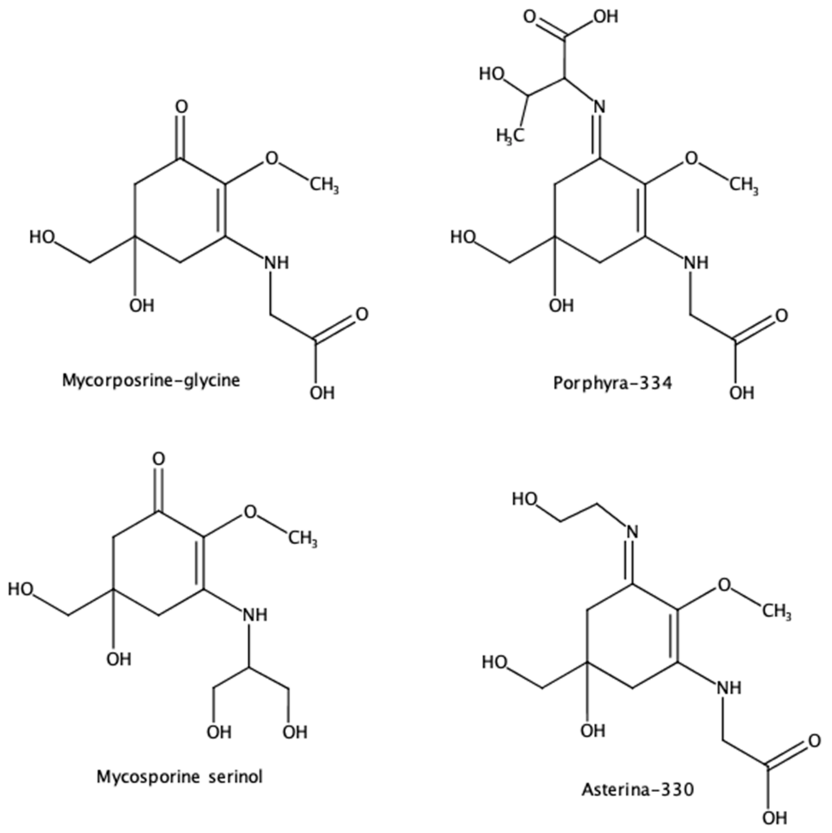
In general terms, UV radiation has both direct and indirect effects on human skin. In everyday activity, we receive UV radiation from both natural (solar radiation) and artificial sources (monitors, fluorescent and halogen light, black light from arc welding, as well as from tanning salons). It is estimated that approximately 50% of solar damage is due to the formation of free radicals, while the other 50% is responsible for direct damage to the nuclear structures of cells and other mechanisms that have not been elucidated. UV radiation is responsible for approximately 90% of all skin cancers by damaging cellular DNA. It is estimated that in the last ten years, skin cancer has increased by 8.3%, essentially due to indiscriminate exposure to the sun [83]. According to the World Health Organization (WHO), direct and excessive exposure to the sun has generated around 130,000 cases of malignant melanoma each year globally, of which around 66,000 cases (50.77%) result in death. In addition, there is evidence to suggest that environmental levels of UV radiation may suppress cellular immunity and thus increase the risk of infectious diseases and the effectiveness of vaccines [84].
Commercially available sunscreens contain synthetic organic and inorganic UVR filters and since many synthetic UV filters have a low photostability and an undesirable effect on the environment, the search for a new generation of UV filters has been ongoing for the last decade. In recent years, many compounds with photoprotective properties have been evaluated and, within this wide range, natural products have gained special interest due to the shift towards environmental safety and the impact of global warming on consumer awareness of the use of environmentally friendly compounds. However, many bioactive compounds present disadvantages (solubility, stability, penetration degree, interactions with biomolecules, etc.) for their application in products for human consumption, and nanotechnology is looking for different alternatives that can provide solutions to these disadvantages.
Premature aging due to UVA, also known as photo-aging, is an aging of the skin at an early age, which can cause an increased likelihood of skin cancer. This aging manifests itself in the form of wrinkles, skin spots, sagging, and a loss of elasticity. The dermis is located on the underside of our skin and provides mechanical support to the epidermis, which is located on the outside of the skin, giving it protection. In the dermis, the extracellular matrix is mainly composed of three types of collagens: type I, type III, elastin, proteoglycans, and fibronectin (9). Premature aging causes levels of collagen types I and III to decrease due to the depletion of collagen precursors. The mechanism of UVB-induced skin deterioration is not yet known.
Some chemical filters that protect mainly UVB rays are aminobenzoates, cinnamates, and salicylates, among others. These filters are usually combined with UVA filters such as benzophenones, which absorb in the UVA-II region, but have the problem that they can cause dermatitis problems, are not photostable, and can generate ROS, generating concern for their possible carcinogenic and endocrine effects. There are also other UVA protectants, such as anthranilates and ecamsule; although, these are less effective than benzophenones and are therefore less widely used [85].
Mycosporin-like amino acids (MAAs) generally consist of amino acid residues linked to a central ring, aminocyclohexenone (AHO) or aminocyclohexenimine (AHI), forming resonance tautomer, which allows for extended conjugation and facilitates absorption of UV radiation [86,87,88]. Their absorption maxima depend on their core and its substituents (Figure 5); for example, Porphyra-334 (P334) has an AHI core (≈320–360 nm) with carboxylic acid substituents, which gives it an absorbance maximum at 334 nm (UVA).
Conversely, mycosporin-glycine (MGly) has an AHO core (≈310) with an absorbance maximum of 310 nm. MAAs are the most UVA-absorbing natural compounds, with a molar absorptivity between 28,100 to 50,000 M−1 cm−1 [89]. In addition, it has been shown that the antioxidant and antiradical capacity of MAAs may be linked to donating a hydrogen atom from the C-4, C-6, or C-9 methylene to the lipid peroxide radical [90,91]. These properties make mycosporins a potential source of additives for developing highly efficient dermo-cosmetic products to prevent skin damage associated with UV radiation (Table 2).
Algae synthesize MAAs and allow them to protect themselves from the harmful effects of UV radiation since they absorb in the 310–360 nm region, covering almost the entire UVR range and being mainly protective against UVA [92,93,94]. Mycosporins can be considered the first line of defense of Sargassum against UVR in natural habitats [19] and also protect macroalgae when other defense mechanisms fail, such as the protection of cells against UVR-induced cell death. It is involved in photosynthetic carbon fixation and has great potential in photoprotection, minimizing cell damage by the presence of ROS and thymine dimer formation due to UVR. Sung et al. [95] reported that the microalga Chlamydomonas hedleyi contains UV-absorbing MAAs; it was reproved that MAAs act as UV-absorbing compounds, contributing to the modulation of gene expression related to oxidative stress, inflammation, and UV-induced skin aging. The anti-aging and wound-healing properties of these metabolites have also been evaluated in three collagenase inhibition, advanced glycation end-product (AGE) inhibition, and a wound healing assay (scratch assay) [96].
Table 2. Some mycosporin-like amino acids with potential application in photoprotection.
Main MAAs EvaluatedApplicationReferences
Mycosporin-serinol and porphyra-334, shinorineTopical sunscreensde la Coba et al. [97]
Mycosporin-glutamicol, mycosporin-glutaminol-glucoside, mycosporin-serinol, mycosporin-taurine, palythine, palythine-threonine-sulphate, porphyra-334, and usujireneAntioxidant potentialBrowne et al. [98]
ShinorineCommercial sunscreen formulationCandelo and Llewellyn [99]
Porphyra-334, shinorine, asterina-330, palythine, and mycosporine-glycineNatural antioxidantde la Coba et al. [87]
A study of 20 cyanobacterial strains obtained from habitats exposed to strong solar radiation revealed that some of them contained one or more UV-absorbing, mycosporin-like compounds (MAAs). The sun protection capabilities of MAAs suggested that significant, although not complete, protection against UV photodamage could be obtained by possessing MAAs [100]. In another investigation, the protective effect of MAA-containing emulsions on the ear tissue of mice exposed to UV irradiation was determined. It was observed that emulsions containing MAA increased total superoxide dismutase (SOD) and catalase (CAT) activity in tissue exposed to UV irradiation. An increased accumulation of copper/zinc (Cu/Zn)−SOD and/or CAT was also observed in the tissue on which the emulsion containing M2G or P-334 had been applied. In addition, P-334 showed an anti-glycation effect on elastin in vitro. Although MAA-containing emulsions have antioxidant and anti-glycation effects in vitro, no protective effect was observed for AGE accumulation in UV-exposed mouse ears [101]. In the in vitro anti-aging study as collagenase inhibitors, a dose-dependent but moderate inhibition was observed for all substances, and IC50 values of 104.0 to 158.9 µM were determined for selected MAAs. However, the effect on collagenase was reasonable; MAAs could act as anti-aging compounds for the skin [102].
In addition, seaweeds provide a large array of micro- and macro-elements essential for health, such as proteins, minerals, vitamins, dietary fiber, antioxidants, and fatty acids, which have allowed them to be incorporated into the development and formulation of food and pharmaceutical products. Therefore, the addition of algae-derived compounds for the formulation of new natural pharmaceuticals is one of the main objectives that have been developed in recent decades at the pharmacological level. Du et al. [103] demonstrated that S. fusiforme and its extracts are effective herbal treatments for leukemia by stimulating apoptosis in human erythroleukaemia (HEL). The anti-leukemic mechanism of apoptosis may involve intrinsic mitochondrial stimulation of caspases and suppression of the PI3K/AKT signaling pathway. On the other hand, Yadav et al., [104] found that by MTT assay of S. tenerrimum extracts in an acute toxicity study conducted on zebrafish for 96 h showed an LC50 value of 504.7 mg/L, finding histopathological changes at the highest concentrations (800 mg/L) of algal extract. These are some examples of the potential of Sargassum as an alternative cancer treatment; of course, more studies are needed to establish a systematic use in pharmaceutical trials.
Finally, the acute impact of climate change on human health is receiving increasing attention because of its effect on chronic diseases such as cancer; in addition to the above, the depletion of the ozone layer has led to an increase in ambient ultraviolet radiation (UVR) in recent decades and consequently to an intensification in the effects of UVR on the skin, ranging from the aesthetic signs of photoaging, which accumulate with daily exposure, to skin cancer.

6. Conclusions

This review provides an overview of the possible use of Sargassum algae as a source of bioactive ingredients for cancer treatment, including biomass washed up on beaches by sea tides. Nevertheless, it is necessary to include preclinical and clinical trials to enforce these findings. Despite this, we found that glycolipids isolated from this raw material can dose-dependently inhibit the efflux activity of ABCB1 and ABCC1 transporters involved in drug resistance, as well as the remarkable inhibitory capacity of ABCC1, suggesting an increased affinity for this transporter, which could increase the efficacy of chemotherapy strategies to combat the MDR phenotype in cancer. One possible mechanism for the inhibitory activity of glycolipids is associated with structural differences in sulphoglycolipids, such as the size of the carbon chains that make up the fatty acids or the degree of stabilization of the molecule. These differences may affect the binding of these glycolipids to transport sites and, consequently, their efflux. Furthermore, SQDGs, being anionic organic compounds, could be competitive inhibitors and possibly a source of phytotherapeutic agents capable of reversing the MDR phenotype in cancer cells. Additionally, although mycosporins have a high photoprotective and antioxidant potential, there are few related applications, suggesting that there is still a major challenge for the pharmaceutical and cosmetic industries to find innovative solutions in the healthcare field. This starting material could be included in new photoprotection strategies as bioactive compounds in topical products such as sunscreen adjuvants, which have been shown to generate significant protection against oxidative stress and inflammation alone or in combination with conventional sunscreens. In spite of these findings, further studies are still needed to demonstrate that Sargassum could be a promising source for future studies to help address this health problem, as well as to understand the mechanisms by which these natural products have beneficial effects in the treatment of cancer.

Author Contributions

Conceptualization, K.J.M.-L., M.G.-V. and M.A.P.-M.; methodology, K.J.M.-L. and M.G.-V.; formal analysis, K.J.M.-L., M.G.-V. and M.A.P.-M.; investigation, K.J.M.-L. and M.G.-V.; resources, M.A.P.-M.; writing—original draft preparation, K.J.M.-L.; writing—review and editing, K.J.M.-L. and M.A.P.-M.; supervision, M.A.P.-M.; project administration, M.A.P.-M.; funding acquisition, M.A.P.-M. All authors have read and agreed to the published version of the manuscript.

Funding

This research received no external funding.

Institutional Review Board Statement

Not applicable.

Informed Consent Statement

Not applicable.

Data Availability Statement

No new data were created or analyzed in this study. Data sharing is not applicable to this article.

Acknowledgments

This work was supported by the CODI of the Universidad de Antioquia (Project no. 2022-55010 and 2022-52641). We thank Valeria Puertas-Meza for her assistance with the illustrations.

Conflicts of Interest

The authors declare no conflicts of interest.

References

  1. World Health Organization. Cancer. Available online: https://www.who.int/news-room/fact-sheets/detail/cancer (accessed on 8 August 2024).
  2. Wild, C.; Weiderpass, E.; Stewart, B. World Cancer Report: Cancer Research for Cancer Prevention; International Agency for Research on Cancer: Lyon, France, 2020. [Google Scholar]
  3. National Cancer Institute. Understanding Cancer. Available online: https://www.cancer.gov/about-cancer/understanding (accessed on 25 November 2024).
  4. National Cancer Institute. SEER Training Modules: Cancer As a Disease. Available online: https://training.seer.cancer.gov/disease/ (accessed on 28 November 2024).
  5. Bray, F.; Ferlay, J.; Soerjomataram, I.; Siegel, R.L.; Torre, L.A.; Jemal, A. Global cancer statistics 2018: GLOBOCAN estimates of incidence and mortality worldwide for 36 cancers in 185 countries. CA Cancer J. Clin. 2018, 68, 394–424. [Google Scholar] [CrossRef] [PubMed]
  6. Anand, U.; Dey, A.; Chandel, A.K.S.; Sanyal, R.; Mishra, A.; Pandey, D.K.; De Falco, V.; Upadhyay, A.; Kandimalla, R.; Chaudhary, A.; et al. Cancer chemotherapy and beyond: Current status, drug candidates, associated risks and progress in targeted therapeutics. Genes Dis. 2023, 10, 1367–1401. [Google Scholar] [CrossRef]
  7. Thirumaran, R.; Prendergast, G.C.; Gilman, P.B. Chapter 7—Cytotoxic Chemotherapy in Clinical Treatment of Cancer. In Cancer Immunotherapy; Prendergast, G.C., Jaffee, E.M., Eds.; Academic Press: Burlington, VT, USA, 2007; pp. 101–116. [Google Scholar]
  8. Awosika, A.O.; Below, J.; Das, J.M. Vincristine. In StatPearls; StatPearls Publishing LLC.: Treasure Island, FL, USA, 2025. [Google Scholar]
  9. Longley, D.B.; Harkin, D.P.; Johnston, P.G. 5-Fluorouracil: Mechanisms of action and clinical strategies. Nat. Rev. Cancer 2003, 3, 330–338. [Google Scholar] [CrossRef]
  10. Abdullah, L.N.; Chow, E.K.-H. Mechanisms of chemoresistance in cancer stem cells. Clin. Transl. Med. 2013, 2, 3. [Google Scholar] [CrossRef]
  11. Yip, Z.T.; Quek, R.Z.B.; Huang, D. Historical biogeography of the widespread macroalga Sargassum (Fucales, phaeophyceae). J. Phycol. 2020, 56, 300–309. [Google Scholar] [CrossRef]
  12. Fidai, Y.A.; Dash, J.; Tompkins, E.L.; Tonon, T. A systematic review of floating and beach landing records of Sargassum beyond the Sargasso Sea. Environ. Res. Commun. 2020, 2, 122001. [Google Scholar] [CrossRef]
  13. Mohan, P.; Strobl, E. Tourism and marine crises: The impact of Sargassum invasion on Caribbean small island developing sates. Ocean. Coast. Manag. 2024, 251, 107091. [Google Scholar] [CrossRef]
  14. Farobie, O.; Amrullah, A.; Anis, L.A.; Hartulistiyoso, E.; Syaftika, N.; Saefurahman, G.; Bayu, A. Valorization of brown macroalgae Sargassum plagiophyllum for biogas production under different salinity conditions. Bioresour. Technol. Rep. 2023, 22, 101403. [Google Scholar] [CrossRef]
  15. Rivera-Hernández, Y.; Hernández-Eugenio, G.; Balagurusamy, N.; Espinosa-Solares, T. Sargassum-pig manure co-digestion: An alternative for bioenergy production and treating a polluting coastal waste. Renew. Energy 2022, 199, 1336–1344. [Google Scholar] [CrossRef]
  16. Abdool-Ghany, A.A.; Pollier, C.G.L.; Oehlert, A.M.; Swart, P.K.; Blare, T.; Moore, K.; Solo-Gabriele, H.M. Assessing quality and beneficial uses of Sargassum compost. Waste Manag. 2023, 171, 545–556. [Google Scholar] [CrossRef]
  17. Rossignolo, J.A.; Felicio Peres Duran, A.J.; Bueno, C.; Martinelli Filho, J.E.; Savastano Junior, H.; Tonin, F.G. Algae application in civil construction: A review with focus on the potential uses of the pelagic Sargassum spp. biomass. J. Environ. Manag. 2022, 303, 114258. [Google Scholar] [CrossRef]
  18. Digala, P.; Saravanan, M.; Dhanraj, M.; Pamarthi, J.; Muralidharan, S.; Narikimelli, A.; Dinakaran, K.P.; Arokiyaraj, S.; Vincent, S. Optimized extraction of sulfated polysaccharide from brown seaweed Sargassum polycystum and its evaluation for anti-cancer and wound healing potential. S. Afr. J. Botany. 2022, 151, 345–359. [Google Scholar] [CrossRef]
  19. Sun, Q.L.; Li, Y.; Ni, L.Q.; Li, Y.X.; Cui, Y.S.; Jiang, S.L.; Xie, E.Y.; Du, J.; Deng, F.; Dong, C.X. Structural characterization and antiviral activity of two fucoidans from the brown algae Sargassum henslowianum. Carbohydr. Polym. 2020, 229, 115487. [Google Scholar] [CrossRef] [PubMed]
  20. Zhang, R.; Zhang, X.; Tang, Y.; Mao, J. Composition, isolation, purification and biological activities of Sargassum fusiforme polysaccharides: A review. Carbohydr. Polym. 2020, 228, 115381. [Google Scholar] [CrossRef] [PubMed]
  21. Alzarea, S.I.; Elmaidomy, A.H.; Saber, H.; Musa, A.; Al-Sanea, M.M.; Mostafa, E.M.; Hendawy, O.M.; Youssif, K.A.; Alanazi, A.S.; Alharbi, M.; et al. Potential Anticancer Lipoxygenase Inhibitors from the Red Sea-Derived Brown Algae Sargassum cinereum: An In-Silico-Supported In-Vitro Study. Antibiotics 2021, 10, 416. [Google Scholar] [CrossRef]
  22. Jiang, H.; Kong, L.; Tang, H.; Wang, Z.; Liu, C.; Zhang, J.; Chen, Y.; Shen, J.; Zhou, Y. Study on the preparation and enzyme inhibitory activity of polyphenols from Sargassum pallidum. PLoS ONE 2024, 19, e0297434. [Google Scholar] [CrossRef] [PubMed]
  23. Shobharani, P.; Nanishankar, V.H.; Halami, P.M.; Sachindra, N.M. Antioxidant and anticoagulant activity of polyphenol and polysaccharides from fermented Sargassum sp. Int. J. Biol. Macromol. 2014, 65, 542–548. [Google Scholar] [CrossRef] [PubMed]
  24. Zhang, J.; Wang, S.; Yang, M.; Ding, J.; Huang, Y.; Zhu, Y.; Zhou, M.; Yan, B. Antiviral activity of a polysaccharide from Sargassum fusiforme against respiratory syncytial virus. Int. J. Biol. Macromol. 2024, 279, 135267. [Google Scholar] [CrossRef] [PubMed]
  25. Uma, V.S.; Usmani, Z.; Sharma, M.; Diwan, D.; Sharma, M.; Guo, M.; Tuohy, M.G.; Makatsoris, C.; Zhao, X.; Thakur, V.K.; et al. Valorisation of algal biomass to value-added metabolites: Emerging trends and opportunities. Phytochem. Rev. 2022, 22, 1015–1040. [Google Scholar] [CrossRef] [PubMed]
  26. Rebucci, M.; Michiels, C. Molecular aspects of cancer cell resistance to chemotherapy. Biochem. Pharmacol. 2013, 85, 1219–1226. [Google Scholar] [CrossRef]
  27. Raguz, S.; Yagüe, E. Resistance to chemotherapy: New treatments and novel insights into an old problem. Br. J. Cancer 2008, 99, 387–391. [Google Scholar] [CrossRef]
  28. Bar-Zeev, M.; Livney, Y.D.; Assaraf, Y.G. Targeted nanomedicine for cancer therapeutics: Towards precision medicine overcoming drug resistance. Drug Resist. Updates 2017, 31, 15–30. [Google Scholar] [CrossRef] [PubMed]
  29. Jaramillo, A.C.; Saig, F.A.; Cloos, J.; Jansen, G.; Peters, G.J. How to overcome ATP-binding cassette drug efflux transporter-mediated drug resistance? Cancer Drug Resist. 2018, 1, 6–29. [Google Scholar] [CrossRef]
  30. Ween, M.P.; Armstrong, M.A.; Oehler, M.K.; Ricciardelli, C. The role of ABC transporters in ovarian cancer progression and chemoresistance. Crit. Rev. Oncol. Hematol. 2015, 96, 220–256. [Google Scholar] [CrossRef] [PubMed]
  31. Pan, S.-T.; Li, Z.-L.; He, Z.-X.; Qiu, J.-X.; Zhou, S.-F. Molecular mechanisms for tumour resistance to chemotherapy. Clin. Exp. Pharmacol. Physiol. 2016, 43, 723–737. [Google Scholar] [CrossRef] [PubMed]
  32. Amin, M.L. P-glycoprotein Inhibition for Optimal Drug Delivery. Drug Target Insights 2013, 7, 27–34. [Google Scholar] [CrossRef]
  33. Zingales, B.; Araujo, R.G.; Moreno, M.; Franco, J.; Aguiar, P.H.; Nunes, S.L.; Silva, M.N.; Ienne, S.; Machado, C.R.; Brandao, A. A novel ABCG-like transporter of Trypanosoma cruzi is involved in natural resistance to benznidazole. Mem. Inst. Oswaldo Cruz 2015, 110, 433–444. [Google Scholar] [CrossRef] [PubMed]
  34. Palmeira, A.; Sousa, E.; Vasconcelos, M.H.; Pinto, M.M. Three decades of P-gp inhibitors: Skimming through several generations and scaffolds. Curr. Med. Chem. 2012, 19, 1946–2025. [Google Scholar] [CrossRef] [PubMed]
  35. Nobili, S.; Landini, I.; Giglioni, B.; Mini, E. Pharmacological strategies for overcoming multidrug resistance. Curr. Drug Targets 2006, 7, 861–879. [Google Scholar] [CrossRef] [PubMed]
  36. Ambudkar, S.V.; Kimchi-Sarfaty, C.; Sauna, Z.E.; Gottesman, M.M. P-glycoprotein: From genomics to mechanism. Oncogene 2003, 22, 7468–7485. [Google Scholar] [CrossRef] [PubMed]
  37. Di Filippo, R.; Bursi, O.S.; di Maggio, R. Global warming and ozone depletion potentials caused by emissions from HFC and CFC banks due structural damage. Energy Build. 2022, 273, 112385. [Google Scholar] [CrossRef]
  38. Zhumadilova, A.; Zhigitova, S.; Turalina, M. The impact of greenhouse gases on climate change. Sci. Horiz. 2023, 26, 97–109. [Google Scholar] [CrossRef]
  39. Grigorieva, E.; Livenets, A.; Stelmakh, E. Adaptation of Agriculture to Climate Change: A Scoping Review. Climate 2023, 11, 202. [Google Scholar] [CrossRef]
  40. Hiatt, R.A.; Beyeler, N. Cancer and climate change. Lancet Oncol. 2020, 21, e519–e527. [Google Scholar] [CrossRef]
  41. Bernhard, G.H.; Neale, R.E.; Barnes, P.W.; Neale, P.J.; Zepp, R.G.; Wilson, S.R.; Andrady, A.L.; Bais, A.F.; McKenzie, R.L.; Aucamp, P.J.; et al. Environmental effects of stratospheric ozone depletion, UV radiation and interactions with climate change: UNEP Environmental Effects Assessment Panel, update 2019. Photochem. Photobiol. Sci. 2020, 19, 542–584. [Google Scholar] [CrossRef] [PubMed]
  42. Chen, X.M.; Sharma, A.; Liu, H. The Impact of Climate Change on Environmental Sustainability and Human Mortality. Environments 2023, 10, 165. [Google Scholar] [CrossRef]
  43. Kimlin, M.G.; Youlden, D.R.; Brodie, A.M.; DiSipio, T.; Youl, P.; Nair-Shalliker, V.; Baade, P.D. Risk of Second Primary Cancer in Survivors of In Situ Melanoma. J. Investig. Dermatol. 2019, 139, 842–847. [Google Scholar] [CrossRef]
  44. Yu, P.; Xu, R.; Yang, Z.; Ye, T.; Liu, Y.; Li, S.; Abramson, M.J.; Kimlin, M.; Guo, Y. Cancer and Ongoing Climate Change: Who Are the Most Affected? ACS Environ. Au 2023, 3, 5–11. [Google Scholar] [CrossRef]
  45. WHO. Climate and Health Country Profile—Italy; United Nations Framework Convention on Climate Change: Rio de Janeiro, Brazil, 2018. [Google Scholar]
  46. D’Orazio, J.; Jarrett, S.; Amaro-Ortiz, A.; Scott, T. UV radiation and the skin. Int. J. Mol. Sci. 2013, 14, 12222–12248. [Google Scholar] [CrossRef] [PubMed]
  47. Tang, X.; Yang, T.; Yu, D.; Xiong, H.; Zhang, S. Current insights and future perspectives of ultraviolet radiation (UV) exposure: Friends and foes to the skin and beyond the skin. Environ. Int. 2024, 185, 108535. [Google Scholar] [CrossRef]
  48. Arnold, M.; de Vries, E.; Whiteman, D.C.; Jemal, A.; Bray, F.; Parkin, D.M.; Soerjomataram, I. Global burden of cutaneous melanoma attributable to ultraviolet radiation in 2012. Int. J. Cancer 2018, 143, 1305–1314. [Google Scholar] [CrossRef] [PubMed]
  49. Tse, B.C.Y.; Ferguson, A.L.; Koay, Y.C.; Grau, G.E.; Don, A.S.; Byrne, S.N. Exposure to solar ultraviolet radiation establishes a novel immune suppressive lipidome in skin-draining lymph nodes. Front. Immunol. 2023, 13, 1045731. [Google Scholar]
  50. Jansen, R.; Wang, S.Q.; Burnett, M.; Osterwalder, U.; Lim, H.W. Photoprotection: Part I. Photoprotection by naturally occurring, physical, and systemic agents. J. Am. Acad. Dermatol. 2013, 69, 853.e1–853.e12. [Google Scholar] [CrossRef] [PubMed]
  51. Paul, S.P. Ensuring the Safety of Sunscreens, and Their Efficacy in Preventing Skin Cancers: Challenges and Controversies for Clinicians, Formulators, and Regulators. Front. Med. 2019, 6, 195. [Google Scholar] [CrossRef] [PubMed]
  52. Krutmann, J.; Passeron, T.; Gilaberte, Y.; Granger, C.; Leone, G.; Narda, M.; Schalka, S.; Trullas, C.; Masson, P.; Lim, H.W. Photoprotection of the future: Challenges and opportunities. J. Eur. Acad. Dermatol. Venereol. 2020, 34, 447–454. [Google Scholar] [CrossRef]
  53. Baertschi, S.W.; Alsante, K.M.; Tønnesen, H.H. A Critical Assessment of the ICH Guideline on Photostability Testing of New Drug Substances and Products (Q1B): Recommendation for Revision. J. Pharm. Sci. 2010, 99, 2934–2940. [Google Scholar] [CrossRef] [PubMed]
  54. Deitrick, R.; Goldblatt, C. Effects of ozone levels on climate through Earth history. Clim. Past 2023, 19, 1201–1218. [Google Scholar] [CrossRef]
  55. Fleming, E.L.; Newman, P.A.; Liang, Q.; Daniel, J.S. The Impact of Continuing CFC-11 Emissions on Stratospheric Ozone. J. Geophys. Res. Atmos. 2020, 125, e2019JD031849. [Google Scholar] [CrossRef]
  56. Trošt Sedej, T.; Erznožnik, T.; Rovtar, J. Effect of UV radiation and altitude characteristics on the functional traits and leaf optical properties in Saxifraga hostii at the alpine and montane sites in the Slovenian Alps. Photochem. Photobiol. Sci. 2020, 19, 180–192. [Google Scholar] [CrossRef] [PubMed]
  57. Medeiros, V.L.; Lim, H.W. Sunscreens in the management of photodermatoses. Ski. Ther. Lett. 2010, 15, 1–3. [Google Scholar]
  58. Kaddurah, H.; Braunberger, T.L.; Vellaichamy, G.; Nahhas, A.F.; Lim, H.W.; Hamzavi, I.H. The Impact of Sunlight on Skin Aging. Curr. Geriatr. Rep. 2018, 7, 228–237. [Google Scholar] [CrossRef]
  59. Subhadarshani, S.; Athar, M.; Elmets, C.A. Photocarcinogenesis. Curr. Dermatol. Rep. 2020, 9, 189–199. [Google Scholar] [CrossRef]
  60. Madan, K.; Nanda, S. In-vitro evaluation of antioxidant, anti-elastase, anti-collagenase, anti-hyaluronidase activities of safranal and determination of its sun protection factor in skin photoaging. Bioorgan. Chem. 2018, 77, 159–167. [Google Scholar] [CrossRef] [PubMed]
  61. Shin, J.-W.; Kwon, S.-H.; Choi, J.-Y.; Na, J.-I.; Huh, C.-H.; Choi, H.-R.; Park, K.-C. Molecular Mechanisms of Dermal Aging and Antiaging Approaches. Int. J. Mol. Sci. 2019, 20, 2126. [Google Scholar] [CrossRef] [PubMed]
  62. Bhattacharya, S.; Sherje, A.P. Development of resveratrol and green tea sunscreen formulation for combined photoprotective and antioxidant properties. J. Drug Deliv. Sci. Technol. 2020, 60, 102000. [Google Scholar] [CrossRef]
  63. Rastogi, R.P.; Sonani, R.R.; Madamwar, D. UV Photoprotectants From Algae-Synthesis and Bio-Functionalities. In Algal Green Chemistry: Recent Progress in Biotechnology; Elsevier: Amsterdam, The Netherlands, 2017; pp. 17–38. [Google Scholar]
  64. Torres, A.E.; Luk, K.M.; Lim, H.W. Botanicals for photoprotection. Plast. Aesthetic Res. 2020, 7, 57. [Google Scholar] [CrossRef]
  65. Mejía-Giraldo, J.C.; Scaiano, J.C.; Gallardo-cabrera, C.; Puertas-Mejía, M.A. Photoprotection and Photostability of a New Hybrid Biomaterial. Antioxidants 2021, 10, 1904. [Google Scholar] [CrossRef]
  66. Dewanjee, S.; Dua, T.K.; Bhattacharjee, N.; Das, A.; Gangopadhyay, M.; Khanra, R.; Joardar, S.; Riaz, M.; Feo, V.D.; Zia-Ul-Haq, M. Natural Products as Alternative Choices for P-Glycoprotein (P-gp) Inhibition. Molecules 2017, 22, 871. [Google Scholar] [CrossRef]
  67. Castañeda-Gómez, J.; Lavias-Hernández, P.; Fragoso-Serrano, M.; Lorence, A.; Pereda-Miranda, R. Acylsugar diversity in the resin glycosides from Ipomoea tricolor seeds as chemosensitizers in breast cancer cells. Phytochem. Lett. 2019, 32, 77–82. [Google Scholar] [CrossRef]
  68. Pham, T.H.; Thomas, R.; Schwab, C.; Martinez, M.M.; Vidal, N.P. Unraveling the neutral and polar lipidome of Nordic brown macroalgae: A sustainable source of functional lipids. Food Chem. 2024, 459, 140415. [Google Scholar] [CrossRef] [PubMed]
  69. Guo, S.-s.; Wang, Z.-g. Glyceroglycolipids in marine algae: A review of their pharmacological activity. Front. Pharmacol. 2022, 13, 1008797. [Google Scholar] [CrossRef] [PubMed]
  70. Abdelrheem, D.A.; Rahman, A.A.; Elsayed, K.N.M.; Abd El-Mageed, H.R.; Mohamed, H.S.; Ahmed, S.A. Isolation, characterization, in vitro anticancer activity, dft calculations, molecular docking, bioactivity score, drug-likeness and admet studies of eight phytoconstituents from brown alga Sargassum platycarpum. J. Mol. Struct. 2021, 1225, 129245. [Google Scholar] [CrossRef]
  71. Plouguerné, E.; de Souza, L.M.; Sassaki, G.L.; Hellio, C.; Trepos, R.; da Gama, B.A.P.; Pereira, R.C.; Barreto-Bergter, E. Glycoglycerolipids From Sargassum vulgare as Potential Antifouling Agents. Front. Mar. Sci. 2020, 7, 116. [Google Scholar] [CrossRef]
  72. Fomenko, S.E.; Kushnerova, N.F.; Sprygin, V.G.; Drugova, E.S.; Lesnikova, L.N.; Merzlyakov, V.Y.; Momot, T.V. Lipid Composition, Content of Polyphenols, and Antiradical Activity in Some Representatives of Marine Algae. Russ. J. Plant Physiol. 2019, 66, 942–949. [Google Scholar] [CrossRef]
  73. Santos, J.; Guihéneuf, F.; Fleming, G.; Chow, F.; Stengel, D. Temporal stability in lipid classes and fatty acid profiles of three seaweed species from the north-eastern coast of Brazil. Algal Res. 2019, 41, 101572. [Google Scholar] [CrossRef]
  74. Logvinov, S.; Gerasimenko, N.; Esipov, A.; Denisenko, V.A. Examination of the structures of several glycerolipids from marine macroalgae by NMR and GC-MS. J. Phycol. 2015, 51, 1066–1074. [Google Scholar] [CrossRef] [PubMed]
  75. Rahelivao, M.P.; Gruner, M.; Andriamanantoanina, H.; Bauer, I.; Knölker, H.-J. Brown Algae (Phaeophyceae) from the Coast of Madagascar: Preliminary Bioactivity Studies and Isolation of Natural Products. Nat. Prod. Bioprospecting 2015, 5, 223–235. [Google Scholar] [CrossRef]
  76. Ma, A.-C.; Chen, Z.; Wang, T.; Song, N.; Yan, Q.; Fang, Y.-C.; Guan, H.-S.; Liu, H.-B. Isolation of the molecular species of monogalactosyldiacylglycerols from brown edible seaweed Sargassum horneri and their inhibitory effects on triglyceride accumulation in 3T3-L1 adipocytes. J. Agric. Food Chem. 2014, 62, 11157–11162. [Google Scholar] [CrossRef]
  77. Tsai, C.-J.; Sun Pan, B. Identification of sulfoglycolipid bioactivities and characteristic fatty acids of marine macroalgae. J. Agric. Food Chem. 2012, 60, 8404–8410. [Google Scholar] [CrossRef]
  78. Sanina, N.M.; Kostetsky, E.Y.; Shnyrov, V.L.; Tsybulsky, A.V.; Novikova, O.D.; Portniagina, O.Y.; Vorobieva, N.S.; Mazeika, A.N.; Bogdanov, M.V. The influence of monogalactosyldiacylglycerols from different marine macrophytes on immunogenicity and conformation of protein antigen of tubular immunostimulating complex. Biochimie 2012, 94, 1048–1056. [Google Scholar] [CrossRef] [PubMed]
  79. Plouguerné, E.; Ioannou, E.; Georgantea, P.; Vagias, C.; Roussis, V.; Hellio, C.; Kraffe, E.; Stiger-Pouvreau, V. Anti-microfouling activity of lipidic metabolites from the invasive brown alga Sargassum muticum (Yendo) Fensholt. Mar. Biotechnol. 2010, 12, 52–61. [Google Scholar] [CrossRef] [PubMed]
  80. Kim, Y.H.; Kim, E.-H.; Lee, C.; Kim, M.-H.; Rho, J.-R. Two new monogalactosyl diacylglycerols from brown alga Sargassum thunbergii. Lipids 2007, 42, 395–399. [Google Scholar] [CrossRef]
  81. Hossain, Z.; Kurihara, H.; Hosokawa, M.; Takahashi, K. Growth inhibition and induction of differentiation and apoptosis mediated by sodium butyrate in Caco-2 cells with algal glycolipids. In Vitro Cell. Dev. Biology Anim. 2005, 41, 154–159. [Google Scholar] [CrossRef] [PubMed]
  82. Figueroa-González, G.; Jacobo-Herrera, N.; Zentella-Dehesa, A.; Pereda-Miranda, R. Reversal of multidrug resistance by morning glory resin glycosides in human breast cancer cells. J. Nat. Prod. 2012, 75, 93–97. [Google Scholar] [CrossRef] [PubMed]
  83. Rydz, E.; Harper, A.; Leong, B.; Arrandale, V.H.; Kalia, S.; Forsman-Phillips, L.; Holness, D.L.; Tenkate, T.; Peters, C.E. Solar ultraviolet radiation exposure among outdoor workers in Alberta, Canada. Environ. Res. 2020, 189, 109902. [Google Scholar] [CrossRef]
  84. World Health Organization. Radiation and health. Available online: https://www.who.int/news-room/questions-and-answers/item/radiation-and-health (accessed on 10 December 2024).
  85. Garnacho Saucedo, G.M.; Salido Vallejo, R.; Moreno Giménez, J.C. Effects of solar radiation and an update on photoprotection. An. Pediatría 2020, 92, 377.e1–377.e9. [Google Scholar] [CrossRef]
  86. De la Coba, F.; Aguilera, J.; de Gálvez, M.V.; Álvarez, M.; Gallego, E.; Figueroa, F.L.; Herrera, E. Prevention of the ultraviolet effects on clinical and histopathological changes, as well as the heat shock protein-70 expression in mouse skin by topical application of algal UV-absorbing compounds. J. Dermatol. Sci. 2009, 55, 161–169. [Google Scholar] [CrossRef] [PubMed]
  87. De la Coba, F.; Aguilera, J.; Figueroa, F.L.; de Gálvez, M.V.; Herrera, E. Antioxidant activity of mycosporine-like amino acids isolated from three red macroalgae and one marine lichen. J. Appl. Phycol. 2009, 21, 161–169. [Google Scholar] [CrossRef]
  88. Torres, A.; Enk, C.D.; Hochberg, M.; Srebnik, M. Porphyra-334, a potential natural source for UVA protective sunscreens. Photochem. Photobiol. Sci. 2006, 5, 432–435. [Google Scholar] [CrossRef] [PubMed]
  89. Wada, N.; Sakamoto, T.; Matsugo, S. Mycosporine-like amino acids and their derivatives as natural antioxidants. Antioxidants 2015, 4, 603–646. [Google Scholar] [CrossRef]
  90. Nishida, Y.; Saburi, W.; Miyabe, Y.; Kishimura, H.; Kumagai, Y. Characterization of Antioxidant Activity of Heated Mycosporine-like Amino Acids from Red Alga Dulse Palmaria palmata in Japan. Mar. Drugs 2022, 20, 184. [Google Scholar] [CrossRef]
  91. Tarasuntisuk, S.; Palaga, T.; Kageyama, H.; Waditee-Sirisattha, R. Mycosporine-2-glycine exerts anti-inflammatory and antioxidant effects in lipopolysaccharide (LPS)-stimulated RAW 264.7 macrophages. Arch. Biochem. Biophys. 2019, 662, 33–39. [Google Scholar] [CrossRef] [PubMed]
  92. Fernandes, S.C.M.; Alonso-Varona, A.; Palomares, T.; Zubillaga, V.; Labidi, J.; Bulone, V. Exploiting Mycosporines as Natural Molecular Sunscreens for the Fabrication of UV-Absorbing Green Materials. ACS Appl. Mater. Interfaces 2015, 7, 16558–16564. [Google Scholar] [CrossRef] [PubMed]
  93. Lalegerie, F.; Lajili, S.; Bedoux, G.; Taupin, L.; Stiger-Pouvreau, V.; Connan, S. Photo-protective compounds in red macroalgae from Brittany: Considerable diversity in mycosporine-like amino acids (MAAs). Mar. Environ. Res. 2019, 147, 37–48. [Google Scholar] [CrossRef] [PubMed]
  94. Lawrence, K.P.; Long, P.F.; Young, A.R. Mycosporine-Like Amino Acids for Skin Photoprotection. Curr. Med. Chem. 2017, 25, 5512–5527. [Google Scholar] [CrossRef] [PubMed]
  95. Suh, S.-S.; Hwang, J.; Park, M.; Seo, H.H.; Kim, H.-S.; Lee, J.H.; Moh, S.H.; Lee, T.-K. Anti-Inflammation Activities of Mycosporine-Like Amino Acids (MAAs) in Response to UV Radiation Suggest Potential Anti-Skin Aging Activity. Mar. Drugs 2014, 12, 5174–5187. [Google Scholar] [CrossRef]
  96. Orfanoudaki, M.; Hartmann, A.; Alilou, M.; Gelbrich, T.; Planchenault, P.; Derbré, S.; Schinkovitz, A.; Richomme, P.; Hensel, A.; Ganzera, M. Absolute Configuration of Mycosporine-Like Amino Acids, Their Wound Healing Properties and In Vitro Anti-Aging Effects. Mar. Drugs 2020, 18, 35. [Google Scholar] [CrossRef] [PubMed]
  97. De la Coba, F.; Aguilera, J.; Korbee, N.; de Gálvez, M.V.; Herrera-Ceballos, E.; Álvarez-Gómez, F.; Figueroa, F.L. UVA and UVB Photoprotective capabilities of topical formulations containing mycosporine-like amino acids (maas) through different biological effective protection factors (BEPFs). Mar. Drugs 2019, 17, 55. [Google Scholar] [CrossRef]
  98. Browne, N.; Otero, P.; Murray, P.; Saha, S.K. Rapid Screening for Mycosporine-like Amino Acids (MAAs) of Irish Marine Cyanobacteria and Their Antioxidant Potential. Sustainability 2023, 15, 3792. [Google Scholar] [CrossRef]
  99. Candelo, V.; Llewellyn, C.A. Separating and Purifying Mycosporine-like Amino Acids from Cyanobacteria for Application in Commercial Sunscreen Formulations. BioTech 2023, 12, 16. [Google Scholar] [CrossRef] [PubMed]
  100. Garcia-Pichelt, F.; Castenholz, R.W. Occurrence of UV-Absorbing, Mycosporine-Like Compounds among Cyanobacterial Isolates and an Estimate of Their Screening Capacity. Appl. Environ. Microbiol. 1993, 59, 163–169. [Google Scholar] [CrossRef] [PubMed]
  101. Waditee-Sirisattha, R.; Kageyama, H. Protective effects of mycosporine-like amino acid-containing emulsions on UV-treated mouse ear tissue from the viewpoints of antioxidation and antiglycation. J. Photochem. Photobiol. B 2021, 223, 112296. [Google Scholar] [CrossRef] [PubMed]
  102. Hartmann, A.; Gostner, J.; Fuchs, J.E.; Chaita, E.; Aligiannis, N.; Skaltsounis, L.; Ganzera, M. Inhibition of Collagenase by Mycosporine-like Amino Acids from Marine Sources. Planta Medica 2015, 81, 813–820. [Google Scholar] [CrossRef] [PubMed]
  103. Du, H.; Jin, X.; Jin, S.; Zhang, D.; Chen, Q.; Jin, X.; Wang, C.; Qian, G.; Ding, H. Anti-Leukemia Activity of Polysaccharide from Sargassum fusiforme via the PI3K/AKT/BAD Pathway In Vivo and In Vitro. Mar. Drugs 2023, 21, 289. [Google Scholar] [CrossRef]
  104. Yadav, R.; Kumaravelu, P.; Umamaheswari, S.; Subramanian, V.; Kantipudi, S.J. Evaluation of Preclinical Toxicity of Methanolic Extract of Sargassum tenerrimum using the Zebrafish Model. J. Clin. Diagn. Res. 2024, 18, 1–6. [Google Scholar] [CrossRef]
Figure 1. Some biological properties of Sargassum seaweeds [21,22,23,24].
Futurepharmacol 05 00005 g001
Figure 2. Overview of the mechanism of cancer drug resistance (Adapted from [28]).
Futurepharmacol 05 00005 g002
Figure 3. A general relationship between human activities, climate change, and skin cancer [41,42,43,44].
Futurepharmacol 05 00005 g003
Figure 4. Types of glycolipids from the genus Sargassum. The terms R1 and R2 designate the location of the long-chain fatty acids that compose the hydrophilic moiety of the glycolipid.
Futurepharmacol 05 00005 g004
Figure 5. Some structures of amino acid-type mycosporins (MAAs). Structures on the left correspond to an aminoacylcohexenone ring; and those on the right to an aminocyclohexenimine ring.
Futurepharmacol 05 00005 g005
Table 1. Biological features of glycolipids from Sargassum species.
SpeciesCharacterization TechniqueKey Features
S. cinereum [21]UV and NMRThe researchers found a glucopyranosylglycerol type glycolipid that can inhibit the growth of HepG2, MCF-7, and Caco-2 cells. The compound selectively inhibits the enzymes 5-LOX and 15-LOX, which are involved in the development of several types of cancer. In silico tests, including docking, MDS, and free energy binding, reveal that the amphipathic character of the compound is crucial in the interaction with the active sites of both enzymes, which explains its moderate antiproliferative activity.
S. platycarpum [70]NMR, EI/MS, and GC/MSA glycolipid of the MGDG-type, which was previously reported, exhibited significant cytotoxic activity against HepG-2 cells when compared to the standard 5-fluorouracil.
S. vulgare [71]NMR, ESI-MS, and CID-MSThe studies showed the chemical characterization of one MGDG (3), one DGDG (4), and six SQDGs (5–10).
S. pallidum [72]GLCThe study of the lipid complex of the extract showed that glycolipids constituted 41.5% of the total lipids. The analysis of fatty acids revealed that the C16:0, C18:2, and C20:4 acids are the most prevalent. Additionally, the extract contains a significant amount of n-6 PUFA, representing 41.3%, with C18 to C20 carbon atoms.
S. vulgare [73]GC/MSDThe extract showed high levels of SFA, with the C16:0 and C14:0 acids being the most prominent. Among the PUFAs, C18:2 and C18:3 acids stood out, while MUFAs mainly contained the C18:1 acid. The lipid analysis did not show any significant differences between the different collection times.
S. pallidum [74]GC-MS and NMRThe chloroform/methanol extract contained different types of glycerolipids, which were identified through analysis of C-H coupling using NMR. Chemical shifts revealed the presence of galactolipids (MGMG, MGDG, and DGDG) and sulfoglycolipids (SQDG), especially the hydrophilic part. The fatty acid composition of the glycerol lipids was investigated. The main fatty acids in MGMG were the C18:2, C16:0, and C20:5 acids; in MGDG, C16:0 and C18:2; in DGDG, C20:5 and C18:3s; and in SQDG, C16:0.
S. incisifolium [75] UV-VIS, ATR, NMR, GC, and ESI-MSExtraction with ethyl acetate resulted in the isolation of two glycolipid-enriched fractions, one of which was identified as MGDG. The sugar residue was identified as β-D-galactose and the fatty acid chains were identified as the C18:3 and C16:0 acids, which were reported for the first time for this species.
S. horneri [76]NMR, GC-FID, HPLC−MS/MS, and ESI−QITMSA total of ten molecular species of MGDGs were characterized by comparing the NMR spectra with previously reported data. The MGDGs identified in the present study mainly contained the C14:0 and C16:0 SFAs in the sn-1 position and the C18 and C16 UFAs at the sn-2 position of the glycerol backbone.
S. crassifolium and S. cristaefolium [77]GCAnalysis of the FA composition of the extract revealed that C16:0 acid was the predominant SFA, while the most prominent UFAs were C18:1, C18:4, C20:4, and C20:5.
S. pallidum [78]GCThe polar lipids were characterized at two different collection times (summer and winter), and the main constituents identified were MGDG, DGDG, and SQDG. During the summer season, C16:0, C18:2, C20:4, and C20:5 were the most abundant FAs for MGDG and DGDG. However, for SQDG, the predominant FAs were C16:0 and C17:0. In summer, MGDG and DGDG had a higher proportion of SFA and MUFA, while SQDG showed this trend in winter. Additionally, PUFAs were more abundant in winter for all glycolipid types.
S. muticum [79]GC-MS and NMRThe chloroform extract was purified, resulting in twelve fractions containing both saturated and unsaturated linear hydrocarbons. Upon analyzing a subfraction in detail, MGDG was identified. The FA analysis of this galactolipid showed that the C14:0, C16:0, and C18:1 acids were the most abundant. In addition, the study found that SFAs were more prevalent than MUFAs and 1PUFAs.
S. thunbergia [80]CID-MS/MS and NMRFour MGDGs were isolated from the polar fraction of the methanolic extract. Compounds 21 and 22 were isolated for the first time, while the others were identified by comparing their spectral and physical data with previously reported compounds.
S. horneri [81]GCConventional chromatography techniques were used to identify DGDG and SQDG. The lipid analysis revealed that the main FAs in DGDG were C16:0, C18:4, C20:4, and C20:5, while those in SQDG were the C16:0, C18:1, C18:4, C20:1, C20:4, and C20:5 acids. Glycolipids showed a potent ability to inhibit Caco-2 growth when co-administered with NaBT (an anticancer drug). In addition, the glycolipids had no toxic effects on normal human colon cell lines.
1 FAs = fatty acids; SFAs = saturated fatty acids; UFAs = unsaturated fatty acids; MUFAs = monounsaturated fatty acids; and PUFAs = polyunsaturated fatty acids.
Disclaimer/Publisher’s Note: The statements, opinions and data contained in all publications are solely those of the individual author(s) and contributor(s) and not of MDPI and/or the editor(s). MDPI and/or the editor(s) disclaim responsibility for any injury to people or property resulting from any ideas, methods, instructions or products referred to in the content.

