Article

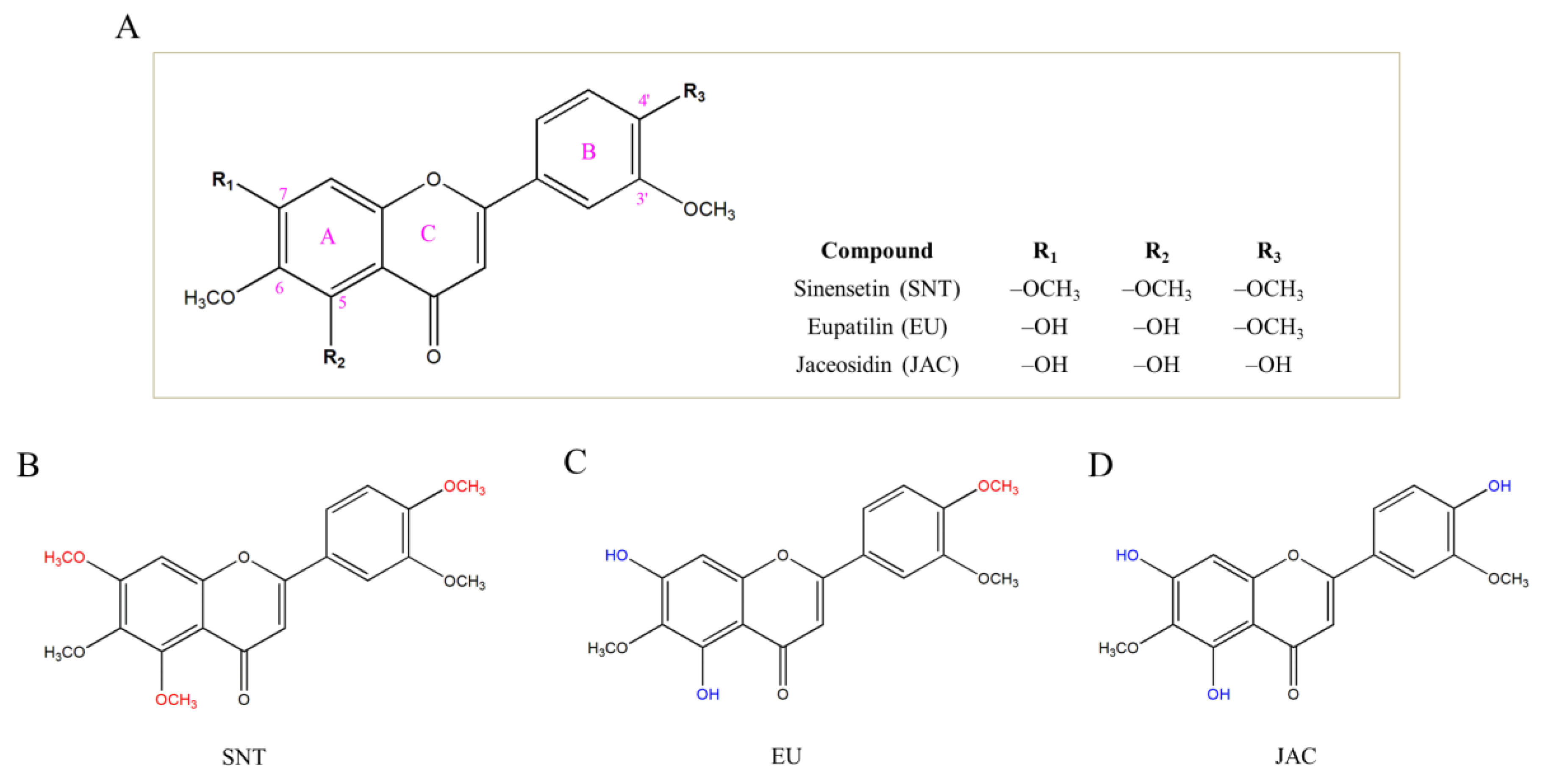
Effects of Sinensetin, Eupatilin, and Jaceosidin on Human Melanogenesis: A Pilot Study

1
Department of Biochemistry and Cell Biology, Stony Brook University, Stony Brook, NY 11794-5215, USA
2
Department of Biomedical Engineering, Stony Brook University, Stony Brook, NY 11794-5281, USA
Future Pharmacol. 2025, 5(1), 12; https://doi.org/10.3390/futurepharmacol5010012
Submission received: 14 January 2025 / Revised: 1 March 2025 / Accepted: 6 March 2025 / Published: 14 March 2025

Abstract

:
Background/Objectives: Flavones, a class of plant-based flavonoids, have demonstrated conflicting anti-melanogenic activities in mouse and human melanocytes. Sinensetin (SNT), a polymethoxyflavone, has shown pro-melanogenic activity in B16F10 mouse melanoma (MM) cells, while eupatilin (EU) and jaceosidin (JAC), two flavones that are structural analogs of SNT, have not been evaluated for their effects on melanogenesis yet. Methods: Herein, the effects of SNT, EU, and JAC on melanogenesis in MNT-1 cells (human melanoma) and HEMn-DP cells (primary human melanocytes) have been examined. The mushroom tyrosinase (TYR) activity was tested in cell-free conditions, followed by examination of the cytotoxicity of the compounds via the Alamar Blue (AB) assay. Cellular melanin production and TYR activity were estimated in MNT-1 cells. The compounds were further examined in primary human melanocytes for melanin production, TYR activity, and protein levels. Results: Our findings show that SNT was a potent inhibitor of TYR activity in a cell-free assay, while EU and JAC had no effect. However, both SNT and EU were shown to exhibit anti-melanogenic activity (that was reversible) in human cells, while JAC was ineffective and cytotoxic. Conclusions: SNT and EU are potential novel candidates for hyperpigmentation treatment without cytotoxicity. Additional studies are warranted to elucidate the signaling mechanisms that govern their anti-melanogenesis action. Future research is necessary to assess the anti-melanogenic effectiveness of SNT/EU using 3D skin tissue equivalents and to select the optimal candidate.

Graphical Abstract

1. Introduction

Melanin has a significant function in safeguarding the skin from the adverse impacts of ultraviolet radiation (UVR) [1]. This pigment is produced by melanocytes inside organelles known as melanosomes, which are subsequently released and transported to keratinocytes. While melanin serves an essential role in photoprotection for skin, excess production can result in the formation of pigmented areas, termed hyperpigmentation, causing conditions of lentigo senilis, melasma, chloasma, and post-inflammatory hyperpigmentation associated with esthetic displeasure and psychosocial implications [2,3]. In extreme cases, consistent UV radiation exposure may be linked to an increased vulnerability to malignant melanoma, a cancer affecting melanocytes [4]. Darkly pigmented skin has a higher likelihood of developing hyperpigmentation disorders in comparison to lightly pigmented skin [5]. Common skin depigmenting treatments, including kojic acid (KA), hydroquinone, and arbutin, have adverse effects such as contact allergy, genotoxicity, and carcinogenicity, resulting in restrictions on their use [6,7,8]. Tyrosinase (TYR) is a binuclear copper-containing enzyme that facilitates the two stages involved in melanin synthesis inside melanosomes. It catalyzes the conversion of tyrosine to 3,4-dihydroxyphenylalanine, DOPA (monophenolase activity), followed by the oxidation of DOPA to dopaquinone (diphenolase activity) [9]. Microphthalmia transcription factor (MITF) is another critical melanogenic protein that is the master regulator of transcription [10].
Recently, bioactives have been discovered to minimize hyperpigmentation without significant adverse effects. Because of their natural origin, safety, accessibility, and superior biocompatibility, plant-based and naturally occurring compounds for whitening and pigmentation spot elimination have garnered significant interest in recent years [11,12]. Due to their many bioactivities—antioxidant, anti-inflammatory, anticancer, metabolic regulating, immunoregulatory, neuroprotective, skin protecting, and so on—flavonoids have attracted growing interest. Flavones, a class of flavonoids, comprise two phenyl rings A and B, and a γ-pyrone heterocyclic ring C, making up the C6-C3-C6 carbon structure (Figure 1A). Polymethoxyflavones (PMFs), which are exclusively found in citrus peels and fruits, are characterized by four or more methoxy (-OCH3) groups on the benzo–pyrone skeleton [13]. Sinensetin (SNT), a PMF, is typically found in byproducts obtained from citrus processing, including the residues of orange oil [14] and peels of sweet orange Citrus sinensis in the Rutaceae family [15,16,17]. In addition, SNT is present in the leaves of Citrus aurantium (Rutaceae) [17] and leaves of the plant Orthosiphon stamineus (Lamiaceae) [18,19]. SNT enhanced adipogenesis in mouse preadipocytes, 3T3-L1 cells [20,21], inhibited angiogenesis [22] and exhibited anticancer activity [23] and anti-inflammatory activity [24,25]. Eupatilin (EU) and jaceosidin (JAC) are flavones that have been identified from medicinal plants belonging to the genus Artemisia [26,27]. These flavones have been shown to have beneficial pharmacological activities. EU is an active component of the marketed drug (Stillen®) that has been used for the treatment of gastrointestinal (GI) disorders [28]. EU has demonstrated a multitude of biological efficacies that include anti-inflammatory effects [29,30], antioxidant effects [31], anti-psoriatic effects [32], cardioprotective effects [33], neuroprotective effects [34,35], bone-loss mitigatory effects [36], and anticancer activity [37,38]. JAC has demonstrated antioxidant activity [39] and anti-inflammatory activity [39,40,41]. JAC also showed anticancer activity in glioblastoma cells [42], and inhibited NF-kB and nitric oxide in LPS-stimulated macrophages [39].
SNT, EU, and JAC flavones are structural analogs, and are formed by changes in substitution of the groups at the 4′, 5, and 7 positions of the rings B and A (Figure 1A). The structure of SNT (3′,4′,5,6,7-pentamethoxyflavone) consists of five methoxy groups, as shown in Figure 1B. EU (5,7-dihydroxy-3′,4′,6-trimethoxyflavone) has two hydroxy groups substituted at the 5 and 7 positions instead of methoxy compared to SNT (Figure 1C). Conversely, JAC (4′,5,7-trihydroxy-3′,6-dimethoxyflavone) is a further modification of EU where the methoxy group at the 4′ position is substituted by a hydroxy group (Figure 1D).
Numerous investigations into different flavonoids, such as quercetin, luteolin, and nobiletin, have exhibited contrasting impacts on melanogenesis in murine and human cells. For example, quercetin and luteolin diminished melanin production in B16F10 mouse melanoma (MM) cells, but stimulated melanin production in melanoma cells from humans [43,44,45,46]. Nobiletin (3′,4′,5,6,7,8-hexamethoxyflavone), a PMF and a structural analog of SNT, has shown stimulatory effects on melanogenesis in B16F10 cells [47], but suppressed melanin production in human melanocytes [48]. Currently, there are limited reports on the impact of SNT on melanogenesis. A prior report [49] indicated that an extract from orange peels, which included SNT and four additional PMFs, inhibited the activity of mushroom TYR. However, the authors did not analyze the individual components present in the extract. In another study, SNT treatment increased melanogenesis in B16F10 MM cells [50]. Whether SNT will similarly promote melanogenesis in human melanocytes remains to be determined. This is especially significant to investigate, as flavones are notorious for exhibiting contrasting effects on the process of melanogenesis in murine and human cellular models. A prior study demonstrated that a 70% ethanolic extract of the plant species Inula sarana (Asteraceae), which is abundant in flavonoids EU and JAC, along with the flavonoid cirsiliol, exhibited antioxidant and tyrosinase inhibitory properties [51]. Conversely, another study indicated that an extract from a Chrysanthemum cultivar containing EU, along with many other flavonoids and bioactives, exhibited low efficacy and correlation as a TYR inhibitor and antioxidant, despite demonstrating significant potency as a sunscreen agent [52]. However, an additional investigation assessing pure EU demonstrated its tyrosinase inhibitory and antioxidant properties [53]. To date, there have been no reports of TYR inhibition by purified JAC. Furthermore, the impact of EU and JAC, structural analogs of SNT, on melanogenesis in cell cultures remains unexplored. Hence, in this study, the three flavones (SNT, EU, and JAC) were initially examined separately for their ability to suppress TYR activity using mushroom TYR. Subsequently, they were evaluated for their effects on melanogenesis in cell cultures utilizing human melanoma cells (MNT-1 cells) and primary human melanocytes (HEMn-DP cells).

2. Materials and Methods

2.1. Materials

EU and JAC (99% purity) were purchased from Cayman Chemicals (Ann Arbor, MI, USA). SNT (99.22% purity) was procured from MedChem Express (Monmouth Junction, NJ, USA). Pyrocatechol violet (PV), copper sulfate, kojic acid, mushroom TYR, and the two substrates (L-DOPA and L-tyrosine) were procured from Sigma Aldrich (St. Louis, MO, USA). The Alamar Blue (AB) dye and bicinchoninic acid (BCA) assay were purchased from Thermo Fisher (Waltham, MA, USA). Penicillin–streptomycin antibiotic mixture, minimum essential medium (MEM), Gibco™ DMEM, Medium 254, TrypLE Express enzyme (1×), HBSS (Hyclone™), human melanocyte growth supplement (HMGS), and NaOH (1N) were acquired from Thermo Fisher. R&D Systems Inc. (Minneapolis, MN, USA) supplied heat-inactivated fetal bovine serum (HI-FBS). LSBio (Seattle, WA, USA) provided cell-based TYR and MITF enzyme-linked immunosorbent assay (ELISA) kits.

2.2. Cell-Free TYR Activity

The direct impacts of compounds on the first step of TYR enzyme activity (monophenolase activity) were assessed using a TYR enzyme (purified from mushroom Agaricus bisporus) with an L-tyrosine substrate. A total of 80 µL of the compounds were combined with 0.6 mM L-tyrosine in a 96-well plate. Next, 20 µL of mushroom TYR was introduced, resulting in a final enzyme concentration of 12.5 µg/mL in the wells. The maximum concentration of DMSO across all wells was 0.4%. After an incubation of 20 min, the 96-well plate was placed in a microplate reader (Versamax™, Molecular Devices, San Jose, CA, USA), and the reaction’s progress was recorded at 475 nm for 30 min. The slope’s linear range was used to determine the monophenolase activity and presented as a percentage of the control. The impact of the flavone compounds on the second step of TYR activity (diphenolase activity) were determined similarly to the method described in our earlier studies [54,55].

2.3. Pyrocatechol Violet (PV) Copper Chelation Assay

Pyrocatechol violet (PV) was used to assess copper-ion chelation activity, as described in our previous studies [56,57]. Briefly, varying SNT, EU, and JAC concentrations prepared in sodium acetate buffer were treated with copper sulfate solution in a 96-well plate and incubated with PV solution. Subsequently, the absorbance was determined at 632 nm and represented as a percentage of the control.

2.4. Cell Culture

MNT-1 cells (ATCC CRL-3450), ATCC, Manassas, VA, USA [58], were kindly donated by Dr. Michael Marks of the University of Pennsylvania in Philadelphia (PA, USA), and were cultivated using DMEM containing 1% MEM, 1% antibiotics, 18% HI-FBS, and 10% AIM-V medium (Invitrogen). HEMn-DP cells, which are primary human melanocytes isolated from the neonatal darkly pigmented foreskin (Cascade Biologics™, C2025C), were purchased from Thermo Fisher (Waltham, MA, USA) [59] and were cultured using medium 254 enriched with HMGS and antibiotics at a concentration of 1%. HaCaT human keratinocyte cell line (AddexBio #T0020001) was purchased from AddexBio, San Diego, CA, USA [60], and were cultivated in DMEM that contained 10% HI-FBS with 1% antibiotics. B16F10 mouse melanoma (MM) cells (ATCC CRL-6475) were procured from ATCC (Manassas, VA, USA) [61].

2.5. Cytotoxicity Assay

To examine the impacts of SNT, EU, and JAC on melanogenesis, the noncytotoxic concentrations of the three compounds were determined with the well-validated and highly sensitive resazurin-based AB cytotoxicity assay [62]. MNT-1 cells or HEMn-DP cells (both at 2 × 10⁴ cells per well) and HaCaT cells (1 × 10⁴ cells per well) were plated in 96-well plates and cultured for 24 h. After the 24 h interval, the flavones (SNT, EU, and JAC) were introduced into the cultures, ensuring a uniform final DMSO concentration of 0.4% throughout all groups, including the control group. Following a 3-day incubation period with the test compounds, the culture media was substituted with 100 μL of new media corresponding to the respective cells. AB (10 μL) was introduced and incubated for 2 h, after which the fluorescence readings was measured at excitation/emission wavelengths of 570/585 nm using a Gemini™ fluorescent microplate reader (Molecular Devices, San Jose, CA, USA). The assessment of cell viability was performed using the fluorescence readings compared to control and reported as a percentage.

2.6. Cellular Melanin Assay

MNT-1 cells (1.4 × 10⁵ cells in 1.5 mL medium per well) or HEMn-DP cells (1.5 × 10⁵ cells in 1.5 mL medium per well) were seeded in 12-well tissue culture plates and grown for 24 h. Subsequently, the medium was replaced with the flavones SNT (5–30 µM), EU (5–30 µM) or JAC (5 µM). KA at 1 mM concentration was employed as a positive control in the case of HEMn-DP cells. The cultures were kept in a CO2 incubator (Forma, Thermo Fisher Scientific) for 3 days. The treatments ended with trypsinization and PBS washing of cell pellets. To dissolve melanin, after aspiration, 250 microliters of sodium hydroxide (1N) was added, and the pellets underwent heating in a temperature-controlled water bath maintained at 70 °C. The absorbance of lysates (200 µL) was measured at 475 nm and was normalized to the total protein contents. This value was presented as a percentage of the control to denote the relative melanin contents across groups.

2.7. Cellular TYR Activity

MNT-1 cells (2 × 10⁵ cells in 2 mL medium per well) cultured in 6-well tissue culture plates were treated with the flavones SNT (5–30 µM), EU (5–30 µM), and JAC (5 µM) for 3 days. After this, the harvested cells were lysed, and 50 µL of the lysates were mixed with 100 µL of L-DOPA substrate solution (at a concentration of 6 mM) in a 96-well microplate and incubated for 20 min. Following this, the plate was read at 475 nm in a Versamax™ microplate reader for 45 min, with readings taken at 30 s intervals. TYR activity was estimated from the ratio of the linear slope of inhibitory velocities normalized to the total protein content.
For the HEMn-DP cells, a total of 0.72 × 10⁵ cells in 1.5 mL medium per well were grown in 12-well culture plates for a duration of 72 h, after which the medium was replaced with the three flavones SNT (5–30 µM), EU (5–30 µM), and positive control KA (1 mM), and the cell cultures were sustained for 3 days. Subsequently, the activities of TYR in the lysates of these cells were assessed using the methodology described in our prior study [63].

2.8. ELISA for TYR and MITF Protein

HEMn-DP cells were grown in a 96-well tissue culture plate and treated with compounds for 3 days. Subsequently, cells were processed based on the manufacturer’s instructions and the levels of TYR protein and levels of MITF protein were determined as per the ELISA kit instructions for both kits. The absorbance values were normalized to the staining of crystal violet stain as per the instructions, and relative levels of TYR and MITF protein were presented as a percentage of control.

2.9. Recovery Experiment for Melanogenesis

HEMn-DP cells (1 × 10⁵ cells/well inoculated in a six-well plate) underwent treatment with the compounds, SNT (30 µM) or EU (30 µM) for a duration of 3 days, referred to as the exposure phase, after which cultures were rinsed with HBSS and maintained for an additional 4 days in a compound-free medium, termed the recovery period. This was conducted to examine whether removing compounds from the culture medium reverses their effects on HEMn-DP cell melanogenesis. The relative melanin contents after the 3-day exposure period and 4-day recovery period were assayed using the NaOH lysis method outlined in Section 2.6.

2.10. Statistical Analysis

GraphPad Prism software version 10.4.0 (Boston, MA, USA) was used to analyze the data. Student’s t-test was used to compare two groups, while one-way analysis of variance (ANOVA) with Dunnett’s post hoc test was utilized for comparisons involving three or more groups. The differences were deemed statistically significant at a level of p < 0.05. The different levels of significance were shown as follows: * p < 0.05, ** p < 0.01, $ p < 0.001, and # p < 0.0001 vs. control. All data are reported as mean ± SD.

3. Results

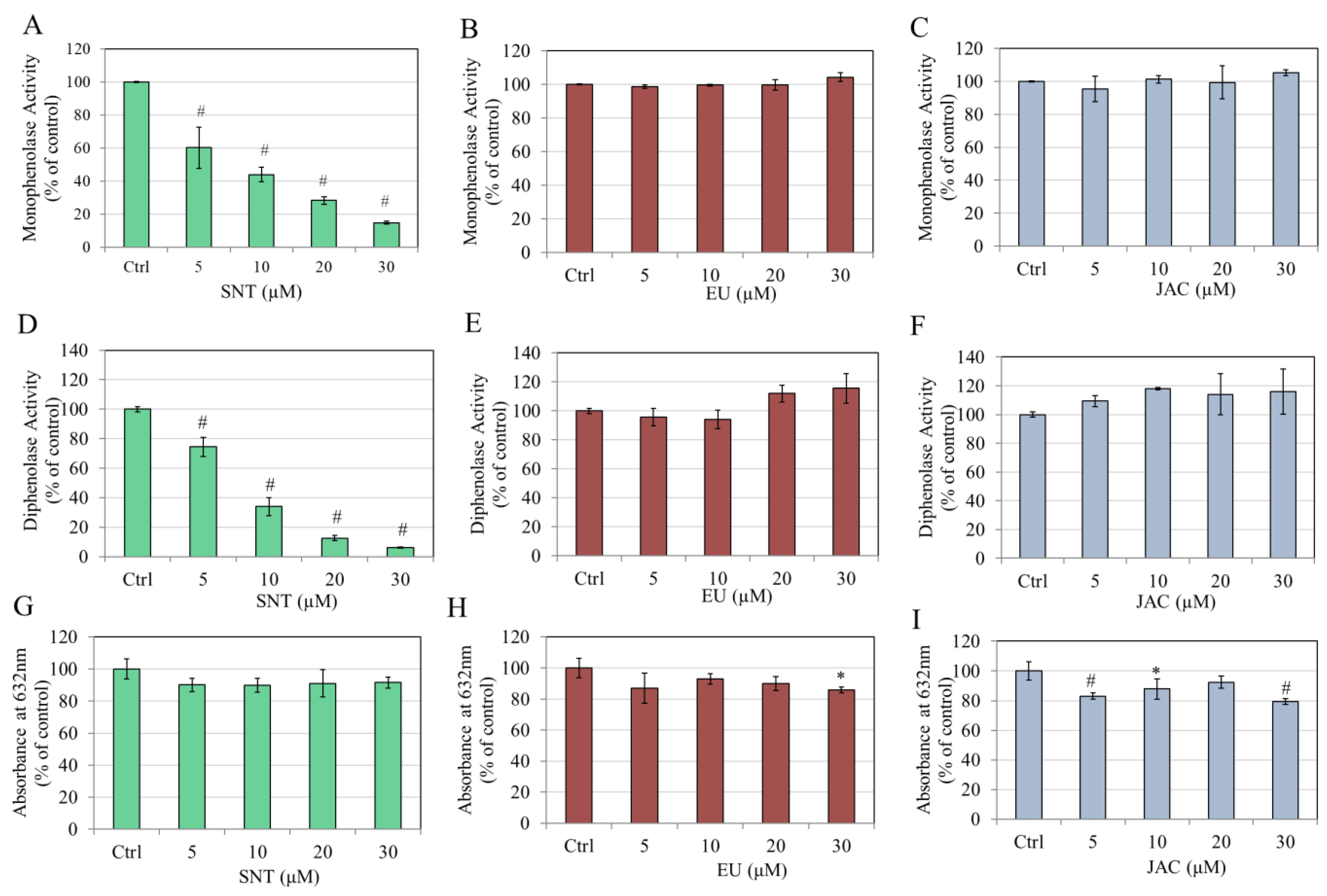
3.1. Direct Effects of Flavones on Mushroom TYR Activity and Copper Chelation

The three flavones (SNT, EU, and JAC) were initially evaluated in a cell-free system with a TYR enzyme to identify any direct effects of the flavones on TYR enzyme activity. For the same, low micromolar concentration ranges of 5–30 µM were selected for all three compounds. SNT showed a potent concentration-dependent inhibitory impact on the monophenolase activity of TYR, with significant inhibitions of 39.81%, 56.06%, 71.73%, and 85.13% at 5, 10, 20, and 30 µM concentrations, respectively (Figure 2A). However, neither EU (Figure 2B) nor JAC (Figure 2C) demonstrated any influence on the monophenolase activity compared to the control.
Next, the findings of diphenolase activity also revealed a robust concentration-dependent inhibition by SNT, with significant inhibitions of 25.55% at 5 µM, 66.15% at 10 µM, 87.44% at 20 µM, and 93.80% at 30 µM (Figure 2D). Conversely, EU (Figure 2E) and JAC (Figure 2F) showed no influence on the diphenolase activity at any concentration. The IC50 value for the monophenolase activity of SNT was estimated to be 7.35 μM, and the IC50 value for the diphenolase activity of SNT was 6.24 μM.
The effects of the three flavones were next evaluated on copper chelation ability. SNT did not show any copper chelation activity (Figure 2G). Unexpectedly, EU showed copper chelation at 30 µM by 14.08% (Figure 2H), whereas JAC showed copper chelation at 5, 10, and 30 µM by 16.94%, 12.12%, and 20.78%, respectively (Figure 2I).
Together, these results show that only the flavone SNT had a potent inhibitory effect on both steps of the TYR-catalyzed reactions in a cell-free system, and these effects do not involve copper chelation at the enzyme active site.

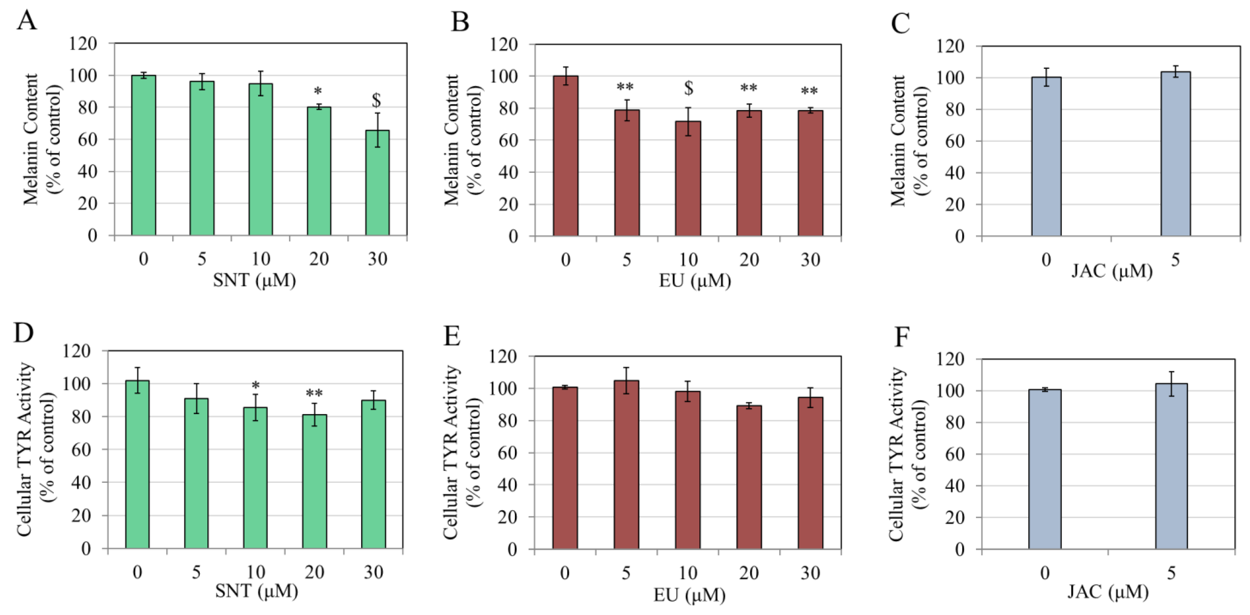
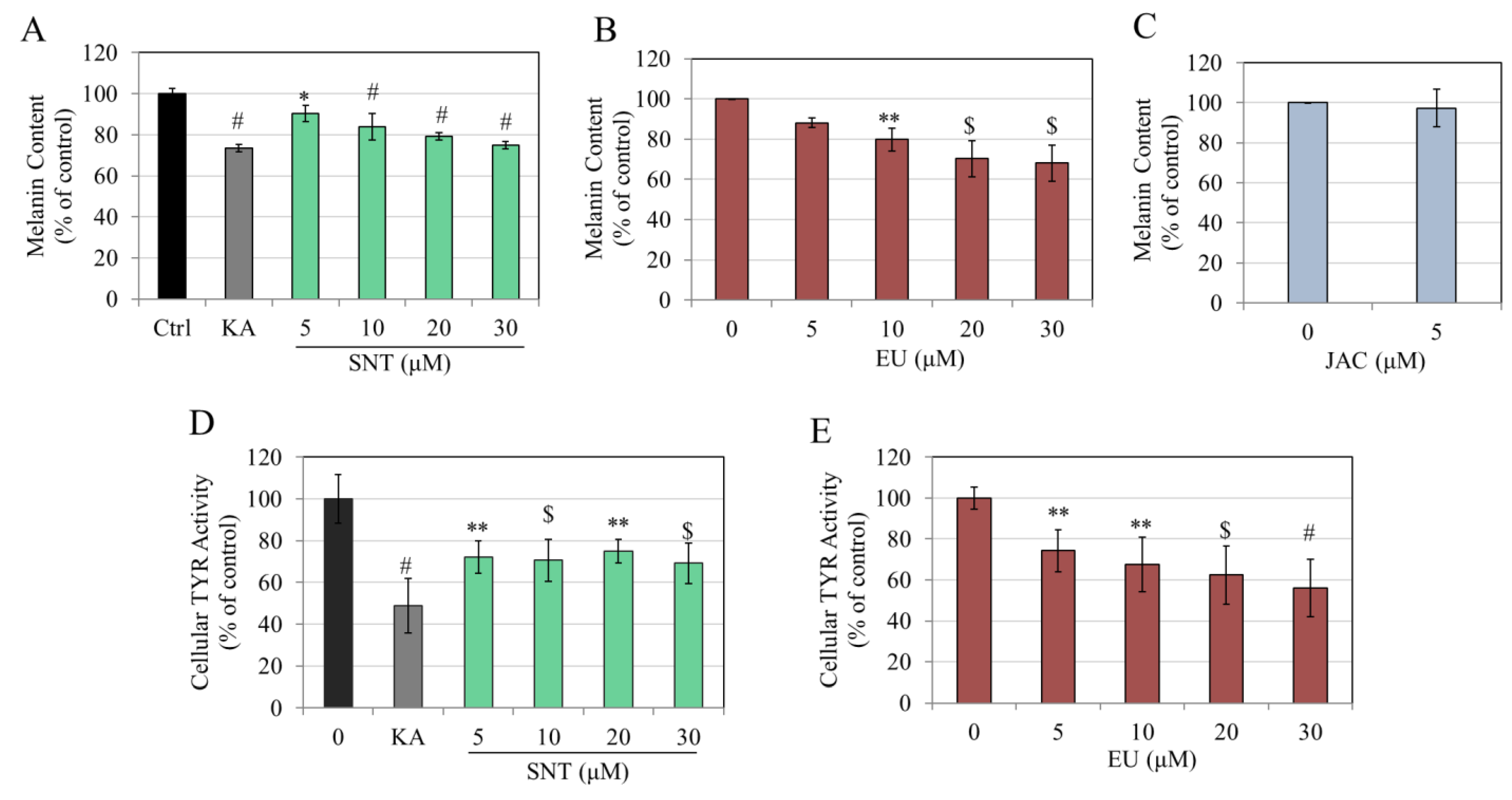
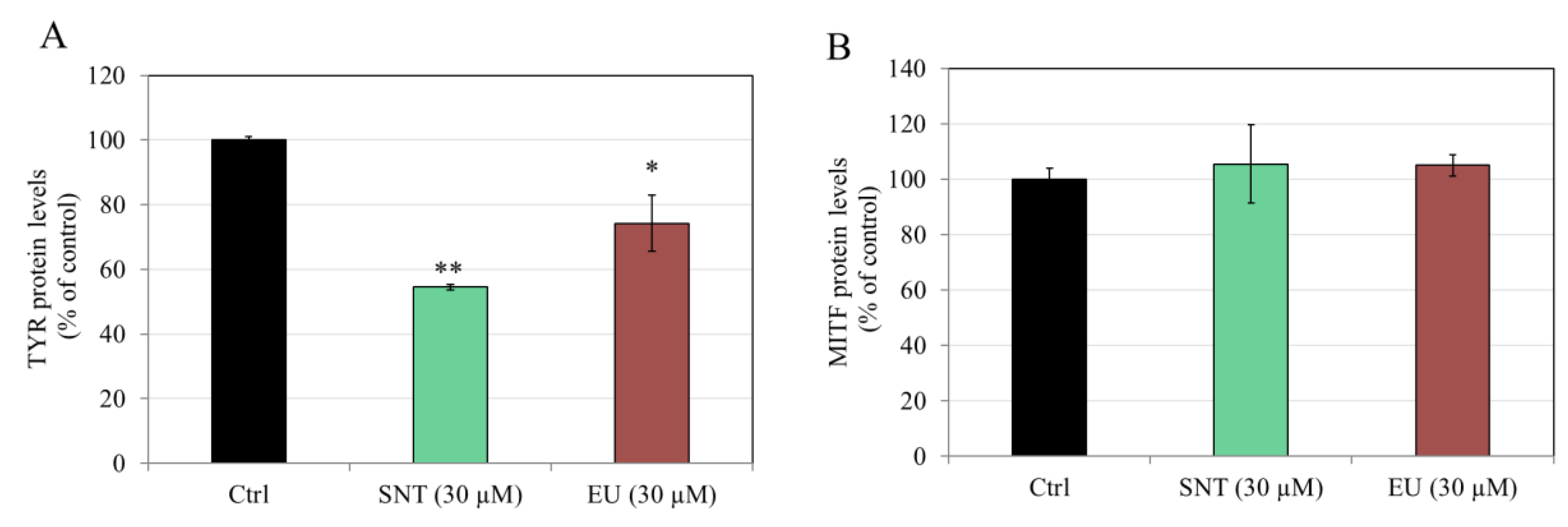
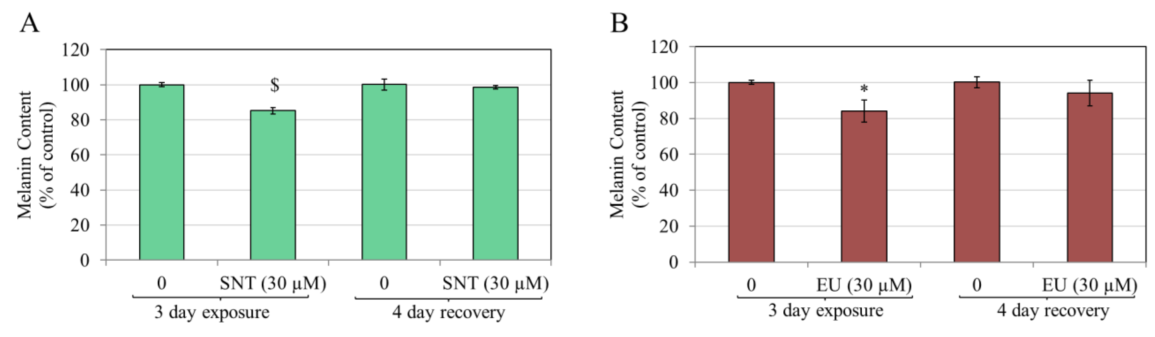
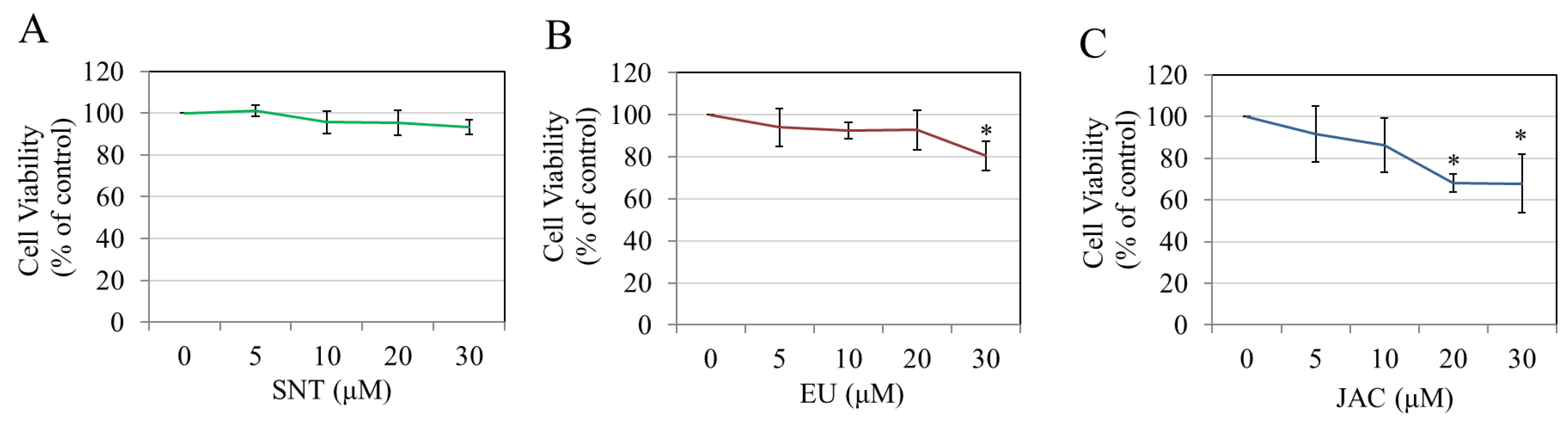
3.2. Effects of Flavones in MNT-1 Human Melanoma Cells

The three flavones (SNT, EU, and JAC) were assessed for cytotoxicity to MNT-1 cells through the AB assay to identify noncytotoxic concentrations for subsequent testing on melanogenesis. The results showed that both SNT (Figure 3A) and EU (Figure 3B) showed no signs of cytotoxicity across all the concentrations, while JAC significantly lowered cell viability by 13.52% at 10 µM, 13.60% at 20 µM, and 21.41% at 30 µM (Figure 3C). Accordingly, SNT and EU were selected at concentrations between 5 and 30 µM, while JAC was selected at 5 µM for further experiments in MNT-1 cells.
SNT treatment significantly lowered the melanin content of MNT-1 cells by 19.70% at 20 μM and 34.25% at 30 μM (Figure 4A). EU treatment led to a significant lowering of melanin content by 21.21% at 5 µM, 28.41% at 10 µM, 21.51% at 20 µM, and 21.48% at 30 µM, although there were no concentration-dependent effects (Figure 4B). Treatment with 5 µM JAC had no effect on melanin production (Figure 4C).
SNT led to a significant inhibition of the TYR activity of MNT-1 cells by 16.51% at 10 μM and by 20.87% at 20 μM, although there was no significant effect at the concentration of 30 μM (Figure 4D). Conversely, EU (Figure 4E) and JAC (Figure 4F) did not show any influence on the cellular TYR activity.
These results suggest that EU has superior anti-melanogenic activity compared to SNT in MNT-1 cells, while JAC was ineffective. Furthermore, the anti-melanogenic action of SNT, as opposed to EU, is associated with suppressing TYR enzyme activity.

3.3. Effects of Flavones in Darkly Pigmented Primary Human Melanocytes (HEMn-DP Cells)

There was no significant effects from either SNT (Figure 5A) or EU (Figure 5B) on the viability of the HEMn-DP cells. However, JAC induced cytotoxicity at concentrations of 10, 20, and 30 µM, with significantly lowered residual viabilities of 73.85%, 79%, and 56.23%, respectively (Figure 5C).
The data of HEMn-DP cells’ melanin content revealed that treatment with SNT significantly suppressed melanin production across all concentrations, with the effect being concentration-dependent. Specifically, SNT significantly decreased melanin content by 9.77% at 5 µM, 16.38% at 10 µM, 20.94% at 20 µM, and 25.25% at 30 µM (Figure 6A). The positive control KA demonstrated a decrease in melanin content by 26.67% at a concentration of 1 mM, suggesting that SNT exhibited greater potency compared to KA (Figure 6A). Next, the treatment of HEMn-DP cells with EU revealed a significant suppression of melanin production; specifically, EU at concentrations of 10, 20, and 30 µM significantly decreased melanin contents by 20.31%, 29.89%, and 32.07%, respectively (Figure 6B). Conversely, the treatment of HEMn-DP cells with JAC at 5 μM exhibited no variation in melanin production in comparison to the control (Figure 6C).
The effects of SNT and EU were next examined on the TYR activities of HEMn-DP cells to identify if their anti-melanogenic effect might be linked to the inhibition of TYR activity. JAC was excluded as it did not show any impact on melanin production. The results revealed that SNT significantly inhibited intracellular TYR activity by 27.82% at 5 μM, 29.41% at 10 μM, 25.09% at 20 μM, and 30.68% at 30 μM (Figure 6D). Kojic acid (KA) showed suppression of 51.24% at the concentration of 1 mM (Figure 6D). Next, the treatment of HEMn-DP cells with EU also inhibited the cellular TYR activity in a concentration-dependent manner, with significant suppressions of 25.74%, 32.49%, 37.55%, and 43.97% at 5, 10, 20, and 30 μM concentration, respectively (Figure 6E).
The effects of the highest concentrations of SNT and EU (30 µM) on the TYR and MITF protein amounts were also evaluated. Treatment with SNT (30 µM) significantly attenuated the TYR protein levels of HEMn-DP cells by 45.55%, whereas treatment with EU (30 µM) also led to the significant attenuation of the TYR protein by 25.78% (Figure 7A). Neither SNT nor EU affected the amounts of MITF protein (Figure 7B).
We next examined whether the melanogenesis-suppressing effects of SNT and EU treatment in HEMn-DP cells were reversible. For this, HEMn-DP cells were treated with the highest concentrations (30 µM) of SNT or EU for 3 days, and then separate cultures continued for additional 4 days in flavone-free medium, denoted by the recovery period. The results for SNT showed that the significantly diminished melanin content in HEMn-DP cells completely recovered after the 4-day recovery period (Figure 8A). Similarly, a complete recovery of melanin levels was obtained for EU (Figure 8B). Collectively, these findings indicate that both SNT and EU suppress melanin content, which is completely restored with discontinuation of treatment.

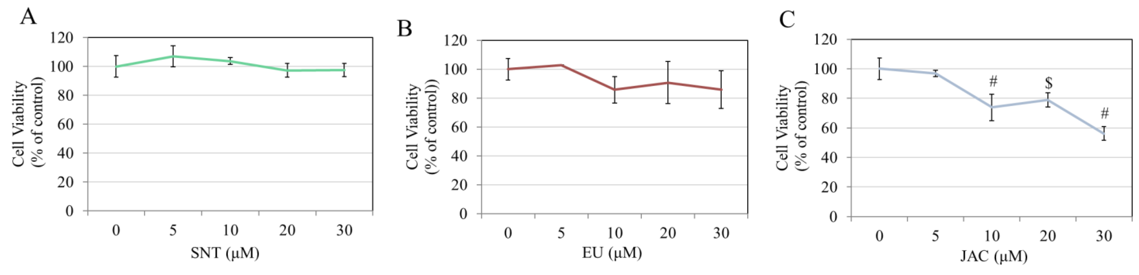
3.4. Effects of Flavones on Keratinocyte Viability

SNT was noncytotoxic to human keratinocytes within the concentrations of 5–30 µM (Figure 9A). However, EU significantly lowered cell viability by 19.56% at 30 µM (Figure 9B). Conversely, JAC induced high cytotoxicity, with viability significantly lowered by 31.96% and 32.10% at 20 and 30 µM concentrations, respectively (Figure 9C). These data suggest that both SNT and EU are noncytotoxic to keratinocytes at the concentrations at which they inhibited melanin production, indicative of their safety for topical use.

4. Discussion

Identifying efficient treatments for skin hyperpigmentation derived from natural products provides enhanced safety and tends to have fewer toxicities and side effects compared to synthetic chemical alternatives. Flavonoids are popular natural molecules for controlling pigmentation disorders because of their beneficial biological activities and minimal toxicity. Nonetheless, various flavonoids demonstrate conflicting impacts on melanin production in both mouse and human cells [43,44,45,47,48,64]. To date, there is only one study [50] that has shown that SNT robustly increased melanin production in B16F10 cells, while EU and JAC have not yet been examined in B16F10 cells. We evaluated the three flavones on B16F10 cells melanogenesis as a preliminary experiment (Supplementary Materials). SNT (Figure S1A) and EU (Figure S1B) showed no alterations in B16F10 cell viability across 5–30 µM, whereas interestingly, JAC only significantly diminished B16F10 cell viability at the maximum concentration of 30 µM (Figure S1C). The results of B16F10 cell melanin production showed that SNT robustly enhanced melanin production at concentrations of 5, 10, 20, and 30 µM by 60.14%, 78.60%, 92.99%, and 76.04%, respectively (Figure S2A). EU enhanced melanin production at 5, 10, 20, and 30 µM concentrations by 22.06%, 28.29%, 48.28%, and 92.13%, respectively (Figure S2B). Conversely, JAC did not exhibit any impact at 5 or 10 µM, but enhanced melanin production by 18.88% at 20 µM (Figure S2C). The order of melanogenesis stimulation in B16F10 cells by the three flavones was SNT > EU > JAC. Our results from B16F10 cells show that SNT and EU significantly enhanced melanin production at 5 and 10 µM, whereas JAC exhibited no effect at those concentrations. It should be noted that both SNT and EU contain a 4′-methoxy group on their B-ring, which is absent in JAC, which contains a hydroxyl group instead. Our results of increased melanin production by the flavones in B16F10 cells are consistent with previous findings that have documented that the 4′-methoxy group on the B-ring of flavonoids results in the stimulation of melanogenesis in a B16F10 cell model, even when various substituents are present on other positions of the ring [65,66]. Moreover, our current study demonstrates that SNT and EU exhibit anti-melanogenic capacity in human melanoma cells (MNT-1) and normal human melanocytes (HEMn-DP), which further corroborates that flavones display opposite effects on murine cell melanogenesis in comparison to human cell melanogenesis.
In our study, SNT suppressed the mushroom TYR enzymatic activity, but SNT, EU, and JAC enhanced melanogenesis to varying degrees in B16F10 cells. In contrast, in human melanoma cells (MNT-1) and normal melanocytes, they suppressed melanogenesis. A similar result was observed in another study, where increased melanogenesis was observed in B16F10 cells, while it was inhibited in MNT-1 cells after treatment with citric acid [67]. Moreover, this conflicting effect in mouse B16F10 cells and normal human melanocytes is not surprising, given that, closely related PMFs, such as nobiletin (formed from SNT by the addition of one methoxy group at 8 position of ring A) and tangeretin (formed from nobiletin by the removal of one methoxy group at 3′ position of ring B), have also exhibited a similar discrepancy in their effects. For example, nobiletin suppressed mushroom TYR activity [68,69], but in B16F10 cells, it potently stimulated melanogenesis with an increase of 200% at 20 µM after a 2-day treatment [47]. However, in HM3KO human melanoma cells [70] and primary melanocytes, nobiletin behaved as an anti-melanogenic candidate [48]. Similarly, tangeretin increased melanogenesis by ~100% at 25 µM in B16F10 cells [71], but it suppressed melanin production in human melanoma cells [70]. The IC50 values for SNT’s monophenolase and diphenolase activities were 7.35 and 6.24 μM, respectively. KA, a known skin-whitening compound, inhibits TYR activity via copper chelation. Under similar experimental conditions, KA shows IC50 of about 70.87 μM for diphenolase activity [54], which shows that SNT has an 11.35-fold lower IC50 than KA. Moreover, KA at 500 µM shows 64.60% copper chelation [54], while SNT does not show copper chelation activity. Thus, the mode of inhibition of SNT differs from KA. TYR is the principal rate-limiting enzyme in the melanogenesis cascade whose active site comprises two atoms of copper; hence, substances capable of chelating copper may suppress TYR activity and offer an attractive target for depigmentation [72].
Due to the increased number of methoxy groups, PMFs exhibit greater hydrophobicity compared to hydroxylated flavones [73]; this characteristic facilitates their ability to penetrate the subcellular membranes of cells, leading to potential alterations. Nobiletin differs from SNT solely by the presence of an additional methoxy group at the 8-position of ring A. Our findings of SNT reflect those observed with nobiletin, which has demonstrated similarly opposing effects on melanogenesis. While nobiletin was not included in our experiments since the aim was not to compare SNT with nobiletin, a previous study [68] indicated that the IC50 values for diphenolase activity of mushroom TYR for nobiletin and KA, a recognized TYR inhibitor were 46.2 µM and 77.4 µM, respectively. Although the experimental method differed from our method, it is reasonable to compare the IC50 values of nobiletin and SNT in our current investigation since the IC50 values of KA reported in the previous study [68] match rather closely with those derived from our experimental approach (KA IC50: 70.87 μM for diphenolase activity [54]). Our experiments demonstrate that SNT exhibited a 7.4-fold greater potency as an inhibitor of diphenolase activity, with an IC50 value of 6.24 µM, compared to nobiletin. Moreover, a prior study [22] that evaluated SNT and nobiletin in a zebrafish model showed that the presence of the 8-methoxy group, as seen in nobiletin, induced high systemic toxicity to zebrafish in comparison to SNT. KA (1 mM) was employed as a positive control in the cellular tyrosinase activity assay, based on our previous research [63]. KA was utilized as a positive control for our assays at a 1 mM concentration, which is not comparable to the low micromolar concentrations of flavones (5–30 µM) employed in the assay. Nevertheless, our objective was not to evaluate the anti-melanogenic efficacy of flavones in direct comparison to equivalent concentrations of KA. Previous reports substantiate this observation, indicating that higher concentrations of KA are generally required to achieve a significant decrease in cellular melanin content [74,75,76]. For example, in a prior study [75], it was observed that KA at higher concentrations (175–350 µM) decreased B16F10 cell melanin content by ~20% following a 2-day treatment. In human MNT-1 cells, it was observed that KA at a concentration of 100 µg/mL (704 µM) did not result in any decrease in melanin content following a 2-day treatment [74]. Conversely, another study [76] involving primary human melanocytes revealed that while KA at 10 µM exhibited no alterations, a concentration of 100 µM resulted in a modest, yet statistically significant, decrease in ~11% in cellular melanin content, accompanied by a ~14% inhibition of cell tyrosinase activity after a 5-day treatment.
Our results indicating the absence of inhibition on mushroom TYR activity by EU contradict a previous study [53] that demonstrated EU’s suppression of the diphenolase activity of mushroom TYR. Our findings are somewhat analogous to another study [52], which indicated that an extract from the cultivar of Chrysanthemum morifolium Ramat. (Asteraceae) containing EU and other bioactive compounds demonstrated limited activity and correlation as a TYR inhibitor. No reports exist on the impact of pure JAC on TYR activity; however, a previous study [51] indicated that an extract from the plant species Inula sarana (Asteraceae), which included JAC among other chemicals, suppressed mushroom TYR activity. These discrepancies may be attributed to the utilization of pure compounds vs. mixtures and variations in the experimental settings. Our results indicate that SNT showed a potent concentration-dependent suppression of TYR activity in a direct system, but did not show concentration-dependent suppression of TYR activity in MNT-1 cells, where concentrations of 5 and 30 µM showed no change. However, SNT suppressed TYR activity in HEMn-DP cells at all concentrations (5–30 µM). EU failed to show TYR inhibition directly in a cell-free system and MNT-1 cells. However, EU showed a potent effect in suppressing TYR activity in HEMn-DP cells. The divergent effects seen in MNT-1 cells and HEMn-DP cells could be linked to the inherent differences in them since MNT-1 are immortalized and derived from a malignant melanoma of an adult, while HEMn-DP are primary melanocytes derived from a neonate with no malignancy. The TYR enzyme present in humans shows differences when compared to those identified in mushrooms [77,78]. Consequently, the activity of TYR assessed through TYR obtained from purified mushrooms may vary from that of mammalian TYRs. The robust inhibition of mushroom TYR by SNT, contrasted with the absence of similar effects from EU, may be attributed to their structural differences. Given that SNT possesses methoxy groups at the 5 and 7 positions, unlike EU, which contains hydroxy groups at those locations, it is plausible that this structural difference may facilitate SNT’s binding with the TYR enzyme’s active site. The methoxy group’s electron-donating nature may affect the distribution of electrons throughout the molecule, potentially enhancing its interaction in contrast to EU [79]. The current study did not focus on expanding the analysis utilizing docking studies. To investigate the molecular interactions of the TYR enzyme with SNT/EU in greater depth, it may be essential to conduct additional studies incorporating molecular docking [80].
Our results demonstrate that of the three flavones, only SNT and EU show a promising capacity to suppress increased melanin production, while JAC is ineffective owing to the cytotoxic effects across all cells studied. The pronounced cytotoxic effects of JAC observed in our study align with the existing literature, which indicates that JAC exhibits cytotoxicity across various cell types, including cells from breast carcinoma [81], ovarian carcinoma [82], and twelve variants of gastric carcinoma [83]. A comparison of the structure–activity relationship (SAR) of flavones suggests that the 4′-OH group (on the B-ring) is responsible for the heightened cytotoxicity and antiproliferative effects. The analysis of EU and JAC, which vary solely by the substitution of one -OCH3 group in EU with a hydroxy group (resulting in EU being more lipophilic than JAC), reveals that JAC exhibits significant cytotoxicity, whereas EU demonstrates strong efficacy without any associated cytotoxicity. Flavones have demonstrated the capacity to induce anti-proliferative effects in various cells through their ability to modify membrane rigidity via interactions with cellular lipids [84,85]. The variation in lipophilicity may lead to distinct alterations in cell membrane organization and fluidity and alter signaling pathways that are crucial for growth, proliferation, and apoptosis in cells. Flavonoids with -OH groups tend to be more cytotoxic than the methoxy derivatives [86]. Moreover, a prior investigation [87] indicated that -OH groups at 3′ and 4′ positions (in B-ring) of flavonoids (like quercetin and luteolin) led to an enhanced anti-proliferative effect. In our study, JAC features an -OH group at the 4′ position, whereas SNT and EU possess -OCH3 group at the 4′ position. Consequently, the increased cytotoxicity of JAC aligns with the findings of the abovementioned studies. JAC failed to inhibit tyrosinase activity in cell-free assay but exhibited copper chelation; however, the response was non-linear. The precise rationale for this non-linear impact is unknown and was not investigated since JAC did not show promise as an anti-melanogenic candidate.
Our findings indicate that SNT and EU displayed anti-melanogenic properties, but JAC displayed significant cytotoxicity without any anti-melanogenic effects. Thus, the SAR demonstrates that the 4′-OCH3 group of ring B is essential for anti-melanogenic action in human cells. The substitution of this group with a -OH group leads to the cessation of anti-melanogenic action and induces significant cytotoxicity. A previous study [88] indicated that eupafolin, a flavone that is the subsequent analog to JAC (differing solely by the substitution of the 3′-OCH3 group in ring B with an -OH group), suppressed melanin production in B16F10 cells without inducing cytotoxicity; however, it remains uncertain whether it can similarly suppress melanogenesis in human melanocytes. Consequently, based on our findings regarding JAC and the previous study’s results on eupafolin [88], it can be further inferred that the 4′-OH group of ring B contributes to cytotoxicity and the absence of anti-melanogenic efficacy, whereas substituting the 3′-OCH3 group of ring B with a -OH group could decrease cytotoxicity and reinstate anti-melanogenic properties. However, it is important to acknowledge that our work was limited to three flavone analogs; to better understand SARs, it may be essential to include a diverse library of flavone analogs with various substitutions and assess their effects on human melanocytes.
Similarly to EU, SNT also suppressed melanin production, but appears to show a distinct mechanism from EU as it can also act directly on the first and second steps of TYR-catalyzed reactions. However, our findings showed that SNT and EU could downregulate TYR protein levels in HEMn-DP cells, although SNT had a higher capacity. Interestingly, neither SNT nor EU affected the levels of MITF, another critical melanogenic protein that is the master regulator of transcription [10]. The role of maturation of TYR by glycosylation in the melanin synthesis cascade has been established [89,90,91]. Hence, compounds that impede glycosylation by inhibiting the activity of α-glucosidase can also suppress melanin production [92]. Previous studies have reported the α-glucosidase inhibitory activity of both SNT [93,94] and EU [95]. Therefore, it is conceivable that SNT and EU inhibit melanogenesis, in part, through their effect on α-glucosidase enzyme activity; however, additional research is required to substantiate this idea. Both SNT and EU have been shown to demonstrate antioxidant activities [53,96]. As compounds can show efficacy as melanogenesis inhibitors by their capacity to scavenge cellular reactive oxygen species (ROS) [97], we also evaluated if the flavones SNT or EU might decrease ROS generation in HEMn-DP cells since they suppressed melanogenesis. However, neither SNT (Figure S3A) nor EU (Figure S3B) showed any effect on cellular ROS production.
The mitogen-activated protein kinase (MAPK) pathway is essential in melanin production, mainly by activating the P38, ERK, and Akt pathways [98]. In a previous study [88], the flavone eupafolin exhibited the capacity to activate the p38 and ERK pathways while simultaneously inhibiting the Akt pathway. This was demonstrated by an increased p38 MAPK and ERK1/2 phosphorylation and decreased Akt phosphorylation. In another study [99], the suppressive effects of a natural compound, gomisin N, on melanogenesis were correlated with the activation of the P13K/Akt and MAPK/ERK pathways, as it enhanced the phosphorylation of both Akt and ERK. Although our study did not examine the signaling pathways that could play a role in the anti-melanogenic effects of SNT and EU, additional research is necessary to elucidate the possible roles of the ERK, Akt, and p38 pathways. The induction of autophagy has been identified as a mechanism through which certain compounds can suppress cellular melanin levels [100,101,102,103]. It is noteworthy that EU has demonstrated the ability to activate autophagy and increase the expression of autophagy markers across various cell types [104,105]. Furthermore, SNT has shown the ability to induce autophagy in a range of cell types in earlier studies [106,107]. We speculate that the suppression of cellular melanin levels by SNT and EU could be associated, at least in part, to the capacity of these flavones to break down pre-existing melanin through the activation of autophagy. This hypothesis is partially supported by our findings (Figure 8), which indicate a lower efficacy in the suppression of cellular melanin by SNT and EU in HEMn-DP cells in exposure–recovery experiments. In these experiments, the cell density employed was lower compared to that in the 3-day treatment studies, ensuring that cells did not reach over-confluence during the experiments while maintaining viability throughout the recovery period of extended cultures. This indicates that in melanocytes grown at high density (which may not be producing new melanin), SNT and EU could be more effective at breaking down existing melanin as compared to suppressing the synthesis of new melanin. In the context of exposure–recovery experiments, the introduction of compounds into the cultures during the 3-day incubation period may yield limited efficacy in suppressing melanin content. This is due to the fact that some cells may be in the division phase, while others could be synthesizing increased amounts of melanin. Further investigations into the induction of autophagy and the testing of protein markers associated with this process may provide valuable insights into this hypothesis.
SNT and EU have shown efficacy as anticancer agents for human melanoma in previous studies [108,109,110]. For example, SNT exhibited antiproliferative effects in SK-MEL5 human melanoma cells [110]. EU showed anticancer activity in A375 human melanoma cells [108,109], although no research has been conducted on its impact on melanogenesis yet. Interestingly, compounds that inhibit the melanogenesis of melanoma cells make them more sensitive to the therapeutic approaches of immunotherapy, chemotherapy, and radiation [111,112,113]. Moreover, radiation employed in cancer therapy has also been demonstrated to cause hyperpigmentation [114]. In light of the fact that SNT and EU demonstrated the ability to diminish melanin synthesis in MNT-1 human melanoma cells, our findings suggest that these two compounds also have the potential to be used as adjuvants in anti-melanoma pharmacotherapy.
The reversibility of topical skin-lightening products is essential from a safety perspective. In a previous study [115], deoxyarbutin, a compound that is a TYR inhibitor, exhibited skin-lightening effects in an in vivo model of a hairless pigmented guinea pig. Notably, this effect was entirely reversible within eight weeks following the cessation of topical use. Our results, albeit obtained from in vitro cell cultures, demonstrate that HEMn-DP cells exhibited diminished melanin production following treatment with SNT or EU and that, upon removing these compounds, cellular melanin levels fully returned to baseline, suggesting safety. Additionally, both SNT and EU showed no cytotoxic effects for human keratinocytes, which demonstrates the safety of topical care use in cosmetics without any irreversible changes in melanocyte pigment production levels. Our findings show promise for the use of purified bioactives SNT and EU for the pharmacological management of hyperpigmentation. Interestingly, both compounds have previously shown ease of formulation with several topical carriers. For example, EU has been used as a topical formulation in in vivo studies in mice, where it showed efficacy in the treatment of dermatitis-like skin lesions [116] and skin psoriasis [30]. SNT has been delivered through the skin at a 38% amount in an extract of Orthosiphon stamineus (Lamiaceae) plant-based nano-formulation [117]. In our current investigation, SNT and EU compounds demonstrated a comparable and significant inhibition of melanogenesis despite their differing effects on the TYR enzyme in a cell-free system. Future investigations are necessary to assess the effectiveness of SNT and EU utilizing a 3D skin tissue equivalent, Melanoderm™ [118], to ascertain which of these two compounds exhibits superior anti-melanogenic properties.

5. Conclusions

The results of this investigation provide proof-of-principle that of the three flavones, SNT and EU show promise in suppressing human cell melanin production without cytotoxicity. Our findings indicate that the anti-melanogenic mechanism of SNT and EU in primary human melanocytes was partially associated with the suppression of TYR enzyme activity and protein levels, with SNT also demonstrating a direct inhibitory impact on the TYR enzyme, as shown by cell-free assays. Furthermore, the anti-melanogenic effects of both SNT and EU were reversible in primary human melanocytes. In contrast, the flavone JAC was ineffective, owing to high cytotoxicity. Therefore, SNT and EU are potential therapeutic compounds for treating disorders related to hyperpigmentation in humans. Future investigations utilizing 3D skin tissues or in vivo studies will be essential to determine the anti-melanogenic effectiveness of SNT and EU and identify the most suitable candidate. Moreover, further studies delineating the signaling mechanisms behind the anti-melanogenic effect of these flavones are warranted.

Supplementary Materials

The following supporting information can be downloaded at: https://www.mdpi.com/article/10.3390/futurepharmacol5010012/s1, Figure S1: Viability of B16F10 cells after treatment with varying concentrations of (A) SNT, (B) EU, and (C) JAC for a duration of 72 h; # p < 0.0001 vs. untreated control; data are mean ± SD of quadruplicate determinations from one experiment (n = 4 per group); Figure S2: Relative melanin contents of B16F10 cells after treatment with varying concentrations of (A) SNT, (B) EU, and (C) JAC for a duration of 72 h; * p < 0.05, ** p < 0.01, $ p < 0.001, and # p < 0.0001 vs. untreated control; data are mean ± SD of triplicate determinations (n = 3 per group); Figure S3: DCF fluorescence of HEMn-DP cells after treatment with varying concentrations of (A) SNT, and (B) EU for a duration of 72 h; data are mean ± SD (n = 3–4 per group). Refs. [119,120] were cited in Supplementary Materials.

Funding

This research was funded in part, by the Research Foundation of the State University of New York (85184–1155067).

Institutional Review Board Statement

Not applicable.

Informed Consent Statement

Not applicable.

Data Availability Statement

The raw data supporting the conclusions of this article will be made available by the author on request.

Acknowledgments

The author acknowledges Michael S. Marks (University of Pennsylvania, Philadelphia, PA, USA) for their kind gift of MNT-1 cells and Sanford R. Simon (Department of Biochemistry and Cell Biology, Stony Brook University, NY, USA) for the use of resources.

Conflicts of Interest

The author declares no conflicts of interest.

References

  1. Brenner, M.; Hearing, V.J. The protective role of melanin against UV damage in human skin. Photochem. Photobiol. 2008, 84, 539–549. [Google Scholar] [CrossRef] [PubMed]
  2. Rigopoulos, D.; Gregoriou, S.; Katsambas, A. Hyperpigmentation and melasma. J. Cosmet. Dermatol. 2007, 6, 195–202. [Google Scholar] [CrossRef] [PubMed]
  3. Grimes, P.E. Disorders of pigmentation: Global issues of major cosmetic concern. West. J. Med. 1998, 169, 226–227. [Google Scholar]
  4. Kanavy, H.E.; Gerstenblith, M.R. Ultraviolet radiation and melanoma. Semin. Cutan. Med. Surg. 2011, 30, 222–228. [Google Scholar] [CrossRef]
  5. Vashi, N.A.; Wirya, S.A.; Inyang, M.; Kundu, R.V. Facial hyperpigmentation in skin of color: Special considerations and treatment. Am. J. Clin. Dermatol. 2017, 18, 215–230. [Google Scholar] [CrossRef]
  6. Gaskell, M.; McLuckie, K.I.; Farmer, P.B. Genotoxicity of the benzene metabolites para-benzoquinone and hydroquinone. Chem.-Biol. Interact. 2005, 153, 267–270. [Google Scholar] [CrossRef]
  7. Hirose, M.; Imai, T.; Mitsumori, K. Carcinogenicity of kojic acid in rodents. JSM Mycotoxins 2004, 2003, 59–67. [Google Scholar] [CrossRef]
  8. Nakagawa, M.; Kawai, K.; Kawai, K. Contact allergy to kojic acid in skin care products. Contact Dermat. 1995, 32, 9–13. [Google Scholar] [CrossRef]
  9. Bae-Harboe, Y.-S.C.; Park, H.-Y. Tyrosinase: A central regulatory protein for cutaneous pigmentation. J. Investig. Dermatol. 2012, 132, 2678–2680. [Google Scholar] [CrossRef]
  10. Vachtenheim, J.; Borovanský, J. “Transcription physiology” of pigment formation in melanocytes: Central role of MITF. Exp. Dermatol. 2010, 19, 617–627. [Google Scholar] [CrossRef]
  11. Peng, X.; Ma, Y.; Yan, C.; Wei, X.; Zhang, L.; Jiang, H.; Ma, Y.; Zhang, S.; Xing, M.; Gao, Y. Mechanism, formulation, and efficacy evaluation of natural products for skin pigmentation treatment. Pharmaceutics 2024, 16, 1022. [Google Scholar] [CrossRef] [PubMed]
  12. Feng, D.; Fang, Z.; Zhang, P. The melanin inhibitory effect of plants and phytochemicals: A systematic review. Phytomedicine Int. J. Phytother. Phytopharm. 2022, 107, 154449. [Google Scholar] [CrossRef] [PubMed]
  13. Wang, Y.; Qian, J.; Cao, J.; Wang, D.; Liu, C.; Yang, R.; Li, X.; Sun, C. Antioxidant capacity, anticancer ability and flavonoids composition of 35 citrus (Citrus reticulata Blanco) varieties. Molecules 2017, 22, 1114. [Google Scholar] [CrossRef] [PubMed]
  14. Manthey, J.A. Fourier transform infrared spectroscopic analysis of the polymethoxylated flavone content of orange oil residues. J. Agric. Food Chem. 2006, 54, 3215–3218. [Google Scholar] [CrossRef]
  15. Nakanishi, M.; Hino, M.; Yoshimura, M.; Amakura, Y.; Nomoto, H. Identification of sinensetin and nobiletin as major antitrypanosomal factors in a citrus cultivar. Exp. Parasitol. 2019, 200, 24–29. [Google Scholar] [CrossRef]
  16. Li, S.; Lo, C.-Y.; Ho, C.-T. Hydroxylated polymethoxyflavones and methylated flavonoids in sweet orange (Citrus sinensis) peel. J. Agric. Food Chem. 2006, 54, 4176–4185. [Google Scholar] [CrossRef]
  17. Haggag, E.; Mahmoud, I.; Abou-Moustafa, E.; Mabry, T. Flavonoids from the leaves of Citrus aurantium (sour orange) and Citrus sinensis (sweet orange). Asian J. Chem. 1999, 11, 707–714. [Google Scholar]
  18. Hossain, M.A.; Rahman, S.M. Isolation and characterisation of flavonoids from the leaves of medicinal plant Orthosiphon stamineus. Arab. J. Chem. 2015, 8, 218–221. [Google Scholar] [CrossRef]
  19. Hossain, M.A.; Ismail, Z. Quantification and enrichment of sinensetin in the leaves of Orthosiphon stamineus. Arab. J. Chem. 2016, 9, S1338–S1341. [Google Scholar] [CrossRef]
  20. Kang, S.I.; Shin, H.S.; Kim, S.J. Sinensetin enhances adipogenesis and lipolysis by increasing cyclic adenosine monophosphate levels in 3T3-L1 adipocytes. Biol. Pharm. Bull. 2015, 38, 552–558. [Google Scholar] [CrossRef]
  21. Kang, S.I.; Shin, H.S.; Ko, H.C.; Kim, S.J. Effects of sinensetin on lipid metabolism in mature 3T3-L1 adipocytes. Phytother. Res. PTR 2013, 27, 131–134. [Google Scholar] [CrossRef] [PubMed]
  22. Lam, I.K.; Alex, D.; Wang, Y.H.; Liu, P.; Liu, A.L.; Du, G.H.; Lee, S.M. In vitro and in vivo structure and activity relationship analysis of polymethoxylated flavonoids: Identifying sinensetin as a novel antiangiogenesis agent. Mol. Nutr. Food Res. 2012, 56, 945–956. [Google Scholar] [CrossRef] [PubMed]
  23. Dong, Y.; Ji, G.; Cao, A.; Shi, J.; Shi, H.; Xie, J.; Wu, D. Effects of sinensetin on proliferation and apoptosis of human gastric cancer AGS cells. Zhongguo Zhongyao Zazhi = China J. Chin. Mater. Medica 2011, 36, 790–794. [Google Scholar]
  24. Shin, H.S.; Kang, S.I.; Yoon, S.A.; Ko, H.C.; Kim, S.J. Sinensetin attenuates LPS-induced inflammation by regulating the protein level of IkappaB-alpha. Biosci. Biotechnol. Biochem. 2012, 76, 847–849. [Google Scholar] [CrossRef]
  25. Laavola, M.; Nieminen, R.; Yam, M.F.; Sadikun, A.; Asmawi, M.Z.; Basir, R.; Welling, J.; Vapaatalo, H.; Korhonen, R.; Moilanen, E. Flavonoids eupatorin and sinensetin present in Orthosiphon stamineus leaves inhibit inflammatory gene expression and STAT1 activation. Planta Med. 2012, 78, 779–786. [Google Scholar] [CrossRef]
  26. Sun, K.J. Quantitative analysis of eupatilin and jaceosidin in Artemisia herba. Korean J. Crop Sci. 2004, 49, 452–456. [Google Scholar]
  27. Malhotra, H.; Ashri, A.; Singla, R.K.; Gautam, R.K. Eupatilin: Sources, extraction, derivatives, and pharmacological activity. In Handbook of Dietary Flavonoids; Springer: Berlin/Heidelberg, Germany, 2023; pp. 1–50. [Google Scholar]
  28. Ryoo, S.-B.; Oh, H.-K.; Yu, S.A.; Moon, S.H.; Choe, E.K.; Oh, T.Y.; Park, K.J. The Effects of Eupatilin (Stillen) on Motility of Human Lower Gastrointestinal Tracts. Korean J. Physiol. Pharmacol. 2014, 18, 383–390. [Google Scholar] [CrossRef]
  29. Lee, B.E.; Park, S.J.; Kim, G.H.; Joo, D.C.; Lee, M.W. Anti-inflammatory effects of eupatilin on Helicobacter pylori CagA-induced gastric inflammation. PLoS ONE 2024, 19, e0313251. [Google Scholar] [CrossRef]
  30. Giangaspero, A.; Ponti, C.; Pollastro, F.; Del Favero, G.; Della Loggia, R.; Tubaro, A.; Appendino, G.; Sosa, S. Topical anti-inflammatory activity of eupatilin, a lipophilic flavonoid from mountain wormwood (Artemisia umbelliformis Lam.). J. Agric. Food Chem. 2009, 57, 7726–7730. [Google Scholar] [CrossRef]
  31. Du, L.; Chen, J.; Xing, Y.Q. Eupatilin prevents H2O2-induced oxidative stress and apoptosis in human retinal pigment epithelial cells. Biomed. Pharmacother. 2017, 85, 136–140. [Google Scholar] [CrossRef]
  32. Bai, D.; Cheng, X.; Li, Q.; Zhang, B.; Zhang, Y.; Lu, F.; Sun, T.; Hao, J. Eupatilin inhibits keratinocyte proliferation and ameliorates imiquimod-induced psoriasis-like skin lesions in mice via the p38 MAPK/NF-κB signaling pathway. Immunopharmacol. Immunotoxicol. 2023, 45, 133–139. [Google Scholar] [CrossRef] [PubMed]
  33. Lu, Y.; Min, Q.; Zhao, X.; Li, L.; Zhao, G.; Dong, J. Eupatilin attenuates doxorubicin-induced cardiotoxicity by activating the PI3K-AKT signaling pathway in mice. Mol. Cell. Biochem. 2024, 479, 869–880. [Google Scholar] [CrossRef] [PubMed]
  34. Sapkota, A.; Gaire, B.P.; Cho, K.S.; Jeon, S.J.; Kwon, O.W.; Jang, D.S.; Kim, S.Y.; Ryu, J.H.; Choi, J.W. Eupatilin exerts neuroprotective effects in mice with transient focal cerebral ischemia by reducing microglial activation. PLoS ONE 2017, 12, e0171479. [Google Scholar] [CrossRef] [PubMed]
  35. Cai, M.; Phan, P.T.; Hong, J.G.; Kim, D.H.; Kim, J.M.; Park, S.J.; Liu, X.; Han, J.E.; Park, H.; Choi, J.W.; et al. The neuroprotective effect of eupatilin against ischemia/reperfusion-induced delayed neuronal damage in mice. Eur. J. Pharmacol. 2012, 689, 104–110. [Google Scholar] [CrossRef]
  36. Hong, L.; Yang, C. Eupatilin ameliorates postmenopausal osteoporosis via elevating microRNA-211-5p and repressing Janus kinase 2/Signal transducer and activator of transcription 3 pathway. Mol. Cell. Biochem. 2024, 479, 2471–2481. [Google Scholar] [CrossRef]
  37. Wang, Y.; Hou, H.; Li, M.; Yang, Y.; Sun, L. Anticancer effect of eupatilin on glioma cells through inhibition of the Notch-1 signaling pathway. Mol. Med. Rep. 2016, 13, 1141–1146. [Google Scholar] [CrossRef]
  38. Park, B.B.; Yoon, J.; Kim, E.; Choi, J.; Won, Y.; Choi, J.; Lee, Y.Y. Inhibitory effects of eupatilin on tumor invasion of human gastric cancer MKN-1 cells. Tumour Biol. J. Int. Soc. Oncodev. Biol. Med. 2013, 34, 875–885. [Google Scholar] [CrossRef]
  39. Kim, M.J.; Han, J.M.; Jin, Y.Y.; Baek, N.I.; Bang, M.H.; Chung, H.G.; Choi, M.S.; Lee, K.T.; Sok, D.E.; Jeong, T.S. In vitro antioxidant and anti-inflammatory activities of Jaceosidin from Artemisia princeps Pampanini cv. Sajabal. Arch. Pharmacal Res. 2008, 31, 429–437. [Google Scholar] [CrossRef]
  40. Nam, Y.; Choi, M.; Hwang, H.; Lee, M.G.; Kwon, B.M.; Lee, W.H.; Suk, K. Natural flavone jaceosidin is a neuroinflammation inhibitor. Phytother. Res. 2013, 27, 404–411. [Google Scholar] [CrossRef]
  41. Clavin, M.; Gorzalczany, S.; Macho, A.; Munoz, E.; Ferraro, G.; Acevedo, C.; Martino, V. Anti-inflammatory activity of flavonoids from Eupatorium arnottianum. J. Ethnopharmacol. 2007, 112, 585–589. [Google Scholar] [CrossRef]
  42. Khan, M.; Yu, B.; Rasul, A.; Al Shawi, A.; Yi, F.; Yang, H.; Ma, T. Jaceosidin induces apoptosis in U87 glioblastoma cells through G2/M phase arrest. Evid.-Based Complement. Altern. Med. 2011, 2012, 703034. [Google Scholar] [CrossRef] [PubMed]
  43. Yang, Y.M.; Son, Y.O.; Lee, S.A.; Jeon, Y.M.; Lee, J.C. Quercetin inhibits α-MSH-stimulated melanogenesis in B16F10 melanoma cells. Phytother. Res. 2011, 25, 1166–1173. [Google Scholar] [CrossRef] [PubMed]
  44. Takekoshi, S.; Nagata, H.; Kitatani, K. Flavonoids enhance melanogenesis in human melanoma cells. Tokai J. Exp. Clin. Med. 2014, 39, 116–121. [Google Scholar] [PubMed]
  45. An, S.M.; Kim, H.J.; Kim, J.E.; Boo, Y.C. Flavonoids, taxifolin and luteolin attenuate cellular melanogenesis despite increasing tyrosinase protein levels. Phytother. Res. 2008, 22, 1200–1207. [Google Scholar] [CrossRef]
  46. Nagata, H.; Takekoshi, S.; Takeyama, R.; Homma, T.; Yoshiyuki Osamura, R. Quercetin enhances melanogenesis by increasing the activity and synthesis of tyrosinase in human melanoma cells and in normal human melanocytes. Pigment. Cell Res. 2004, 17, 66–73. [Google Scholar] [CrossRef]
  47. Yoon, H.S.; Kim, I.-J. Nobiletin induces differentiation of murine B16/F10 melanoma cells. J. Korean Soc. Appl. Biol. Chem. 2011, 54, 353–361. [Google Scholar] [CrossRef]
  48. Kim, H.J.; Yonezawa, T.; Teruya, T.; Woo, J.T.; Cha, B.Y. Nobiletin, a polymethoxy flavonoid, reduced endothelin-1 plus SCF-induced pigmentation in human melanocytes. Photochem. Photobiol. 2015, 91, 379–386. [Google Scholar] [CrossRef]
  49. Guo, C.; Shan, Y.; Yang, Z.; Zhang, L.; Ling, W.; Liang, Y.; Ouyang, Z.; Zhong, B.; Zhang, J. Chemical composition, antioxidant, antibacterial, and tyrosinase inhibition activity of extracts from Newhall navel orange (Citrus sinensis Osbeck cv. Newhall) peel. J. Sci. Food Agric. 2020, 100, 2664–2674. [Google Scholar] [CrossRef]
  50. Yoon, H.S.; Ko, H.-C.; Kim, S.-J.; Kim, S.S.; Choi, Y.H.; An, H.J.; Lee, N.H.; Hyun, C.-G. Stimulatory effects of a 5, 6, 7, 3′, 4′-pentamethoxyflavone, sinensetin, on melanogenesis in B16/F10 murine melanoma cells. Lat. Am. J. Pharm. 2015, 34, 1087–1092. [Google Scholar]
  51. Zengin, G.; Nilofar; Yildiztugay, E.; Bouyahya, A.; Cavusoglu, H.; Gevrenova, R.; Zheleva-Dimitrova, D. A Comparative study on UHPLC-HRMS profiles and biological activities of Inula sarana different extracts and its beta-cyclodextrin complex: Effective insights for novel applications. Antioxidants 2023, 12, 1842. [Google Scholar] [CrossRef]
  52. Wang, F.; Liu, H.; Huang, Z.; Zhang, Y.; Lu, Y.; Zhou, Y. Evaluation of whitening effects and identification of potentially active compounds based on untargeted metabolomic analysis in different chrysanthemum cultivar extracts. Antioxidants 2024, 13, 1557. [Google Scholar] [CrossRef] [PubMed]
  53. Okhundedaev, B.S.; Bacher, M.; Mukhamatkhanova, R.F.; Shamyanov, I.J.; Zengin, G.; Böhmdorfer, S.; Mamadalieva, N.Z.; Rosenau, T. Flavone glucosides from Artemisia juncea. Nat. Prod. Res. 2019, 33, 2169–2175. [Google Scholar] [CrossRef] [PubMed]
  54. Goenka, S.; Johnson, F.; Simon, S.R. Novel chemically modified curcumin (CMC) derivatives inhibit tyrosinase activity and melanin synthesis in B16f10 mouse melanoma cells. Biomolecules 2021, 11, 674. [Google Scholar] [CrossRef]
  55. Goenka, S.; Simon, S.R. Comparative study of curcumin and its hydrogenated metabolites, tetrahydrocurcumin, hexahydrocurcumin, and octahydrocurcumin, on melanogenesis in B16F10 and MNT-1 cells. Cosmetics 2021, 8, 4. [Google Scholar] [CrossRef]
  56. Goenka, S.; Simon, S.R. Cmt-308, a nonantimicrobial chemically-modified tetracycline, exhibits anti-melanogenic activity by suppression of melanosome export. Biomedicines 2020, 8, 411. [Google Scholar] [CrossRef]
  57. Goenka, S.; Simon, S.R. Asoprisnil, a selective progesterone receptor modulator (SPRM), inhibits melanosome export in B16F10 cells and HEMn-DP melanocytes. Molecules 2020, 25, 3581. [Google Scholar] [CrossRef]
  58. ATCC. MNT-1, CRL-3450 TM. Available online: https://www.atcc.org/products/crl-3450 (accessed on 20 February 2025).
  59. Human Epidermal Melanocytes, Neonatal, Darkly Pigmented Donor, (HEMn-DP). Available online: https://www.thermofisher.com/order/catalog/product/C2025C (accessed on 24 December 2024).
  60. AddexBio. HaCaT Cells, Catalog #:T0020001. Available online: https://addexbio.com/productdetail?pid=117 (accessed on 20 February 2025).
  61. ATCC. B16-F10, CRL-6475 TM. Available online: https://www.atcc.org/products/crl-6475 (accessed on 20 February 2025).
  62. O’brien, J.; Wilson, I.; Orton, T.; Pognan, F. Investigation of the Alamar Blue (resazurin) fluorescent dye for the assessment of mammalian cell cytotoxicity. Eur. J. Biochem. 2000, 267, 5421–5426. [Google Scholar] [CrossRef]
  63. Goenka, S.; Simon, S.R. Novel Chemically Modified Curcumin (CMC) analogs exhibit anti-melanogenic activity in primary human melanocytes. Int. J. Mol. Sci. 2021, 22, 6043. [Google Scholar] [CrossRef]
  64. Takeyama, R.; Takekoshi, S.; Nagata, H.; Yoshiyuki Osamura, R.; Kawana, S. Quercetin-induced melanogenesis in a reconstituted three-dimensional human epidermal model. J. Mol. Histol. 2004, 35, 157–165. [Google Scholar] [CrossRef]
  65. Horibe, I.; Satoh, Y.; Shiota, Y.; Kumagai, A.; Horike, N.; Takemori, H.; Uesato, S.; Sugie, S.; Obata, K.; Kawahara, H.; et al. Induction of melanogenesis by 4′-O-methylated flavonoids in B16F10 melanoma cells. J. Nat. Med. 2013, 67, 705–710. [Google Scholar] [CrossRef]
  66. Kumagai, A.; Horike, N.; Satoh, Y.; Uebi, T.; Sasaki, T.; Itoh, Y.; Hirata, Y.; Uchio-Yamada, K.; Kitagawa, K.; Uesato, S.; et al. A potent inhibitor of SIK2, 3, 3′, 7-trihydroxy-4′-methoxyflavon (4′-O-methylfisetin), promotes melanogenesis in B16F10 melanoma cells. PLoS ONE 2011, 6, e26148. [Google Scholar] [CrossRef] [PubMed]
  67. Zhou, S.; Sakamoto, K. Citric acid promoted melanin synthesis in B16F10 mouse melanoma cells, but inhibited it in human epidermal melanocytes and HMV-II melanoma cells via the GSK3β/β-catenin signaling pathway. PLoS ONE 2020, 15, e0243565. [Google Scholar] [CrossRef] [PubMed]
  68. Sasaki, K.; Yoshizaki, F. Nobiletin as a tyrosinase inhibitor from the peel of Citrus fruit. Biol. Pharm. Bull. 2002, 25, 806–808. [Google Scholar] [CrossRef]
  69. Zhang, C.; Lu, Y.; Tao, L.; Tao, X.; Su, X.; Wei, D. Tyrosinase inhibitory effects and inhibition mechanisms of nobiletin and hesperidin from citrus peel crude extracts. J. Enzym. Inhib. Med. Chem. 2007, 22, 83–90. [Google Scholar] [CrossRef]
  70. Yoshizaki, N.; Hashizume, R.; Masaki, H. A polymethoxyflavone mixture extracted from orange peels, mainly containing nobiletin, 3, 3′, 4′, 5, 6, 7, 8-heptamethoxyflavone and tangeretin, suppresses melanogenesis through the acidification of cell organelles, including melanosomes. J. Dermatol. Sci. 2017, 88, 78–84. [Google Scholar] [CrossRef]
  71. Yoon, H.S.; Ko, H.-C.; Kim, S.S.; Park, K.J.; An, H.J.; Choi, Y.H.; Kim, S.-J.; Lee, N.-H.; Hyun, C.-G. Tangeretin triggers melanogenesis through the activation of melanogenic signaling proteins and sustained extracellular signal-regulated kinase in B16/F10 murine melanoma cells. Nat. Prod. Commun. 2015, 10, 389–392. [Google Scholar] [CrossRef]
  72. Chang, T.-S. An updated review of tyrosinase inhibitors. Int. J. Mol. Sci. 2009, 10, 2440–2475. [Google Scholar] [CrossRef]
  73. Owis, A.I. Citrus polymethoxyflavones: Biofunctional molecules of therapeutic interest. Stud. Nat. Prod. Chem. 2018, 59, 509–530. [Google Scholar]
  74. Jamaluddin, A.; Yusof, N.; Abdul Rahman, S.; Pilus, N. Effect of Aspergilus oryzae-fermented broken rice, brewers’ rice and rice bran on melanogenesis in highly pigmented human melanoma, MNT-1. Food Res. 2023, 6, 81–89. [Google Scholar] [CrossRef]
  75. Wang, W.; Gao, Y.; Wang, W.; Zhang, J.; Yin, J.; Le, T.; Xue, J.; Engelhardt, U.H.; Jiang, H. Kojic acid showed consistent inhibitory activity on tyrosinase from mushroom and in cultured B16F10 cells compared with arbutins. Antioxidants 2022, 11, 502. [Google Scholar] [CrossRef]
  76. Kim, D.-S.; Kim, S.-Y.; Chung, J.-H.; Kim, K.-H.; Eun, H.-C.; Park, K.-C. Delayed ERK activation by ceramide reduces melanin synthesis in human melanocytes. Cell. Signal. 2002, 14, 779–785. [Google Scholar] [CrossRef] [PubMed]
  77. Oyama, T.; Yoshimori, A.; Ogawa, H.; Shirai, Y.; Abe, H.; Kamiya, T.; Tanuma, S.-i. The structural differences between mushroom and human tyrosinase cleared by investigating the inhibitory activities of stilbenes. J. Mol. Struct. 2023, 1272, 134180. [Google Scholar] [CrossRef]
  78. Mann, T.; Gerwat, W.; Batzer, J.; Eggers, K.; Scherner, C.; Wenck, H.; Stäb, F.; Hearing, V.J.; Röhm, K.-H.; Kolbe, L. Inhibition of human tyrosinase requires molecular motifs distinctively different from mushroom tyrosinase. J. Investig. Dermatol. 2018, 138, 1601–1608. [Google Scholar] [CrossRef]
  79. Lee, Y.-J.; Hyun, C.-G. Mechanistic Insights into the Stimulatory Effect of Melanogenesis of 4-Methylcoumarin Derivatives in B16F10 Melanoma Cells. Int. J. Mol. Sci. 2024, 25, 12421. [Google Scholar] [CrossRef]
  80. Alsantali, R.I.; Mughal, E.U.; Naeem, N.; Alsharif, M.A.; Sadiq, A.; Ali, A.; Jassas, R.S.; Javed, Q.; Javid, A.; Sumrra, S.H.; et al. Flavone-based hydrazones as new tyrosinase inhibitors: Synthetic imines with emerging biological potential, SAR, molecular docking and drug-likeness studies. J. Mol. Struct. 2022, 1251, 131933. [Google Scholar] [CrossRef]
  81. Ojulari, O.V.; Chae, J.-B.; Lee, S.G.; Min, K.; Kwon, T.K.; Nam, J.-O. Apoptotic effect of jaceosidin on MCF-7 human breast cancer cells through modulation of ERK and p38 MAPK pathways. Nat. Prod. Res. 2021, 35, 6049–6053. [Google Scholar] [CrossRef]
  82. Lv, W.; Sheng, X.; Chen, T.; Xu, Q.; Xie, X. Jaceosidin induces apoptosis in human ovary cancer cells through mitochondrial pathway. J. Biomed. Biotechnol. 2008, 2008, 394802. [Google Scholar] [CrossRef]
  83. Liu, J.; Li, S.-M.; Tang, Y.-J.; Cao, J.-L.; Hou, W.-S.; Wang, A.-Q.; Wang, C.; Jin, C.-H. Jaceosidin induces apoptosis and inhibits migration in AGS gastric cancer cells by regulating ROS-mediated signaling pathways. Redox Rep. 2024, 29, 2313366. [Google Scholar] [CrossRef]
  84. Selvaraj, S.; Krishnaswamy, S.; Devashya, V.; Sethuraman, S.; Krishnan, U.M. Influence of membrane lipid composition on flavonoid–membrane interactions: Implications on their biological activity. Prog. Lipid Res. 2015, 58, 1–13. [Google Scholar] [CrossRef]
  85. Tsuchiya, H. Structure-dependent membrane interaction of flavonoids associated with their bioactivity. Food Chem. 2010, 120, 1089–1096. [Google Scholar] [CrossRef]
  86. Jeong, J.-M.; Choi, C.-H.; Kang, S.-K.; Lee, I.-H.; Lee, J.-Y.; Jung, H. Antioxidant and chemosensitizing effects of flavonoids with hydroxy and/or methoxy groups and structure-activity relationship. J. Pharm. Pharm. Sci. 2007, 10, 537–546. [Google Scholar] [CrossRef] [PubMed]
  87. Kanadaswami, C.; Lee, L.-T.; Lee, P.-P.H.; Hwang, J.-J.; Ke, F.-C.; Huang, Y.-T.; Lee, M.-T. The antitumor activities of flavonoids. Vivo 2005, 19, 895–909. [Google Scholar]
  88. Ko, H.-H.; Chiang, Y.-C.; Tsai, M.-H.; Liang, C.-J.; Hsu, L.-F.; Li, S.-Y.; Wang, M.-C.; Yen, F.-L.; Lee, C.-W. Eupafolin, a skin whitening flavonoid isolated from Phyla nodiflora, downregulated melanogenesis: Role of MAPK and Akt pathways. J. Ethnopharmacol. 2014, 151, 386–393. [Google Scholar] [CrossRef] [PubMed]
  89. Choi, H.; Ahn, S.; Chang, H.; Cho, N.S.; Joo, K.; Lee, B.G.; Chang, I.; Hwang, J.S. Influence of N-glycan processing disruption on tyrosinase and melanin synthesis in HM3KO melanoma cells. Exp. Dermatol. 2007, 16, 110–117. [Google Scholar] [CrossRef]
  90. Ando, H.; Kondoh, H.; Ichihashi, M.; Hearing, V.J. Approaches to identify inhibitors of melanin biosynthesis via the quality control of tyrosinase. J. Investig. Dermatol. 2007, 127, 751–761. [Google Scholar] [CrossRef]
  91. Mikami, M.; Sonoki, T.; Ito, M.; Funasaka, Y.; Suzuki, T.; Katagata, Y. Glycosylation of tyrosinase is a determinant of melanin production in cultured melanoma cells. Mol. Med. Rep. 2013, 8, 818–822. [Google Scholar] [CrossRef]
  92. Bin, B.H.; Seo, J.; Yang, S.H.; Lee, E.; Choi, H.; Kim, K.H.; Cho, E.G.; Lee, T.R. Novel inhibitory effect of the antidiabetic drug voglibose on melanogenesis. Exp. Dermatol. 2013, 22, 541–546. [Google Scholar] [CrossRef]
  93. Liu, D.; Cao, X.; Kong, Y.; Mu, T.; Liu, J. Inhibitory mechanism of sinensetin on α-glucosidase and non-enzymatic glycation: Insights from spectroscopy and molecular docking analyses. Int. J. Biol. Macromol. 2021, 166, 259–267. [Google Scholar] [CrossRef]
  94. Mohamed, E.A.; Siddiqui, M.J.; Ang, L.F.; Sadikun, A.; Chan, S.H.; Tan, S.C.; Asmawi, M.Z.; Yam, M.F. Potent alpha-glucosidase and alpha-amylase inhibitory activities of standardized 50% ethanolic extracts and sinensetin from Orthosiphon stamineus Benth as anti-diabetic mechanism. BMC Complement. Altern. Med. 2012, 12, 176. [Google Scholar] [CrossRef]
  95. Shah, S.W.A.; Ghias, M.; Shoaib, M.; Ali, N.; Shah, I.; Umar, M.N.; Shah, S.M.M.; Shah, S.M.H.; Khan, W.; Khan, S.; et al. Antidiabetic potential of flavonoids from Artemisia macrocephalla Jaquem in streptozotocin-induced diabetic rats: Pharmacological and biochemical approach. Pak. J. Pharm. Sci. 2019, 32, 2865–2871. [Google Scholar]
  96. Yao, X.; Xu, X.; Fan, G.; Qiao, Y.; Cao, S.; Pan, S. Determination of synergistic effects of polymethoxylated flavone extracts of Jinchen orange peels (Citrus sinensis Osberk) with amino acids and organic acids using chemiluminescence. Eur. Food Res. Technol. 2009, 229, 743–750. [Google Scholar] [CrossRef]
  97. Manap, A.S.A.; Lum, Y.K.; Ong, L.H.; Tang, Y.-Q.; Gew, L.T.; Chia, A.Y.Y. Perspective approaches on melanogenesis inhibition. Dermatol. Sin. 2021, 39, 1–12. [Google Scholar] [CrossRef]
  98. D’Mello, S.A.; Finlay, G.J.; Baguley, B.C.; Askarian-Amiri, M.E. Signaling pathways in melanogenesis. Int. J. Mol. Sci. 2016, 17, 1144. [Google Scholar] [CrossRef] [PubMed]
  99. Chae, J.K.; Subedi, L.; Jeong, M.; Park, Y.U.; Kim, C.Y.; Kim, H.; Kim, S.Y. Gomisin N inhibits melanogenesis through regulating the PI3K/Akt and MAPK/ERK signaling pathways in melanocytes. Int. J. Mol. Sci. 2017, 18, 471. [Google Scholar] [CrossRef]
  100. Kim, E.S.; Shin, J.H.; Seok, S.H.; Kim, J.B.; Chang, H.; Park, S.J.; Jo, Y.K.; Choi, E.S.; Park, J.-S.; Yeom, M.H.; et al. Autophagy mediates anti-melanogenic activity of 3′-ODI in B16F1 melanoma cells. Biochem. Biophys. Res. Commun. 2013, 442, 165–170. [Google Scholar] [CrossRef]
  101. Hseu, Y.-C.; Gowrisankar, Y.V.; Wang, L.-W.; Zhang, Y.-Z.; Chen, X.-Z.; Huang, P.-J.; Yen, H.-R.; Yang, H.-L. The in vitro and in vivo depigmenting activity of pterostilbene through induction of autophagy in melanocytes and inhibition of UVA-irradiated α-MSH in keratinocytes via Nrf2-mediated antioxidant pathways. Redox Biol. 2021, 44, 102007. [Google Scholar] [CrossRef]
  102. Park, H.J.; Jo, D.S.; Choi, H.; Bae, J.-E.; Park, N.Y.; Kim, J.B.; Choi, J.Y.; Kim, Y.H.; Oh, G.S.; Chang, J.H.; et al. Melasolv induces melanosome autophagy to inhibit pigmentation in B16F1 cells. PLoS ONE 2020, 15, e0239019. [Google Scholar] [CrossRef]
  103. Cho, Y.H.; Park, J.E.; Lee, J.S. Tranexamic acid inhibits melanogenesis by activating the autophagy system in cultured melanoma cells. J. Dermatol. Sci. 2017, 88, 96–102. [Google Scholar] [CrossRef]
  104. Lee, M.; Yang, C.; Song, G.; Lim, W. Eupatilin impacts on the progression of colon cancer by mitochondria dysfunction and oxidative stress. Antioxidants 2021, 10, 957. [Google Scholar] [CrossRef]
  105. Lou, Y.; Wu, J.; Liang, J.; Yang, C.; Wang, K.; Wang, J.; Guo, X. Eupatilin protects chondrocytes from apoptosis via activating sestrin2-dependent autophagy. Int. Immunopharmacol. 2019, 75, 105748. [Google Scholar] [CrossRef]
  106. Kong, Z.; Lv, W.; Wang, Y.; Huang, Y.; Che, K.; Nan, H.; Xin, Y.; Wang, J.; Chen, J.; Wang, Y.; et al. Sinensetin ameliorates high glucose-induced diabetic nephropathy via enhancing autophagy in vitro and in vivo. J. Biochem. Mol. Toxicol. 2023, 37, e23445. [Google Scholar] [CrossRef] [PubMed]
  107. Xiong, Y.-J.; Deng, Z.-B.; Liu, J.-N.; Qiu, J.-J.; Guo, L.; Feng, P.-P.; Sui, J.-R.; Chen, D.-P.; Guo, H.-S. Enhancement of epithelial cell autophagy induced by sinensetin alleviates epithelial barrier dysfunction in colitis. Pharmacol. Res. 2019, 148, 104461. [Google Scholar] [CrossRef] [PubMed]
  108. Rosa, A.; Piras, F.; Pollastro, F.; Sogos, V.; Appendino, G.; Nieddu, M. Comparative evaluation of anticancer activity of natural methoxylated flavones xanthomicrol and eupatilin in A375 skin melanoma cells. Life 2024, 14, 304. [Google Scholar] [CrossRef] [PubMed]
  109. Shawi, A.; Rasul, A.; Khan, M.; Iqbal, F.; Tonghui, M. Eupatilin: A flavonoid compound isolated from the artemisia plant, induces apoptosis and G2/M phase cell cycle arrest in human melanoma A375 cells. Afr. J. Pharm. Pharmacol. 2011, 5, 582–588. [Google Scholar] [CrossRef]
  110. Manthey, J.A.; Guthrie, N. Antiproliferative activities of citrus flavonoids against six human cancer cell lines. J. Agric. Food Chem. 2002, 50, 5837–5843. [Google Scholar] [CrossRef]
  111. Brozyna, A.; Jozwicki, W.; Roszkowski, K.; Filipiak, J.; Slominski, A. Melanin content in melanoma metastases affects the outcome of radiotherapy. Oncotarget 2016, 7, 17844–17853. [Google Scholar] [CrossRef]
  112. Slominski, A.; Zbytek, B.; Slominski, R. Inhibitors of melanogenesis increase toxicity of cyclophosphamide and lymphocytes against melanoma cells. Int. J. Cancer 2009, 124, 1470–1477. [Google Scholar] [CrossRef]
  113. Brożyna, A.A.; VanMiddlesworth, L.; Slominski, A.T. Inhibition of melanogenesis as a radiation sensitizer for melanoma therapy. Int. J. Cancer 2008, 123, 1448–1456. [Google Scholar] [CrossRef]
  114. Chu, C.-N.; Hu, K.-C.; Wu, R.S.-C.; Bau, D.-T. Radiation-irritated skin and hyperpigmentation may impact the quality of life of breast cancer patients after whole breast radiotherapy. BMC Cancer 2021, 21, 330. [Google Scholar] [CrossRef]
  115. Boissy, R.E.; Visscher, M.; DeLong, M.A. DeoxyArbutin: A novel reversible tyrosinase inhibitor with effective in vivo skin lightening potency. Exp. Dermatol. 2005, 14, 601–608. [Google Scholar] [CrossRef]
  116. Lee, J.H.; Lee, Y.J.; Lee, J.Y.; Park, Y.M. Topical application of eupatilin ameliorates atopic dermatitis-like skin lesions in NC/Nga mice. Ann. Dermatol. 2017, 29, 61–68. [Google Scholar] [CrossRef] [PubMed]
  117. Suhaimi, S.; Hasham, R.; Rosli, N. Effects of formulation parameters on particle size and polydispersity index of Orthosiphon stamineus loaded nanostructured lipid carrier. J. Adv. Res. Appl. Sci. Eng. Technol. 2015, 1, 36–39. [Google Scholar]
  118. Hayden, P.J.; Bachelor, M.; Ayehunie, S.; Letasiova, S.; Kaluzhny, Y.; Klausner, M.; Kandárová, H. Application of MatTek in vitro reconstructed human skin models for safety, efficacy screening, and basic preclinical research. Appl. Vitr. Toxicol. 2015, 1, 226–233. [Google Scholar] [CrossRef]
  119. Goenka, S. Cyclocurcumin, a minor curcuminoid, is a novel candidate for hypopigmentary skin disorders with melanogenesis-stimulating capacity. Drugs Drug Candidates 2024, 3, 410–436. [Google Scholar] [CrossRef]
  120. Goenka, S.; Simon, S.R. Organogold drug Auranofin exhibits anti-melanogenic activity in B16F10 and MNT-1 melanoma cells. Arch. Dermatol. Res. 2020, 312, 213–221. [Google Scholar] [CrossRef]
Figure 1. (A) Basic flavone structure showing the description of the R1, R2, and R3 groups that result in three structural analogs, (B) sinensetin; SNT, (C) eupatilin; EU, and (D) jaceosidin; JAC. The pink, red, and blue colors denote the flavone structure rings, OCH3 group, and OH group, respectively.
Futurepharmacol 05 00012 g001
Figure 2. Determination of the monophenolase activity of flavones (A) SNT, (B) EU, and (C) JAC, and the diphenolase activity of flavones (D) SNT, (E) EU, and (F) JAC, using mushroom TYR enzyme; the copper chelation assay of flavones (G) SNT, (H) EU, and (I) JAC; * p < 0.05 and # p < 0.0001 vs. Ctrl, data are mean ± SD of triplicate determinations.
Futurepharmacol 05 00012 g002
Figure 3. MNT-1 cell viability measured by AB assay following treatment with varying concentrations (5–30 µM) of (A) SNT, (B) EU, and (C) JAC; ** p < 0.01 and $ p < 0.001, all data are mean ± SD of three independent experiments (n = 3).
Futurepharmacol 05 00012 g003
Figure 4. Intracellular melanin of MNT-1 cells following treatment with (A) SNT, (B) EU, and (C) JAC; TYR activity of MNT-1 cells after (D) SNT, (E) EU, and (F) JAC treatments; data for (AC) are mean ± SD of one representative experiment in triplicates of two separate experiments (n = 3), data for (D) are mean ± SD of values combined from two independent experiments (n = 5), * p < 0.05, ** p < 0.01, and $ p < 0.001, Data for (E,F) are mean ± SD of three independent experiments (n = 3).
Futurepharmacol 05 00012 g004
Figure 5. Viability of HEMn-DP cells that were treated with (5–30 µM) of (A) SNT, (B) EU, and (C) JAC; $ p < 0.001 and # p < 0.0001, all data are mean ± SD of quadruplicates (n = 4) from one representative of two independent experiments.
Futurepharmacol 05 00012 g005
Figure 6. Intracellular melanin in HEMn-DP cell cultures estimated following treatment with (A) SNT, (B) EU, and (C) JAC; TYR activity of HEMn-DP cells after treatment with (D) SNT and (E) EU; the positive control KA is used at 1 mM. The black and grey columns denote control and KA groups, respectively, * p < 0.05, ** p < 0.01, $ p < 0.001, and # p < 0.0001, Data for (A) is mean ± SD of one representative experiment in triplicates of two separate experiments (n = 3), Data for (B,C) are mean ± SD of three independent experiments (n = 3), and data for (D,E) are mean ± SD of values combined from two independent experiments (n = 5).
Futurepharmacol 05 00012 g006
Figure 7. Assessment of (A) TYR and (B) MITF proteins in HEMn-DP cells after treatment with 30 µM of SNT and EU for 3 days; one-way ANOVA with Dunnett’s test; * p < 0.05 and ** p < 0.01, all data are mean ± SD of triplicates (n = 3) from one experiment.
Futurepharmacol 05 00012 g007
Figure 8. HEMn-DP cells’ melanin contents for 3-day exposure and 4-day recovery for the compounds (A) SNT and (B) EU. * p < 0.05 and $ p < 0.001, All data are mean ± SD of triplicates (n = 3); Students’ t-test.
Futurepharmacol 05 00012 g008
Figure 9. Viability of HaCaT cells after a 3-day treatment with different concentrations (5–30 µM) of (A) SNT, (B) EU, and (C) JAC; * p < 0.05, all data are mean ± SD of three independent experiments.
Futurepharmacol 05 00012 g009
Disclaimer/Publisher’s Note: The statements, opinions and data contained in all publications are solely those of the individual author(s) and contributor(s) and not of MDPI and/or the editor(s). MDPI and/or the editor(s) disclaim responsibility for any injury to people or property resulting from any ideas, methods, instructions or products referred to in the content.

