Article

Lipid Composition-, Medium pH-, and Drug-Concentration-Dependent Membrane Interactions of Ibuprofen, Diclofenac, and Celecoxib: Hypothetical Association with Their Analgesic and Gastrointestinal Toxic Effects

1
Department of Anesthesiology, Central Japan International Medical Center, Minokamo, Gifu 505-8510, Japan
2
Department of Dental Basic Education, Asahi University School of Dentistry, Mizuho, Gifu 501-0296, Japan
*
Author to whom correspondence should be addressed.
Future Pharmacol. 2024, 4(2), 437-448; https://doi.org/10.3390/futurepharmacol4020024
Submission received: 15 May 2024 / Revised: 15 June 2024 / Accepted: 17 June 2024 / Published: 20 June 2024

Abstract

:
Among nonsteroidal anti-inflammatory drugs, ibuprofen, diclofenac, and celecoxib have been frequently used in multimodal analgesia. Recent studies challenge the conventional theory that they exhibit activity and toxicity by acting on cyclooxygenase selectively. We compared their membrane interactions that may be associated with analgesic and gastrointestinal toxic effects. Biomimetic membranes suspended in buffers of different pH were prepared with 1-palmitoyl-2-oleoylphosphatidylcholine, sphingomyelin, and cholesterol to mimic neuronal membranes and with 1,2-dipalmitoylphosphatidylcholine to mimic gastrointestinal mucosae. The membrane interactivity was determined by measuring fluorescence polarization. At pH 7.4, the drugs interacted with neuro-mimetic membranes to decrease membrane fluidity at pharmacokinetically-relevant 0.5–100 μM. Celecoxib was most potent, followed by ibuprofen and diclofenac. At pH 4.0 and 2.5, however, the drugs increased the fluidity of 1,2-dipalmitoylphosphatidylcholine membranes at 0.1–1 mM, corresponding to gastroduodenal lumen concentrations after administration. Their membrane fluidization was greater at gastric pH 2.5 than at duodenal pH 4.0. Low-micromolar ibuprofen, diclofenac, and celecoxib structure specifically decrease neuronal membrane fluidity, which hypothetically could affect signal transmission of nociceptive sensory neurons. Under gastroduodenal acidic conditions, high-micromolar ibuprofen, diclofenac, and celecoxib induce fluidity increases of membranous phosphatidylcholines that are hypothetically associated with gastrointestinal toxic effects, which would enhance acid permeability of protective mucosal membranes.

1. Introduction

Nonsteroidal anti-inflammatory drugs are one of the most common over-the-counter and prescribed medicines for relieving pain and suppressing inflammation. In addition to their use as a popular anti-inflammatory analgesic, ibuprofen (IBU), diclofenac (DIC), and celecoxib (CEL) (Figure 1) have frequently been used in multimodal analgesia for the purpose of potentiating analgesic effects and sparing opioids [1,2]. These drugs exhibit beneficial analgesic and anti-inflammatory effects when used properly, but otherwise adverse toxic effects to cause gastrointestinal complications (inflammatory injury, ulceration, bleeding, etc.) and cardiovascular complications (myocardial infarction, myocardial ischemia, abnormal bleeding tendency, etc.) [3,4,5]. Their pharmacological and toxicological mechanisms have been primarily explained by relating to inhibition of prostanoid biosynthesis-relevant enzyme, cyclooxygenase (COX), which consists of constitutive COX-1 responsible for physiological functions and inducible COX-2 upregulated by various pathological conditions [4,6]. However, recent findings do not necessarily support the conventional theory that nonsteroidal anti-inflammatory drugs selectively act on COX-constituting proteins to exert beneficial effects by inhibiting COX-2 and adverse effects by inhibiting COX-1. They include experimental results indicating that selective COX-1 inhibition does not cause gastric damage to preserve mucosal integrity [7] but inhibition of both COX isozymes is required to induce gastric injuries [8] and that gastrointestinal ulceration occurs independently of a prostanoid metabolic pathway [9]. Involvement of causative factors other than COX mediation has been suggested for the mechanisms of gastrointestinal damage [10].
In sensory neurons, the axonal signal transmission and the activity of membrane-bound receptors, ion channels, and enzymes depend on neuronal membrane fluidity. Physicochemical membrane modifiers modulate nociceptive signaling and pain transduction [11]. Membrane-active agents could affect inflammatory pain signaling by altering fluidity or elasticity of the relevant membranes [12]. Since analgesics used for multimodal analgesia such as IBU, DIC, and CEL commonly have amphiphilic structures, they are expected to act on lipid bilayers. Even if nonsteroidal anti-inflammatory drugs mechanistically inhibit COX, their interactions with the monotopic membrane enzyme COX are considered to take place in the membrane lipid environment. Drug-induced changes in property of lipid bilayers affect the functions of membrane proteins [13]. In addition to the direct effects on neuronal membranes, IBU, DIC, and CEL may influence the activity of COX through physicochemical modification of membrane lipid bilayers in which COX is embedded.
Membrane fluidity, membrane order, membrane lipid phase transition, membrane elasticity, and membrane permeability are increased and/or decreased by drugs interacting with membranes. Among these parameters, membrane fluidity has been referred to as a determinant for the integrity of biomembranes and the function of membrane-embedded or membrane-bound proteins [14,15]. To date, many different nonsteroidal anti-inflammatory drugs, including IBU, DIC, and CEL, have been suggested to interact with artificial membranes (such as unilamellar vesicles, multilamellar vesicles, liposomal membranes, etc.) and biological membranes (such as erythrocyte membranes, cancer cell membranes, cultured cell membranes, etc.) [16,17,18,19,20,21,22,23,24,25,26,27,28,29,30,31]. Regarding the membrane effects reported previously, however, a considerable number of studies indicated that nonsteroidal anti-inflammatory drugs decrease membrane fluidity [18,19,20,24,25,26,27,28], whereas other studies, increase membrane fluidity [16,21,22,23,29,30,31]. Such opposing membrane effects can be attributed to experimental conditions that are different in membrane lipid composition, reaction medium pH, and used drug concentration.
In the present study, we compared the membrane interactions of IBU, DIC, and CEL by varying lipid composition, medium pH, and drug concentration to associate their membrane effects with analgesic and gastrointestinal toxic effects. For this purpose, we used protein-free biomimetic lipid membranes for neuronal membranes and gastrointestinal protective mucosae to focus on the interactions of drugs with membrane lipid components. While the interactions between drugs and membranes can be investigated by a variety of methodologies, including fluorescence polarization or fluorescence anisotropy, differential scanning calorimetry, electron spin resonance spectroscopy, and Fourier-transform infrared spectroscopy, we employed fluorescence polarization because this method has been most widely applied to membrane experiments and can easily simulate in vivo conditions by varying the lipid compositional ratio of membranes, the pH for reaction media, and the concentration of tested drugs.

2. Materials and Methods

2.1. Chemicals

IBU, DIC, CEL, and reference drugs such as aspirin (ASP), indomethacin (IND), and loxoprofen (LOX) were obtained from Wako Pure Chemicals (Osaka, Japan). We purchased 1,2-Dipalmitoylphosphatidylcholine (DPPC), 1-palmitoyl-2-oleoylphosphatidylcholine (POPC), and porcine brain sphingomyelin (SM) from Avanti Polar Lipids (Alabaster, AL, USA), cholesterol from Wako Pure Chemicals (Osaka, Japan), and diphenyl-1,3,5-hexatriene (DPH) from Molecular Probes (Eugene, OR, USA). The 4-(2-Hydroxyethyl)-1-piperazineethanesulfonic acid (HEPES), acetate, and phosphate buffers were prepared to contain 125 mM NaCl and 25 mM KCl by using reagent products (Wako Pure Chemicals). Dimethyl sulfoxide (DMSO) of spectroscopic grade, ethanol of spectroscopic grade, and water of liquid chromatographic grade, all of which were used for preparing reagent solutions, were obtained from Kishida (Osaka, Japan). All other chemicals were of the highest analytical grade available commercially.

2.2. Preparation of Biomimetic Membranes

DPH-labelled biomimetic membranes were prepared by an ethanol injection method [32], in which a lipid solution of ethanol is injected to a huge excess of buffer to form unilamellar vesicles or single bilayer liposomes [33]. In the present study, the dry film of phospholipids and cholesterol was dissolved with an ethanolic solution of DPH. An aliquot (250 μL) of the resulting solution (total lipids of 10 mM and DPH of 50 μM) was rapidly injected four times into 199 mL of 10 mM HEPES buffer (pH 7.4), 50 mM acetate buffer (pH 4.0), or 20 mM phosphate buffer (pH 2.5) under stirring above the phase transition temperatures of phospholipids as reported previously [34]. The membrane lipid composition was 45 mol% POPC, 10 mol% SM, and 45 mol% cholesterol to mimic neuronal membranes, used as neuro-mimetic membranes [20,34] and 100 mol% DPPC to mimic gastrointestinal protective mucosae, used as DPPC membranes [35]. It is presumed that the particle size of prepared liposomal membranes was approximately 100–400 nm with reference to liposomes prepared by means of the ethanol injection method [36].

2.3. Determination of Membrane Interactivity

Drug-induced changes in membrane fluidity were determined by measuring fluorescence polarization of biomimetic membranes labelled with fluorescent probe DPH. The polarization of fluorescence emitted by a fluorophore incorporated into lipid bilayers reflects its mobility in membrane lipid environments. Fluorescent probes are subject to the rotational restriction imparted by membrane rigidity or order. When drugs decrease membrane fluidity, the induced more rigid membranes disturb the probe rotation to emit the absorbed light in all directions, resulting in an increase in fluorescence polarization. On the contrary, drug-induced more fluid membranes facilitate the probe rotation to emit the absorbed light in all directions, resulting in a decrease in fluorescence polarization.
The tested drugs dissolved in DMSO were added to the membrane preparations so that final concentrations were 0.5 μM to 1.0 mM for IBU, 0.5–200 μM for DIC, 0.5–200 μM for CEL, and 25–200 μM for ASP. The concentration of DMSO was adjusted to be less than 0.5% (v/v) of the total volume so as not to affect the fluidity of intact membranes. Control experiments were conducted by adding an equivalent volume of DMSO. Such DMSO does not have any significant influence on the assay results, as supported by our previous membrane studies [37,38] and different experiments to test the effect of different concentrations of DMSO on the fluorescence polarization assay [39,40]. After reactions at 37 °C for 45 min, DPH fluorescence polarization was measured at 360 nm for excitation wavelength and 430 nm for emission wavelength by an FP-777 spectrofluorometer (Japan Spectroscopic Cooperation; Tokyo, Japan) equipped with a polarizer (Shimadzu; Kyoto, Japan). Polarization values were calculated by the formula (IVVGIVH)/(IVV + GIVH) according to the method of Ushijima et al. [41], in which I is the fluorescence intensity and the subscripts V and H refer to the vertical and horizontal orientation of excitation and emission polarizer, respectively. The grating correction factor (G = IHV/IHH) is the ratio of the detection system sensitivity for vertically and horizontally polarized light, which was used to correct the polarizing effects of a monochromator. Compared with controls, an increase and a decrease in fluorescence polarization indicates a decrease and an increase in membrane fluidity, respectively. When comparing the membrane interactivity between different conditions, the polarization changes (%) relative to control polarization values were used because the polarization values of intact membranes vary depending on lipid composition and medium pH.

2.4. Statistical Analysis

The data were statistically analyzed by one-way ANOVA with a Bonferroni post-hoc comparison using SPSS version 22 (IBM Corporation; Chicago, IL, USA). All results are expressed as means ± SD (n = 8 for each experiment), and values of * p < 0.05 and ** p < 0.01 were considered statistically significant.

3. Results

3.1. Interactions with Neuro-Mimetic Membranes

Firstly, the tested drugs were subjected to the reactions with neuro-mimetic membranes in media of pH 7.4. IBU, DIC, CEL, and ASP structure specifically interacted with neuro-mimetic membranes to decrease membrane fluidity, as shown by polarization increases in Table 1. When comparing the membrane interactivity at 50 μM for each, CEL was most potent, followed by IBU, DIC, and ASP. IND also increased fluorescence polarization at 50–200 μM. However, IND was excluded from the comparative assessment because its natural fluorescence might affect analytical data.
There is a possibility that reaction medium pH may influence the drug and membrane interactions as suggested by previous studies on the pH-dependent membrane interactivity of nonsteroidal anti-inflammatory drugs [30,31]. Therefore, using IBU as a representative of investigated drugs, we confirmed whether its effects on neuro-mimetic membranes are influenced by changing medium pH to 4.0. In contrast to the membrane fluidity decreases induced at pH 7.4, IBU increased membrane fluidity at pH 4.0 with increasing concentrations, as shown by polarization decreases at 100–500 μM in Table 1.

3.2. Interactions with DPPC Membranes

Secondly, drugs were subjected to the reactions with DPPC membranes in media of pH 7.4, 4.0, and 2.5. As shown by representative results in Table 2, IBU, DIC, CEL, and ASP pH dependently interacted with DPPC membranes to change membrane fluidity differently. When reacting at pH 7.4, all the tested drugs decreased DPPC membrane fluidity, as shown by polarization increases. Although LOX similarly changed DPH polarization, it was excluded from the comparative assessment because LOX is a prodrug that is converted into an active metabolite after absorption.
Unlike the membrane effects at pH 7.4, IBU, DIC, CEL and ASP increased membrane fluidity under acidic conditions (pH 4.0 and pH 2.5) at relatively high concentrations, as shown by polarization decreases. The relative potency of DPPC membrane interactivity was CEL > DIC > IBU based on comparative DPH polarization decreases induced by 100 μM for each. DPPC membrane-fluidizing effects of IBU (≥200 μM), DIC (≥100 μM), and CEL (≥50 μM) at pH 2.5 were greater than those at pH 4.0 (pH 4.0 vs. pH 2.5, p < 0.01 for all). In particular, DIC and ASP acted on DPPC membranes more potently at pH 2.5 compared with pH 4.0.

3.3. Membrane Interactivity Depending on Lipid Composition, Medium pH, and Drug Concentration

Finally, we determined whether drug and membrane interactions vary depending on membrane lipid composition, reaction medium pH, and drug concentration.
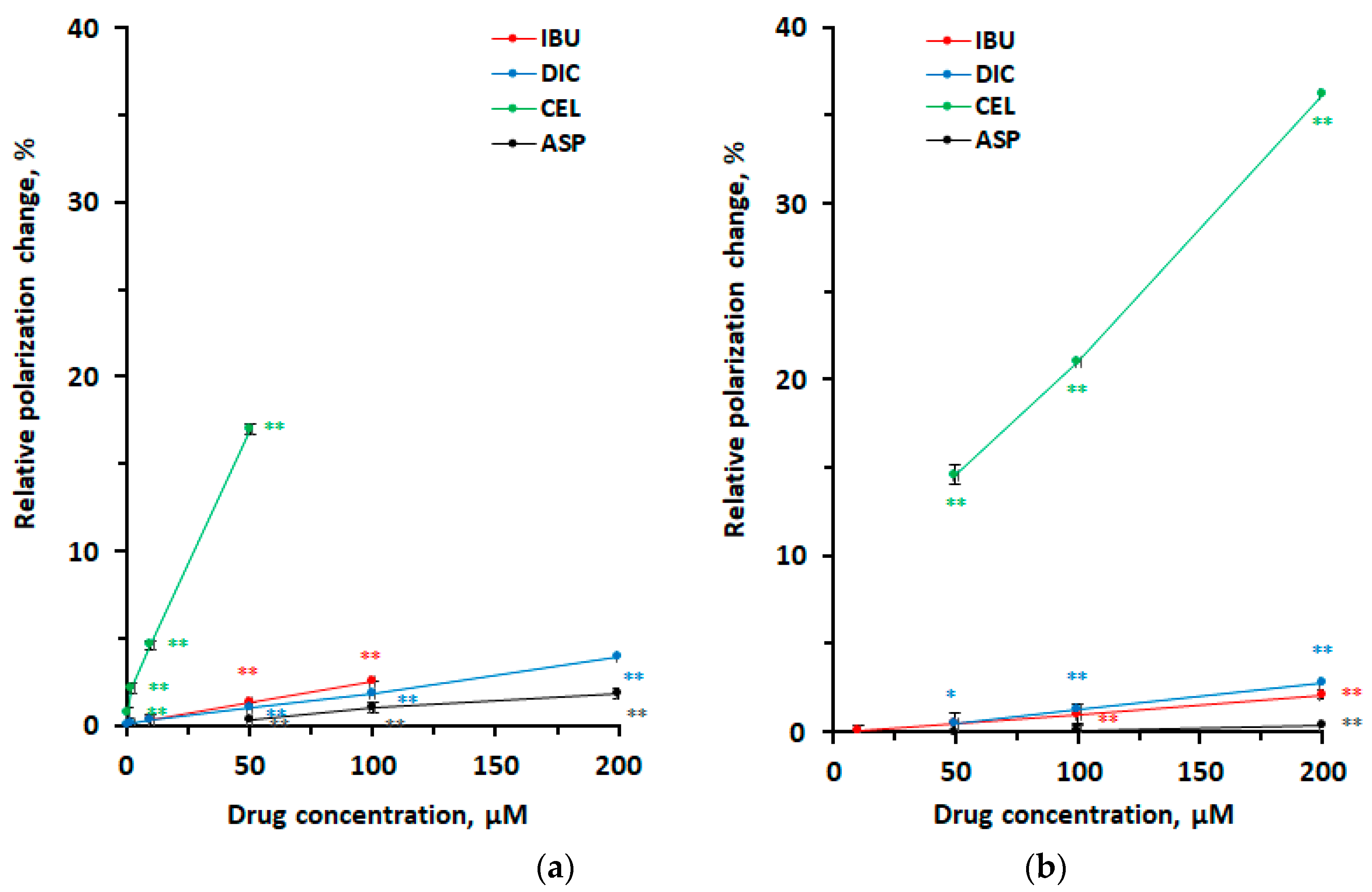
Drugs were subjected to the reactions with membranes of different lipid composition in media of pH 7.4. IBU, DIC, CEL, and ASP interacted with neuro-mimetic membranes and DPPC membranes, as shown by Figure 2. They decreased fluidity of both membranes, but with greater potency to act on neuro-mimetic membranes (Figure 2a) than DPPC membranes (Figure 2b). Fluidity-decreasing effects of IBU, DIC, and CEL on neuro-mimetic membranes were 2.6, 2.0, and 1.2 times greater than those on DPPC membranes, respectively, based on comparative DPH polarization increases induced by 50 μM for each (neuro-mimetic vs. DPPC, p < 0.01 for all).
In order to reveal the pH dependence of drug and membrane interactions, IBU as a representative of investigated drugs was subjected to the reactions with neuro-mimetic membranes and DPPC membranes under neutral and acidic conditions. The results of membrane interactions are shown by Figure 3. Its effects on both membranes significantly depended on medium pH, as polarization increases at pH 7.4 were drastically changed to polarization decreases at pH 4.0. When reacting at pH 7.4, 50–100 μM IBU preferentially interacted with neuro-mimetic membranes to induce greater fluidity decreases than DPPC membranes (neuro-mimetic vs. DPPC, p < 0.01 for all), as shown by different polarization increases (Figure 3a). In contrast to pH 7.4, 100–500 μM IBU preferentially interacted with DPPC membranes to induce greater fluidity increases than neuro-mimetic membranes at pH 4.0 (DPPC vs. neuro-mimetic, p < 0.01 for all), as shown by different polarization decreases (Figure 3b).
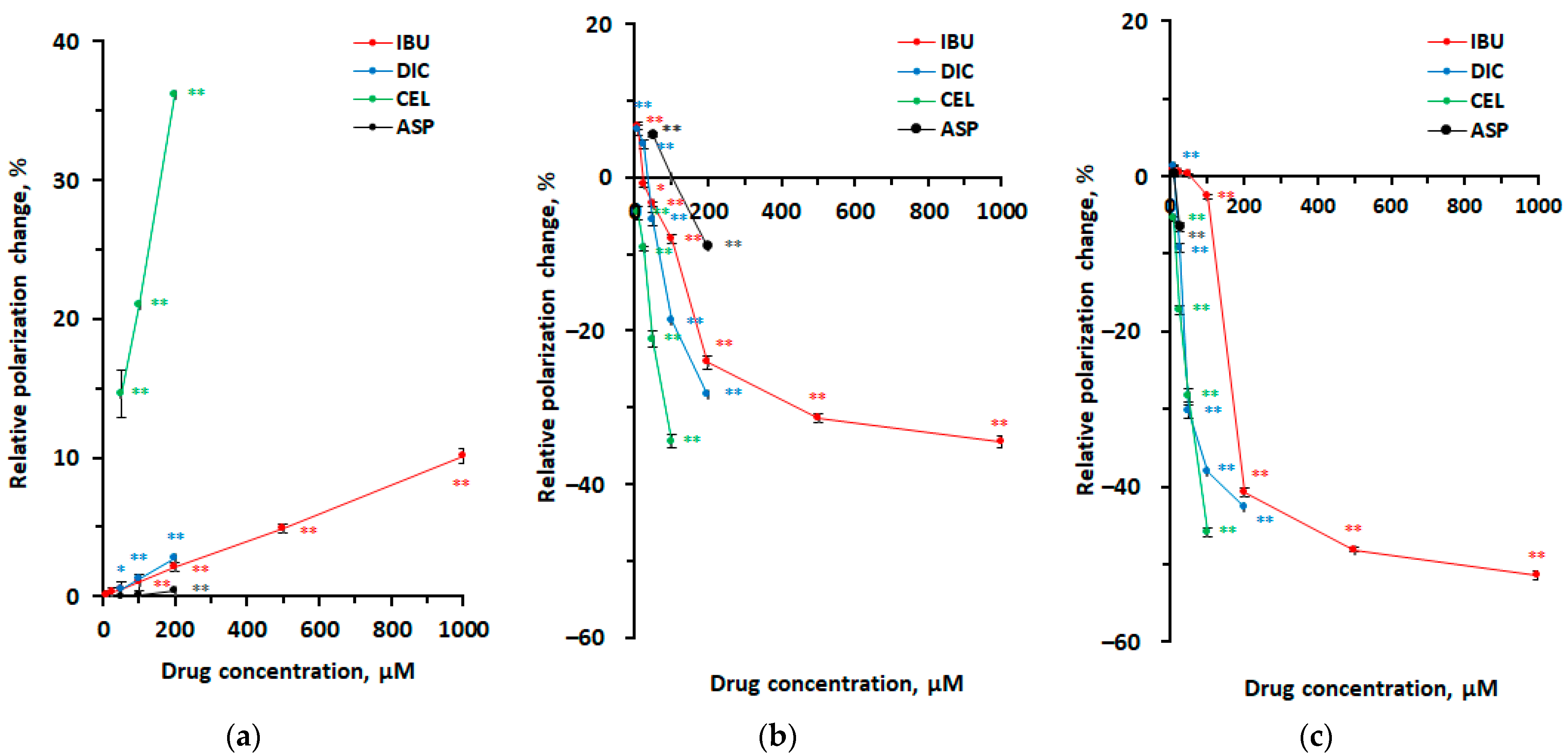
Effects on DPPC membranes under different conditions were investigated by varying drug concentrations. IBU, DIC, CEL, and ASP interacted with DPPC membranes at pH 7.4, 4.0, and 2.5 to change membrane fluidity differently, as shown by Figure 4. At relatively low concentrations, IBU, DIC, CEL, and ASP decreased membrane fluidity at pH 7.4, as shown by polarization increases (Figure 4a). At relatively high concentrations, however, IBU, DIC, CEL, and ASP increased DPPC membrane fluidity at pH 4.0 (Figure 4b) and pH 2.5 (Figure 4c), as shown by polarization decreases. Fluidity-increasing effects of 500 μM IBU, 200 μM DIC, and 100 μM CEL on DPPC membranes at pH 2.5 were 1.5, 1.5, and 1.3 times greater than those at pH 4.0, respectively, based on comparative DPH polarization decreases (pH 2.5 vs. pH 4.0, p < 0.01 for all).

4. Discussion

We have comparatively studied the interactions of IBU, DIC, and CEL frequently used in multimodal analgesia with biomimetic lipid bilayer membranes by varying reaction conditions. Our main findings on drug-induced membrane fluidity changes are as follows: (1) IBU, DIC, and CEL lipid composition-dependently interact with different membranes at pH 7.4. That is, they structure-specifically decrease neuro-mimetic membrane fluidity more potently than DPPC membrane fluidity at relatively low micromolar concentrations; (2) IBU, DIC, and CEL medium pH-dependently interact with biomimetic membranes. That is, they decrease membrane fluidity at pH 7.4, and increase membrane fluidity under acidic conditions.; and (3) IBU, DIC, and CEL concentration-dependently interact with DPPC membranes at pH 4.0 and pH 2.5. That is, they increase membrane fluidity with increasing concentrations to high micromolar ones.
While phospholipids and cholesterol are the major components in biomembranes, sphingolipids are abundant in neuronal membranes and play an important role in the signaling process [42] and the pain perception of nociceptive sensory neurons [43]. In particular, SM is preset at high concentrations for the structural and functional significance. We prepared neuro-mimetic membranes with POPC and cholesterol plus SM. IBU, DIC, and CEL were more effective in interacting with such membranes containing SM than the membranes consisting of DPPC alone. Their membrane effects were so dependent on lipid composition that they interacted with neuro-mimetic membranes more potently than DPPC membranes, producing greater changes in membrane fluidity. Compared with the effects on biomembranes previously prepared with 3.6 mol% SM, phosphatidylcholine, and cholesterol [20], IBU more effectively interacted with the present neuro-mimetic membranes containing 10 mol% SM to induce greater increases of membrane fluidity, suggesting that SM would characterize the drug and neuronal membrane interactions. Since cholesterol expels IBU from the hydrophobic membrane core to localize its molecules in the superficial region [44], the membrane interaction of IBU is also dependent on the presence of cholesterol in membrane lipid bilayers. Cholesterol may be responsible for the lipid-composition-dependent membrane interactions of drugs as well as membrane SM.
When IBU tablet of 400 mg and 800 mg were administered to human subjects, the maximum plasma concentration (Cmax) reached 147 and 305 μM, respectively [45]. DIC showed Cmax of 3.6–7.5 μM after administration of 50-mg tablet [46]. Oral administration of 100-mg CEL or 200-mg CEL-tramadol co-crystal (112 mg CEL and 88 mg tramadol) to healthy subjects resulted in Cmax of CEL ranging from 0.7 to 1.4 μM [47]. Intravenous infusion of 400 and 800 mg Caldolor (IBU injection) for 5–60 min produced Cmax of 190–582 μM for IBU [45] and a single dose of intravenous hydroxypropyl-β-cyclodextrin/DIC of 37.5 mg, Cmax of 24–73 μM for DIC [48]. At concentrations corresponding to these pharmacokinetic parameters, IBU, DIC, and CEL commonly decreased the fluidity of neuro-mimetic membranes at pH 7.4, but with different potencies. Since membrane fluidity regulates signal transmission and neuronal functions in sensory neurons, its alteration is presumed to disturb nociceptive signaling and affect inflammatory pain [11]. Some pain relievers act on lipid bilayer membranes and modify membrane fluidity in addition to acting on channels relevant to pain perception [12]. IBU, DIC, and CEL of pharmacokinetically-relevant low-micromolar concentrations induce structure-specific decreases in neuro-mimetic membrane fluidity, which hypothetically could affect signal transmission of nociceptive sensory neurons, possibly resulting in analgesia.
Enzyme 5-Lipoxygenase, which binds to nuclear membranes for activation, preferentially interacts with fluid (fluidity-increased) membranes, but not with rigid (fluidity-decreased) membranes [14]. Similarly, monotopic membrane enzyme COX may be activated in biomembranes with the relatively high fluidity. The activity of membrane-associated enzymes is considered to be enhanced when the membrane domains have higher fluidity, whereas reduced by decreasing membrane fluidity. We could hypothesize that membrane-rigidifying drugs inhibit COX indirectly through alteration of the membrane lipid environment optimal for COX activity. CEL and DIC are highly and intermediately selective for COX-2, respectively, but IBU is not selective for COX-2, whereas ASP has high selectivity for COX-1. Lucio et al. [49] compared the effects of drugs on lipid bilayers and revealed that COX-2 selective inhibitors change membrane fluidity, but not COX-1 inhibitors. In the present study, the relative membrane-interacting potency was CEL >> IBU >> ASP, suggesting the possibility that the membrane interactivity of drugs may correlate with their selectivity for COX-2. Although the membrane effect of DIC was comparable to or slightly weaker than that of IBU, higher affinity of DIC for COX-2 would enhance its COX-2 selectivity. According to Ki (inhibitory constant) values of CEL, DIC, and IBU, COX-2 binding affinity of DIC is almost similar to CEL but much higher than IBU. A relationship between membrane interactivity and COX-2 selectivity needs to be verified by further studies.
COX-1-selective IND and COX-2-selective CEL both develop gastrointestinal ulcers in COX-1-deficient mice, although COX-1-selective inhibitor SC-560 does not induce intestinal ulceration in wild-type mice [50]. Inhibiting both COX-1 and COX-2 is necessary for nonsteroidal anti-inflammatory drugs to exhibit the gastric toxicity [8]. Human gastric lesions produced by nonsteroidal anti-inflammatory drugs relate to their physicochemical properties (pKa and partition coefficient), but not to their COX-2/COX-1 selectivity [51]. Therefore, it is speculated that the mechanism irrespective of COX inhibition underlies gastrointestinal toxic effects of IBU, DIC, and CEL. For the purpose of investigating the gastrointestinal toxic effects independent of COX inhibition, Lichtenberger et al. [15,52] conducted a series of studies to assess the property of nonsteroidal anti-inflammatory drugs to attenuate the hydrophobic protective barrier of gastrointestinal mucosae. Gastrointestinal tracts are protected against luminal acids by the linings, which are constituted of phospholipids (rich in phosphatidylcholine) monolayers at the interface between mucus gel and luminal fluid, and of phospholipid (primarily phosphatidylcholine) bilayer membranes of epithelial cells [15]. In human gastroduodenal mucosae, the most abundant species of phosphatidylcholine were identified as 16:0/18:1, 16:0/18:2, and 16:0/20:4 [35].
Koenigsknecht et al. [53] administered 800-mg IBU tablet to human subjects to determine pH and IBU in gastrointestinal fluids. They revealed that gastric and duodenal pH are 2.3 and 4–5, respectively, and that intragastric and intraduodenal IBU concentrations are 439 μM after 15 min and 400–900 μM after 3–7 h, respectively. Hens et al. [54] followed up changes in pH of gastrointestinal fluids for 7 h after administration of 800-mg IBU tablet. In the fasting state, gastric and duodenal pH were 1.1–7.5 and 1.7–7.6, respectively.
By reference to previous studies [15,53,54], we assessed the effects of CEL, DIC, and IBU on DPPC (16:0/16:0) membranes as protective mucosae at pH 2.5 and pH 4.0 reflecting the gastroduodenal environments and at high-micromolar concentrations corresponding to drug concentrations in the gastroduodenal lumen after administration. The tested drugs increased the fluidity of DPPC membranes at pH 2.5 more significantly than pH 4.0 with the relative potency being CEL > DIC > IBU. These drugs are efficiently ionized at pH being > pKa, but not at pH being < pKa, so that they are very likely to be present in a nonionized form. Nonionized molecules are considered to effectively interact with phospholipid membranes. The greater membrane interactivity at gastric pH 2.5 than at duodenal pH 4.0 is consistent with the relative gastrointestinal toxicity that the incidence of gastric ulcers is higher than that of duodenal ulcers. Pereira-Leite et al. [55] investigated the effects of DIC on 1,2-dimyristoylphosphatidylcholine liposomal membranes at pH 3–5. The neutral form of DIC displayed greater affinity for phospholipid bilayers to modulate the bilayer structures more effectively than the anionic form. They suggested that nonionized DIC-induced changes in membranous phospholipids at low pH constitute a topical mechanism of the gastric toxicity. Increasing membrane fluidity should increase permeability of the membrane lipid bilayers, which may exert adverse effects on gastroduodenal protective mucosae.
Meta-analysis of gastrointestinal complications indicated that the risk of DIC is 2–3 times higher than IBU [56]. The membrane-interaction-mediated gastrointestinal effect of CEL may have a longer time course as CEL causes gastrointestinal injuries by oral administration for a long term. Nonsteroidal anti-inflammatory drugs also adversely affect the cardiovascular system. Pereira-Leite et al. [57] recently characterized the interactions of cardiotoxic DIC with lipid bilayers as model systems for cell and mitochondrial membranes while comparing with lower cardiotoxic naproxen. Based on drug-induced changes in lipid bilayer membranes, they suggested that the membrane interactions depending on lipid composition, drug concentration, and drug structure may be one of mechanisms underlying the cardiotoxic effects of nonsteroidal anti-inflammatory drugs.

5. Conclusions

Drugs frequently used in multimodal analgesia interact with biomimetic membranes depending on lipid composition, medium pH, and drug concentration. As a result of membrane interactions, IBU, DIC, and CEL of pharmacokinetically-relevant low-micromolar concentrations structure-specifically increase the fluidity of neuro-mimetic membranes at the physiological pH, which hypothetically could affect signal transmission of nociceptive sensory neurons, possibly resulting in analgesic effects. Under gastroduodenal acidic conditions, however, IBU, DIC, and CEL of high-micromolar concentrations corresponding to intragastric and intraduodenal concentrations after oral administration induce fluidity increases of membranous phosphatidylcholines that are hypothetically associated with gastrointestinal toxic effects, which would enhance acid permeability of protective mucosal membranes, possibly causing damage of gastroduodenal tracts.

Author Contributions

M.M. performed the experiments and statistically analyzed the data. H.T. designed and conducted the study. Both M.M. and H.T. wrote and reviewed the manuscript. All authors have read and agreed to the published version of the manuscript.

Funding

The present study was supported by JSPS KAKENHI grant number 20K10152 and JSPS KAKENHI grant number 24K12106.

Institutional Review Board Statement

Not applicable.

Informed Consent Statement

Not applicable because the present study includes neither human nor animal experiments.

Data Availability Statement

All data generated or analyzed during the present study are included in this article. The data supporting the findings are kept at the affiliation of author 1 (M.M.) and are available on request.

Conflicts of Interest

The authors have no conflicts of interest to declare.

References

  1. Young, A.; Buvanendran, A. Recent advances in multimodal analgesia. Anesthesiol. Clin. 2012, 30, 91–100. [Google Scholar] [CrossRef] [PubMed]
  2. Brubaker, L.; Kendall, L.; Reina, E. Multimodal analgesia: A systematic review of local NSAIDs for non-ophthalmologic postoperative pain management. Int. J. Surg. 2016, 32, 158–166. [Google Scholar] [CrossRef] [PubMed]
  3. Rainsford, K.D. Ibuprofen: Pharmacology, efficacy and safety. Inflammopharmacology 2009, 17, 275–342. [Google Scholar] [CrossRef] [PubMed]
  4. Harirforoosh, S.; Asghar, W.; Jamali, F. Adverse effects of nonsteroidal antiinflammatory drugs: An update of gastrointestinal, cardiovascular and renal complications. J. Pharm. Pharm. Sci. 2013, 16, 821–847. [Google Scholar] [CrossRef] [PubMed]
  5. Cooper, C.; Chapurlat, R.; Al-Daghri, N.; Herrero-Beaumont, G.; Bruyère, O.; Rannou, F.; Roth, R.; Uebelhart, D.; Reginster, J.Y. Safety of oral non-selective non-steroidal anti-inflammatory drugs in osteoarthritis: What does the literature say? Drugs Aging 2019, 36 (Suppl. S1), 15–24. [Google Scholar] [CrossRef] [PubMed]
  6. Bindu, S.; Mazumder, S.; Bandyopadhyay, U. Non-steroidal anti-inflammatory drugs (NSAIDs) and organ damage: A current perspective. Biochem. Pharmacol. 2020, 180, 114147. [Google Scholar] [CrossRef] [PubMed]
  7. Perrone, M.G.; Lofrumento, D.D.; Vitale, P.; De Nuccio, F.; La Pesa, V.; Panella, A.; Calvello, R.; Cianciulli, A.; Panaro, M.A.; Scilimati, A. Selective cyclooxygenase-1 inhibition by p6 and gastrotoxicity: Preliminary investigation. Pharmacology 2015, 95, 22–28. [Google Scholar] [CrossRef] [PubMed]
  8. Wallace, J.L.; McKnight, W.; Reuter, B.K.; Vergnolle, N. NSAID-induced gastric damage in rats: Requirement for inhibition of both cyclooxygenase 1 and 2. Gastroenterology 2000, 119, 706–714. [Google Scholar] [CrossRef] [PubMed]
  9. Lim, Y.J.; Dial, E.J.; Lichtenberger, L.M. Advent of novel phosphatidylcholine-associated nonsteroidal anti-inflammatory drugs with improved gastrointestinal safety. Gut Liver 2013, 7, 7–15. [Google Scholar] [CrossRef] [PubMed]
  10. Bjarnason, I.; Scarpignato, C.; Holmgren, E.; Olszewski, M.; Rainsford, K.D.; Lanas, A. Mechanisms of damage to the gastrointestinal tract from nonsteroidal anti-inflammatory drugs. Gastroenterology 2018, 154, 500–514. [Google Scholar] [CrossRef]
  11. Wagner, K.; Vito, S.; Inceoglu, B.; Hammock, B.D. The role of long chain fatty acids and their epoxide metabolites in nociceptive signaling. Prostaglandins Other Lipid Mediat. 2014, 113–115, 2–12. [Google Scholar] [CrossRef]
  12. Matta, J.A.; Miyares, R.L.; Ahern, G.P. TRPV1 is a novel target for omega-3 polyunsaturated fatty acids. J. Physiol. 2007, 578 Pt 2, 397–411. [Google Scholar] [CrossRef] [PubMed]
  13. Escribá, P.V.; González-Ros, J.M.; Goñi, F.M.; Kinnunen, P.K.; Vigh, L.; Sánchez-Magraner, L.; Fernández, A.M.; Busquets, X.; Horváth, I.; Barceló-Coblijn, G. Membranes: A meeting point for lipids, proteins and therapies. J. Cell. Mol. Med. 2008, 12, 829–875. [Google Scholar] [CrossRef] [PubMed]
  14. Pande, A.H.; Qin, S.; Tatulian, S.A. Membrane fluidity is a key modulator of membrane binding, insertion, and activity of 5-lipoxygenase. Biophys. J. 2005, 88, 4084–4094. [Google Scholar] [CrossRef] [PubMed]
  15. Lichtenberger, L.M.; Zhou, Y.; Jayaraman, V.; Doyen, J.R.; O’Neil, R.G.; Dial, E.J.; Volk, D.E.; Gorenstein, D.G.; Boggara, M.B.; Krishnamoorti, R. Insight into NSAID-induced membrane alterations, pathogenesis and therapeutics: Characterization of interaction of NSAIDs with phosphatidylcholine. Biochim. Biophys. Acta 2012, 1821, 994–1002. [Google Scholar] [CrossRef] [PubMed]
  16. Pereira-Leite, C.; Nunes, C.; Reis, S. Interaction of nonsteroidal anti-inflammatory drugs with membranes: In vitro assessment and relevance for their biological actions. Prog. Lipid Res. 2013, 52, 571–584. [Google Scholar] [CrossRef] [PubMed]
  17. Tsuchiya, H.; Mizogami, M. Membrane interactivity of non-steroidal anti-inflammatory drugs: A literature review. J. Adv. Med. Med. Res. 2019, 31, 1–30. [Google Scholar] [CrossRef]
  18. Giraud, M.N.; Motta, C.; Romero, J.J.; Bommelaer, G.; Lichtenberger, L.M. Interaction of indomethacin and naproxen with gastric surface-active phospholipids: A possible mechanism for the gastric toxicity of nonsteroidal anti-inflammatory drugs (NSAIDs). Biochem. Pharmacol. 1999, 57, 247–254. [Google Scholar] [CrossRef] [PubMed]
  19. Suwalsky, M.; Manrique, M.; Villena, F.; Sotomayor, C.P. Structural effects in vitro of the anti-inflammatory drug diclofenac on human erythrocytes and molecular models of cell membranes. Biophys. Chem. 2009, 141, 34–40. [Google Scholar] [CrossRef] [PubMed]
  20. Tsuchiya, H.; Mizogami, M. Discrimination of stereoisomers by their enantioselective interactions with chiral cholesterol-containing membranes. Molecules 2017, 23, 49. [Google Scholar] [CrossRef] [PubMed]
  21. Nunes, C.; Lopes, D.; Pinheiro, M.; Pereira-Leite, C.; Reis, S. In vitro assessment of NSAIDs-membrane interactions: Significance for pharmacological actions. Pharm. Res. 2013, 30, 2097–2107. [Google Scholar] [CrossRef] [PubMed]
  22. Manrique-Moreno, M.; Heinbockel, L.; Suwalsky, M.; Garidel, P.; Brandenburg, K. Biophysical study of the non-steroidal anti-inflammatory drugs (NSAID) ibuprofen, naproxen and diclofenac with phosphatidylserine bilayer membranes. Biochim. Biophys. Acta 2016, 1858, 2123–2131. [Google Scholar] [CrossRef] [PubMed]
  23. Fernandes, E.; Soares, T.B.; Gonçalves, H.; Bernstorff, S.; Real Oliveira, M.E.C.D.; Lopes, C.M.; Lúcio, M. A molecular biophysical approach to diclofenac topical gastrointestinal damage. Int. J. Mol. Sci. 2018, 19, 3411. [Google Scholar] [CrossRef] [PubMed]
  24. Tomisato, W.; Tanaka, K.; Katsu, T.; Kakuta, H.; Sasaki, K.; Tsutsumi, S.; Hoshino, T.; Aburaya, M.; Li, D.; Tsuchiya, T.; et al. Membrane permeabilization by non-steroidal anti-inflammatory drugs. Biochem. Biophys. Res. Commun. 2004, 323, 1032–1039. [Google Scholar] [CrossRef] [PubMed]
  25. Ferreira, H.; Lúcio, M.; Lima, J.L.; Matos, C.; Reis, S. Effects of diclofenac on EPC liposome membrane properties. Anal. Bioanal. Chem. 2005, 382, 1256–1264. [Google Scholar] [CrossRef] [PubMed]
  26. Okamoto, Y.; Kishi, Y.; Ishigami, T.; Suga, K.; Umakoshi, H. Chiral selective adsorption of ibuprofen on a liposome membrane. J. Phys. Chem. B 2016, 120, 2790–2795. [Google Scholar] [CrossRef] [PubMed]
  27. Sade, A.; Tunçay, S.; Cimen, I.; Severcan, F.; Banerjee, S. Celecoxib reduces fluidity and decreases metastatic potential of colon cancer cell lines irrespective of COX-2 expression. Biosci. Rep. 2012, 32, 35–44. [Google Scholar] [CrossRef] [PubMed]
  28. Gamerdinger, M.; Clement, A.B.; Behl, C. Cholesterol-like effects of selective cyclooxygenase inhibitors and fibrates on cellular membranes and amyloid-beta production. Mol. Pharmacol. 2007, 72, 141–151. [Google Scholar] [CrossRef] [PubMed]
  29. Sousa, C.; Nunes, C.; Lúcio, M.; Ferreira, H.; Lima, J.L.; Tavares, J.; Cordeiro-da-Silva, A.; Reis, S. Effect of nonsteroidal anti-inflammatory drugs on the cellular membrane fluidity. J. Pharm. Sci. 2008, 97, 3195–3206. [Google Scholar] [CrossRef] [PubMed]
  30. Nunes, C.; Brezesinski, G.; Lopes, D.; Lima, J.L.; Reis, S.; Lúcio, M. Lipid-drug interaction: Biophysical effects of tolmetin on membrane mimetic systems of different dimensionality. J. Phys. Chem. B 2011, 115, 12615–12623. [Google Scholar] [CrossRef] [PubMed]
  31. Pereira-Leite, C.; Nunes, C.; Lima, J.L.; Reis, S.; Lúcio, M. Interaction of celecoxib with membranes: The role of membrane biophysics on its therapeutic and toxic effects. J. Phys. Chem. B 2012, 116, 13608–13617. [Google Scholar] [CrossRef] [PubMed]
  32. Akbarzadeh, A.; Rezaei-Sadabady, R.; Davaran, S.; Joo, S.W.; Zarghami, N.; Hanifehpour, Y.; Samiei, M.; Kouhi, M.; Nejati-Koshki, K. Liposome: Classification, preparation, and applications. Nanoscale Res. Lett. 2013, 8, 102. [Google Scholar] [CrossRef] [PubMed]
  33. Gouda, A.; Sakr, O.S.; Nasr, M.; Sammour, O. Ethanol injection technique for liposomes formulation: An insight into development, influencing factors, challenges and applications. J. Drug Deliv. Sci. Technol. 2021, 61, 102174. [Google Scholar] [CrossRef]
  34. Mizogami, M.; Tsuchiya, H. Acetaminophen has lipid composition-dependent membrane interactivity that could be related to nephrotoxicity but not to analgesic activity and hepatotoxicity. Med. Princ. Pract. 2022, 31, 111–117. [Google Scholar] [CrossRef] [PubMed]
  35. Ehehalt, R.; Wagenblast, J.; Erben, G.; Lehmann, W.D.; Hinz, U.; Merle, U.; Stremmel, W. Phosphatidylcholine and lysophosphatidylcholine in intestinal mucus of ulcerative colitis patients. A quantitative approach by nanoElectrospray-tandem mass spectrometry. Scand. J. Gastroenterol. 2004, 39, 737–742. [Google Scholar] [CrossRef] [PubMed]
  36. Shaker, S.; Gardouh, A.R.; Ghorab, M.M. Factors affecting liposomes particle size prepared by ethanol injection method. Res. Pharm. Sci. 2017, 12, 346–352. [Google Scholar] [CrossRef] [PubMed]
  37. Tsuchiya, H. Effects of green tea catechins on membrane fluidity. Pharmacology 1999, 59, 34–44. [Google Scholar] [CrossRef] [PubMed]
  38. Tsuchiya, H. Structure-specific membrane-fluidizing effect of propofol. Clin. Exp. Pharmacol. Physiol. 2001, 28, 292–299. [Google Scholar] [CrossRef]
  39. Heine, P.; Witt, G.; Gilardi, A.; Gribbon, P.; Kummer, L.; Plückthun, A. High-throughput fluorescence polarization assay to identify ligands using purified G protein-coupled receptor. SLAS Discov. 2019, 24, 915–927. [Google Scholar] [CrossRef]
  40. Iyamu, I.D.; Huang, R. Development of fluorescence polarization-based competition assay for nicotinamide N-methyltransferase. Anal. Biochem. 2020, 604, 113833. [Google Scholar] [CrossRef] [PubMed]
  41. Ushijima, H.; Tanaka, K.; Takeda, M.; Katsu, T.; Mima, S.; Mizushima, T. Geranylgeranylacetone protects membranes against nonsteroidal anti-inflammatory drugs. Mol. Pharmacol. 2005, 68, 1156–1161. [Google Scholar] [CrossRef] [PubMed]
  42. Grassi, S.; Chiricozzi, E.; Mauri, L.; Sonnino, S.; Prinetti, A. Sphingolipids and neuronal degeneration in lysosomal storage disorders. J. Neurochem. 2019, 148, 600–611. [Google Scholar] [CrossRef] [PubMed]
  43. Sántha, P.; Dobos, I.; Kis, G.; Jancsó, G. Role of gangliosides in peripheral pain mechanisms. Int. J. Mol. Sci. 2020, 21, 1005. [Google Scholar] [CrossRef] [PubMed]
  44. Alsop, R.J.; Armstrong, C.L.; Maqbool, A.; Toppozini, L.; Dies, H.; Rheinstädter, M.C. Cholesterol expels ibuprofen from the hydrophobic membrane core and stabilizes lamellar phases in lipid membranes containing ibuprofen. Soft Matter 2015, 11, 4756–4767. [Google Scholar] [CrossRef] [PubMed]
  45. Smith, H.S.; Voss, B. Pharmacokinetics of intravenous ibuprofen: Implications of time of infusion in the treatment of pain and fever. Drugs 2012, 72, 327–337. [Google Scholar] [CrossRef] [PubMed]
  46. Altman, R.; Bosch, B.; Brune, K.; Patrignani, P.; Young, C. Advances in NSAID development: Evolution of diclofenac products using pharmaceutical technology. Drugs 2015, 75, 859–877. [Google Scholar] [CrossRef] [PubMed]
  47. Cebrecos, J.; Carlson, J.D.; Encina, G.; Lahjou, M.; Sans, A.; Sust, M.; Vaqué, A.; Morte, A.; Gascón, N.; Plata-Salamán, C. Celecoxib-tramadol co-crystal: A randomized 4-way crossover comparative bioavailability study. Clin. Ther. 2021, 43, 1051–1065. [Google Scholar] [CrossRef] [PubMed]
  48. Hamilton, D.A.; Ernst, C.C.; Kramer, W.G.; Madden, D.; Lang, E.; Liao, E.; Lacouture, P.G.; Ramaiya, A.; Carr, D.B. Pharmacokinetics of diclofenac and hydroxypropyl-β-cyclodextrin (HPβCD) following administration of injectable HPβCD-diclofenac in subjects with mild to moderate renal insufficiency or mild hepatic impairment. Clin. Pharmacol. Drug Dev. 2018, 7, 110–122. [Google Scholar] [CrossRef] [PubMed]
  49. Lúcio, M.; Ferreira, H.; Lima, J.L.; Reis, S. Interactions between oxicams and membrane bilayers: An explanation for their different COX selectivity. Med. Chem. 2006, 2, 447–456. [Google Scholar] [CrossRef] [PubMed]
  50. Sigthorsson, G.; Simpson, R.J.; Walley, M.; Anthony, A.; Foster, R.; Hotz-Behoftsitz, C.; Palizban, A.; Pombo, J.; Watts, J.; Morham, S.G.; et al. COX-1 and 2, intestinal integrity, and pathogenesis of nonsteroidal anti-inflammatory drug enteropathy in mice. Gastroenterology 2002, 122, 1913–1923. [Google Scholar] [CrossRef] [PubMed]
  51. Bjarnason, I.; Scarpignato, C.; Takeuchi, K.; Rainsford, K.D. Determinants of the short-term gastric damage caused by NSAIDs in man. Aliment. Pharmacol. Ther. 2007, 26, 95–106. [Google Scholar] [CrossRef] [PubMed]
  52. Lichtenberger, L.M.; Barron, M.; Marathi, U. Association of phosphatidylcholine and NSAIDs as a novel strategy to reduce gastrointestinal toxicity. Drugs Today 2009, 45, 877–890. [Google Scholar] [CrossRef] [PubMed]
  53. Koenigsknecht, M.J.; Baker, J.R.; Wen, B.; Frances, A.; Zhang, H.; Yu, A.; Zhao, T.; Tsume, Y.; Pai, M.P.; Bleske, B.E.; et al. In vivo dissolution and systemic absorption of immediate release ibuprofen in human gastrointestinal tract under fed and fasted conditions. Mol. Pharm. 2017, 14, 4295–4304. [Google Scholar] [CrossRef]
  54. Hens, B.; Tsume, Y.; Bermejo, M.; Paixao, P.; Koenigsknecht, M.J.; Baker, J.R.; Hasler, W.L.; Lionberger, R.; Fan, J.; Dickens, J.; et al. Low buffer capacity and alternating motility along the human gastrointestinal tract: Implications for in vivo dissolution and absorption of ionizable drugs. Mol. Pharm. 2017, 14, 4281–4294. [Google Scholar] [CrossRef] [PubMed]
  55. Pereira-Leite, C.; Jamal, S.K.; Almeida, J.P.; Coutinho, A.; Prieto, M.; Cuccovia, I.M.; Nunes, C.; Reis, S. Neutral diclofenac causes remarkable changes in phosphatidylcholine bilayers: Relevance for gastric toxicity mechanisms. Mol. Pharmacol. 2020, 97, 295–303. [Google Scholar] [CrossRef] [PubMed]
  56. Henry, D.; Lim, L.L.; Garcia Rodriguez, L.A.; Perez Gutthann, S.; Carson, J.L.; Griffin, M.; Savage, R.; Logan, R.; Moride, Y.; Hawkey, C.; et al. Variability in risk of gastrointestinal complications with individual non-steroidal anti-inflammatory drugs: Results of a collaborative meta-analysis. BMJ 1996, 312, 1563–1566. [Google Scholar] [CrossRef] [PubMed]
  57. Pereira-Leite, C.; Figueiredo, M.; Burdach, K.; Nunes, C.; Reis, S. Unraveling the role of drug-lipid interactions in NSAIDs-induced cardiotoxicity. Membranes 2021, 11, 24. [Google Scholar] [CrossRef] [PubMed]
Figure 1. Drugs frequently used in multimodal analgesia.
Futurepharmacol 04 00024 g001
Figure 2. Interactions of drugs at pH 7.4 with neuro-mimetic membranes (a) and 1,2-dipalmitoylphosphatidylcholine membranes (b). Values are means ± SD (n = 8). * p < 0.05 and ** p < 0.01 compared with controls. IBU, ibuprofen; DIC, diclofenac; CEL, celecoxib; ASP, aspirin.
Futurepharmacol 04 00024 g002
Figure 3. Interactions of ibuprofen with neuro-mimetic membranes and 1,2-dipalmitoylphosphatidylcholine membranes at pH 7.4 (a) and pH 4.0 (b). Values are means ± SD (n = 8). * p < 0.05 and ** p < 0.01 compared with controls. DPPC, 1,2dipalmitoylphosphatidylcholine.
Futurepharmacol 04 00024 g003
Figure 4. Interactions of drugs with 1,2-dipalmitoylphosphatidylcholine membranes at pH 7.4 (a), pH 4.0 (b), and pH 2.5 (c). Values are means ± SD (n = 8). * p < 0.05 and ** p < 0.01 compared with controls. IBU, ibuprofen; DIC, diclofenac; CEL, celecoxib; ASP, aspirin.
Futurepharmacol 04 00024 g004
Table 1. Interactions of drugs with neuro-mimetic membranes.
Polarization Change
Drug
Concentration
Membrane Interaction at pH 7.4
IBUDICCELASP
0.5 μM0.0000 ± 0.00080.0000 ± 0.00060.0018 ± 0.0006 *
2 μM0.0002 ± 0.00060.0002 ± 0.00110.0054 ± 0.0011 **
10 μM0.0009 ± 0.00030.0006 ± 0.00030.0121 ± 0.0006 **
50 μM0.0034 ± 0.0000 **0.0027 ± 0.0003 **0.0445 ± 0.0008 **0.0009 ± 0.0000 **
100 μM0.0065 ± 0.0008 **0.0047 ± 0.0000 ** 0.0026 ± 0.0000 **
200 μM 0.0102 ± 0.0006 ** 0.0046 ± 0.0003 **
Membrane Interaction at pH 4.0
IBU
50 μM0.0098 ± 0.0008 **
100 μM−0.0067 ± 0.0006 **
500 μM−0.0270 ± 0.0006 **
Values are means ± SD (n = 8). * p < 0.05, ** p < 0.01 compared with controls. IBU, ibuprofen; DIC, diclofenac; CEL, celecoxib; ASP, aspirin.
Table 2. Interactions of drugs with DPPC membranes at different pH.
Polarization Change
Drug
Concentration
Membrane Interaction at pH 7.4
IBUDICCELASP
50 μM0.0010 ± 0.00060.0010 ± 0.0000 *0.0283 ± 0.0008 **0.0000 ± 0.0003
100 μM0.0018 ± 0.0008 **0.0025 ± 0.0006 **0.0407 ± 0.0025 **0.0002 ± 0.0003
200 μM0.0040 ± 0.0006 **0.0054 ± 0.0008 **0.0703 ± 0.0006 **0.0008 ± 0.0000 **
Membrane Interaction atpH 4.0
IBUDICCELASP
25 μM−0.0017 ± 0.0006 *0.0074 ± 0.0014 **−0.0154 ± 0.0006 **
50 μM−0.0059 ± 0.0006 **−0.0095 ± 0.0014 **−0.0350 ± 0.0020 **0.0098 ± 0.0008 **
100 μM−0.0134 ± 0.0011 **−0.0310 ± 0.0023 **−0.0570 ± 0.0014 **
200 μM−0.0398 ± 0.0011 **−0.0469 ± 0.0006 ** −0.0150 ± 0.0006 **
500 μM−0.0518 ± 0.0011 **
1 mM−0.0562 ± 0.0014 **
Membrane Interaction atpH 2.5
IBUDICCELASP
10 μM0.0010 ± 0.00080.0033 ± 0.0011 **−0.0123 ± 0.0011 **0.0010 ± 0.0008
25 μM0.0009 ± 0.0008−0.0252 ± 0.0011 **−0.0369 ± 0.0011 **−0.0163 ± 0.0017 **
50 μM0.0006 ± 0.0000−0.0832 ± 0.0011 **−0.0634 ± 0.0017 **
100 μM−0.0059 ± 0.0008 **−0.0920 ± 0.0011 **−0.1028 ± 0.0014 **
200 μM−0.0916 ± 0.0014 **−0.1029 ± 0.0017 **
500 μM−0.1081 ± 0.0008 **
1 mM−0.1119 ± 0.0014 **
Values are means ± SD (n = 8). * p < 0.05 and ** p < 0.01 compared with controls. IBU, ibuprofen; DIC, diclofenac; CEL, celecoxib; ASP, aspirin.
Disclaimer/Publisher’s Note: The statements, opinions and data contained in all publications are solely those of the individual author(s) and contributor(s) and not of MDPI and/or the editor(s). MDPI and/or the editor(s) disclaim responsibility for any injury to people or property resulting from any ideas, methods, instructions or products referred to in the content.