Article

Prolonged Cold Ischemia Did Not Impair Mitochondrial Oxygen Consumption or Reactive Oxygen Species Production in Human Uterine Fundus and Horn Myometrium

1
Gynecology Unit, CMCO, Pôle de Gynécologie Obstétrique, Hôpitaux Universitaires de Strasbourg, 67300 Schiltigheim, France
2
Team 3072 “Mitochondria, Oxidative Stress and Muscle Protection”, Faculty of Medicine, University of Strasbourg, CRBS, Translational Medicine Federation of Strasbourg (FMTS), 67000 Strasbourg, France
3
Physiology and Functional Exploration Service, University Hospital of Strasbourg, 67091 Strasbourg, France
*
Author to whom correspondence should be addressed.
Oxygen 2022, 2(1), 12-21; https://doi.org/10.3390/oxygen2010002
Submission received: 8 February 2022 / Revised: 25 February 2022 / Accepted: 26 February 2022 / Published: 28 February 2022

Abstract

:
Uterine transplantation may be a solution for infertility of uterine origin. Nevertheless, only three pregnancies with a live birth have so far been possible involving a uterine transplant from a brain-dead donor. Particularly, the impact of ischemia needs a better understanding. Analysis of mitochondrial respiration and production of reactive oxygen species (ROS) in muscle are of interest since they are pertinent markers of the harmful effects of ischemia. We therefore studied both uterine fundus and horn muscle mitochondrial use of oxygen and ROS production in eight women needing hysterectomy. High resolution respirometry and electron paramagnetic resonance allowed the determination of, respectively, myometrium oxidative capacity, hydrogen peroxide, mitochondrial free radical leak and superoxide anion production early (2 and 7 h) and late (24 h) following surgery. Mitochondrial oxygen consumption of the uterine fundus and horn tended to decrease with time but this was not statistically significant. Concerning ROS production, globally, we observed no significant change for H2O2, superoxide anion and free radical leak. In conclusion, a long period of cold ischemia did not impair myometrium mitochondrial respiration, only generating a transient H2O2 increase in uterine fundus. These data support that cold ischemia, even when prolonged, does not significantly alter uterine muscle oxidative capacity.

1. Introduction

Almost 200 women of reproductive age per million are suffering from uterine infertility that need uterine transplantation to allow pregnancy [1].
Proposed in cases of absent or dys-functionning uterus (agenesia, hysterectomy, diverse malformations, Asherman syndrome, or endometriosis), uterus transplantation is still relatively rare. To date, about 24 healthy children are born after uterine transplantation, resulting from over than 70 attempts worldwide. In general, uterus was obtained from live donors. Brain-dead donors have only given rise to three live birth [2,3,4,5,6,7,8,9,10,11,12,13,14,15,16,17,18,19,20].
This clearly raises the issue uterine viability when subjected to long-lasting ischemia.
Although they are placed in a hypothermic preservation medium, all organs can be significantly impaired by cold ischemia depending on its duration. In fact, organ characteristics modulate the tolerance to cold ischemia. Experimental and human studies investigating the uterine tolerance to cold ischemia demonstrated a lack of significant change in markers of necrosis and apoptosis till 6 and 24 h after cold ischemia [21,22,23]. Interestingly, studies of ischemia reperfusion performed upon skeletal muscle showed an early mitochondrial dysfunction suggesting that such a parameter might also demonstrate an eventual ischemia-induced uterine alteration [24,25].
Accordingly, mitochondrion is an important organelle producing energy and its dysfunction causes depletion in ATP and increased reactive oxygen species (ROS) production, particularly in the case of ischemia reperfusion [26]. Oxygen consumption by mitochondria is therefore widely used as a biological marker of its functionality.
Currently, little is known about the fate of mitochondria in the human uterus during transplantation and the purpose of this study was to determine the kinetics of mitochondrial respiration and of ROS production (2, 7 and 24 h after surgery) in two different segments of uterine myometrium subjected to cold ischemia as potentially observed during the clinical setting. Indeed, in skeletal muscles, the sensitivity to ischemia depend on muscles characteristics [27] and it appears to interestingly challenge the hypothesis of a differential effect of ischemia on either uterine fundus or horn, localizations often involved in embryo implantation.

2. Population and Methods
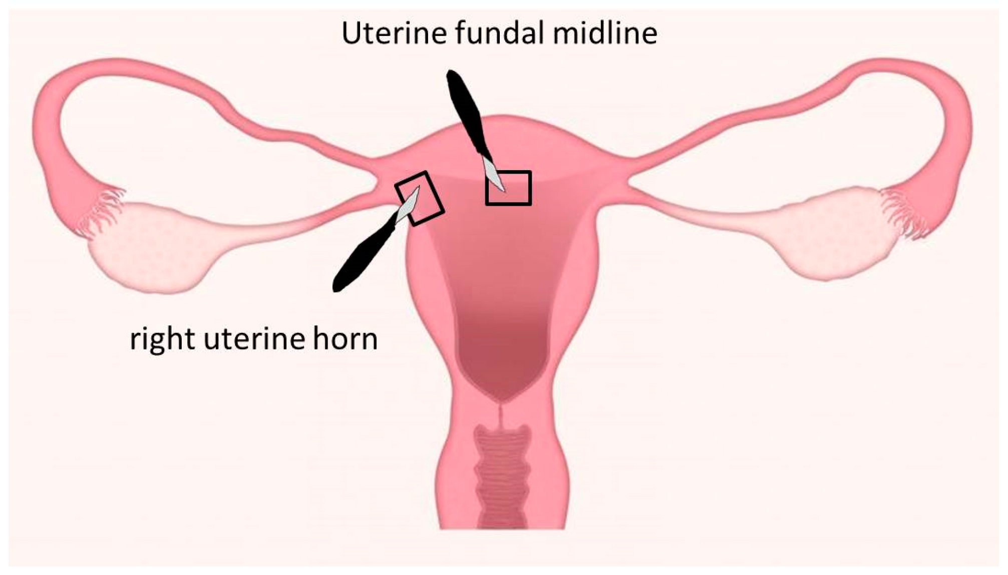
Eight women needing hysterectomy gave their informed consent and participated in the study carried out in the university teaching hospital of Strasbourg CHU and approved by the French Committee for the Protection of Persons (CPP) and registered on clinicaltrials.gov (RIN 2018–HUS N 7222).

2.1. Study Design

Inclusion criteria were patient >18 years old, affiliated to a social security regime and undergoing scheduled hysterectomy for an ostensibly benign pathology.
Tissue samples were provided by the surgeon immediately following surgical removal of the uterus during a total hysterectomy procedure conducted vaginally, laparoscopically or by laparotomy in the gynecological surgical service at the Strasbourg University Hospitals, France. The exact time at which the second uterine artery was ligated was recorded (ischemia T0). The surgeon cut the tissue directly post-operatively, with a cold scalpel to remove two samples of one cm3 myometrium per uterus, one at the fundal midline and the other at the right uterine horn (Figure 1).
These sites were readily identifiable and therefore reproducible, and corresponded to myometrium-rich areas where embryo implantation characteristically takes place. Samples were dissected and consisted only of myometrium, without mucosa or serosa, and any macroscopically pathological myometrium (myoma, adenomyosis) was avoided.
Tissues were immediately placed in Krebs Hepes Buffer (NaCl 99 mM; KCl 4.69 mM; CaCl2 2.5 mM; MgSO4 1.2 mM; NaHCO3 25 mM; KH2PO4 1.03 mM; d(+)glucose 5.6 mM; Na-Hepes 20 mM; pH 7.4) in a beaker in an ice-bucket.
Three samples per tissue were obtained at three different times: 2, 7, and 24 h after cold ischemia.

2.2. Parameters Determined

2.2.1. Mitochondrial Use of Oxygen

A total of 10 mg wet tissue was placed in the oxygraphic chamber at 37 °C with 2 mL stirred MirO5 (Mitochondrial respiration medium) + creatine solution (EGTA (0.5 mM), MgCl2 (3 mM), K lactobionate (60 mM), taurine (20 mM), KH2PO4 (10 mM), Hepes (20 mM), sucrose (110 mM), creatine (20 mM), BSA (1 g/L)), and saponin (125 µg/mL).
Additionally, tissue was permeabilized with saponin directly in the oroboros. Then, mitochondrial respiration was analyzed using a high-resolution oxygraph (Oxygraph-2k; Oroboros Instruments, Innsbruck, Austria) [28].
Thus, glutamate (10 mM) and malate (2.5 mM) were added to support electron flow through complex I (CI) of the electron transport system (ETS). Then, the addition of ADP (2 mM) stimulated respiration and oxidative phosphorylation (OXPHOS by CI). Finally, succinate was added (25 mM) to activate the complex II (OXPHOS by CI&II). DatLab software 4.3 was used to determine the oxygen flow (Oroboros Instruments, Innsbruck, Austria). Data are expressed in pmol O2/s/mg wet weight.

2.2.2. Reactive Oxygen Species Production

Reactive Oxygen Species (ROS) comprise several moieties such as the superoxide anion (O2●−) or hydrogen peroxide (H2O2). Hydrogen peroxide production was measured using combined AmplexRed and horseradish peroxidase (HRP). AmplexRed reacts with H2O2 (1:1) and is catalyzed by HRP into resorufin, a fluorescent molecule. Resorufin is detected at 563/587nm. Fluorescence was measured simultaneously with mitochondrial respiration on the high-resolution oxygraph. AmplexRed (20 µM) and HRP (1 U/mL) were thus added into the Oroboros chambers at the beginning of the experiment. Results are expressed in pmol/min/mg wet weight.
To analyze superoxide anion, electron paramagnetic resonance (EPR) is the gold standard. In this technique, ROS concentration is directly determined using a specific spin probe: 1-hydroxy-3-methoxycarbonyl-2,2,5,5-tetramethylpyrrolidine hydrochloride (CMH).
Tissues were sectioned in Krebs Hepes buffer containing deferoxamine (25 µM) and diethyldithiocarbamate (DETC, 5 µM) and incubated with the spin probe CMH (200 µM) for 30 min. Temperature and oxygen partial pressure were controlled (37 °C, 20 mmHg) using a gas controller (Noxygen Sciences Transfer, Elzach, Germany). Then, 40 µL supernatant was analyzed in a capillary tube, and results recorded using an e-scan spectrometer (Bruker, Elzach, Germany) at 15 °C. EPR settings were: centerfield 3482.579× g, microwave power 21.85 mW, modulation amplitude 2.40 G, sweep time 5.24 s (10 scans), sweep width 60 G, number of lag curve points 1. Tissues were finally harvested and dried at 150 °C for 15 min. ROS production was expressed in µmol/min/mg dry weight, as previously reported [29].

2.2.3. Free Radical Leak

Hydrogen peroxide and O2 consumption were measured simultaneously. This allowed the calculation of the fraction of out-of-sequence electrons serving to oxidize O2 into ROS in the respiratory chain. The Free Radical Leak (FRL) is therefore the rate of H2O2 production divided by twice the rate of O2 consumption, expressed as a percentage.

2.3. Statistical Analysis

All data are expressed as mean ± standard error of the mean (SEM) and were analyzed using Prism software (GraphPad Prism 5, Graph Pad Software, San Diego, CA, USA).We tested normality for all groups, based on the Shapiro-Wilk test. When normality was not observed, the non-parametric test for repeated values (Friedman test) was applied followed by a posttest (Dunn’s multiple tests). A p value < 0.05 was considered significant.

3. Results

3.1. Clinical Characteristics of the Patients

The clinical characteristics of the eight women participating in the study are summarized in Table 1.
The mean patient age was 47.6 years, and mean gravidity 2.4. Two women were treated for hypertension and one was smoker. The indications for hysterectomy were myomatous uterus causing symptoms, symptomatic adenomyosis, and treatment of a prolapse in, respectively, four, three and one patients. A sarcoma was discovered on histological examination in one patient. No patient was menopaused.

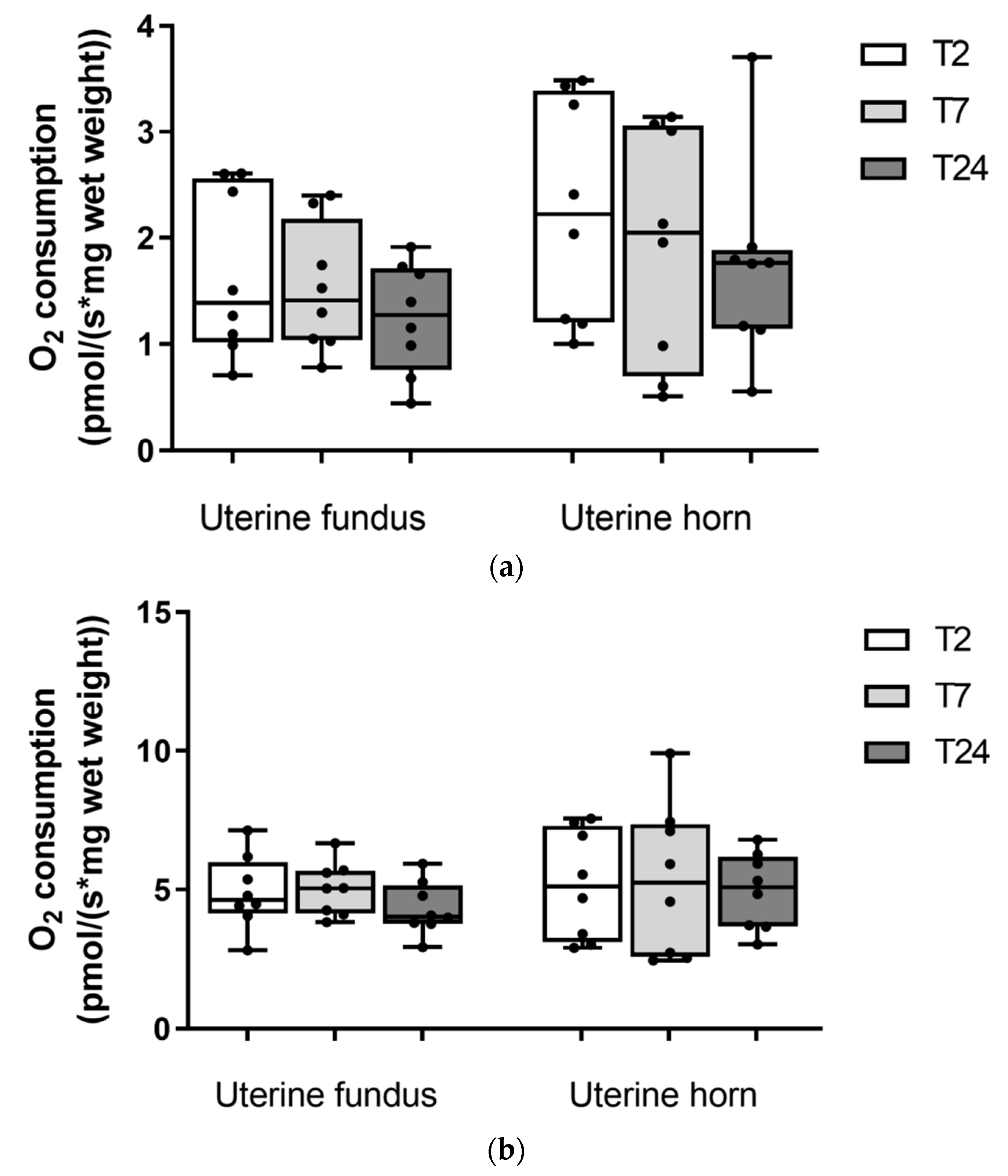
3.2. Mitochondrial Oxygen Consumption in Uterine Fundus and Horn after Two, Seven, and Twenty-Four Hours of Cold Ischemia

3.2.1. Uterine Fundus

OXPHOS by complex I state of the uterine fundus tended to decrease with time but this was not statistically significant (1.65 ± 0.28, 1.52 ± 0.21, 1.25 ± 0.18 pmol/s/mg ww, after 2, 7, and 24 h of cold ischemia, respectively, Figure 2a). Moreover, the OXPHOS by complex I&II state was not altered after 2, 7, and 24 h of cold ischemia (4.90 ± 0.47, 5.03 ± 0.34, 4.32 ± 0.34 pmol/s/mg ww, after 2, 7, and 24 h of cold ischemia, respectively, Figure 2b).

3.2.2. Uterine Horn

Similarly, OXPHOS by complex I state tended to decrease not significantly, after 2, 7, and 24 h’ cold ischemia (2.26 ± 0.37, 1.93 ± 0.39, 1.73 ± 0.33 pmol/s/mg ww, respectively, Figure 2a). Moreover, the OXPHOS by complex I&II state was not altered after 2, 7, and 24 h of cold ischemia (5.18 ± 0.70, 5.33 ± 0.97, 4.95 ± 0.48 pmol/s/mg ww, after 2, 7, and 24 h of cold ischemia, respectively, Figure 2b).

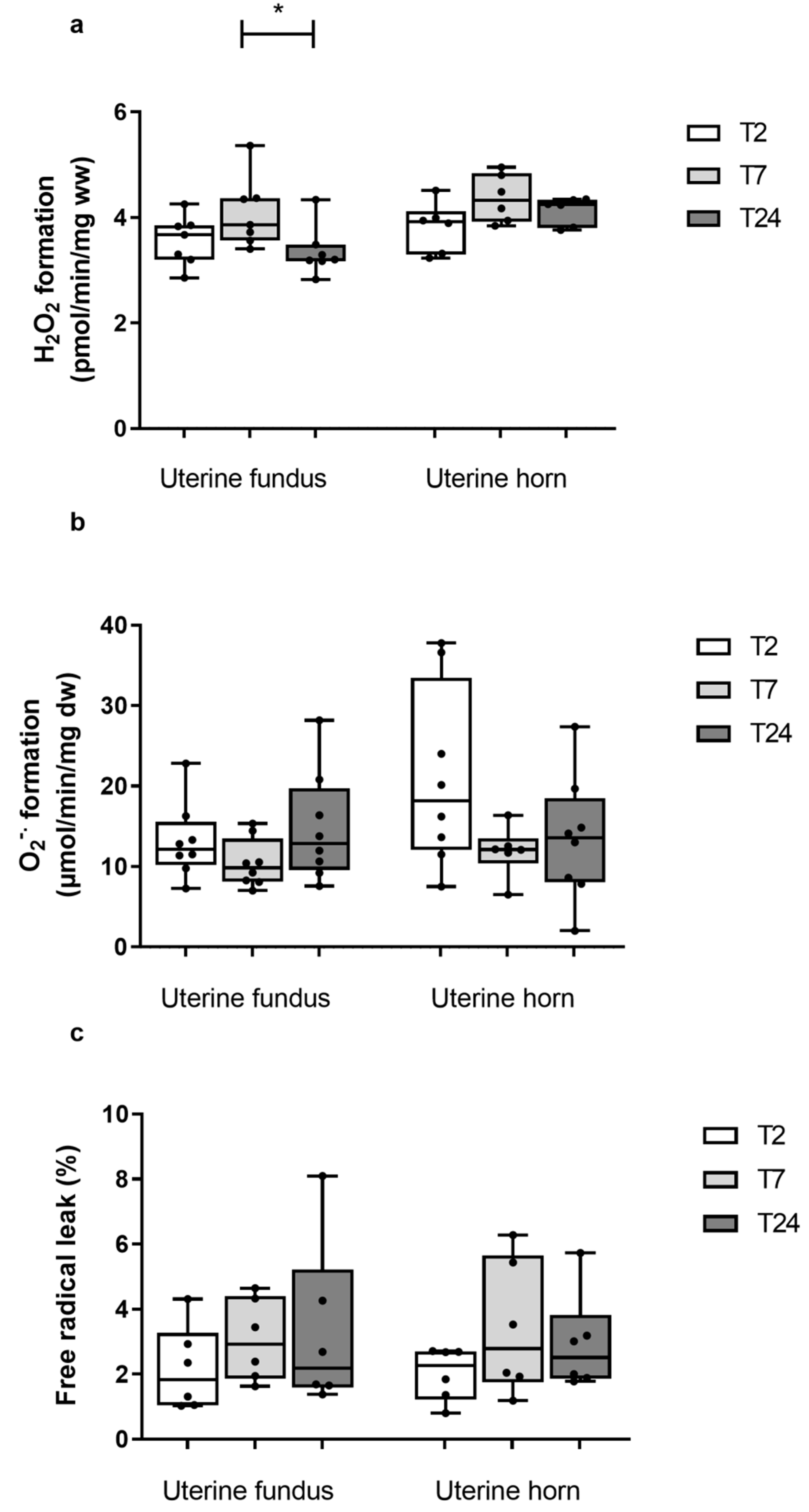
3.3. ROS Production at the Uterine Fundus and Horn after Two, Seven, and Twenty-Four Hours of Cold Ischemia

3.3.1. Uterine Fundus

Concerning H2O2 production, a significant but transient increase was observed after 7 h of ischemia (3.57 ± 0.18, 4.09 ± 0.25 pmol/min/mg ww, p < 0.05), the values at 24 h after ischemia being close then that observed after 2 h of ischemia (3.36 ± 0.17 pmol/min/mg ww, p < 0.01, Figure 3a).
Superoxide anion production was unchanged, regardless of the duration of cold ischemia (13.15 ± 1.66, 10.43 ± 1.07, 14.83 ± 2.42 pmol/min/mg dw, after 2, 7, 24 h of cold ischemia, respectively, Figure 3b).
Finally, the free radical leak (FRL), which indicates the number of oxygen molecules preferentially required to produce ROS (Figure 3c), did not change significantly (2.16 ± 0.53, 3.06 ± 0.52, 3.29 ± 1.05, after 2, 7, and 24 h of cold ischemia, respectively).

3.3.2. Uterine Horn

H2O2 production was not modified regardless of the duration of ischemia (3.81 ± 0.19, 4.36 ± 0.19, 4.12 ± 0.11 pmol/min/mg ww, after 2, 7, and 24 h of ischemia, respectively, Figure 3a).
Superoxide anion production did not change significantly when comparing 2, 7 and 24 h of cold ischemia (20.93 ± 3.97, 11.90 ± 1.28 and 13.44 ± 2.74 pmol/min/mg dw, respectively). (Figure 2b).
Considering the FRL, again no significant change was observed (2.01 ± 0.33, 3.40 ± 0.84, 2.93 ± 0.61 after 2, 7, and 24 h of ischemia, respectively, Figure 3c).

4. Discussion

This study is the first to examine the kinetic of myometrial mitochondrial respiration and reactive oxygen production at both uterine fundus and horn, two, seven and twenty-four h after cold ischemia (CI). The main results are that mitochondrial oxidative capacity remains preserved over a long period, together with a lack of major rise in ROS production. These data support that cold ischemia is relatively well tolerated by human uterine myometrium.
Ischemia is a complex pathophysiological process leading progressively to impairments in the mitochondrial respiratory chain, intracellular edema, acidosis, oxidative stress, and finally cell death. Often studied in many organs such as the heart, brain, kidney, lungs, skeletal muscle etc. [27,30,31,32,33], there are relatively few data investigating uterine muscle ischemia-induced change in mitochondrial function [34]. Accordingly, mainly other methods were used to analyze uterine ischemia effects [35]. However, since to date, no transplant has produced a viable pregnancy after more than 6 h of cold ischemia, there is still an urgent need to better understand CI effect on uterine muscle, and particularly on mitochondria that are largely involved in many key cellular mechanisms.
Interestingly, our results demonstrate that cold ischemia (better corresponding to the clinical setting than warm ischemia) did not significantly alter uterine myometrium mitochondrial respiration, whatever the site analyzed. Thus, although porcine uterine regions might have different sensitivities to hypoxia [36], both human uterine fundus and horn remained unaltered by short term and prolonged CI supporting that if present, an eventual mitochondrial dysfunction should have been minimal. In agreement with these data, Del Priore et al. did not observe any histological modification as assessed by electron microscopy after 12 h of cold ischemia [37]. Similarly, no histological modification or increased apoptosis, as assessed by TUNEL assay, was shown after 24 h of CI [21]. In 2005, Wranning et al. studied changes in energy metabolism (ATP) and in the capacity of human uterus to contract after various lengths of CI [38]. They observed a decrease in the spontaneous contractility, irrespective of the duration of ischemia. No histological modification was observed on electron microscopy, but degeneration of the vesicular cytoplasm and chromatin condensation were observed after 24 h of CI mainly when a protective solution was not used. The authors concluded that that human myometrial tissue can tolerate cold ischaemia for at least 6 h, but mitochondrial respiration determination was not performed. Further, the fact that swollen mitochondria were observed after 8 h of warm uterine ischemia [34] suggests that warm ischemia is likely to be much more harmful than cold ischemia which reduces tissue metabolism.
Oxidative stress is also a key mechanism in ischemia pathophysiology [38]. When investigating glutathione pathway in uterine myometrium in humans or ewes, respectively, an increased oxidative stress was observed mainly after 24 h of CI [39,40]. On the other hand, in our study, despite using three complementary approaches as often suggested, we observed no significant increase in hydrogen peroxide, superoxide anion productions or mitochondrial free radical leak during this experiment except at 7 h in uterine fundus for H2O2. However, such an increase was transient and concerned only one marker of oxidative stress. Therefore, the general picture is that no major change in ROS production occurred in this experiment, although further studies will be useful to determine the antioxidant status in such a setting. This is in agreement with the lack of mitochondrial dysfunction observed since mitochondria are known to be also largely involved in ROS production, particularly in the ones we studied here. Globally, these findings confirm that uterine muscle has a marked ability to tolerate cold ischemia well, corresponding to its physiological exposure to ischemia, particularly during menstrual cycles and labor.

5. Conclusions

In conclusion, although mitochondrial respiration tended to decrease over time, this was not statistically significant and this study shows that uterus mitochondria are relatively resistant to long periods of ischemia. We nevertheless cannot totally exclude evidence of other mitochondrial dysfunction potentially involving mitochondrial dynamic, calcium handling etc. Further, no major increase in ROS production was observed until 24h after surgery. Thus, other predictive markers of uterine function should be investigated, probably including studies on metabolomics or progenitor stem cells of the endometrium which play an essential role in physiological remodeling and regeneration of the human uterus and might be impaired by prolonged ischemia. Additionally, reperfusion studies will be useful since it is a critical time, allowing increased oxidative stress related to oxygen reflow.

Author Contributions

Conceptualization, M.P., A.-L.C., B.G. and O.G.; methodology, A.-L.C., I.G. and F.G.; validation, M.P., A.-L.C., O.G. and B.G.; formal analysis, M.P., A.-L.C., A.B.; investigation, M.P., A.-L.C. and O.G.; writing—original draft preparation, M.P., A.-L.C., F.G., I.G., A.B., B.G. and O.G; writing—review and editing, M.P., A.-L.C., I.G., F.G, A.B., B.G. and O.G. All authors have read and agreed to the published version of the manuscript.

Funding

There is no funding Statement.

Institutional Review Board Statement

The study was conducted according to the guidelines of the Declaration of Helsinki, and approved by the Ethics Committee of French Committee for the Protection of Persons (protocol code RIN 2018–HUS N° 7222, approved the 15 November 2018).

Informed Consent Statement

Informed consent was obtained from all subjects involved in the study. Written informed consent has been obtained from the patient(s) to publish this paper.

Data Availability Statement

The data presented in this study are available on request from the corresponding author.

Acknowledgments

We would like to thank ADIRAL (Association D’aide aux Insuffisants Respiratoires d’Alsace-Lorraine, France) and OCOVAS (Association des Opérés du Cœur et des Vaisseaux à Strasbourg, France) for their help in acquiring Oroboros and EPR equipments. This work was sponsored by the University Hospitals of Strasbourg (DRCI) for regulatory and ethic submission.

Conflicts of Interest

The authors declare no conflict of interest.

References

  1. Milliez, J. Uterine Transplantation: FIGO Committee for the Ethical Aspects of Human Reproduction and Women’s Health. Int. J. Gynecol. Obstet. 2009, 106, 270. [Google Scholar] [CrossRef] [PubMed]
  2. Fageeh, W.; Raffa, H.; Jabbad, H.; Marzouki, A. Transplantation of the Human Uterus. Int. J. Gynecol. Obstet. 2002, 76, 245–251. [Google Scholar] [CrossRef]
  3. Ozkan, O.; Akar, M.E.; Ozkan, O.; Erdogan, O.; Hadimioglu, N.; Yilmaz, M.; Gunseren, F.; Cincik, M.; Pestereli, E.; Kocak, H.; et al. Preliminary Results of the First Human Uterus Transplantation from a Multiorgan Donor. Fertil. Steril. 2013, 99, 470–476.e5. [Google Scholar] [CrossRef] [PubMed]
  4. Brännström, M.; Johannesson, L.; Dahm-Kähler, P.; Enskog, A.; Mölne, J.; Kvarnström, N.; Diaz-Garcia, C.; Hanafy, A.; Lundmark, C.; Marcickiewicz, J.; et al. First Clinical Uterus Transplantation Trial: A Six-Month Report. Fertil. Steril. 2014, 101, 1228–1236. [Google Scholar] [CrossRef]
  5. Flyckt, R.; Kotlyar, A.; Arian, S.; Eghtesad, B.; Falcone, T.; Tzakis, A. Deceased Donor Uterine Transplantation. Fertil. Steril. 2017, 107, e13. [Google Scholar] [CrossRef] [Green Version]
  6. Testa, G.; Koon, E.C.; Johannesson, L.; McKenna, G.J.; Anthony, T.; Klintmalm, G.B.; Gunby, R.T.; Warren, A.M.; Putman, J.M.; de Prisco, G.; et al. Living Donor Uterus Transplantation: A Single Center’s Observations and Lessons Learned from Early Setbacks to Technical Success. Am. J. Transpl. 2017, 17, 2901–2910. [Google Scholar] [CrossRef] [Green Version]
  7. Wei, L.; Xue, T.; Tao, K.-S.; Zhang, G.; Zhao, G.-Y.; Yu, S.-Q.; Cheng, L.; Yang, Z.-X.; Zheng, M.-J.; Li, F.; et al. Modified Human Uterus Transplantation Using Ovarian Veins for Venous Drainage: The First Report of Surgically Successful Robotic-Assisted Uterus Procurement and Follow-up for 12 Months. Fertil. Steril. 2017, 108, 346–356.e1. [Google Scholar] [CrossRef] [Green Version]
  8. Brucker, S.Y.; Brännström, M.; Taran, F.-A.; Nadalin, S.; Königsrainer, A.; Rall, K.; Schöller, D.; Henes, M.; Bösmüller, H.; Fend, F.; et al. Selecting Living Donors for Uterus Transplantation: Lessons Learned from Two Transplantations Resulting in Menstrual Functionality and Another Attempt, Aborted after Organ Retrieval. Arch. Gynecol. Obs. 2018, 297, 675–684. [Google Scholar] [CrossRef]
  9. Puntambekar, S.; Telang, M.; Kulkarni, P.; Jadhav, S.; Sathe, R.; Warty, N.; Puntambekar, S.; Kade, S.; Panse, M.; Agarkhedkar, N.; et al. Laparoscopic-Assisted Uterus Retrieval From Live Organ Donors for Uterine Transplant. J. Minim. Invasive Gynecol. 2018, 25, 571–572. [Google Scholar] [CrossRef]
  10. Puntambekar, S.; Puntambekar, S.; Telang, M.; Kulkarni, P.; Date, S.; Panse, M.; Sathe, R.; Agarkhedkar, N.; Warty, N.; Kade, S.; et al. Novel Anastomotic Technique for Uterine Transplant Using Utero-Ovarian Veins for Venous Drainage and Internal Iliac Arteries for Perfusion in Two Laparoscopically Harvested Uteri. J. Minim. Invasive Gynecol. 2019, 26, 628–635. [Google Scholar] [CrossRef]
  11. Brännström, M.; Enskog, A.; Kvarnström, N.; Ayoubi, J.M.; Dahm-Kähler, P. Global Results of Human Uterus Transplantation and Strategies for Pre-Transplantation Screening of Donors. Fertil. Steril. 2019, 112, 3–10. [Google Scholar] [CrossRef] [PubMed]
  12. Chmel, R.; Novackova, M.; Janousek, L.; Matecha, J.; Pastor, Z.; Maluskova, J.; Cekal, M.; Kristek, J.; Olausson, M.; Fronek, J. Revaluation and Lessons Learned from the First 9 Cases of a Czech Uterus Transplantation Trial: Four Deceased Donor and 5 Living Donor Uterus Transplantations. Am. J. Transpl. 2019, 19, 855–864. [Google Scholar] [CrossRef] [PubMed]
  13. Jones, B.; Saso, S.; Bracewell-Milnes, T.; Thum, M.; Nicopoullos, J.; Diaz-Garcia, C.; Friend, P.; Ghaem-Maghami, S.; Testa, G.; Johannesson, L.; et al. Human Uterine Transplantation: A Review of Outcomes from the First 45 Cases. BJOG Int. J. Obs. Gy. 2019, 126, 1310–1319. [Google Scholar] [CrossRef] [PubMed]
  14. Johannesson, L.; Koon, E.C.; Bayer, J.; McKenna, G.J.; Wall, A.; Fernandez, H.; Martinez, E.J.; Gupta, A.; Ruiz, R.; Onaca, N.; et al. Dallas UtErus Transplant Study: Early Outcomes and Complications of Robot-Assisted Hysterectomy for Living Uterus Donors. Transplantation 2021, 105, 225–230. [Google Scholar] [CrossRef]
  15. Jones, B.P.; Kasaven, L.; Vali, S.; Saso, S.; Jalmbrant, M.; Bracewell-Milnes, T.; Thum, M.-Y.; Quiroga, I.; Friend, P.; Diaz-Garcia, C.; et al. Uterine Transplantation: Review of Livebirths and Reproductive Implications. Transplantation 2021, 105, 1695–1707. [Google Scholar] [CrossRef]
  16. Tummers, P.; Göker, M.; Dahm-Kahler, P.; Brännström, M.; Tullius, S.G.; Rogiers, X.; Van Laecke, S.; Weyers, S. Meeting Report: First State-of-the-Art Meeting on Uterus Transplantation. Transplantation 2019, 103, 455–458. [Google Scholar] [CrossRef]
  17. Ejzenberg, D.; Andraus, W.; Baratelli Carelli Mendes, L.R.; Ducatti, L.; Song, A.; Tanigawa, R.; Rocha-Santos, V.; Macedo Arantes, R.; Soares, J.M.; Serafini, P.C.; et al. Livebirth after Uterus Transplantation from a Deceased Donor in a Recipient with Uterine Infertility. Lancet 2018, 392, 2697–2704. [Google Scholar] [CrossRef]
  18. Chmel, R.; Cekal, M.; Pastor, Z.; Chmel, R.; Paulasova, P.; Havlovicova, M.; Macek, M.; Novackova, M. Assisted Reproductive Techniques and Pregnancy Results in Women with Mayer-Rokitansky-Küster-Hauser Syndrome Undergoing Uterus Transplantation: The Czech Experience. J. Pediatric Adolesc. Gynecol. 2020, 33, 410–414. [Google Scholar] [CrossRef]
  19. Flyckt, R.; Falcone, T.; Quintini, C.; Perni, U.; Eghtesad, B.; Richards, E.G.; Farrell, R.M.; Hashimoto, K.; Miller, C.; Ricci, S.; et al. First Birth from a Deceased Donor Uterus in the United States: From Severe Graft Rejection to Successful Cesarean Delivery. Am. J. Obstet. Gynecol. 2020, 223, 143–151. [Google Scholar] [CrossRef]
  20. Brännström, M.; Bokström, H.; Dahm-Kähler, P.; Diaz-Garcia, C.; Ekberg, J.; Enskog, A.; Hagberg, H.; Johannesson, L.; Kvarnström, N.; Mölne, J.; et al. One Uterus Bridging Three Generations: First Live Birth after Mother-to-Daughter Uterus Transplantation. Fertil. Steril. 2016, 106, 261–266. [Google Scholar] [CrossRef] [Green Version]
  21. Gauthier, T.; Piver, P.; Pichon, N.; Bibes, R.; Guillaudeau, A.; Piccardo, A.; Pesteil, F.; Tricard, J.; Gardet, E.; Laskar, M.; et al. Uterus Retrieval Process from Brain Dead Donors. Fertil. Steril. 2014, 102, 476–482. [Google Scholar] [CrossRef] [PubMed]
  22. Tricard, J.; Ponsonnard, S.; Tholance, Y.; Mesturoux, L.; Lachatre, D.; Couquet, C.; Terro, F.; Yardin, C.; Marquet, P.; Piccardo, A.; et al. Uterus Tolerance to Extended Cold Ischemic Storage after Auto-Transplantation in Ewes. Eur. J. Obstet. Gynecol. Reprod. Biol. 2017, 214, 162–167. [Google Scholar] [CrossRef] [PubMed]
  23. Kisu, I.; Banno, K.; Matoba, Y.; Adachi, M.; Aoki, D. Basic Research on Uterus Transplantation in Nonhuman Primates in Japan: Uterus Transplant Research in Japan. J. Obstet. Gynaecol. Res. 2018, 44, 1871–1881. [Google Scholar] [CrossRef] [PubMed]
  24. Pottecher, J.; Guillot, M.; Belaidi, E.; Charles, A.-L.; Lejay, A.; Gharib, A.; Diemunsch, P.; Geny, B. Cyclosporine A Normalizes Mitochondrial Coupling, Reactive Oxygen Species Production, and Inflammation and Partially Restores Skeletal Muscle Maximal Oxidative Capacity in Experimental Aortic Cross-Clamping. J. Vasc. Surg. 2013, 57, 1100–1108.e2. [Google Scholar] [CrossRef] [Green Version]
  25. Pizzimenti, M.; Riou, M.; Charles, A.-L.; Talha, S.; Meyer, A.; Andres, E.; Chakfé, N.; Lejay, A.; Geny, B. The Rise of Mitochondria in Peripheral Arterial Disease Physiopathology: Experimental and Clinical Data. JCM 2019, 8, 2125. [Google Scholar] [CrossRef] [Green Version]
  26. Paradis, S.; Charles, A.-L.; Meyer, A.; Lejay, A.; Scholey, J.W.; Chakfé, N.; Zoll, J.; Geny, B. Chronology of Mitochondrial and Cellular Events during Skeletal Muscle Ischemia-Reperfusion. Am. J. Physiol.-Cell Physiol. 2016, 310, C968–C982. [Google Scholar] [CrossRef] [Green Version]
  27. Charles, A.-L.; Guilbert, A.-S.; Guillot, M.; Talha, S.; Lejay, A.; Meyer, A.; Kindo, M.; Wolff, V.; Bouitbir, J.; Zoll, J.; et al. Muscles Susceptibility to Ischemia-Reperfusion Injuries Depends on Fiber Type Specific Antioxidant Level. Front. Physiol. 2017, 8, 52. [Google Scholar] [CrossRef] [Green Version]
  28. Diana, M.; Noll, E.; Diemunsch, P.; Dallemagne, B.; Benahmed, M.A.; Agnus, V.; Soler, L.; Barry, B.; Namer, I.J.; Demartines, N.; et al. Enhanced-Reality Video Fluorescence: A Real-Time Assessment of Intestinal Viability. Ann. Surg. 2014, 259, 700–707. [Google Scholar] [CrossRef]
  29. Lejay, A.; Charles, A.-L.; Georg, I.; Goupilleau, F.; Delay, C.; Talha, S.; Thaveau, F.; Chakfé, N.; Geny, B. Critical Limb Ischaemia Exacerbates Mitochondrial Dysfunction in ApoE−/− Mice Compared with ApoE+/+ Mice, but N-Acetyl Cysteine Still Confers Protection. Eur. J. Vasc. Endovasc. Surg. 2019, 58, 576–582. [Google Scholar] [CrossRef]
  30. Murphy, M.P.; Hartley, R.C. Mitochondria as a Therapeutic Target for Common Pathologies. Nat. Rev. Drug Discov. 2018, 17, 865–886. [Google Scholar] [CrossRef] [Green Version]
  31. Kamarauskaite, J.; Baniene, R.; Trumbeckas, D.; Strazdauskas, A.; Trumbeckaite, S. Increased Succinate Accumulation Induces ROS Generation in In Vivo Ischemia/Reperfusion-Affected Rat Kidney Mitochondria. BioMed Res. Int. 2020, 2020, 8855585. [Google Scholar] [CrossRef] [PubMed]
  32. Go, K.L.; Lee, S.; Zendejas, I.; Behrns, K.E.; Kim, J.-S. Mitochondrial Dysfunction and Autophagy in Hepatic Ischemia/Reperfusion Injury. BioMed Res. Int. 2015, 2015, 183469. [Google Scholar] [CrossRef] [PubMed] [Green Version]
  33. Mansour, Z.; Charles, A.L.; Kindo, M.; Pottecher, J.; Chamaraux-Tran, T.N.; Lejay, A.; Zoll, J.; Mazzucotelli, J.P.; Geny, B. Remote Effects of Lower Limb Ischemia-Reperfusion: Impaired Lung, Unchanged Liver, and Stimulated Kidney Oxidative Capacities. BioMed Res. Int. 2014, 2014, 392390. [Google Scholar] [CrossRef] [PubMed]
  34. Adachi, M.; Kisu, I.; Nagai, T.; Emoto, K.; Banno, K.; Umene, K.; Nogami, Y.; Tsuchiya, H.; Itagaki, I.; Kawamoto, I.; et al. Evaluation of Allowable Time and Histopathological Changes in Warm Ischemia of the Uterus in Cynomolgus Monkey as a Model for Uterus Transplantation. Acta Obs. Gynecol Scand. 2016, 95, 991–998. [Google Scholar] [CrossRef] [PubMed] [Green Version]
  35. Tardieu, A.; Dion, L.; Lavoué, V.; Chazelas, P.; Marquet, P.; Piver, P.; Sallée, C.; Aubard, Y.; Barin-Le Guellec, C.; Favreau, F.; et al. The Key Role of Warm and Cold Ischemia in Uterus Transplantation: A Review. JCM 2019, 8, 760. [Google Scholar] [CrossRef] [PubMed] [Green Version]
  36. Kwiatkowska, J.; Wąsowska, B.; Gilun, P. Expression of Hypoxia Inducible Factor 1α and Antioxidant Enzymes: Superoxide Dismutases-1 and -2 in Ischemic Porcine Endometrium. Reprod. Biol. 2017, 17, 289–293. [Google Scholar] [CrossRef] [PubMed]
  37. Del Priore, G.; Stega, J.; Sieunarine, K.; Ungar, L.; Smith, J.R. Human Uterus Retrieval From a Multi-Organ Donor. Obstet. Gynecol. 2007, 109, 101–104. [Google Scholar] [CrossRef]
  38. Guillot, M.; Charles, A.L.; Chamaraux-Tran, T.N.; Bouitbir, J.; Meyer, A.; Zoll, J.; Schneider, F.; Geny, B. Oxidative stress precedes skeletal muscle mitochondrial dysfunction during experimental aortic cross-clamping but is not associated with early lung, heart, brain, liver, or kidney mitochondrial impairment. J. Vasc. Surg. 2014, 60, 1043–1051. [Google Scholar] [CrossRef] [Green Version]
  39. Wranning, C.A.; Mölne, J.; El-Akouri, R.R.; Kurlberg, G.; Brännström, M. Short-Term Ischaemic Storage of Human Uterine Myometrium—Basic Studies towards Uterine Transplantation. Hum. Reprod. 2005, 20, 2736–2744. [Google Scholar] [CrossRef] [Green Version]
  40. Tardieu, A.; Chazelas, P.; Faye, P.-A.; Favreau, F.; Nadal-Desbarats, L.; Sallée, C.; Margueritte, F.; Couquet, C.-Y.; Marquet, P.; Guellec, C.B.-L.; et al. Changes in the Metabolic Composition of Storage Solution with Prolonged Cold Ischemia of the Uterus. J. Assist. Reprod. Genet. 2019, 36, 1169–1178. [Google Scholar] [CrossRef]
Figure 1. Surgical design. Two samples of one cm3 myometrium per uterus was removed, one at the fundal midline and the other at the right uterine horn.
Oxygen 02 00002 g001
Figure 2. Mitochondrial respiration at the uterine fundus and horn, after 2, 7, and 24 h’ cold ischemia. (a) Complex I-linked OXPHOS state in uterine fundus and horn at different time of ischemia. (b) Complex I&II-linked OXPHOS state in uterine fundus and horn at different time of ischemia. Results are presented as mean ± sem.
Oxygen 02 00002 g002
Figure 3. ROS production measured in the different tissues and at different time of ischemia (2, 7, 24 h ischemia). (a) H2O2 production measured by high resolution oxygraphy, during complex I-linked OXPHOS state after 2, 7, and 24 hours’ cold ischemia (n = 8 per group). (b) Superoxide anion production measured by EPR at different times: after 2, 7 and 24 h’cold ischemia. (n = 7–8 per group). (c) Free radical leak at the uterine fundus and horn. These graphs represent the fraction of out-of-sequence electrons required to oxidize O2 into ROS in the respiratory chain (n = 8 per group). Results are presented as mean ± sem. * p < 0.05.
Oxygen 02 00002 g003
Table 1. Patient characteristics.
Clinical DataMean ± SD, Min–Max
Age (mean ± SD, min–max)47.6 ± 3.9 (44–57)
Gravidity (mean ± SD, min–max) *2.37 ± 1.49 (0–5)
Parity (mean ± SD, min–max) **2 ± 1.1 (0–4)
Smoker (n/8)1
Post–menopausal (n/8)0
Cardiovascular disease (n/8)2
Indication (n/8)4 myomas–3 adenomyosis–1 prolapse
Type of hysterectomy (n/8)5 laparoscopic–3 vaginal–0 laparotomy
Histology(n/8)2 myomas–3 adenomyosis
1 sarcoma–1 normal tissue
* number of previous pregnancies; ** number of previous deliveries.
Publisher’s Note: MDPI stays neutral with regard to jurisdictional claims in published maps and institutional affiliations.

