Review

Preanalytical Strategies for Native Mass Spectrometry Analysis of Protein Modifications, Complexes, and Higher-Order Structures

Analytical Development Laboratory, United States Pharmacopeia, Rockville, MD 20852, USA
AppliedChem 2025, 5(4), 35; https://doi.org/10.3390/appliedchem5040035
Submission received: 31 August 2025 / Revised: 19 October 2025 / Accepted: 11 November 2025 / Published: 18 November 2025

Abstract

Proteins are essential biological macromolecules that play key regulatory roles in all biological processes. Abnormalities in these processes are often reflected in proteins, manifesting as changes in their structure, sequence, folding state, stoichiometry, or spatial and temporal distribution. Proteins serve as biological targets for drugs and other therapeutics and can also function as therapeutic agents to restore normal biological functions by treating diseases. Hence, it is essential to study native protein species, their modifications, higher-order structures, and complexes, which can be extremely difficult due to the challenges in preserving their native conditions and the instrumental capability required for such analysis. High-resolution mass spectrometry (HRMS) instruments provide advanced technical capabilities to study intact protein species from their gas phase ions after the protein solution is sprayed into the mass spectrometers. However, there are debates about the gas-phase protein structures obtained through mass spectrometry and the resemblance to their biological native state. This review discusses various techniques for isolating, separating, and enriching intact protein species for their native mass spectrometry (nMS) analysis. Emerging technologies, such as automated sample preparation, ion mobility spectrometry, and ambient surface mass spectrometry, are briefly discussed. This review aims to serve as a general guideline for beginners, primarily focusing on the preanalytical strategies and critical instrument parameters for nMS analysis of intact proteins, proteoforms, protein complexes, and higher-order structures.

1. Introduction

Proteins are essential biomolecules composed of amino acid residues that fold into a specific three-dimensional native structure. Proteins can oligomerize, interact with other molecules, assemble into complexes, and play integral roles in carrying out various tasks critical for the structural integrity, function, and regulation of biological systems. Their native structure also affects their charge distribution and ligand-binding behavior, and is foundational to their ability to perform their intended biological functions [1]. Not all canonical proteins are the biologically active ones, and post-translational modifications (PTMs) are often required for rendering the biological function through regulating molecular interactions, enzymatic activity, and structural stability [2]. Single point mutation, PTM, misfolding, or aggregation can render proteins defective or toxic, disrupting the cellular homeostasis, and often serve as hallmarks of aberrant cell behavior [3]. Changes in the native structure of proteins can disrupt interactions with other molecules and can also significantly affect their utility as drug targets [4], which have been linked to several human diseases, including cancers, immune disorders, and neurodegeneration [5,6]. Thus, conformational and structural characterization of proteins, their complexes, and higher-order structures are essential for understanding normal and disease biology, as well as for advancing drug discovery and development. In addition to their diagnostic value, proteins such as monoclonal antibodies (mAbs) and antibody–drug conjugates (ADCs) have emerged as highly sought-after therapeutics over the past decade [7]. Modification of these therapeutic proteins can result in reduced efficacy as well as serious adverse effects [8]. Hence, determining the native state of the protein has become a forefront of biological research necessitating robust workflows for their isolation, separation, and enrichment under native-like conditions.
Biological activity of proteins cannot be directly measured from their expression level or abundance and depends on their structure, sequence, PTMs, localization, oligomerization state, folding pattern, and spatial distribution [9]. The success of studying endogenous proteins begins with their extraction from the matrix while preserving the native conditions. Hard extraction techniques such as precipitation, liquid–liquid extraction, solid phase extraction, etc., are not suitable due to exposure to non-physiological conditions such as high organics, extreme pH, ionic strength (high salt concentration), temperature, etc., that would alter the native structure of proteins [10] but are commonly used to control interaction and release of small molecule drugs and oligonucleotides. Exposure to harsh conditions may also lead to the generation of artifact proteoforms [11,12]. On the other hand, soft extraction techniques such as centrifugation, native gel electrophoresis, immunoaffinity purification, and ultrafiltration can potentially preserve the native or native-like state of proteins as they do not involve harsh or non-physiological conditions or can be adapted to avoid such conditions. Although some of the soft extraction workflows have limited high-throughput compatibility, they are better suited for native mass spectrometry (nMS) analysis of endogenous proteoforms, protein complexes, and higher-order structures.
Most commonly used biophysical techniques for analyzing the native structure of proteins include light scattering, circular dichroism spectroscopy (CDS), isothermal titration calorimetry (ITC), x-ray crystallography (XRC), nuclear magnetic resonance (NMR) spectroscopy, and mass spectrometry (MS). Light scattering-based approaches provide information about the size and the oligomeric state of a protein [13], whereas CDS and ITC can help in studying protein–protein interactions [14] and protein–small molecule interactions [15]. However, none of these methods can provide residue-specific information. XRC and NMR are both powerful techniques in analyzing the three-dimensional structure of proteins at atomic resolution [16,17]. The former requires high-quality crystals, while the latter requires a higher analyte concentration compared to mass spectrometry. Additionally, XRC and NMR-based approaches struggle to handle large molecular-weight proteins [18,19]. Mass spectrometry, on the other hand, is a highly sensitive analytical technique that offers higher resolution insights into native protein structure, interactions, modifications, subunit stoichiometry, and intersubunit connectivity [20,21] from a minimal amount of materials (picomoles to femtomoles) [22,23,24,25,26,27]. The field of protein mass spectrometry has gained new momentum over the past two decades due to advances in methodologies [28], mass spectrometry hardware [29], and bioinformatics [30], and enabled analysis of non-covalent interactions of proteins during the transition from solution to the gas phase [29,31,32], thereby deepening our understanding of biology and diseases. MS-based intact protein analysis is the most direct available technique to study proteoforms, conformation, interaction, and stoichiometry of proteins and protein complexes [33]. It also enables studying of protein complexes that are too large to analyze by NMR but too small for cryo-electron microscopy (cryo-EM), such as Toyocamycin nitrile hydratase (TNH) (86 kDa) [34] and bacterial biomineralization enzyme Mnx (210 kDa) [35]. Mass spectrometers, however, cannot physically separate biomolecules in the liquid state, but when coupled with a front-end separation, they can provide better resolution and sensitivity. A cleaner sample, free from unwanted matrix components and enriched for target protein species, significantly aids in successful nMS analysis of protein. Advancements have been made to automate some of these steps through direct hyphenation to the mass spectrometers, facilitating high-throughput operation, as discussed later in Section 5.1.
Proteins are primarily composed of proteinogenic amino acid residues [36]. Most eukaryotic proteins contain only L-amino acids; however, prokaryotic proteins, such as those derived from bacteria, have been reported to contain D-amino acids [37,38,39]. With the 20 common proteinogenic amino acids as the building blocks, many proteins have variants that share identical m/z values and charged states despite having similar or different amino acid sequences, e.g., hemoglobin, which has five variants (Hb C, E, D-Punjab, G-Accra, G-Siraj), have the same molecular weight and charge state distribution but differences in amino acid compositions [40], histones with numerous isobaric PTMs combinations [41] etc. Moreover, many proteins in intact form can have similar retention [42] or migration behavior [43], making their separation challenging. In addition, to maintain native-like conditions, specific buffers are often used that are not compatible with mass spectrometry-based analysis, e.g., TRIS, HEPES, PBS, MES, MOPS, etc. [44], and require exchange into an MS-compatible solvent. An abundance of protein of interest is another critical factor, and without enrichment, low-abundant proteins can be challenging to detect in biological samples. These challenges make nMS analysis of intact protein species a daunting task and also indicate that, as a standalone technique, MS may not be sufficient to fully characterize the endogenous protein landscape [33]. To minimize sample handling, online workflows that integrate protein isolation and detection in a single step are preferred. Hyphenated techniques, which combine two or more analytical instruments, can provide a more comprehensive analysis without artifacts, such as the integration of UV, light scattering, and mass spectrometry (MS). Specialized MS technologies, such as hydrogen deuterium exchange mass spectrometry (HDX-MS) [45] and crosslinking mass spectrometry (XL-MS) [46], have been developed for studying protein structure and interactions, but they require labeling of the protein species, and may also involve denaturing conditions [47] or digestion [48]. Kaulich et al. assessed the effect of different sample preparation workflows on proteoform identification by top-down proteomics and proposed a decision tree that is based on sample availability, desired throughput, physicochemical properties of proteins (mass and isoelectric point), and application [12]. Similar empirical analysis for native proteomics is not available to date. In this article, label-free workflows for the isolation, separation, and enrichment of intact proteins for nMS analysis are discussed. These workflows aim to facilitate the discovery of the native structure of proteins, protein complexes, higher-order structures, and endogenous proteoforms. For simplicity, native protein complexes, higher-order structures, and proteoforms are collectively referred to as protein species in this article. Offline techniques, such as immunoprecipitation, gel- and free-flow electrophoresis, and differential and density gradient centrifugation, are discussed, along with routinely hyphenated online techniques with mass spectrometers like liquid chromatography and capillary electrophoresis. Mass spectrometry techniques that have demonstrated success and could further synergize native protein analysis, such as ion mobility spectrometry (IMS) and ambient surface mass spectrometry (ASMS), are briefly reviewed. Additionally, a fully automated mass spectrometry workflow for protein isolation and analysis is discussed. For comprehensive protein purification workflows and best practices in top-down mass spectrometry-based intact protein analysis, readers are encouraged to consult available excellent review articles [49,50,51,52,53].

2. General Considerations for Native Mass Spectrometry Analysis of Protein Species

Due to the size of protein oligomers, high-molecular-weight protein complexes, and therapeutics (mAbs), their analysis requires a fast and high-resolution mass spectrometer (HRMS) that offers high-efficiency ionization, desolvation, transmission, and fragmentation, and can operate at extremely low pressure. Unit resolution or older generation HRMSs do not provide these performances. In addition, under native-like conditions, charges on proteins remain significantly lower compared to the denatured state, as can be seen for the denatured and native mass spectra of carbonic anhydrase in Figure 1 [54] i.e., native proteins generate higher m/z values, and their analysis requires a quadrupole with an extended mass range.
Under native-like conditions, proteins remain folded, and their inner part remains inaccessible for taking up protons during ionization. Additionally, during the oligomerization of proteins, the loss of outer sections can lead to a reduction in charges relative to the increase in molecular weight. The net result is a slower charge increase in protein species under native-like conditions, which require mass spectrometers with extended mass range, larger low-frequency quadrupole, increased ion guide pressure, and higher acceleration potential [56]. Because of all these reasons, analysis of intact protein species under native-like conditions requires dedicated mass spectrometry instrumentation, and the readers are encouraged to review the article by Campuzano and Loo to learn more about the evolution of mass spectrometers for high m/z ion analysis [57]. Three main components for successful molecular analysis are sample preparation, instrument conditions for data acquisition, and data analysis. Best practices for sample preparation and instrument operation conditions for intact proteins are benchmarked by Donnelly et al. [53]. Kirshenbaum et al. reported a step-by-step protocol for protein complex analysis by structural mass spectrometry, highlighted the importance of instrument parameter optimization, and discussed strategies to disassemble protein complexes on the fly via application of collision energy, organic solvents, and pH regulation [58]. Tamara et al. comprehensively discussed different gas phase ion activation strategies as well as various aspects of data analysis for intact proteins, their modifications, and complexes [59]. These resources are excellent guides for nMS analysis of intact protein species. In this section, sample preparation, instrument conditions, and data analysis for intact protein species are briefly reviewed.

2.1. Sample Preparation

Native MS analysis of intact protein species generally employs reagents and methodologies that help to preserve the native-like conditions, such as milder buffer and softer ionization (e.g., electrospray ionization, ESI) [44]. The selection of the sample carrier solution and the mobile phase is critical, as their composition and pH will directly affect the native state of the protein species. Buffers like ammonium acetate, formate, and bicarbonate are commonly used in MS analysis to control the pH of the mobile phase due to their volatile nature [60]. Although ammonium acetate, formate, and bicarbonate solutions in water have a pH range of 6.50–8.00, ammonium acetate has been widely used in nMS analysis of protein species, as they are nondenaturing and do not form any unwanted non-volatile adducts [53,61,62]. However, the nondenaturing nature of ammonium acetate depends on its concentration, which must be carefully monitored during nMS analysis. Additionally, neither of these buffers has buffering capacity at pH 7.00, and ammonium acetate and bicarbonate can lead to CO2 bubble formation that can unfold proteins [63]. Charge-reducing agents such as trimethylammonium acetate, triethylammonium acetate, and ethylenediamine diacetate can be used to control the protein unfolding in the gas phase, generate lower charge states, and achieve greater conformational stability, all of which can be useful for nMS analysis of protein species [64,65,66]. Marchand and Gabelica reported the use of trimethylammonium acetate in analyzing polymorphic human telomeric DNA G-quadruplexes in the presence of KCl, which is a physiologically relevant cation [67]. Lemaire et al. demonstrated the benefits of using triethylammonium bicarbonate (TEAB) in analyzing heme/myoglobin complex, which was significantly more stable than those formed in ammonium bicarbonate and acetate solution [68]. Native proteins tend to generate a lower charge state, which is used as a diagnostic feature to determine the suitability of the buffer in preserving their native structure. Hadavi et al. identified 4-ethymorpholinium acetate as a promising alternative buffer to ammonium acetate [69]. During the isolation and enrichment of target protein species from biological systems, reagents or conditions are often used that can disrupt the native state of the protein species. For example, the use of detergents for cell lysis, mechanical agitation for tissue homogenization, the use of organic solvents for solubility, etc., which can ensure success in analyzing protein via denatured top-down proteomics analysis, would destroy native protein structure and result in artifacts [70]. Hence, these conditions need to be carefully controlled for nMS analysis of intact protein, its modification, complexes, and higher-order structure.

2.2. Instrument Parameters for Data Acquisition

Mass spectrometry parameters depend on the type of mass spectrometer and the acquisition strategy. Preserving the native state of the protein species from solution to the gas phase is crucial for successful nMS analysis, ensuring the retention of higher-order structure and avoiding the creation of artifacts that do not represent their natural state. Vimer et al. discussed various MS acquisition strategies for intact protein analysis [71]. Desolvation of intact protein and transfer of charges from the solvent to the protein molecules are critical to ensure analytical success and are generally achieved through ESI, during which protein samples are sprayed under high electric potential, ionized via solvent evaporation, and the continued shrinking of the electrosprayed droplets [72,73]. Ideally, each electrospray droplet should contain one protein species during the final evaporation step, which can potentially be achieved by maintaining low per-droplet protein solution-state concentrations in the low micromolar range, e.g., by using nano-ESI that produces smaller droplets compared to conventional ESI [72]. For small molecules, ion activation via application of collision energy (CE) fragments the analytes, generates product ions, and provides analyte-specific unique transitions that enable their accurate identification. However, for large molecules such as proteins, CE also assists in dislodging solvent molecules from the protein complex, unfolding of the protein molecules, and can also be used to drive dissociation of ligands from the protein–ligand complex [74,75]. To preserve the native state of the protein species, specifically the noncovalent protein complexes, gentle tuning conditions are used, which can lead to insufficient desolvation and peak broadening [33].
Desolvation can also be enhanced by increasing collision energy in the source or collision cell, increasing gas pressure, or capillary temperature [53]. Activating protein oligomers via gas collision and dissociating them into monomeric subunits has been commonly used to study protein complex subunit interaction, connectivity, and stoichiometry [76]. This strategy is known as collision-induced dissociation (CID), collisionally activated dissociation (CAD), or higher-energy collisional dissociation (HCD) in some instruments [77,78]. However, CID preferentially ejects peripheral subunits of a multimeric protein while other subunits may remain with the complex, leading to incomplete dissociation [79,80]. Lantz et al. demonstrated the utility of CAD in ejecting monomer from yeast alcohol dehydrogenase (ADH) homotetramer (147 kDa), which was then fragmented using HCD, yielding only 12% coverage [81]. Surface-induced dissociation (SID) is often preferred for analyzing protein complexes due to its rapid and more complete dissociating capability compared to CID [82]. It has been successfully used to study subunit connectivity [83], topology [84], assembly mechanisms [84], and ligand-binding localization [82]. However, caution must be taken to not overactivate the protein complexes, which may generate artifacts [83]. Fragmentation techniques such as electron capture/transfer/activated dissociation (ECD/ETD/EAD collectively termed as ExD) [84,85,86], ultraviolet photodissociation (UVPD) [87], or hybrid techniques such as EThcD (sequential ETD and HCD) [88] are reported to provide extensive fragmentation of intact protein species and better sequence coverage than HCD/CID, which enable confident identification of protein modifications. Among these fragmentation techniques, ExD and UVPD have been reported to generate fragments of protein complexes without disrupting the higher-order structure [33], with the latter yielding fragments from the more inaccessible regions of proteins [89]. More comprehensive discussions on the performance and application of these fragmentation techniques for intact protein species are reviewed in [33,59,90,91,92,93]. The addition of supercharging agents, such as meta-nitrobenzyl alcohol, can boost signal intensities of intact proteins while preserving their native state [94] and tends to benefit CID. In contrast, charge-reducing agents tend to help SID by increasing structurally informative products [95]. However, increasing desolvation may lead to disruption of labile modifications and non-covalent adducts, hence it needs to be critically optimized [53].
Analysis of intact protein species requires high-resolution MS analyzers, which are primarily of two types based on how they regulate ion movements, such as (i) ion trapping MS analyzers: Fourier-Transform Ion Cyclotron Resonance (FT-ICR) and Orbitrap, and (ii) moving ion MS analyzer: time-of-flight (TOF) [59], both of which can transmit and analyze large protein complexes and higher-order structures without excessive activation [33]. TOF-MS can detect many ions simultaneously that have distributions over several charged states. In contrast, FT-ICR and Orbitraps detect both m/z and z in parallel, but one ion at a time. Hence, TOF is better for relatively smaller protein molecules in a homogenous sample, whereas FT-ICR and Orbitraps are better suited for larger proteins and inhomogeneous samples. The operational principles and structures of these mass spectrometers vary, as do the parameters associated with them. Critical parameters such as gas flow, voltages (e.g., source/ion spray/capillary/cone, etc.), temperature, scan rate, cycle time, resolution, pressure, and ion activation need to be carefully assessed and optimized. Table 1 lists parameters for different analyzers for intact protein species nMS analysis and highlights different instrument parameters for three scenarios: (I) same protein complex analyzed by different mass spectrometers (FT-ICR vs. TOF) (entry 1 and entry 2, subscript 1), (II) different protein complexes (protein higher-order structure vs. protein–ligand complex) analyzed by same mass spectrometer (TOF) (entry two, subscript 1 and subscript 2), and (III) intact protein vs. protein complex analyzed by same mass spectrometer (TOF) (entry five, subscript 3 and subscript 4).
As shown in Table 1, MS parameters vary among the types of MS and the manufacturer, and they need to be carefully assessed and optimized based on the protein species. For example, the first two entries of Table 1 show the variation in instrument parameters between an FT-ICR and a Q-TOF MS for a hemotetrameric membrane protein complex, AquaporinZ. Entry two, subscript 1 and 2, demonstrates that for similar types of analytes (e.g., protein complexes), the MS parameters can have similarities as observed for a Q-TOF HRMS for the analysis of homotetrameric AquaporinZ and various noncovalent protein–ligand complexes (Lysozyme and tri-N-acetylchitotriose (NAG3), Trypsin/Pefabloc, Carbonic Anhydrase II/Chlorothiazide, β-Lactoglobulin A/Lauric Acid). Finally, subscripts 3 and 4 of the last entry in Table 1 highlight the differences among various MS parameters for a Q-TOF HRMS when used for analyzing protein complexes vs. an intact protein.

2.3. Data Analysis

Intact protein mass spectra acquired via ESI are different when compared to a small molecule mass spectra and are composed of multiple peaks that represent multiply charged species of the same protein. For large-molecular-weight proteins, protein complexes, and their higher-order structure, ESI is recommended over MALDI [53]. Hence, the focus of this section will be limited to data acquired via ESI-based approaches. As intact protein mass spectra generate multiple peaks of different mass-to-charge (m/z) ions, they need to be deconvoluted to yield the mass of the protein. PTMs and ligand-bound proteins are also represented among the multiply charged m/z peaks [98], and the visualization of the final mass spectra depends on the abundance of the species in the sample, as well as the ionization efficiency and the resolving power of the mass spectrometer. The presence of isotopes makes protein mass spectrometry data analysis more complex, as, unlike small molecules, where the most abundant isotopic peak typically corresponds to the monoisotopic peak, proteins have a complex isotopic distribution, often with no observable monoisotopic peak [99]. Additionally, overlapping signals in both precursor and product ion spectra, along with complex tandem mass spectrometry spectra, further complicate the analysis of intact protein MS data. Finally, confident identification and quantification of detected molecular species require spectral matching, which depends on high mass accuracy, sequence coverage, and database quality. Higher-order structures of protein also have high heterogeneity, which leads to broad and poorly resolved peaks and makes it extremely challenging to determine their charge and the accurate mass [100,101]. Several packages have been developed to date that offer solutions for analyzing MS data derived from intact proteins, their modifications, complexes, and higher order structures, such as ProSight Suite (PC/PD/PTM/Lite/Native) [102,103,104], MassExplorer [105], Informed-Proteomics [106], TopPIC [107], MS-Align+ [108], etc. Due to the complexity of the native protein MS data analysis, perhaps a targeted approach on a specific protein species or a group of relevant proteins is better than a discovery protein approach, for which it is imperative to adopt the right strategy for sample preparation. Critical analysis and comparison of listed software packages will be extremely resourceful; however, it is out of the scope of this review, the focus of which is on the preanalytical strategies for nMS analysis of intact protein species to ensure that the sample solution is clean, enriched for target protein species, and handled with caution to avoid artifacts. In the following sections, different strategies for isolation, separation, and enrichment of protein species for native mass spectrometry analysis are discussed.

3. Offline Strategies for Isolation, Separation, and Enrichment of Intact Protein Species for nMS Analysis

3.1. Immunoprecipitation (IP)

Target protein species can be enriched via a capture entity with high affinity and specificity for the target, a procedure known as affinity purification [109]. Enrichment of the target protein species can also be achieved through the removal of interfering components from the sample matrix, which is known as immunodepletion or immunoaffinity subtraction chromatography. In affinity purification, a bait molecule (e.g., small molecule ligand, antibody, receptor, enzyme, or nucleic acid) is immobilized on a solid support (e.g., magnetic beads) that interacts with the target protein species while other matrix components remain unbound and are cleared off by washing [109]. The protein of interest can finally be eluted by disrupting the interaction between the protein and the bait molecule via changes in pH, ionic strength, or polarity of the eluting agent. Immunoprecipitation (IP) is an affinity purification technique where an antibody is used as bait, which is typically immobilized on magnetic or agarose beads [110]. Bait antibody recognizes and binds to a specific amino acid sequence (epitope) of the target protein species, which is then eluted by disrupting the antigen–antibody interaction. IP has been successfully used for the isolation of target protein species for downstream mass spectrometry analysis. Care should be taken during the IP procedure, as changes in buffer composition, pH, the presence of organic reagents, extreme temperatures, etc., can perturb the native state of the protein species. The native variant of IP, known as native co-immunoprecipitation (Native co-IP), allows for the co-isolation of proteins that interact with the protein of interest and is used to study protein–protein interactions and protein complexes [111]. Native co-IP has been successfully used to study DNA-binding proteins (also known as chromatin) under native-like conditions [112]. Native co-IP can isolate chromatin-associated proteins via sequential extraction using Triton X-100, 0.5 M MgCl2, 0.5 M DTT, and 1 M NaCl, and was reported to be successful for studying proteins that interact with Topoisomerase I from mouse B-lymphocytic leukemia cell lines [111]. Chromatin immunoprecipitation (ChIP) demonstrated successful isolation of DNA-binding protein using conditions up to 1.5% SDS [113]. In addition to the challenges regarding the maintenance of the native-like conditions while ensuring sufficient recovery, other critical parameters that need careful assessment include binding affinity, non-specific binding, cross-reactivity, and suboptimal elution of the target proteins [114]. Antibodies can be engineered to target specific epitopes of the bait protein and proteoforms with particular modifications of specific amino acids, e.g., methylation of arginine and lysine motifs [115]. Guo et al. developed highly specific antibodies that successfully identified the methylation of 1000 arginine and 160 lysine residues in proteins from a human cell line (HCT116) and mouse tissues [115]. Antibodies that can selectively bind to proteins of different oligomeric states and sizes are also available; for example, antibodies with preferential binding to different amyloid beta oligomers (AβOs) were developed and used to isolate different AβOs via immunoprecipitation [116]. In recent years, automated high-throughput immunoprecipitation workflows have been established, as discussed in Section 5.1.
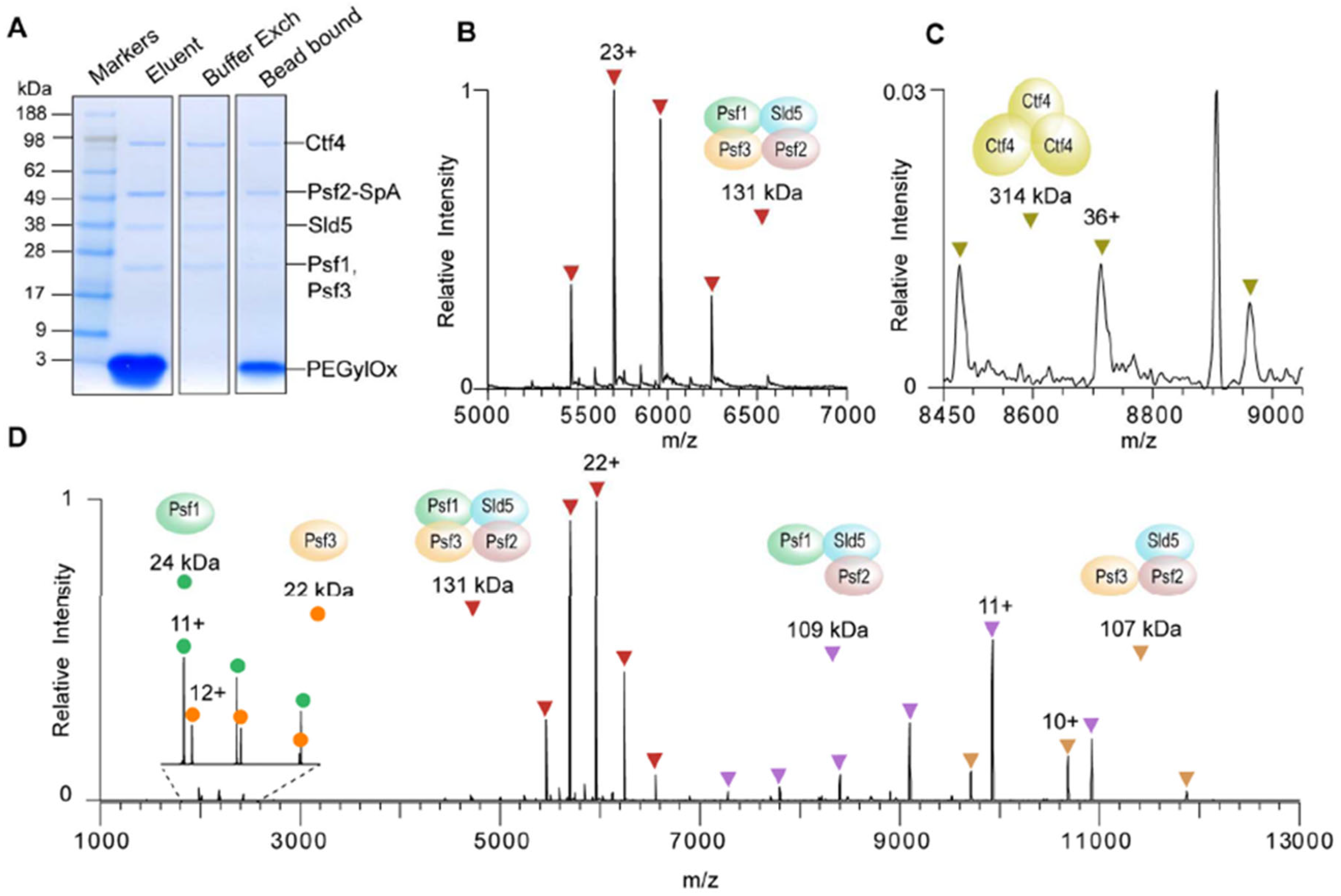
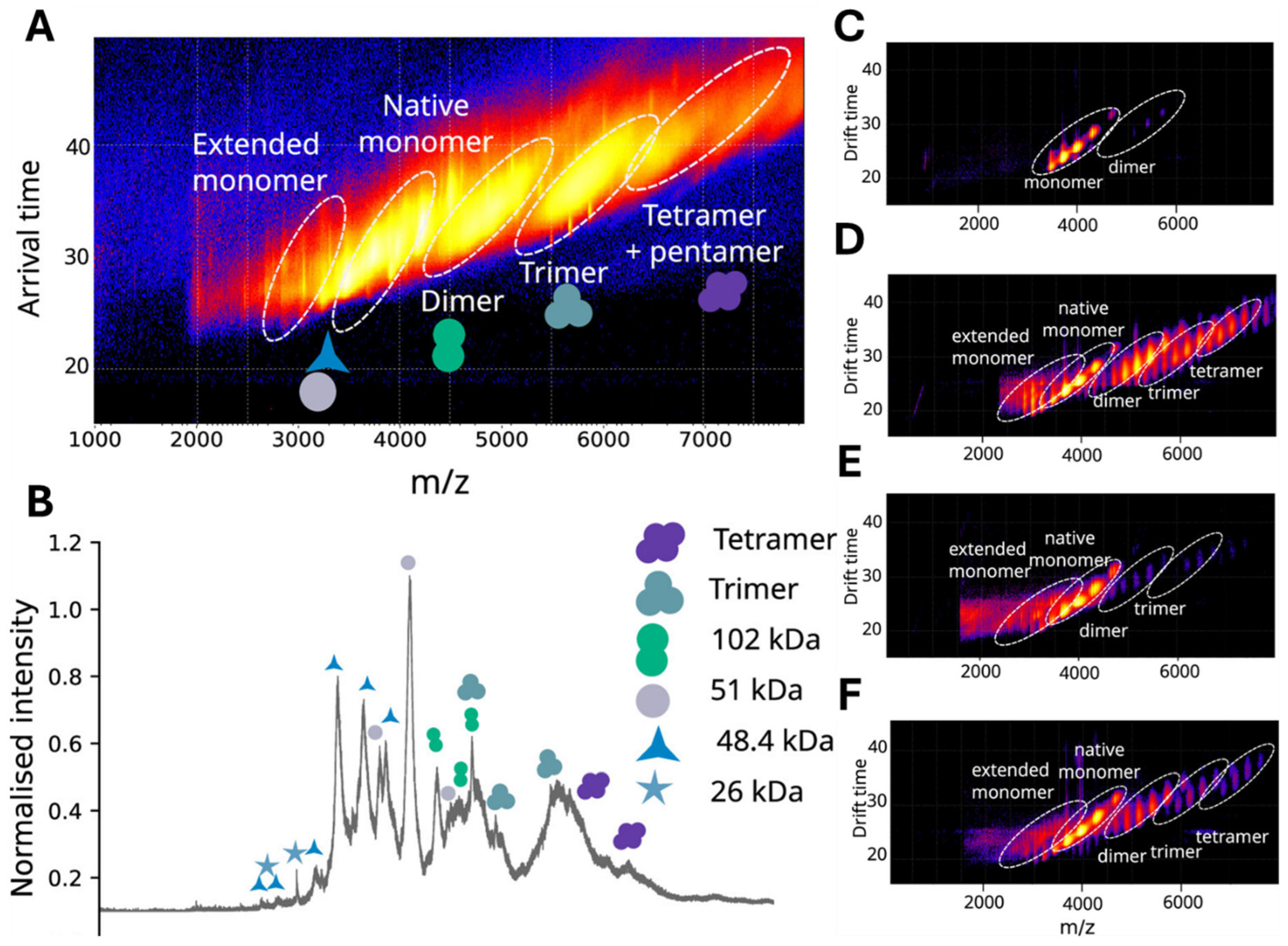
Affinity purification can also be adapted for epitope tagging, where specific tags such as FLAG, TAP, c-myc, glutathione S-transferase (GST), poly-histidine (His-Tag), and short peptide sequence (Strep-Tag) are fused to the target protein during expression using gene editing technology, e.g., CRISPR-Cas9 [117], and can be eluted more efficiently with an anti-epitope antibody [118]. These tagged proteins are used as baits to capture interacting proteins from complex biological samples to study protein–protein interaction, as reported by the BioPlex study, which identified 56,533 interactions with 10,961 proteins in HEK233T cells [119,120]. These tags are mostly C-terminal peptides and generally do not interfere with the function of the tagged protein [121] and are most cost-effective. Isolation of protein species via tagging, although reported to preserve their function, does not result in tagged protein species that resemble their endogenous form [122]. However, this approach can be used for studying subunit stoichiometry and intersubunit connectivity as demonstrated by Olinares et al., who used a Staphylococcus aureus-derived (SpA) tag to successfully purify a yeast-derived GINS complex (a protein complex comprising 4 proteins, Sld5, Psf1, Psf2, and Psf3) from 1 g of cryomilled yeast powder, due to the high affinity of the SpA tag for the Fc-domain of the antibody [121]. For nMS analysis, they employed collisional dissociation to disassemble the protein complex (Figure 2). This protocol can be adapted to automation from affinity purification to nMS analysis and is an excellent example of stoichiometry analysis of native protein complexes.
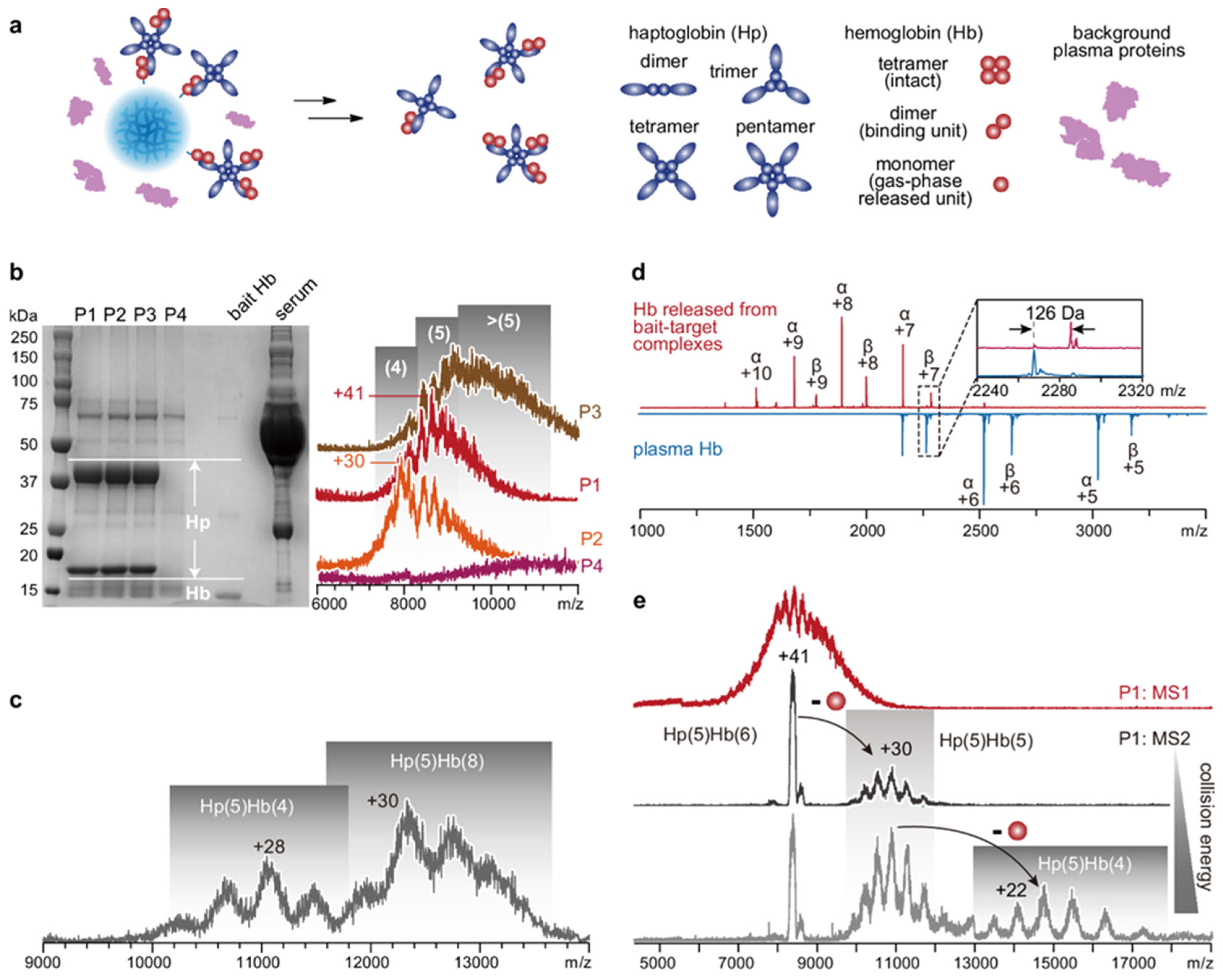
Achieving high recovery via affinity purification under native-like conditions could be challenging, as disrupting the antibody–antigen interaction may require nonphysiological pH, high salt concentration, or reagents that could denature the protein species [123]. Additionally, the nonspecific binding of the target protein on the solid support or beads leads to additional loss. Although blocking buffers containing albumin or milk protein could help prevent this, the addition of such external blocking agents may have an unprecedented effect on the native state of the protein species. Multiple washing and elution steps can dissociate weak protein complexes and cannot be used to determine the binding affinities [124]. Shao et al. recently developed a biofunctionalized dissolvable hydrogel microbead-based stationary phase for native affinity purification and mass spectrometry analysis of protein complexes, called Stationary-phase-dissolvable Native Affinity Purification and Mass Spectrometric characterization (SNAP-MS), which dissolves after capturing the target protein, relieving the need for elution, and ensuring complete recovery of the target protein with pico-mole level starting input, with a sample-to-data time as brief as 2 h [125]. In this workflow, the linear polyacrylamide chains from the dissolvable bead matrix were removed by SEC before MS analysis. They demonstrated the application of SNAP-MS in native analysis of various oligomeric states of avidin and haptoglobin, as well as integrity, subunit composition, and topology analysis of 20S proteasome (20S), a 28-mer protein complex (consisting of two outer layers of heptameric α subunits and two inner layers of heptameric β subunits) from Thermoplasma acidophilum (Figure 3).
As the beads are dissolvable, they eliminate the need for an elution step and prevent loss of target protein due to inefficient elution or protein adsorption, ensuring 100% recovery. The bait molecule can be released from the native protein complexes by collisional dissociation in the gas phase after being subjected to MS analysis, or could be subjected to gas phase separation, such as via ion mobility spectrometry. Adopting this technology in protein pulldown assays can potentially assist in the successful analysis of low-abundant proteins.

3.2. Gel Electrophoresis (GE)

Gel electrophoresis (GE) is primarily used for separating large molecules based on their charge and size as they migrate through a gel in the presence of an electrical field. Two types of gels are used for protein electrophoresis: agarose (for high-molecular-weight proteins, >200 kDa) [126] and polyacrylamide (for proteins in the range of 5–200 kDa) [127]. In principle, the larger the protein, the lower the percentage of the gel should be to achieve better separation. Unlike chromatography, gel-electrophoresis enables simultaneous and relatively faster separation, followed by straightforward visual representation of the resolved protein species without the need for complex instrumentation and data processing workflows. Primarily, there are two mechanisms to elute proteins from gel while maintaining native-like conditions, namely, (i) passive elution that works via diffusion and (ii) electroelution, where proteins elute due to the presence of an electric field forcing molecules out of the gel [128]. During passive elution, the region of interest of the gel is cut and exposed to water to allow the diffusion of proteins from the gel [129]. However, this method leads to low and variable recovery, longer incubation times, co-diffusion of polymers, and other contaminants necessitating a second step for their removal, and is reported to only work for molecules below 60 kDa [129]. Electroelution, on the other hand, is more efficient in eluting proteins from the gel and can potentially be used for nMS analysis when operated under nondenaturing conditions [130]. Readers are encouraged to review the work by Seelert and Krause [131] for exploring various types of GE conditions for the isolation of various proteins and protein complexes. Additionally, continuous elution devices have been developed where the proteins migrate and eventually move out of the gel into a buffer in the presence of an electric field; this has been successfully used in isolating protein complexes like photosystems, ATP-dependent enzymes, and active respiratory supercomplexes [131]. Isoelectric focusing (IEF) is another technique for the separation of protein species that avoids harsher conditions and has demonstrated success in separating membrane proteins in their native conditions with intact enzyme activity [132]. During native GE, separation of proteins is regulated by their shape, besides charge and size, and resolved proteins can be visualized by enzyme-linked staining [133]. Neutral to slightly alkaline electrophoretic buffer is used in native GE, where negatively charged proteins can be recovered by either passive diffusion or electroelution for mass spectrometry analysis. Although the presence of detergent is reported to be safe for preserving the covalent structural features of a protein, such use should be assessed case-by-case to ensure preservation of non-covalent interactions of proteins to ligands, cofactors, and metal ions [134]. Detergents should be removed before MS analysis, and the effect of detergent removal on gas-phase protein structure needs to be assessed. Additionally, omitting any heating step during gel electrophoresis is essential to help preserve the native-like conditions.
Blue-Native Polyacrylamide Gel Electrophoresis (BN-PAGE) provides a simple one-stop isolation of proteins and protein complexes from biological matrices and is suitable for 2D crystallization, electron microscopy, native electroblotting, or immunodetection [135]. Nowakowski et al. demonstrated retention of the protein–metal native interaction isolated from LLC-PK1 cell-derived protein (5–25 μg) using the native Sodium dodecyl sulfate polyacrylamide gel electrophoresis (SDS-PAGE) and Laser ablation inductively coupled plasma mass spectrometry (LA-ICP-MS) [134]. BN-PAGE has a fixed running pH of 7.50, reflecting the physiological pH, which helps to preserve the native state of the proteins and does not use any harsh detergents, but Coomassie Brilliant Blue (CBB) to impart negative charges to the proteins [136]. However, removal of CBB is also essential before MS analysis, which can be challenging without perturbing the native structure of the protein. Taurodeoxycholic acid is a milder detergent that is active at pH 7.50 and can be used as an alternative for some proteins upon optimization [137]. Zinc-imidazole staining is a milder technique recommended for visualizing proteins in the BN-PAGE workflow. In this method, the zinc ions and imidazole can be removed without applying harsher conditions, allowing for the recovery of proteins in native conditions using passive elution [138]. However, proteins with a high isoelectric point (pI) but a net negative charge, such as cytochrome C (pI: 10.0–10.5), face challenges migrating through the gel because they cannot overcome the substantial positive charge. Clear-native PAGE (CN-PAGE) resolves this limitation, which has demonstrated separation of acidic water-soluble and membrane proteins and complexes (pI < 7) while replacing the Coomassie dye with non-colored mixtures of anionic and neutral detergents, omitting the requirement of dye removal [139]. During CN-PAGE, proteins migrate based on their intrinsic charge and the pore size of the gel under milder conditions, demonstrating utility in studying the catalytic activity of mitochondrial ATP synthase [140]. Sarkozy and Guttman developed a technology by combining native and SDS capillary gel electrophoresis coupled to ESI-MS via a closed-circuit coaxial sheath flow reactor interface with online removal of SDS using γ-cyclodextrin in the sheath liquid, enabling intact protein analysis using 3.5 µg standard protein under native-like conditions [141].
GE-based approaches can also enable the identification of protein complexes and post-translational modifications of proteins under native-like conditions [142,143,144,145]. Pieper et al. demonstrated identification of 3700 distinct protein spots via two-dimensional gel-electrophoresis (2-DE) from human serum post-immunodepletion and anion exchange/size-exclusion chromatography, many of which were post-translationally modified [146]. Munawar et al. used native-GE to survey cytoplasmic, nuclear, and chromatin fractions of HEK293 cells and identified 217 protein complexes containing various subunits (3–15), demonstrating its utility as an upstream separation step for analyzing protein complexes and determining their cellular distribution [147]. More recently, four native membrane proteins, a potassium channel (KvAP), a sodium channel (NavMs), a water channel (GlpF), and a urea channel (HpUreI) were reported to be analyzed via CN-PAGE with the help of Glyco-DIBMA, a negatively charged amphiphilic copolymer-encapsulated nanodisc that helps the protein to avoid exposure to detergent [148]. These examples demonstrated the utility of gel-electrophoresis for isolating, visualizing, and analyzing native protein species as well as their potential in studying protein interactions, structure, and PTMs when combined with nMS.
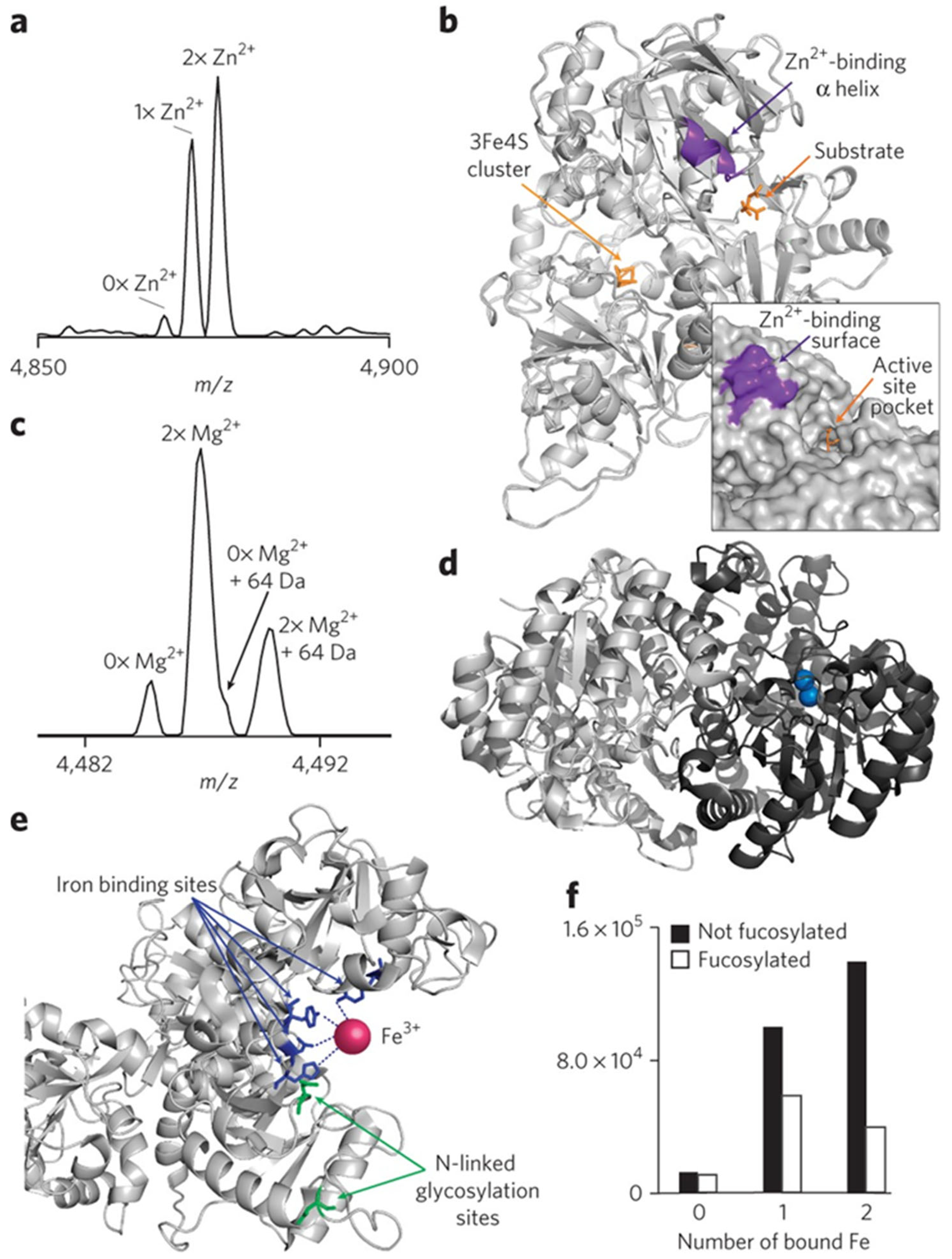
Stabilizers can help in preserving fragile protein complexes, but their addition into the buffer needs to be carefully assessed to ensure optimum migration and integrity of native proteins. Additionally, radicals of acrylamide can remain trapped inside the gel, leading to the oxidation of specific amino acids such as tryptophan, histidine, methionine, lysine, and cysteine, which damages the native structure of the protein [149]. Oxidative damage to the proteins can be prevented by antioxidant pretreatment [150,151]. But the effect of such a procedure on the native structure of the proteins needs to be assessed. The key step to utilize GE for nMS analysis is to obtain a relatively pure protein sample in sufficient quantity and in liquid form in an MS-friendly solvent. Several elution methods and devices for the extraction of proteins from gels have been developed, including electroelution into membrane traps from gel slices [152], continuous electroelution during gel electrophoresis [153], diffusion out of homogenized gel slices [154] etc. However, each of them suffers from various drawbacks, such as steps prone to inconsistency (e.g., gel slicing), large recovery volume, incompatibility for larger proteins (greater than 60 kDa), and high sample preparation time. These problems were addressed by a variant of a continuous electroelution device based on a semidry blotter that allows simultaneous elution of proteins from one or two-dimensional gels [155,156,157]. This device demonstrated the recovery of proteins up to 120 kDa in a 200 µL volume of 20 mM Tris HCl buffer (pH 8.00) or 2 mM phosphate buffer (pH 6.80), with minimal loss and preservation of their biological activity. Based on similar technology, Tran and Doucette developed a continuous elution tube gel electrophoresis for proteome fractionation to achieve effective separation of proteins over a broad mass range in the solution phase with high recovery from submicrogram to milligram level sample loading for MS analysis [158]. Although this device uses a gel column for separation, the proteins ultimately elute from the column in the gel-free solution phase; hence, the device is named Gel-Eluted Liquid Fraction Entrapment Electrophoresis (GELFrEE). Skinner et al. adopted the GELFrEE approach for native proteomics analysis (Native-GELFrEE) and demonstrated its application to separate proteins from complex mixtures (mouse heart extracts and fungal secretome) for nMS analysis [159] and reported the intact mass of cytoplasmic malate dehydrogenase (a homodimeric enzyme with a key role in the malate-aspartate shuttle). Building on these technologies, Skinner et al. characterized 125 intact endogenous protein complexes, 217 distinct proteoforms, and 28 metal-binding (Zn2+, Cu+, Mg2+, Ca2+, Fe3+) (Figure 4) proteins from mouse heart tissue and human cancer cell lines [160].
Skinner et al. also demonstrated amino acid and isoform variants using native top-down mass spectrometry (nTDMS) and characterized two mouse hemoglobin subunit β-1 chains each as a heterodimer, differing by three amino acids and bound to a hemoglobin α-chain. PTM identification can be achieved by denatured top-down mass spectrometry (dTDMS) [161]; however, conformational information as observed for mouse hemoglobin and the metal binding to murine aconitase obtained via this study would be unattainable in the former. This clearly demonstrates the power of nMS to identify an enzyme, its modification, and interactions with cofactors in a single experiment under normal physiological conditions. The ability to study any deviations from these can help predict abnormalities in a biological system. GELFrEE technique, however, requires detergents which need to be removed before mass spec acquisition, e.g., by using HiPPR detergent removal columns. Later, the manufacturer was acquired by Life Technologies, merged with Thermo Fisher, and then discontinued. Gel-based approaches for native protein separation, followed by nMS, although they provided the power of analyzing intact protein species while preserving the native or native-like state, the workflow required a high sample amount compared to bottom-up, denatured top-down proteomics [162] or crosslinking MS [163]. Additionally, the workflow from protein isolation to MS measurement is challenging to automate and directly hyphenate to mass spectrometers.

3.3. Free-Flow Electrophoresis (FFE)

Electromigration in free liquid has been known for several decades [164]; however, integration of this principle for high-throughput protein isolation lagged. Gel-free electrophoresis, also known as free-flow electrophoresis (FFE) or carrier-free electrophoresis (CFE), allows for comparable resolution to gel electrophoresis without the use of a matrix (i.e., gel), where separation occurs in a liquid state. During FFE, the analytes separate as they flow through a planar channel with a hydrodynamically pumped buffer stream (i.e., carrier buffer) perpendicular to which an electric field is applied that deflects the analytes laterally according to their size, charge, migration properties, buffer conductivity, pH, and the electric field strength [165,166]. The gentler condition used in FFE helps preserve the native integrity of the proteins and protein complexes [167] and offers higher analyte recovery than gel-based electrophoresis [168]. The major advantage of gel-free approaches is the removal of the transfer step from gel to liquid for mass spectrometry analysis, making it a desirable method for separating proteins under native-like conditions. Additionally, being in the liquid phase during separation enables easy integration into a chromatographic or flow injection system and compatibility with automation for direct hyphenation to the mass spectrometers. Various liquid fractionators that separate proteins based on free-flow electrophoresis have been commercialized, such as Elphor VAP, Octopus FFE, and ProTeam™ FFE, which can operate under both denatured and native-like conditions. Lee and Kwon reviewed the µ-FFE for prefractionation, enrichment, and purification of proteins from biological systems, and the readers are encouraged to consult this excellent review for further information on the utility of this technology [169]. However, FFE-based native separation suffers from buffer instabilities due to thermal convection, which leads to hydrodynamic and electrodynamic distortion affecting the separation efficiencies [168].
To overcome some of these shortcomings, miniaturized FFE devices such as microfluidic FFE (µ-FFE) technology were developed, offering rapid separation of proteins (<2 min) from a simple protein mixture in analytical scale (µL range) [169,170]. However, fabrication of µ-FFE can be complex, and they often suffer from bubble formation, the removal of which can be challenging [36,171]. Jeon et al. reported a pressure-driven flow-induced miniaturizing FFE (PDF-induced µ-FFE) device that overcomes this limitation, where the movement of particles is navigated by pressure in addition to the electrical field [172]. In this system, the electric field is applied parallel to the fluidic stream, enabling the separation of target particles from the sample in the opposite direction. This approach contrasts with traditional µ-FFE devices and demonstrates 97% efficiency in separating micro- and nano-sized particles. Using a similar principle, Ayan et al. developed a technology using electrophoresis and steady-state counter-current pressure-driven flow to extract two intact oleosomes (highly stable oil-storing organelles), cruciferin (a 230–300 kDa hexameric protein), and napins (a 12–17 kDa monomeric protein) from rapeseed oilseeds without the need for using organic solvents and high temperature [173]. Newer FFE-based approaches have been used in downstream imaging experiments, but their hyphenation to MS for native analysis of intact protein species remains to be evaluated.
FFE combined with isoelectric focusing (IEF) demonstrated success in analyzing intact protein species under denatured and native-like conditions [174]. Native MS analysis of very high-molecular-weight proteins via direct infusion mass spectrometry is now feasible, and FFE can be a viable preanalytical sample fractionation technique for nMS analysis. Despite the challenges of keeping hydrophobic proteins such as membrane proteins soluble after separation under native conditions, Weber et al. demonstrated the use of FF-IEF for keeping peroxisomal membrane proteins soluble post-separation [175]. FF-IEF coupled to FT-ICR-MS exhibited the capability to identify protein isoforms which has been challenging to distinguish due to minimal mass differences [176]. Unfortunately, despite the successful separation of different protein species under native-like conditions, the fabrication of FFE devices for robust nMS analysis at a reasonable cost has remained a challenge, limiting their widespread utilization. With 3D printing devices and new-generation mass spectrometers, gel-free approaches should be revisited for isolating native proteins for mass spectrometry analysis.

4. Online Strategies for Isolation, Separation, and Enrichment of Intact Protein Species for nMS Analysis

4.1. Liquid Chromatography (LC)

The most common LC separation technique, reverse-phase (RP), is not amenable to native protein analysis due to the presence of organic mobile phases and modifiers that denature the proteins and disrupt their native state [177]. There are other LC approaches which can separate protein species based on size (size exclusion chromatography, SEC), charge (ion exchange chromatography, IEX), and hydrophobicity (hydrophobic interaction chromatography, HIC) while preserving their native state [178], which are discussed below.

4.1.1. Size Exclusion Chromatography (SEC)

Size exclusion chromatography (SEC) is a liquid chromatography technique that separates proteins based on the hydrodynamic volume, which is directly related to their size. SEC can be useful in separating protein aggregates, such as oligomeric protein structures and non-covalent protein complexes, using native buffers [179]. Conventional SEC is a low-resolution separation technique where proteins in the range of 50 kDa apart elute as a primary peak and a shoulder [180]. Conventional SEC columns have longer (>100 mm) and wider dimensions (>4 mm), which require a higher flow rate and can disrupt the native state due to heat and stress if not carefully controlled [181]. The column pore size, composition, pH, and ionic strength of the carrier buffer play a critical role in the success of SEC-based protein separation under native-like conditions. For faster nMS analysis of protein species, pore sizes of 200 and 300 Å, ammonium acetate-based buffers with an ionic strength of 20 mM up to 500 mM, and silica-based SEC columns have been recommended for a size range of 10–200 kDa [182,183,184]. High salt in the SEC buffers and high flow rates require high desolvation gas flow and source temperature, which can disrupt the native state and lead to the degradation of the protein complexes. These challenges have been addressed by narrow SEC columns with sub-3 µm particles [185,186]. Ventouri et al. developed an integrated system coupling online ion-exchange-based solid phase extraction (SPE) to microflow (15 µL/min) SEC-nMS for analyzing higher order structure of protein complexes up to 230 kDa with a 20-fold enhanced signal compared to conventional SEC-based workflows [187]. They comprehensively discussed the effects of injection volume, concentration, flow rate, and mobile phase composition on mass spectra quality and signal-to-noise ratio, making it an excellent resource for SEC-based nMS analysis.
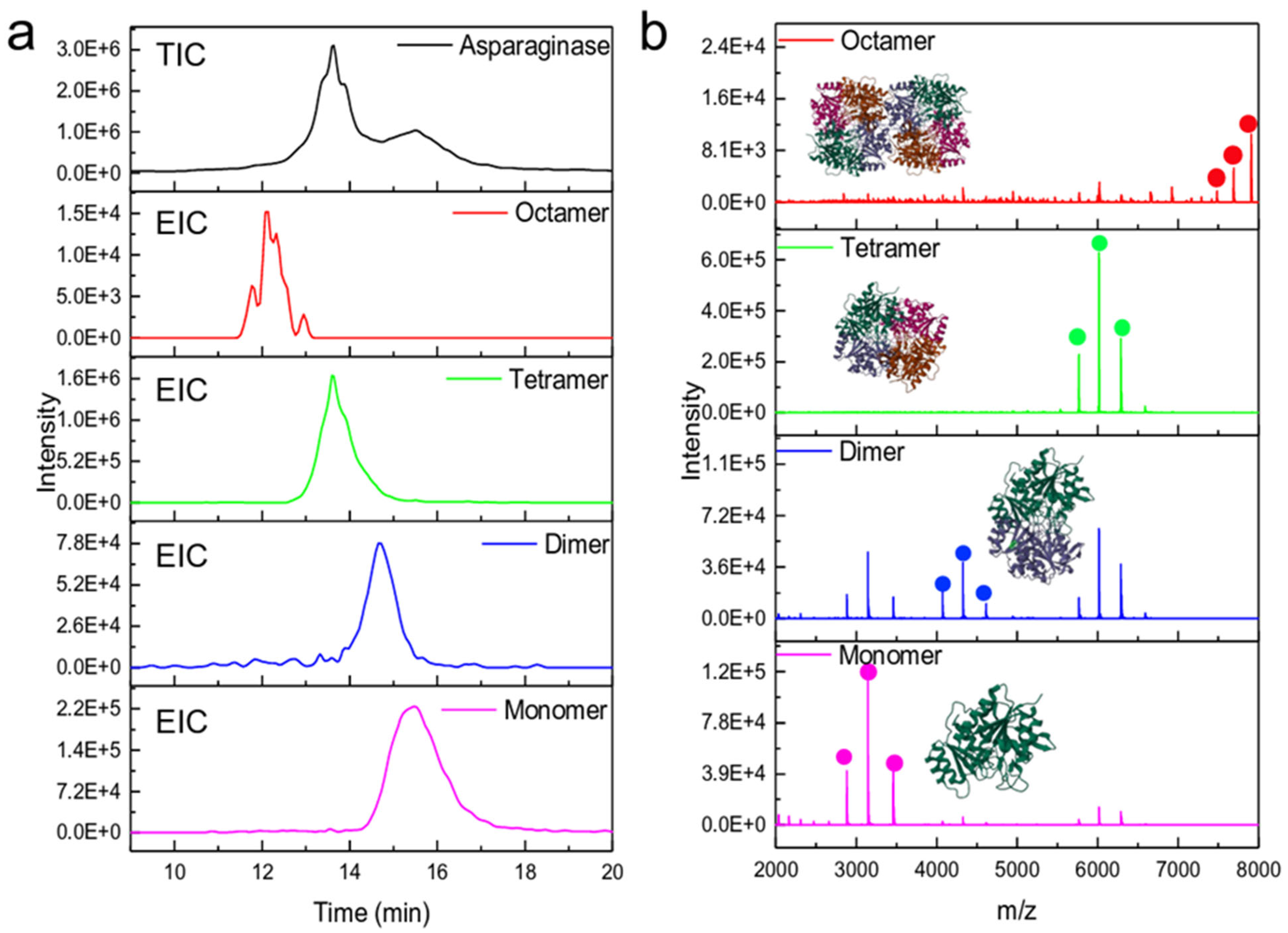
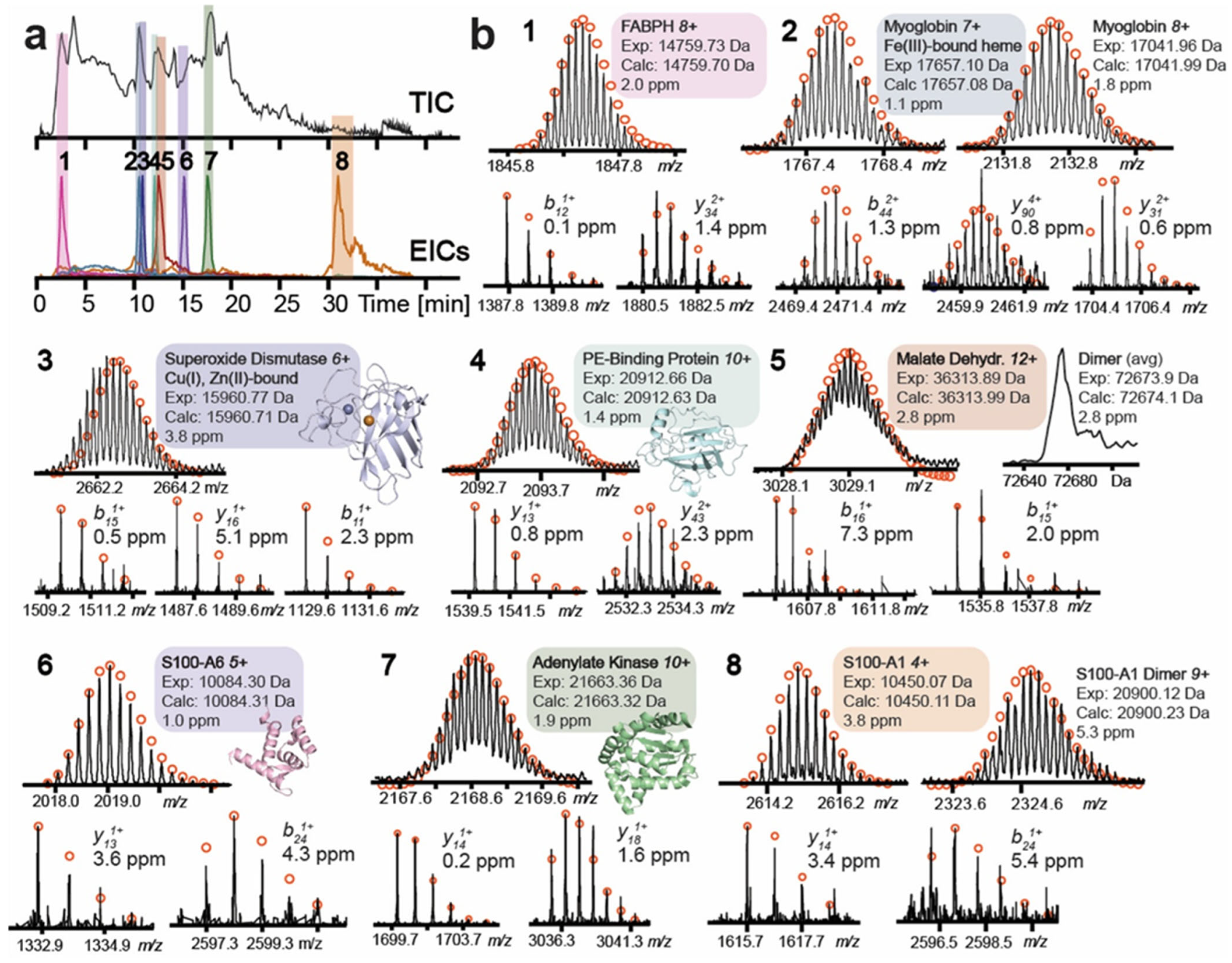
The coupling of SEC with other liquid phase and gaseous phase separation techniques for intact protein analysis was reported. For example, Shen et al. reported hyphenation of SEC and MWCO-based buffer exchange to capillary zone electrophoresis (CZE) (autosampler operated) for native MS analysis of a complex mixture (600 µg E. coli proteome) and discovered 4 protein homodimers, 16 protein–metal complexes, two protein–[2Fe-2S] complexes, and one protein–glutamine complex [188]. Other groups have reported integrated SEC-buffer exchange-ion-mobility spectrometry (IMS) for nMS analysis of intact antibody and demonstrated the preservation of structural integrity and higher order structure [183,189]. Gargano group who previously developed microflow SEC with a SPE trap for nMS analysis recently reported a new nanoflow SEC-nMS technology using 200 µm internal diameter SEC columns operating at 500 nL/min with a combined 80-fold gain in sensitivity and demonstrated its application in resolving monomer, dimer, tetramer, and octamer of L-asparaginase in a 6 min effective separation (retention time in 11–17 min window) as can be seen in the extracted ion chromatograms (EICs) in Figure 5a and their corresponding mass spectra in Figure 5b [190]. They used an autosampler for CZE operation and demonstrated success in analyzing higher-order structures of proteins from only 50 ng of injection, which is an excellent example of a sensitive and high-throughput platform for intact protein nMS analysis.

4.1.2. Ion Exchange Chromatography (IEC)

Ion Exchange Chromatography (IEC) separates molecules based on their charge via interaction with a stationary phase that has the opposite charge. IEC can help in separating charge variants of proteins that can form due to post-translational modification, such as deamidation, pyroglutamation, carbamylation, etc. [191]. The negatively charged stationary phase can interact with positively charged proteins through electrostatic interactions, which is why it is known as a cation exchanger (CEX). In contrast, a positively charged stationary phase can interact with a protein with a net negative charge, hence known as an anion exchanger (AEX). An incremental increase in ionic strength and the pH of the elution buffer disrupts the electrostatic interaction, leading to the separation of protein species. The gradient of salts in IEC-based separation presents challenges in MS-based analysis on the back end due to ion suppression and low ionization efficiency, leading to reduced sensitivity, adduct formation, and excessive contamination of the MS source and quadrupole. Continuous removal of salt and other interfering molecules through buffer exchange or complexation with cyclodextrin [141] can be explored to fully utilize the potential of IEX for nMS analysis of protein species. Several studies reported a pH gradient elution of a low-ionic-strength volatile buffer to directly couple IEC to HRMS without the need for any buffer exchange for the analysis of PTMs in mAbs [192,193,194,195]; however, protein measurement in these studies was based on digestion. In recent years, several studies have demonstrated the application of mixed-mode SEC-nMS (mmSEC-nMS) analysis of antibodies and intact proteins. Yan et al. reported the utility of strong cation exchange chromatography to purify bispecific antibodies (2–10 µg) from homodimer impurities (up to 0.01%), followed by mmSEC-nMS analysis using an ammonium acetate- and ammonium bicarbonate-based mobile phase of varying concentrations (30–450 mM) [184]. This approach can help quantify the homodimer impurities in therapeutic bispecific antibody formulations, which is critical to support their development and quality control. Fischer et al. reported nMS analysis of endogenous metal–protein complexes and oligomeric protein species up to 146 kDa using a mixed-bed IEC from 5.5 to 12.6 µg proteome from a complex mixture (human heart tissues) using a salt gradient (10–800 mM ammonium acetate) (Figure 6) [196], highlighting the utility of this approach for high-throughput analysis of endogenous protein complexes while preserving the tertiary structures of proteins and the noncovalent interactions.

4.1.3. Hydrophobic Interaction Chromatography (HIC)

Hydrophobic interaction chromatography (HIC) is a non-denaturing technique that separates proteins under native-like conditions based on their hydrophobicity. In HIC, more hydrophobic proteins bind to the stationary phase more strongly and elute later using a salt gradient [197]. Adsorption of proteins via the non-polar surface and the hydrophobic stationary phase increases during high salt concentration, and the elution of the protein occurs by disrupting that interaction by using a decreasing salt concentration in the eluent [198]. PTMs that impart hydrophobicity to the protein molecules are good candidates for HIC-based separation [199]. A decreasing salt gradient, such as 1.8–0.02 M ammonium sulfate, is commonly used in HIC to elute proteins while preserving the native-like conditions [200]; however, its nonvolatile nature complicates direct coupling to mass spectrometers. Ammonium acetate has not performed as well as ammonium sulfate, which also requires a high concentration, leading to decreased MS sensitivity [201,202]. Strategies to address these challenges involve the use of ammonium tartrate [200] and online buffer exchange, such as SEC [203] or other technologies such as complexation (e.g., via cyclodextrin) [141]. Kempen et al. used ammonium tartrate to analyze the drug-to-antibody ratio (DAR) in different ADC formulations from 50 µg injection via online HIC-SEC-nMS analysis, demonstrating comparable performance to ammonium sulfate, which is the preferred salt for HIC due to its selectivity [204]. Furthermore, to prevent salt contamination of the ESI source of the MS, they integrated an online SEC-based desalting cleanup step prior to MS analysis, which provided superior performance than a traditional heartcut setup. This study also reported the detection of several glycoforms of the antibody, in addition to DAR values of 0, 2, 4, 6, and 8, within a 20 min run. HIC, as a standalone separation technique for nMS analysis of intact protein species, has been reported in recent years. Wei et al. reported the detection of non-oxidized, mono, and di-oxidized mAbs via online HIC using a isocratic flow of 300 mM ammonium acetate to HRMS [205]. HIC has been a popular method of choice for mAbs, ADCs, and related product analyses, and the readers are encouraged to consult the excellent review by Fekete et al. for a comprehensive discussion on HIC-based method development for native protein analysis [199].

4.1.4. Affinity Liquid Chromatography (ALC)

Affinity Liquid Chromatography (ALC) separates proteins based on their binding to an immobilized ligand or receptors on the stationary phase and is often used to study the binding characteristics of proteins to other molecules [206]. Binding occurs under native-like conditions, while the elution occurs by changing the pH or the ionic strength of the mobile phase [207]. The review articles by Banks et al. [208] and LaCava et al. [209] discuss the critical parameters for developing affinity purification workflows for proteins and protein complexes. In recent years, affinity LC-MS for antibody characterization has gained popularity and demonstrated the identification of several glycoforms using increasing [210] or decreasing [211] pH gradient. Prinston et al. used ALC in determining the PTMs of therapeutic antibody, specifically glycosylation of the antigen-binding site (Fab), a critical quality attribute (CQA) for drug product quality, as this modification affects the binding of the therapeutic antibody to the target receptor and its therapeutic efficacy [212]. Cotham et al. reported an affinity LC-UV-nMS analysis of a therapeutic mAb using three different affinity columns, namely, Protein A (ProA) (pH gradient of 6.9–2.8), FcγRIIIa (pH gradient of 6.5–4.5), and FcRn (pH gradient of 5.5–8.8) using 15 mM ammonium acetate [213]. They reported the identification of proteoforms of a purified antibody, NISTmAb, due to oxidative stress (Figure 7a). Additionally, they detected homodimer impurities from harvested cell culture using a ProA affinity column and identified several glycoforms using an FcγRIIIa affinity column (Figure 7b). They also demonstrated the application of the FcRn affinity column for comparative analysis of a therapeutic antibody and its isoform differing by 3 amino acids in the sequence, with injections ranging from 10 to 25 µg (Figure 7c). Their setup included a splitting valve post-affinity column elution, which enabled conventional analytical flow (100–400 µL/min) for affinity purification and sub-µL/min flow rate for nanospray ESI-HRMS analysis. This integrated platform not only facilitated the direct coupling of affinity chromatography to mass spectrometry but also enabled optimal conditions for maximum performance in both affinity purification and MS analysis. Additionally, they also demonstrated how the modification of the mAbs alters their interaction with the affinity column.

4.2. Capillary Electrophoresis (CE)

Separation of molecules via capillary electrophoresis is based on their electrophoretic mobility, which depends on the charge of the molecules. Among the electromigration techniques for nMS analysis of intact protein species are capillary zone electrophoresis (CZE), mobility capillary electrophoresis (MoCE), affinity capillary electrophoresis (ACE), and capillary isoelectric focusing (CIEF), and the readers are encouraged to consult the reviews by Schwenzer et al. [214] and Shen et al. [215] for critical method development parameters and various MS fragmentation techniques for native CE-MS analysis, respectively. In this section, recent applications of native CE-MS-based approaches for analyzing intact protein species are discussed briefly.

4.2.1. Capillary Zone Electrophoresis (CZE)

Capillary zone electrophoresis (CZE) is a separation technique that separates molecules based on differences in charge and hydrodynamic radii, which determines the migration of the molecules in a free solution through a capillary under an electrical field. Abnormal physiological conditions (e.g., disease) may affect the native structure of proteins, altering their charge, which can be probed using CZE-based separation prior to nMS analysis. Separation of proteins in CZE occurs in aqueous-based buffers, making CZE a great alternative to LC-based approaches for nMS analysis of protein species [216]. Sheath solution is often used in CZE-based analysis, which helps with ionization and can be based on nMS-friendly aqueous buffer with low salt contents, such as acetate or formate; however, it can also be mixed with some organic solvents for controlled disassembly of protein aggregates or complexes before ionization. Sheathless CZE-MS analysis operating under nL/min flow rate of background electrolytes (BGE) is preferred for native MS analysis of proteins due to increased sensitivity and more efficient ionization [217]. One critical aspect of CZE-MS analysis is the net charge of the inner surface of the separation capillary, which determines the adsorption of protein species. Hence, the composition of the capillary, whether bare or coated with polybrene (adsorptive), polyethyleneimine (covalently bound), polyacrylamide, or cellulose, needs to be carefully assessed and selected for nMS analysis of protein species [218].
As the mechanism of separation in CZE is based on charge and the hydrodynamic radii of the proteins, which are directly related to their structures, CZE allows for obtaining information about the protein higher-order structures (HOS), from a limited sample amount [219]. Application of CZE-MS for proteomics applications [215] and higher-order structure analysis [219] is well covered and reviewed comprehensively by several papers [215,219]. CZE-nMS analysis of intact proteins, although demonstrated the power of this approach in studying proteoforms, achieving optimum resolution for closely related proteoforms can still be an analytical challenge [220] as it depends on numerous factors, including the composition, pH, and ionic strength of the conductive liquid (BGE), separation temperature, voltage, and ionization conditions, which also affect the preservation of the native-like conditions [214,221,222], and often require additional separation before CZE. As stated before in Section 4.1.1, Shen et al. reported integration of online SEC hyphenation before CZE-nMS analysis for native proteomics analysis of E. coli lysate [188]. Sun group later demonstrated the application of CZE-nMS without additional separation workflow for analyzing proteoforms, including oligomeric species of proteins and protein–metal complexes (30–400 kDa range) from a complex sample mixture (E. coli cell lysate) using only a 50 ng sample load in a single run (Figure 8) [223].
However, the authors report that further improvement of the workflow is needed by incorporating better identification (MS/MS fragmentation), sensitivity (online stacking or isoelectric focusing), and resolution (peak capacity). Jooß et al. demonstrated the utility of CZE-nMS for analysis of endogenous nucleosomes, a DNA–protein complex, by controlled de-assembly of the complex that allowed analysis of the intact nucleosome, followed by ejection and analysis of the constituent histone proteins [224]. Although CZE-MS-based approaches provide better sensitivity than LC-based approaches with picolitre (pL) injections onto the capillary, the workflow is more prone to shifts in the migration time of the analytes [225]. Slight variations in sample or instrument conditions affect the reproducibility of the data and require lengthy cleanup procedures. Blockage of the capillary or analyte loss due to adsorption leads to further complications [226]. Although commercial equipment is available for automated injections, they have not seen widespread application in nMS analysis of intact protein species. Sadeghi et al. reported reproducible results for 67 injections of yeast cell proteome with ammonium hydroxide-based capillary cleanup, after which the peak intensity substantially decreased, warranting a cleanup before further analysis [226]. However, reproducibility after cleaning has not been reported. Results from this study indicate the challenges of CZE-MS for large-scale analysis, especially when compared with LC-MS-based workflows. Despite the obstacles, the sensitivity offered by CZE-MS and its potential for proteoform level resolution, it remains an active area of research for studying protein modifications and complexes.

4.2.2. Mobility Capillary Electrophoresis (MoCE)

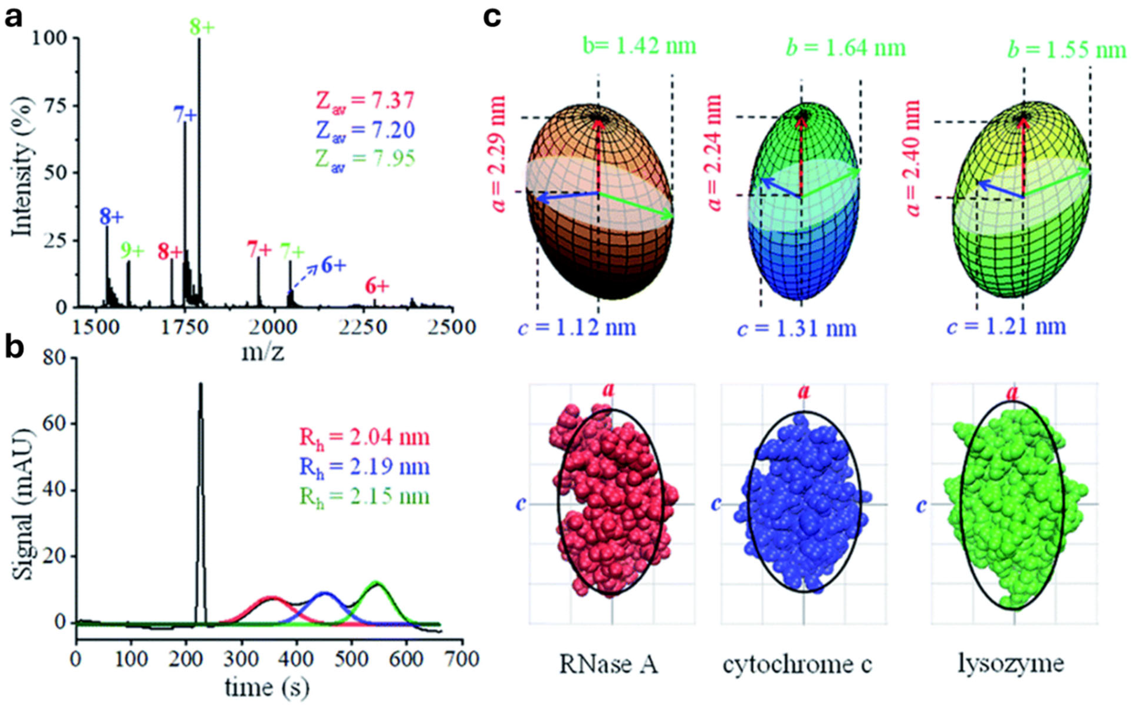
During conventional CZE-MS analysis, information about the effective charge or the hydrodynamic radii of the protein species cannot be determined, and only migration time information is available. To address this, mobility capillary electrophoresis (MoCE) has been developed, which can be integrated into CZE-nMS analysis to enable measurement of charge, hydrodynamic radii, as well as the solvent accessible surface area, leading to the achievement of three-dimensional conformation analysis of protein species under native-like conditions [227]. Wu et al. reported the use of MoCE for CZE-nMS analysis of protein mixtures (nanospray ESI infusion of 1 mg/mL) in determining their 3-D structure based on the charged states through calculation of effective charge, Gaussian curve fitting, and hydrodynamic radii calculations, as can be seen for RNase A, cytochrome c, and lysozyme (Figure 9) [227].
The integration of MoCE into CZE-nMS for conformational analysis of protein species relies on accurate measurement of hydrodynamic radii and effective charge, which in turn require controlled laminar liquid flow and precise information on the flow rate [228] and can significantly advance the 3D structure analysis of endogenous protein species.

4.2.3. Affinity Capillary Electrophoresis (ACE)

During Affinity Capillary Electrophoresis (ACE), the ligand is kept free-flowing either as part of the sample (pre-equilibrium and kinetic capillary electrophoresis) or the BGE (mobility shift) with which the protein interacts upon migration through the capillary, leading to a change in their electrophoretic mobility compared to the uninteracted protein [229]. ACE has demonstrated success in determining the ligand-binding constant and in analyzing protein–ligand binding due to the ability to measure the molecular mass of unbound species, intact protein, and ligand, as well as the bound protein–ligand complex, all of which have different electrophoretic mobility [230,231]. ACE is a preferred technique to determine the ligand–protein binding over ALC due to having both the ligand and the free protein under the native-like conditions mimicking the physiological conditions closely, whereas in the latter, the ligands are bound to the stationary phase [232]. Although direct coupling of ACE to MS requires the use of MS-compatible buffers, which differ from the non-volatile buffers commonly used in other binding assays, they have been reported to exert minimal effect on protein–ligand interactions during native MS analysis [231,233]. ACE-MS has been successfully used in studying protein interactions, including enzymatic inhibition, stoichiometry analysis, binding, and dissociation constant determination, such as the binding of trypsinogen to aprotinin [231], matrix metalloproteinase-9 (MMP-9) to tetracycline and a few natural products [234], and human tissue transglutaminase TG2 to several synthetic compounds [235], chemokine stromal cell-derived factor-1 to SDF-1) [236] and cytochrome P450 isoform 2C9) to diclofenac [237].
The success of ACE-MS in determining the binding of the protein to the ligand depends on the concentration of each species and how they are introduced into the system during analysis. They need to be critically monitored for an accurate readout of the binding. Additionally, the internal environment of the capillary plays a vital role in determining the success of protein–ligand binding, as protein adsorption into the capillary inner surface can lead to erroneous results. Domínguez-Vega et al. demonstrated a capillary coating of polybrene–dextran sulfate–polybrene (PB–DS–PB) to prevent adsorption of the proteins to the capillary wall during trypsinogen-aprotinin binding [231]. Davoine and Fillet demonstrated the use of n-methylmorpholine (NMM) acetate buffer, which increased the peak shape at physiological pH, and polydopamine-based coating, which is easy to prepare and stable at physiological pH, in determining the dissociation constant of p-aminobenzamidine (PABZM) from PABZM-coagulation factor XIIa (FXIIa) complex by ACE-MS [238].

4.2.4. Capillary Isoelectric Focusing (cIEF)

Capillary Isoelectric Focusing (cIEF) separates proteins in solution while migrating via a capillary under an electric field using a pH gradient, based on their isoelectric point (pI), which is the pH at which the protein has a net charge of zero [239]. CIEF only works if the molecules of interest are amphoteric in nature, i.e., they must contain functional groups that can be protonated and deprotonated, making proteins and peptides ideal candidates for this workflow. Based on the pI of the protein of interest, the pH gradient can be shallow or steep, which can be controlled by the addition of small, charged molecules such as water, amino acids, bicarbonate, metal oxide, and superoxide. [240,241]. It is amenable to automation, allowing high-throughput analysis of biomolecules compared to gel-based isoelectric focusing. Isoelectric point (pI) depends not only on the charge of the proteins but also on the three-dimensional structure; hence, cIEF-based bioanalytical approaches are preferred for conformational analysis of proteins and protein complexes. The success of cIEF depends on the selection of the appropriate ampholyte, carrier and sample composition (pI, concentration), capillary (non-coated vs. coated), focusing current and focusing time, additives and their stability at extreme pH as well as their binding capacity to inner capillary surface (e.g., polymers), their ability in solubilizing proteins or preventing precipitation or aggregation (e.g., glycerol, ethylene glycol, propylene glycol, zwitterionic and neutral detergents) and pI marker (pI close to the target protein) [241]. However, online hyphenation to MS analysis will necessitate removal of these molecules before MS injection as their presence can lead to significant ion suppression, yielding to reduced sensitivity as well as contamination of the MS. Additionally, desalting is recommended before separation by cIEF, as high salt concentration can lead to high current, damage to the capillary, and Joule heating leading to protein denaturation in addition to the challenges with MS analysis. The use of salt at low concentration demonstrated improved resolution compared to pure water [242]. Compared to CZE, cIEF requires a higher sample load [214].
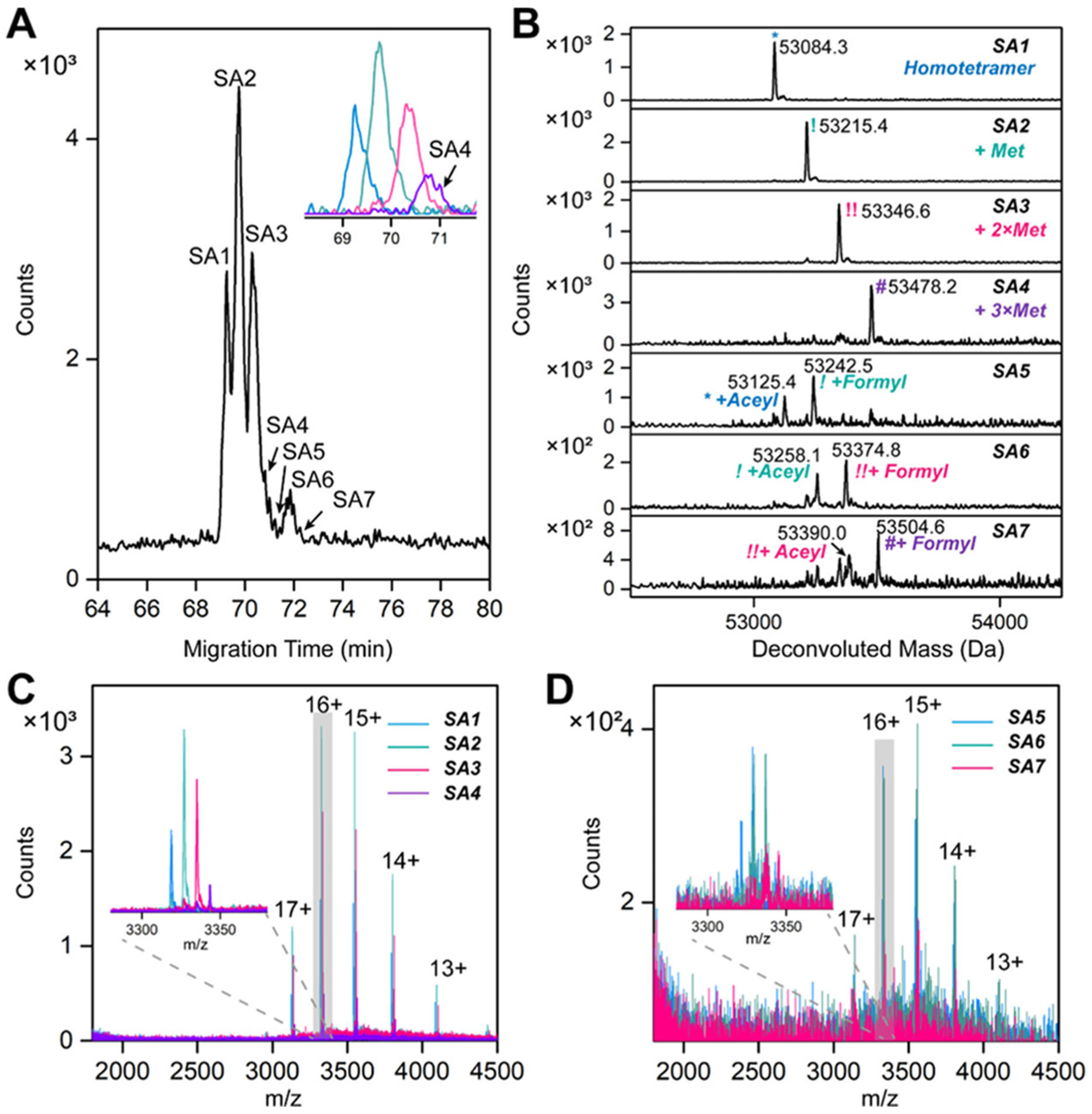
Fonslow et al. demonstrated cIEF-based analysis of rapamycin complex (mTORC1 and 2), their subcomplexes, phosphorylation states, and their disassembly with a temperature gradient (15–25 °C) and ampholyte-induced pH manipulation using HIS- and FLAG-tag purified protein complexes [243]. Przybylski et al. reported characterization of highly basic cytokine human interferon-gamma (IFN-γ) as a non-covalent homodimer and its mutations [244]. Xu et al. developed an automated nondenaturing cIEF-MS (ncIEF-MS) workflow that demonstrated superior performance than nCZE-MS in characterizing streptavidin homotetramer proteoforms, e.g., methylation, formylation, and acetylation (Figure 10) [245]. Using this technology, they also calculated the precise readouts of pI of proteoforms due to modifications, e.g., for streptavidin homotetramer, for which presence of N-term Met reduced pI by 0.1, addition of one acetyl group reduced pI by 0.4, incorporation of one formyl group reduced pI by 0.3 whereas loading 2 more drug molecules on one antibody–drug conjugate (ADC) increased the pI by 0.1 [245]. These examples demonstrated the application of cIEF-based approaches to study protein complexes and proteoforms via native MS.
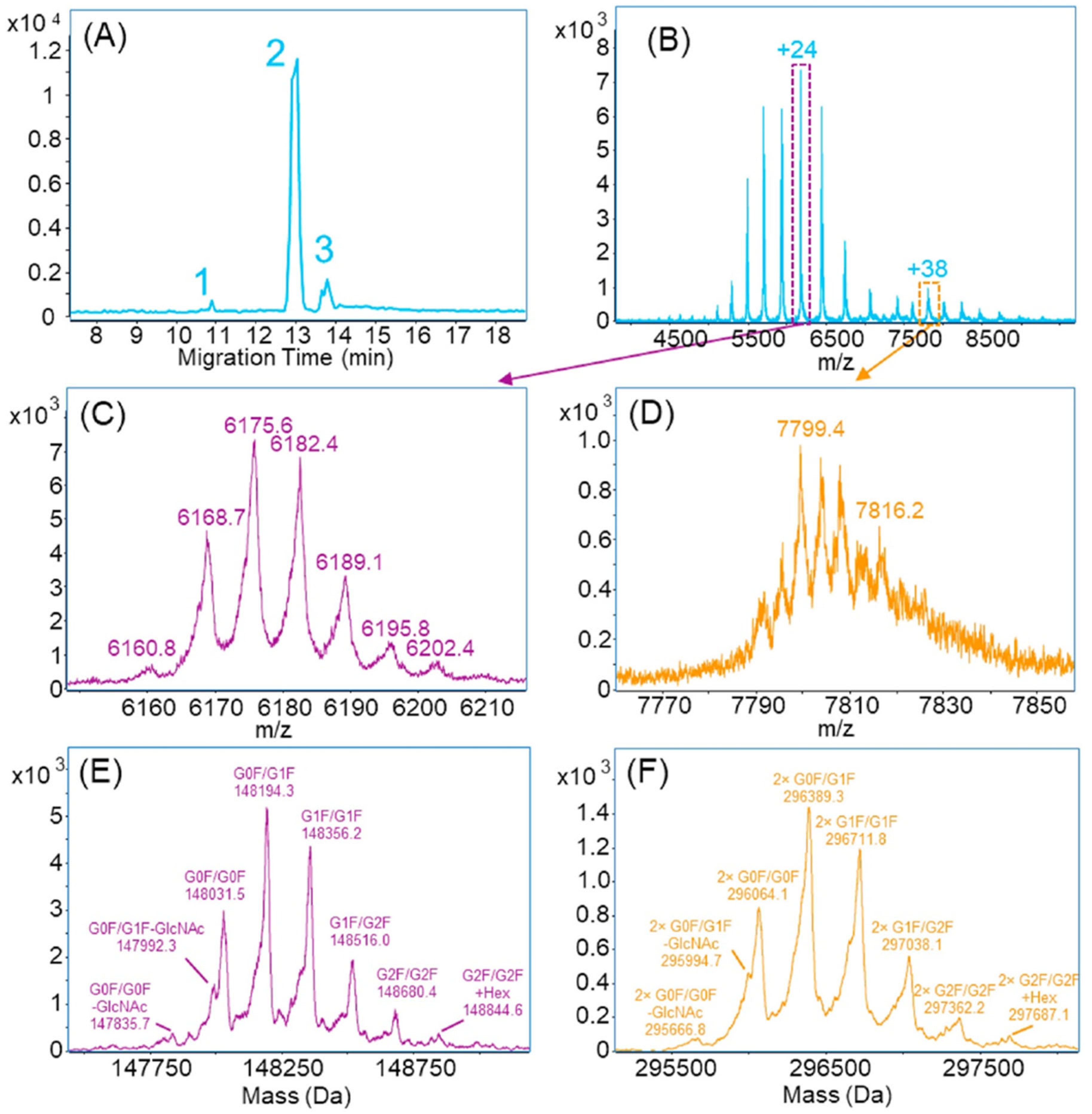
cIEF-based analytical approaches have found greater application in characterizing mAbs, especially in identifying modifications that play a critical role in determining the efficacy and safety of these therapeutics. In this regard, the work by Dai et al. is notable due to their comprehensive method development and optimization of numerous parameters, including composition of anolyte, catholyte, sample mixture, sheath liquid, and cIEF running parameters such as injection amount, field strength, and applied pressure [246] for nMS analysis. They analyzed trastuzumab, bevacizumab, infliximab, and cetuximab, identifying both basic and acidic variants for all the mAbs, as well as the main peak for each. Shen et al. developed an integrated cIEF-assisted CZE-nMS method using a new linear carbohydrate polymer-based neutral coating with increased sample loading capability and an automated CE sampler and identified various glycol-proteoforms and a homodimer of sigmaMAb and NIST mAb (Figure 11) [220].
Naghdi et al. reported a chemical mobilization-based cIEF-MS workflow with a resolution of 0.1 pI unit, established superior performance compared to pressure-assisted chemical mobilization, and demonstrated separation of Fc-conjugated insulins (62 kDa) differing in only one amino acid [247]. A major hurdle of all capillary-based separations is the physical movement of the molecules through the capillary to the detector, which is primarily performed by chemical or pressure mobilization [248] and leads to several critical performance issues, including distorted peak shapes, inconsistent resolution, increased run time, aggregation, and/or precipitation of the sample, among others [249]. Wu et al. discussed these challenges with examples demonstrating that the lack of real-time observation during capillary electrophoresis makes optimization very challenging [250,251,252]. This major limitation of capillary electrophoresis-based bioanalysis was addressed by integrating whole column imaging, termed as imaged capillary isoelectric focusing (icIEF), which was first proposed by Wu and Pawliszyn using refractive index (RI) and ultraviolet spectroscopy (UV) [250,251,252] that eliminated the mobilization step and increased the throughput by reducing the analysis time to 5–15 min from 40 to 60 min per sample [253]. IcIEF-based bioanalytical methods are being successfully used to determine protein stability, charge heterogeneity, proteoforms, e.g., glycation and glycosylation (sugar is used as a stabilizer in protein formulations), allowing critical observation during process development of protein therapeutics, including mAbs [254,255], ADCs [256], and vaccines [257,258]. Readers are encouraged to review the excellent resources by Bo et al. [259], Ghizzani et al. [260], and Wu et al. [261] to explore more about this topic.

5. New Frontiers of Native Mass Spectrometry and Proteomics

5.1. Automated Purification, Buffer Exchange, and Individual Ion Mass Spectrometry

Among the major limitations of mass spectrometry analysis of intact protein species are the resolving power of the mass spectrometer, ion dissociation, and the dampening of ion motions, which cause signal decay. High resolution mass analyzers, such as time-of-flight (TOF) MSs, offer orthogonal acceleration and/or increased ion-flight time, whereas modern FT-ICR mass spectrometer contains newer and improved cell design and higher magnetic fields, all leading to higher resolving power [262]. During intact protein mass spectrometry analysis, signals of multiply charged ions in the individual spectra are summed into a composite spectrum demonstrating resolved isotopic distribution. Makarov et al. demonstrated the ability of the Orbitrap mass analyzer to detect and collect single ion events using myoglobin with only 20 charges [263]. Continuing on this, Kafader et al. developed individual ion mass spectrometry technology (I2MS, commercially known as direct mass technology by Thermo Fisher Scientific) and demonstrated its applications in measuring individual ions of +37 charge states of transferrin (~80 kDa) and +49 charge state of NIST antibody (~150 kDa) using Q Exactive Plus (Standard Orbitrap analyzer) and Q Exactive HF (ultrahigh-field Orbitrap analyzer) [264]. The Kelleher research group pioneered this technology and later demonstrated its application in analyzing proteins and proteoforms from complex samples. They reported identification of 550 proteoforms in the range of 0–30 kDa from HEK-293 cell extract, tetramer of pyruvate kinase (232 kDa), 14-mer of GroEL (~801 kDa), and virus-like particles from viral capsids carrying varying amounts of DNA and mRNA cargo loads (3.19 MDa and 0.99 MDa) using Q Exactive Plus and Q Exactive Ultra High Mass Range (UHMR) mass spectrometers [265]. Sample introduction for these I2MS measurements was performed via direct infusion using either standard protein samples or GELFrEE fractions of cell extracts. Quantitative performance of I2MS technology is yet to be seen.
Purified protein and protein complexes obtained via immunoprecipitation often contain high salt or contaminants, which need to be removed from the samples prior to introduction to MS. Ultrafiltration is a pressure-driven membrane-based separation technique that separates proteins and other particles based on size. Membranes used in ultrafiltration typically have a pore size of 10 to 500 Å [266]. It is a routine method often used in a laboratory to remove salt, exchange the buffer to bring the protein into an MS-friendly solvent, and concentrate samples. Based on the molecular weight cutoff of the membrane, a mixture of proteins can be obtained in either the retentate or the filtrate for MS analysis. Ultrafiltration is rapid and can preserve the native structure of proteins; hence, it can be used as a low-resolution first step for nMS, or it can be hyphenated with immunoprecipitation to remove unwanted molecules from samples that managed to escape antibody-based pulldown. Ultrafiltration follows the direction of gravity and is commonly seen as spin columns and microwell formats that are compatible with different scales of protein isolation. However, it necessitates offline handling of the samples, which not only limits throughput but also leads to sample loss. Tangential flow-based filtration uses membranes with varying retention ranges (1 kDa to 1000 kDa) and can be made from low protein-binding materials such as cellulose, cellulose acetate, polysulphone, polyacrylonitrile–polyvinylchloride copolymers, and modified polyethersulphone [267]. Kelleher lab developed a novel interface, named SampleStream, using a membrane-based ultrafiltration strategy for proteome sample cleanup. This method is amenable to both denatured and native top-down proteomics analyses and operates with a throughput of up to 15 s/sample, providing sensitivity comparable to liquid chromatographic methods [268]. Continuing on this pursuit, Des Soye and Kelleher developed a fully automated platform, Auto-Ig-MS, integrating automated immunoprecipitation using robotic purification system (KingFisher) to purify antibodies from 58 test subjects in response to SARS-CoV-2 antigen to SampleStream for sample cleanup and injection and I2MS data acquisition on a Q Exactive HF mass spectrometer powered with automatic ion control (AIC) for adjusting ion injection times per proteoforms [269] that can process over 100 samples per day (Figure 12) [270].

5.2. Ion Mobility Spectrometry (IMS)

Ion mobility spectrometry (IMS) is a gas-phase technique that separates ions in gaseous states based on their three-dimensional structure, specifically their collisional cross-section, which is theoretically the best strategy to separate native proteins prior to MS analysis. Additionally, IMS can differentiate between conformers present within a single oligomeric state, which can provide valuable mechanistic information about the assembly and evolution of the oligomers [271]. Application of IMS in analyzing disease-related oligomeric protein structures, such as those in Alzheimer’s disease [272,273], type II diabetes [274], and dialysis-related amyloidosis [275] as well as protein complexes such as the noncovalent 28-subunit 20S proteasome [276] and trp RNA binding protein, TRAP [277] demonstrated its utility in studying the size, topology, and stoichiometry of native proteins via mass spectrometry. Ion Mobility Spectrometry–Mass Spectrometry (IMS-MS) has also been used in studying multiprotein complexes, such as the DNA clamp loader assembly from E. coli [278], eukaryotic translation factor eIF3 [279], and RNA polymerase I and II complexes [280]. IMS-MS assisted in monitoring the assembly of viral capsid proteins and their intermediates [281]. Cyclic IMS has demonstrated its utility to study distinct oligomeric conformers (compact vs. elongated), demonstrating the power of gas-phase separation in providing structural information about particular subpopulations in an aggregating system in response to changes prior to ionization [271]. IMS also enables deposition of the protein in solution for further studies, demonstrating that the proteins and protein complexes can survive ionization, gas-phase purification, and soft landing that presents an exciting possibility for high-resolution structure determination of complex modalities like AβOs [282]. Overall, gas-phase separation techniques like IMS can assist in studying the protein’s higher-order structure without the need to fix the native profiles using complex strategies like crosslinking from the solution-state samples. However, for complex mixtures (e.g., biological samples or samples with high racemic contents) without any purification and/or without any front-end separation, IMS may not be sufficient as a standalone separation technique [283]. If coupled to a sample purification step, it can enhance endogenous protein complex and proteoform analyses.
During the preparation of this manuscript, an article was published by Vickers et al., who used IMS integrated top-down MS to discover several oligomeric states of canonical alpha-1-antityrpsin isolated from a liver-cirrhosis patient post-transplantation that were absent in wild type (Figure 13) [284]. This study further demonstrates the power of IMS-MS in characterizing higher oligomeric species and misfolded proteins, which may be associated with serious disease conditions. This study also highlights the necessity for orthogonal validation to confirm the gas-phase oligomeric structures, as sample processing conditions can result in artifacts, as observed in Figure 13D and Figure 13F due to heating and pH changes, respectively.

5.3. Ambient Surface Mass Spectrometry (ASMS)

Surface sampling via ESI over matrix-assisted laser desorption (MALDI)-based approaches is preferred for larger protein molecules due to the ability of the former to generate multiply charged ions, which the MALDI-based approaches lack. Sample preparation for ambient surface MS analysis, such as liquid extraction surface analysis mass spectrometry (LESA-MS) and desorption electrospray ionization mass spectrometry (DESI-MS), involves minimal sample handling at atmospheric pressure, where solvents (with or without charge) are deposited on the sample and the analytes are directly extracted, sent, and analyzed by mass spectrometers [285]. For nMS analysis, this provides the added benefit of minimal sample preparation and a reduced likelihood of artifacts. However, the most significant limitation of ASMS workflows is the matrix effect, which leads to reduced sensitivity, spatial resolution, and poor recovery of the analytes [286]. The Cooper group demonstrated nMS analysis of proteins directly from dried blood spots (on filter paper) and tissues, including mouse liver and kidney sections (mounted on glass slides), via LESA-MS under native-like conditions using ammonium acetate and reported an intact hemoglobin oligomeric complex [287,288,289]. They also demonstrated the integration of IMS into the LESA-MS workflow for the spatial and conformational analysis of intact protein species, including β-thymosin (4.9 kDa), ubiquitin (8.5 kDa), and tetrameric hemoglobin (64 kDa), from a mouse kidney section under native-like conditions [290]. The overall sample preparation steps involved in ASMS inherently favor native protein analysis; however, they are limited by the abundance and size of the protein of interest [291,292]. Efforts are being undertaken to address some of these challenges, potentially enabling spatial and conformational analysis of low-abundant intact proteins under native-like conditions from complex samples via ASMS.
Ambrose et al. first demonstrated the DESI-MS workflow under native-like conditions to study an intact canonical protein (hen egg-white lysozyme) and its interaction with a substrate (N-acetyl-glucosamine, NAG) [292]. They successfully used this approach to analyze different oligomeric states of membrane protein and their interactions, e.g., outer-membrane protein F (OmpF) and the stoichiometry of binding of ferric-pyoverdine (Pvd) to the outer membrane protein receptor FpvA from Pseudomonas aeruginosa. A version of DESI-MS, nanoDESI-MS, has recently gained traction in the successful imaging of intact proteins. The Cooper group also reported a series of studies demonstrating the utility of nanoDESI-MS for native mass spectrometry imaging, which offers improved resolution compared to LESA-MS-based workflows in identifying numerous canonical proteins and their oligomeric states. They have utilized nanoDESI-MSI for native analysis of numerous low-abundant proteins, noncovalent dimer of S100-A6, adipocyte lipid-binding protein (ALBP), and phosphatidylethanolamine-binding protein (PEBP1) from rat kidney tissues using ammonium acetate (200 mM) + 0.125% C8E4 detergent, and a combination of collisional activation and proton transfer charge reduction [293]. These techniques can not only enhance the spatial analysis of endogenous proteoforms and protein complexes but can also be further explored for nMS analysis from solution-phase samples.

6. Assessment of nMS Sample Resemblance to the Native State

Biological sample preparation often involves reagents and conditions that do not reflect the native biological conditions. However, many such conditions may not be non-perturbative at the level of proteins, e.g., flash freezing, cryomilling and grinding of tissues, 0.1% detergents (e.g., Tween-20 and CHAPS), buffer exchange through molecular weight-cutoff filtration, and centrifugation [121,294]. Ammonium acetate has been a buffer of choice for native mass spectrometry analysis of intact protein species. Although ammonium acetate is generally considered to keep the protein in its native folded state through stabilization by arranging water molecules surrounding it, this effect is concentration-dependent, and higher concentrations can interfere with the native assembly of the proteins [295]. Trialkylammonium salts such as trimethylammonium acetate, triethylammonium acetate, and bicarbonate can be more suitable for nMS analysis and should be carefully evaluated as discussed in Section 2.1. Additionally, the cellular environment is made up of many other molecules, salts, and ions; hence, the native-like state that is assumed to have been achieved during nMS analysis by using a buffer at pH 7.00 is not a true reflection of native conditions [296]. Care needs to be taken to consider the type and strength of buffers for nMS analysis, and they should be validated by orthogonal techniques to ensure that the proteins have not been denatured.
The presence of certain detergents, such as up to 0.01% of Tween-20 (non-ionic) or 0.0375% of SDS (ionic), has been used for protein recovery. Their role in preserving the native structure of proteins requires careful investigation, as their effects are concentration-dependent and generally considered safe up to 0.1%, as discussed earlier. Non-ionic detergents such as maltosides, glucosides, NP-40, Triton X-100, digitonin, etc., are choices that could be considered [297]. Behnke and Urner discussed that heterogeneous detergents with varied critical aggregation concentration (CAC), reduced charge, and hydroxyl groups, and the presence of basic or chelating groups are crucial properties to consider assessing [297]. Solvent additives such as NaCl, arginine hydrochloride, glycine, sucrose, caprylic acid, N-acetyl tryptophan, gallic acid, etc., have been used to stabilize proteins, offer protection, and reduce their aggregation [298,299] and could also potentially be explored to preserve native conditions for mass spectrometry analysis.
The effect of any condition or reagent used in the solution can be analyzed using additional techniques to identify artifacts, which are difficult to detect once the sample solution is sprayed into the MS and becomes gaseous. The process of transferring analytes from solution to a high-vacuum state is inherently disruptive [73]. Breuker and McLafferty reported that dehydration of a protein sample causes proteins to transition from their natural folded state through unfolding, resulting in a stable gas phase structure that has little resemblance to the native folded structure, and these transitions are affected by temperature and pH [73]. McLafferty and coworkers also reported that these gas-state protein transitions may occur within minutes and nMS data obtained from the gas-state proteins may not have a close structural relationship with the native structure of protein species [300]. Schennach and Breuker commented that electrostatic interactions primarily drive gas-phase folding of proteins and that the gas-phase unfolding of more hydrophobic proteins, such as membrane proteins, may be more similar to their folding in solution and their gas-phase folding may be slower (>400 ms), allowing more time to acquire MS data [301]. Wyttenbach and Bowers reported that the native state of protein in solution form was preserved in gaseous form for more than 100 ms [302]. Hence, the structures obtained from the gaseous state of an intact protein depend on the sample and instrument conditions, as well as the time taken to reach the mass analyzer [303] and these need to be carefully tested for nMS analysis and data interpretation.
Despite these uncertainties, studies utilizing ion-mobility and ion-soft landing experiments reported that the degree of nativeness of ions from intact proteins is consistent with that obtained with XRC and NMR analysis, with some exceptions [304,305]. Electron microscopy imaging [306], infrared spectroscopy [307], and electron transfer dissociation mass spectrometry [308] also demonstrated the preservation of native-like protein structures during ionization, dehydration, and soft landing, supporting the idea that gas-phase structures may truly reflect many features of native solution structures of proteins [33]. Based on these mixed opinions, it is imperative to use orthogonal techniques such as XRC, NMR, cryo-EM, Small-Angle X-ray and Neutron Scattering (SAXS and SANS), surface plasmon resonance microscopy, CDS, ITC, and/or computational methods to establish the native state of the intact proteins based on nMS data [309]. Additionally, HDX-MS or XL-MS may help to support the nMS data of intact proteins. More cost-effective orthogonal techniques are analytical centrifugation [310], 2-DE [311], and light scattering [312,313,314], which can help determine the native state of the protein in solution to compare with the nMS data, which is based on the gaseous state. The choice of technique to support nMS data of intact proteins, proteoforms, complexes, or higher-order structures will depend on resource accessibility, but it must accompany the nMS data to ensure it is a true reflection of the native state of the proteins.

7. Importance and Challenges of Native Mass Spectrometry Analysis of Protein Species

Proteins are essential biological regulators, and their modifications have profound biological effects. For subunit stoichiometry analysis of protein complexes, their direct measurement is desirable. As stated earlier, analyzing intact proteins in their native conditions is important because they reflect the actual endogenous state associated with biological function and often serve as a hallmark of a disease condition. For example, amyloid beta peptide (Aβ) is an enzymatically cleaved product of amyloid-beta precursor protein (APP) that is toxic to nerve cells [315]. Low-molecular-weight oligomers, such as trimers and tetramers, are associated with synaptic damage and impaired plasticity, whereas high-molecular-weight oligomers cause cognitive deficits [316]. Determining the type of amyloid beta oligomers (AβOs) that exist in vivo has been extremely challenging, not only because the mechanism of oligomerization remains unknown, but also due to their inherent instability, which makes it difficult to distinguish their native structure from the artificially induced one. However, the AβO hypothesis emphasizes the significance of studying native AβOs, as they are responsible for causing brain damage that leads to Alzheimer’s disease [317]. Another example is aquaporin-0 (AQP0), which is the most abundant membrane protein in the eye lens that functions as a water channel, and regulates the water permeability by interacting with various types of lipids (sphingomyelin and cholesterol) that have recently been elucidated via nMS analysis, highlighting its potential in studying biological phenomena and processes [318]. Small modifications in protein-based biotherapeutics, such as mAbs in ADCs, can cause substantial changes in their aggregation behavior, degradation pathways, or payload binding, leading to ineffective outcomes or toxicity in patients upon administration [319]. These studies highlight the importance of studying intact protein species in native-like conditions for a better understanding of health, disease, and therapeutics. Many factors can affect the native structure and stability of proteins, including temperature, pH, freeze–thaw cycles, storage, handling, and the presence of other molecules such as detergents, additives, and cryoprotectants. [320]. Hence, native MS analysis of protein species requires careful sample handling, preparation, injections, acquisition, and analysis techniques.
To understand the etiology of the modifications and their implications, it is imperative to accurately identify the changes they embraced. Native protein mass spectrometry can identify changes and alterations in protein sequences when compared to their canonical forms. The success of native MS analysis of intact proteins relies on their isolation, separation, and enrichment from the biological system. Proteins or protein complexes can be floating in the cell or can be embedded into the membrane, leading to micro-compartmentalization [321]. Isolating such species from a biological system and then systematically disassembling them in a gas phase for mass spectrometry analysis is challenging. Finally, the success of studying native proteins also depends on the hardware and acquisition settings of the mass spectrometers, including ionization, fragmentation, operating pressure, and scanning speed, among other factors. The capability needed to enable such analysis needs sophisticated instrumentation that is costly and needs specialized training. The instrument conditions listed in Table 1 are general guidelines only and may require further optimization for specific protein species and applications. Moreover, nMS analysis of intact proteins also comes with additional risk of a lack of detailed molecular structure at the atomic level, and orthogonal assays are critical to confirm and support nMS data. Many simulation tools are available to predict the native structure of a protein and help analyze its interactions with other molecules. There is a large body of literature reporting advanced hardware configuration, innovative sample preparation, and data acquisition technologies to achieve success in analyzing bigger proteins, protein complexes, and higher-order structures. However, many studies lack orthogonal validation and focus on achieving success in analyzing higher-molecular-weight proteins, faster acquisition times, and lower sample inputs, among other objectives.
The inception of a physiological abnormality ultimately results in a disease, and the changes in genomes alone do not always provide adequate information to understand the irregularities in biological pathways and processes, as many disease developments do not follow a classical genotype-to-phenotype model [322]. However, when the genes are translated into proteins, they are more involved in regulating the biological processes and functions, which helps to understand the biochemical and biomolecular deviation from normal physiological conditions [323]. Simply put, there are more variations to measure at the protein level than there are at the gene level, such as mutations in their sequence, post-translational modifications, splice variants, folding patterns, cleaving, oligomerization, spatial distribution, topology, stoichiometry, etc., and the ability to measure such features provides important information to understand disease biology. Many of these protein molecules are also the target of therapeutic agents; hence, the ability to study protein–ligand binding can assist with new therapeutic discovery. Furthermore, investigating any changes to protein therapeutics once they are administered to a biological system helps determine their therapeutic success. It is therefore important to have technologies available to achieve these capabilities, and hence, despite the challenges in analyzing intact proteins in their native context, it has remained an active research area. It should also be kept in mind that confirming the existence of a protein, a proteoform, or a lack thereof based on a standalone proteogenomic approach, which is unsupervised and relies on matching mass spectrometry data to a database that likely does not contain all proteoforms’ sequences, could risk losing critical information. Native MS data supported by orthogonal techniques can unleash a wealth of crucial information regulating vital biological processes such as signal transduction, energy conversion, utilization, or transportation of molecules within and between cells that can uncover new insights into what happens when abnormality arises, help in understanding disease progression, and identify novel approaches for disease management and cure.

8. Conclusions

This review article discusses various strategies developed for extracting intact protein from its native environment while maintaining native-like conditions for mass spectrometry analysis. While some of the discussed strategies, e.g., (GELFrEE), are obsolete, they were included to illustrate their potential, as elements of these methods could be adapted to build new technology for successful native mass spectrometry analysis of intact protein species. Immunopurification followed by offline two-dimensional gel electrophoresis (2-DE) and LC- or CE-based separation may represent a viable approach for nMS analysis of intact proteoforms, protein complexes, and higher-order structures. Immunopurification may provide a pure sample that can also be directly infused into the mass spectrometer. However, immunopurification needs a higher amount of starting protein (several hundred micrograms up to 1 mg) for maximum purification [324]. Incorporating electroelution after 2-DE could potentially eliminate the need for immunopurification and provide similar purity. A protein’s pI is primarily determined by its amino acid sequence and the post-translational modifications—for example, point mutations that replace acidic residues with basic ones [325], or modifications such as phosphorylation and acetylation that alter the charge of the proteins, thereby shifting their pI [326]. 2-DE can detect proteoforms with altered isoelectric points resulting from modification of the canonical amino acid sequence, thus aiding in their confident separation and identification. As an alternative to gel elution, adding another dimension of separation, such as ion exchange/hydrophobic interaction, low pH/high pH [327], or capillary isoelectric focusing [328], could be a possible alternative.
Nanoflow SEC demonstrated success in nMS analysis from only 50 ng of protein in 25 min using an autosampler, and demonstrated application in analyzing protein higher-order structure, as well as its application in direct analysis of human chorionic gonadotropin (hCG) hormone from a complex sample without any additional purification or enrichment, analysis of which has been challenging due to extensive and variable glycosylation [190]. Analysis of protein modifications, complexes, and higher-order structures from complex mixtures can benefit from the coupling of SEC to CZE, as demonstrated by Shen et al. [188] or spin filtration followed by CZE-MS, as demonstrated by Wang et al., from a 50 ng E. coli proteome [223], both of which used an automated CE autosampler. Technologies like SNAP-MS could greatly aid in achieving complete recovery of target proteins and overcoming challenges such as non-specific binding, incomplete elution, and sensitivity constraints.
To investigate endogenous protein complex or native oligomeric state, it may be useful to split the samples into two; one portion analyzed by 2-DE to reveal the size and the pI, and the other directed to the mass spectrometer with front-end SEC and/or ion mobility separation under native-like conditions. Collisional cross sections (CCSs) may play an increasingly important role in distinguishing native protein structure, as the CCS values directly reflect the shape of the molecule [329] and help characterize protein complexes and oligomers. Ion mobility spectrometry can benefit proteoform analysis, as the CCS values differ among the proteoforms of the same protein [330]. An integrated nanoflow SEC-ESI-IMS may offer a one-stop approach without any prefractionation and/or purification. For antibodies and other protein therapeutics, affinity-based liquid- or electro-chromatography coupled to native mass spectrometry, capillary isoelectric focusing, and imaged capillary isoelectric focusing (icIEF), which provides a resolution of 0.1 pI unit, may enable comprehensive characterization of the proteoform landscape. Affinity capillary electrophoresis coupled to nMS can be a good approach for studying protein interactions, including enzymatic inhibition, stoichiometry analysis, binding, and dissociation constant determination. For spatial analysis of native proteins, the IMS-enabled nanoDESI workflow can provide critical information about protein localization.
Given the challenges discussed in this article, adopting nMS workflows for the analysis of intact proteins, endogenous proteoforms, higher-order structure, and protein complexes requires careful cost–benefit evaluation. Additionally, as discussed before, orthogonal experimentations, e.g., analytical ultracentrifugation, light scattering, NMR, X-ray crystallography, cryo-EM, surface plasmon resonance microscopy, CDS, ITC, etc., should be considered to validate information obtained from nMS analysis, as can be seen in studying the interaction between viral protein VP30 and human ubiquitin ligase RBBP6 [331] or determining the active structure and ligand-binding mechanism of apolipoprotein E4 [332]. Notably, while non-mass spectrometry-based native protein studies often use MS for orthogonal validation, this practice is frequently lacking in MS-based literature, a gap that should be reduced. Mass spectrometry, together with other orthogonal technologies, can delineate the high-resolution atomic structure of proteins in their biological context [333], which can provide insights into topology, spatial distribution, stoichiometry, interaction networks, and subunit packing—insights that could enhance our understanding of disease biology, accelerate drug discovery and development, and bring us closer to observing biology in real time, perhaps through proteins, transcripts, and metabolites.

Funding

This research received no external funding.

Institutional Review Board Statement

Not applicable.

Informed Consent Statement

Not applicable.

Data Availability Statement

No new data were created or analyzed in this study. Data sharing is not applicable to this article.

Conflicts of Interest

The author declares no conflicts of interest. The content of this article is not an endorsement of the author’s employer.

References

  1. Ward, J.J.; Sodhi, J.S.; McGuffin, L.J.; Buxton, B.F.; Jones, D.T. Prediction and Functional Analysis of Native Disorder in Proteins from the Three Kingdoms of Life. J. Mol. Biol. 2004, 337, 635–645. [Google Scholar] [CrossRef] [PubMed]
  2. Tak, I.-u.-R.; Ali, F.; Dar, J.S.; Magray, A.R.; Ganai, B.A.; Chishti, M.Z. Chapter 1-Posttranslational Modifications of Proteins and Their Role in Biological Processes and Associated Diseases. In Protein Modificomics; Dar, T.A., Singh, L.R., Eds.; Academic Press: New York, NY, USA, 2019; pp. 1–35. [Google Scholar]
  3. Ross, C.A.; Poirier, M.A. Protein aggregation and neurodegenerative disease. Nat. Med. 2004, 10, S10–S17. [Google Scholar] [CrossRef] [PubMed]
  4. Li, D.; Ji, B. Protein conformational transitions coupling with ligand interactions: Simulations from molecules to medicine. Med. Nov. Technol. Devices 2019, 3, 100026. [Google Scholar] [CrossRef]
  5. Ramazi, S.; Dadzadi, M.; Darvazi, M.; Seddigh, N.; Allahverdi, A. Protein modification in neurodegenerative diseases. MedComm 2024, 5, e674. [Google Scholar] [CrossRef]
  6. Xiong, D.; Lee, D.; Li, L.; Zhao, Q.; Yu, H. Implications of disease-related mutations at protein-protein interfaces. Curr. Opin. Struct. Biol. 2022, 72, 219–225. [Google Scholar] [CrossRef]
  7. Lagassé, H.A.; Alexaki, A.; Simhadri, V.L.; Katagiri, N.H.; Jankowski, W.; Sauna, Z.E.; Kimchi-Sarfaty, C. Recent advances in (therapeutic protein) drug development. F1000Research 2017, 6, 113. [Google Scholar] [CrossRef]
  8. Jenkins, N. Modifications of therapeutic proteins: Challenges and prospects. Cytotechnology 2007, 53, 121–125. [Google Scholar] [CrossRef]
  9. Schmidt, C.; Robinson, C.V. Dynamic protein ligand interactions—Insights from MS. FEBS J. 2014, 281, 1950–1964. [Google Scholar] [CrossRef]
  10. Visser, N.F.C.; Lingeman, H.; Irth, H. Sample preparation for peptides and proteins in biological matrices prior to liquid chromatography and capillary zone electrophoresis. Anal. Bioanal. Chem. 2005, 382, 535–558. [Google Scholar] [CrossRef]
  11. Filipe, V.; Hawe, A.; Carpenter, J.F.; Jiskoot, W. Analytical approaches to assess the degradation of therapeutic proteins. TrAC Trends Anal. Chem. 2013, 49, 118–125. [Google Scholar] [CrossRef]
  12. Kaulich, P.T.; Jeong, K.; Kohlbacher, O.; Tholey, A. Influence of different sample preparation approaches on proteoform identification by top-down proteomics. Nat. Methods 2024, 21, 2397–2407. [Google Scholar] [CrossRef] [PubMed]
  13. Lorber, B.; Fischer, F.; Bailly, M.; Roy, H.; Kern, D. Protein analysis by dynamic light scattering: Methods and techniques for students. Biochem. Mol. Biol. Educ. 2012, 40, 372–382. [Google Scholar] [CrossRef] [PubMed]
  14. Greenfield, N.J. Using circular dichroism spectra to estimate protein secondary structure. Nat. Protoc. 2006, 1, 2876–2890. [Google Scholar] [CrossRef] [PubMed]
  15. Pierce, M.M.; Raman, C.S.; Nall, B.T. Isothermal Titration Calorimetry of Protein–Protein Interactions. Methods 1999, 19, 213–221. [Google Scholar] [CrossRef]
  16. Papageorgiou, A.C.; Poudel, N.; Mattsson, J. Protein Structure Analysis and Validation with X-Ray Crystallography. Methods Mol. Biol. 2021, 2178, 377–404. [Google Scholar] [CrossRef]
  17. Hu, Y.; Cheng, K.; He, L.; Zhang, X.; Jiang, B.; Jiang, L.; Li, C.; Wang, G.; Yang, Y.; Liu, M. NMR-Based Methods for Protein Analysis. Anal. Chem. 2021, 93, 1866–1879. [Google Scholar] [CrossRef]
  18. Parois, P.; Cooper, R.I.; Thompson, A.L. Crystal structures of increasingly large molecules: Meeting the challenges with CRYSTALS software. Chem. Cent. J. 2015, 9, 30. [Google Scholar] [CrossRef]
  19. Huang, Y.; Eliezer, D. Sharpening the lens of NMR spectroscopy to study large proteins. Nat. Chem. 2025, 17, 786–788. [Google Scholar] [CrossRef]
  20. Artigues, A.; Nadeau, O.W.; Rimmer, M.A.; Villar, M.T.; Du, X.; Fenton, A.W.; Carlson, G.M. Protein Structural Analysis via Mass Spectrometry-Based Proteomics. In Modern Proteomics–Sample Preparation, Analysis and Practical Applications; Mirzaei, H., Carrasco, M., Eds.; Springer International Publishing: Cham, Germany, 2016; pp. 397–431. [Google Scholar]
  21. Edwards, A.N.; Hsu, K.-L. Emerging opportunities for intact and native protein analysis using chemical proteomics. Anal. Chim. Acta 2025, 1338, 343551. [Google Scholar] [CrossRef]
  22. Xie, Y.; Zhang, J.; Yin, S.; Loo, J.A. Top-Down ESI-ECD-FT-ICR Mass Spectrometry Localizes Noncovalent Protein-Ligand Binding Sites. J. Am. Chem. Soc. 2006, 128, 14432–14433. [Google Scholar] [CrossRef]
  23. Yin, S.; Loo, J.A. Top-down mass spectrometry of supercharged native protein–ligand complexes. Int. J. Mass Spectrom. 2011, 300, 118–122. [Google Scholar] [CrossRef]
  24. Zhang, H.; Cui, W.; Wen, J.; Blankenship, R.E.; Gross, M.L. Native electrospray and electron-capture dissociation in FTICR mass spectrometry provide top-down sequencing of a protein component in an intact protein assembly. J. Am. Soc. Mass Spectrom. 2010, 21, 1966–1968. [Google Scholar] [CrossRef] [PubMed]
  25. Zhang, H.; Cui, W.; Wen, J.; Blankenship, R.E.; Gross, M.L. Native Electrospray and Electron-Capture Dissociation FTICR Mass Spectrometry for Top-Down Studies of Protein Assemblies. Anal. Chem. 2011, 83, 5598–5606. [Google Scholar] [CrossRef] [PubMed]
  26. Rose, R.J.; Damoc, E.; Denisov, E.; Makarov, A.; Heck, A.J.R. High-sensitivity Orbitrap mass analysis of intact macromolecular assemblies. Nat. Methods 2012, 9, 1084–1086. [Google Scholar] [CrossRef] [PubMed]
  27. Michalski, W.P.; Shiell, B.J. Strategies for analysis of electrophoretically separated proteins and peptides. Anal. Chim. Acta 1999, 383, 27–46. [Google Scholar] [CrossRef]
  28. Hernández, H.; Robinson, C.V. Determining the stoichiometry and interactions of macromolecular assemblies from mass spectrometry. Nat. Protoc. 2007, 2, 715–726. [Google Scholar] [CrossRef]
  29. Sobott, F.; Hernández, H.; McCammon, M.G.; Tito, M.A.; Robinson, C.V. A tandem mass spectrometer for improved transmission and analysis of large macromolecular assemblies. Anal. Chem. 2002, 74, 1402–1407. [Google Scholar] [CrossRef]
  30. Xu, T.; Wang, Q.; Wang, Q.; Sun, L. Mass spectrometry-intensive top-down proteomics: An update on technology advancements and biomedical applications. Anal. Methods 2024, 16, 4664–4682. [Google Scholar] [CrossRef]
  31. Robinson, C.V. Protein complexes take flight. Nat. Struct. Biol. 2002, 9, 505–506. [Google Scholar] [CrossRef]
  32. Pukala, T.L.; Ruotolo, B.T.; Zhou, M.; Politis, A.; Stefanescu, R.; Leary, J.A.; Robinson, C.V. Subunit architecture of multiprotein assemblies determined using restraints from gas-phase measurements. Structure 2009, 17, 1235–1243. [Google Scholar] [CrossRef]
  33. Zhou, M.; Lantz, C.; Brown, K.A.; Ge, Y.; Paša-Tolić, L.; Loo, J.A.; Lermyte, F. Higher-order structural characterisation of native proteins and complexes by top-down mass spectrometry. Chem. Sci. 2020, 11, 12918–12936. [Google Scholar] [CrossRef]
  34. Blackwell, A.E.; Dodds, E.D.; Bandarian, V.; Wysocki, V.H. Revealing the Quaternary Structure of a Heterogeneous Noncovalent Protein Complex through Surface-Induced Dissociation. Anal. Chem. 2011, 83, 2862–2865. [Google Scholar] [CrossRef]
  35. Romano, C.A.; Zhou, M.; Song, Y.; Wysocki, V.H.; Dohnalkova, A.C.; Kovarik, L.; Paša-Tolić, L.; Tebo, B.M. Biogenic manganese oxide nanoparticle formation by a multimeric multicopper oxidase Mnx. Nat. Commun. 2017, 8, 746. [Google Scholar] [CrossRef]
  36. Ayon, N.J. Features, roles and chiral analyses of proteinogenic amino acids. Aims Mol. Sci. 2020, 7, 229–268. [Google Scholar] [CrossRef]
  37. Vemula, H.; Ayon, N.J.; Gutheil, W.G. Cytoplasmic peptidoglycan intermediate levels in Staphylococcus aureus. Biochimie 2016, 121, 72–78. [Google Scholar] [CrossRef] [PubMed]
  38. Vemula, H.; Ayon, N.J.; Burton, A.; Gutheil, W.G. Antibiotic Effects on Methicillin-Resistant Staphylococcus aureus Cytoplasmic Peptidoglycan Intermediate Levels and Evidence for Potential Metabolite Level Regulatory Loops. Antimicrob. Agents Chemother. 2017, 61, 10. [Google Scholar] [CrossRef] [PubMed]
  39. Ayon, N.J.; Sharma, A.D.; Gutheil, W.G. LC-MS/MS-Based Separation and Quantification of Marfey’s Reagent Derivatized Proteinogenic Amino Acid dl-Stereoisomers. J. Am. Soc. Mass Spectrom. 2019, 30, 448–458. [Google Scholar] [CrossRef]
  40. Chen, A.; Aquino, R.M.; Vidal, H.A.; Wong, C.V.; Luo, R.Y. A liquid chromatography-high-resolution mass spectrometry method for separation and identification of hemoglobin variant subunits with mass shifts less than 1 Da. J. Mass Spectrom. Adv. Clin. Lab 2025, 35, 1–7. [Google Scholar] [CrossRef]
  41. El Kennani, S.; Crespo, M.; Govin, J.; Pflieger, D. Proteomic Analysis of Histone Variants and Their PTMs: Strategies and Pitfalls. Proteomes 2018, 6, 29. [Google Scholar] [CrossRef]
  42. Bults, P.; Spanov, B.; Olaleye, O.; van de Merbel, N.C.; Bischoff, R. Intact protein bioanalysis by liquid chromatography–High-resolution mass spectrometry. J. Chromatogr. B 2019, 1110–1111, 155–167. [Google Scholar] [CrossRef]
  43. Hajba, L.; Jeong, S.; Chung, D.S.; Guttman, A. Capillary Gel Electrophoresis of Proteins: Historical overview and recent advances. TrAC Trends Anal. Chem. 2023, 162, 117024. [Google Scholar] [CrossRef]
  44. Barth, M.; Schmidt, C. Native mass spectrometry—A valuable tool in structural biology. J. Mass Spectrom. 2020, 55, e4578. [Google Scholar] [CrossRef] [PubMed]
  45. Masson, G.R.; Burke, J.E.; Ahn, N.G.; Anand, G.S.; Borchers, C.; Brier, S.; Bou-Assaf, G.M.; Engen, J.R.; Englander, S.W.; Faber, J.; et al. Recommendations for performing, interpreting and reporting hydrogen deuterium exchange mass spectrometry (HDX-MS) experiments. Nat. Methods 2019, 16, 595–602. [Google Scholar] [CrossRef] [PubMed]
  46. Leitner, A.; Walzthoeni, T.; Kahraman, A.; Herzog, F.; Rinner, O.; Beck, M.; Aebersold, R. Probing native protein structures by chemical cross-linking, mass spectrometry, and bioinformatics. Mol. Cell. Proteom. MCP 2010, 9, 1634–1649. [Google Scholar] [CrossRef]
  47. Ozohanics, O.; Ambrus, A. Hydrogen-Deuterium Exchange Mass Spectrometry: A Novel Structural Biology Approach to Structure, Dynamics and Interactions of Proteins and Their Complexes. Life 2020, 10, 286. [Google Scholar] [CrossRef]
  48. Leitner, A.; Faini, M.; Stengel, F.; Aebersold, R. Crosslinking and Mass Spectrometry: An Integrated Technology to Understand the Structure and Function of Molecular Machines. Trends Biochem. Sci. 2016, 41, 20–32. [Google Scholar] [CrossRef]
  49. Liu, S.; Li, Z.; Yu, B.; Wang, S.; Shen, Y.; Cong, H. Recent advances on protein separation and purification methods. Adv. Colloid Interface Sci. 2020, 284, 102254. [Google Scholar] [CrossRef]
  50. Thomas, S.L.; Thacker, J.B.; Schug, K.A.; Maráková, K. Sample preparation and fractionation techniques for intact proteins for mass spectrometric analysis. J. Sep. Sci. 2021, 44, 211–246. [Google Scholar] [CrossRef]
  51. Masse, F.; Parat, M.; Matte, A.; Thauvette, L.; Hélie, G.; Durocher, Y.; Vercauteren, F. Parallelized protein purification: Opportunities and challenges in earlystage biotherapeutics research & development. Am. Pharm. Rev. 2017, 20. Available online: https://www.americanpharmaceuticalreview.com/Featured-Articles/345445-Parallelized-Protein-Purification-Opportunities-and-Challenges-in-EarlyStage-Biotherapeutics-Research-Development/ (accessed on 31 August 2025).
  52. Matte, A. Chapter 9-High-throughput, parallelized and automated protein purification for therapeutic antibody development. In Approaches to the Purification, Analysis and Characterization of Antibody-Based Therapeutics; Matte, A., Ed.; Elsevier: Amsterdam, The Netherlands, 2020; pp. 181–198. [Google Scholar]
  53. Donnelly, D.P.; Rawlins, C.M.; DeHart, C.J.; Fornelli, L.; Schachner, L.F.; Lin, Z.; Lippens, J.L.; Aluri, K.C.; Sarin, R.; Chen, B.; et al. Best practices and benchmarks for intact protein analysis for top-down mass spectrometry. Nat. Methods 2019, 16, 587–594. [Google Scholar] [CrossRef]
  54. Heck, A.J.R. Native mass spectrometry: A bridge between interactomics and structural biology. Nat. Methods 2008, 5, 927–933. [Google Scholar] [CrossRef]
  55. Kafader, J.O.; Melani, R.D.; Schachner, L.F.; Ives, A.N.; Patrie, S.M.; Kelleher, N.L.; Compton, P.D. Native vs Denatured: An in Depth Investigation of Charge State and Isotope Distributions. J. Am. Soc. Mass Spectrom. 2020, 31, 574–581. [Google Scholar] [CrossRef]
  56. Hyung, S.-J.; Ruotolo, B.T. Integrating mass spectrometry of intact protein complexes into structural proteomics. Proteomics 2012, 12, 1547–1564. [Google Scholar] [CrossRef] [PubMed]
  57. Campuzano, I.D.G.; Loo, J.A. Evolution of Mass Spectrometers for High m/z Biological Ion Formation, Transmission, Analysis and Detection: A Personal Perspective. J. Am. Soc. Mass Spectrom. 2025, 36, 632–652. [Google Scholar] [CrossRef] [PubMed]
  58. Kirshenbaum, N.; Michaelevski, I.; Sharon, M. Analyzing large protein complexes by structural mass spectrometry. J. Vis. Exp. JoVE 2010, 1954. [Google Scholar] [CrossRef]
  59. Tamara, S.; den Boer, M.A.; Heck, A.J.R. High-Resolution Native Mass Spectrometry. Chem. Rev. 2022, 122, 7269–7326. [Google Scholar] [CrossRef]
  60. Susa, A.C.; Xia, Z.; Williams, E.R. Native Mass Spectrometry from Common Buffers with Salts That Mimic the Extracellular Environment. Angew. Chem. Int. Ed. 2017, 56, 7912–7915. [Google Scholar] [CrossRef]
  61. Konermann, L.; Liu, Z.; Haidar, Y.; Willans, M.J.; Bainbridge, N.A. On the Chemistry of Aqueous Ammonium Acetate Droplets during Native Electrospray Ionization Mass Spectrometry. Anal. Chem. 2023, 95, 13957–13966. [Google Scholar] [CrossRef]
  62. Ventouri, I.K.; Malheiro, D.B.A.; Voeten, R.L.C.; Kok, S.; Honing, M.; Somsen, G.W.; Haselberg, R. Probing Protein Denaturation during Size-Exclusion Chromatography Using Native Mass Spectrometry. Anal. Chem. 2020, 92, 4292–4300. [Google Scholar] [CrossRef]
  63. Konermann, L. Addressing a Common Misconception: Ammonium Acetate as Neutral pH “Buffer” for Native Electrospray Mass Spectrometry. J. Am. Soc. Mass Spectrom. 2017, 28, 1827–1835. [Google Scholar] [CrossRef]
  64. Pacholarz, K.J.; Barran, P.E. Use of a charge reducing agent to enable intact mass analysis of cysteine-linked antibody-drug-conjugates by native mass spectrometry. EuPA Open Proteom. 2016, 11, 23–27. [Google Scholar] [CrossRef]
  65. Zhuang, X.; Gavriilidou, A.F.M.; Zenobi, R. Influence of Alkylammonium Acetate Buffers on Protein–Ligand Noncovalent Interactions Using Native Mass Spectrometry. J. Am. Soc. Mass Spectrom. 2017, 28, 341–346. [Google Scholar] [CrossRef] [PubMed]
  66. Song, J.H.; Wagner, N.D.; Yan, J.; Li, J.; Huang, R.Y.C.; Balog, A.J.; Newitt, J.A.; Chen, G.; Gross, M.L. Native mass spectrometry and gas-phase fragmentation provide rapid and in-depth topological characterization of a PROTAC ternary complex. Cell Chem. Biol. 2021, 28, 1528–1538.e1524. [Google Scholar] [CrossRef] [PubMed]
  67. Marchand, A.; Gabelica, V. Native Electrospray Mass Spectrometry of DNA G-Quadruplexes in Potassium Solution. J. Am. Soc. Mass Spectrom. 2014, 25, 1146–1154. [Google Scholar] [CrossRef] [PubMed]
  68. Lemaire, D.; Marie, G.; Serani, L.; Laprévote, O. Stabilization of Gas-Phase Noncovalent Macromolecular Complexes in Electrospray Mass Spectrometry Using Aqueous Triethylammonium Bicarbonate Buffer. Anal. Chem. 2001, 73, 1699–1706. [Google Scholar] [CrossRef]
  69. Hadavi, D.; Ng, C.Y.; Zhao, Y.; Mathew, A.; Anthony, I.G.M.; Cillero-Pastor, B.; Cuypers, E.; Siegel, T.P.; Honing, M. Buffer 4-Ethylmorpholinium/Acetate: Exploring a New Alternative Buffer for Native Mass Spectrometry. Rapid Commun. Mass Spectrom. 2025, 39, e10048. [Google Scholar] [CrossRef]
  70. Brown, K.A.; Melby, J.A.; Roberts, D.S.; Ge, Y. Top-down proteomics: Challenges, innovations, and applications in basic and clinical research. Expert Rev. Proteom. 2020, 17, 719–733. [Google Scholar] [CrossRef]
  71. Vimer, S.; Ben-Nissan, G.; Sharon, M. Mass Spectrometry Analysis of Intact Proteins from Crude Samples. Anal. Chem. 2020, 92, 12741–12749. [Google Scholar] [CrossRef]
  72. Benesch, J.L.P.; Ruotolo, B.T.; Simmons, D.A.; Robinson, C.V. Protein Complexes in the Gas Phase:  Technology for Structural Genomics and Proteomics. Chem. Rev. 2007, 107, 3544–3567. [Google Scholar] [CrossRef]
  73. Breuker, K.; McLafferty, F.W. Stepwise evolution of protein native structure with electrospray into the gas phase, 10−12 to 102. Proc. Natl. Acad. Sci. USA 2008, 105, 18145–18152. [Google Scholar] [CrossRef]
  74. Stiving, A.Q.; VanAernum, Z.L.; Busch, F.; Harvey, S.R.; Sarni, S.H.; Wysocki, V.H. Surface-Induced Dissociation: An Effective Method for Characterization of Protein Quaternary Structure. Anal. Chem. 2019, 91, 190–209. [Google Scholar] [CrossRef] [PubMed]
  75. Laganowsky, A.; Reading, E.; Hopper, J.T.S.; Robinson, C.V. Mass spectrometry of intact membrane protein complexes. Nat. Protoc. 2013, 8, 639–651. [Google Scholar] [CrossRef] [PubMed]
  76. Aquilina, J.A.; Benesch, J.L.P.; Bateman, O.A.; Slingsby, C.; Robinson, C.V. Polydispersity of a mammalian chaperone: Mass spectrometry reveals the population of oligomers in αB-crystallin. Proc. Natl. Acad. Sci. USA 2003, 100, 10611–10616. [Google Scholar] [CrossRef] [PubMed]
  77. Rostom, A.A.; Sunde, M.; Richardson, S.J.; Schreiber, G.; Jarvis, S.; Bateman, R.; Dobson, C.M.; Robinson, C.V. Dissection of multi-protein complexes using mass spectrometry: Subunit interactions in transthyretin and retinol-binding protein complexes. Proteins Struct. Funct. Bioinform. 1998, 33, 3–11. [Google Scholar] [CrossRef]
  78. Schwartz, B.L.; Bruce, J.E.; Anderson, G.A.; Hofstadler, S.A.; Rockwood, A.L.; Smith, R.D.; Chilkoti, A.; Stayton, P.S. Dissociation of tetrameric ions of noncovalent streptavidin complexes formed by electrospray ionization. J. Am. Soc. Mass Spectrom. 1995, 6, 459–465. [Google Scholar] [CrossRef]
  79. Fändrich, M.; Tito, M.A.; Leroux, M.R.; Rostom, A.A.; Hartl, F.U.; Dobson, C.M.; Robinson, C.V. Observation of the noncovalent assembly and disassembly pathways of the chaperone complex MtGimC by mass spectrometry. Proc. Natl. Acad. Sci. USA 2000, 97, 14151–14155. [Google Scholar] [CrossRef]
  80. Ilag, L.L.; Videler, H.; McKay, A.R.; Sobott, F.; Fucini, P.; Nierhaus, K.H.; Robinson, C.V. Heptameric (L12)6/L10 rather than canonical pentameric complexes are found by tandem MS of intact ribosomes from thermophilic bacteria. Proc. Natl. Acad. Sci. USA 2005, 102, 8192–8197. [Google Scholar] [CrossRef]
  81. Lantz, C.; Wei, B.; Zhao, B.; Jung, W.; Goring, A.K.; Le, J.; Miller, J.; Loo, R.R.O.; Loo, J.A. Native Top-Down Mass Spectrometry with Collisionally Activated Dissociation Yields Higher-Order Structure Information for Protein Complexes. J. Am. Chem. Soc. 2022, 144, 21826–21830. [Google Scholar] [CrossRef]
  82. Busch, F.; VanAernum, Z.L.; Ju, Y.; Yan, J.; Gilbert, J.D.; Quintyn, R.S.; Bern, M.; Wysocki, V.H. Localization of Protein Complex Bound Ligands by Surface-Induced Dissociation High-Resolution Mass Spectrometry. Anal. Chem. 2018, 90, 12796–12801. [Google Scholar] [CrossRef]
  83. Quintyn, R.S.; Zhou, M.; Yan, J.; Wysocki, V.H. Surface-Induced Dissociation Mass Spectra as a Tool for Distinguishing Different Structural Forms of Gas-Phase Multimeric Protein Complexes. Anal. Chem. 2015, 87, 11879–11886. [Google Scholar] [CrossRef]
  84. Zubarev, R.A.; Kelleher, N.L.; McLafferty, F.W. Electron Capture Dissociation of Multiply Charged Protein Cations. A Nonergodic Process. J. Am. Chem. Soc. 1998, 120, 3265–3266. [Google Scholar] [CrossRef]
  85. Syka, J.E.P.; Coon, J.J.; Schroeder, M.J.; Shabanowitz, J.; Hunt, D.F. Peptide and protein sequence analysis by electron transfer dissociation mass spectrometry. Proc. Natl. Acad. Sci. USA 2004, 101, 9528–9533. [Google Scholar] [CrossRef]
  86. Searfoss, R.M.; Zahn, E.; Lin, Z.; Garcia, B.A. Establishing a Top-Down Proteomics Platform on a Time-of-Flight Instrument with Electron-Activated Dissociation. J. Proteome Res. 2025, 24, 1230–1240. [Google Scholar] [CrossRef] [PubMed]
  87. Shaw, J.B.; Li, W.; Holden, D.D.; Zhang, Y.; Griep-Raming, J.; Fellers, R.T.; Early, B.P.; Thomas, P.M.; Kelleher, N.L.; Brodbelt, J.S. Complete Protein Characterization Using Top-Down Mass Spectrometry and Ultraviolet Photodissociation. J. Am. Chem. Soc. 2013, 135, 12646–12651. [Google Scholar] [CrossRef] [PubMed]
  88. Brunner, A.M.; Lössl, P.; Liu, F.; Huguet, R.; Mullen, C.; Yamashita, M.; Zabrouskov, V.; Makarov, A.; Altelaar, A.F.M.; Heck, A.J.R. Benchmarking Multiple Fragmentation Methods on an Orbitrap Fusion for Top-down Phospho-Proteoform Characterization. Anal. Chem. 2015, 87, 4152–4158. [Google Scholar] [CrossRef] [PubMed]
  89. O’Brien, J.P.; Li, W.; Zhang, Y.; Brodbelt, J.S. Characterization of Native Protein Complexes Using Ultraviolet Photodissociation Mass Spectrometry. J. Am. Chem. Soc. 2014, 136, 12920–12928. [Google Scholar] [CrossRef]
  90. Reid, G.E.; McLuckey, S.A. ‘Top down’ protein characterization via tandem mass spectrometry. J. Mass Spectrom. 2002, 37, 663–675. [Google Scholar] [CrossRef]
  91. Qi, Y.; Volmer, D.A. Electron-based fragmentation methods in mass spectrometry: An overview. Mass Spectrom. Rev. 2017, 36, 4–15. [Google Scholar] [CrossRef]
  92. Fornelli, L.; Srzentić, K.; Toby, T.K.; Doubleday, P.F.; Huguet, R.; Mullen, C.; Melani, R.D.; dos Santos Seckler, H.; DeHart, C.J.; Weisbrod, C.R.; et al. Thorough Performance Evaluation of 213 nm Ultraviolet Photodissociation for Top-down Proteomics. Mol. Cell. Proteom. 2020, 19, 405–420. [Google Scholar] [CrossRef]
  93. Lanzillotti, M.; Brodbelt, J.S. Comparison of Top-Down Protein Fragmentation Induced by 213 and 193 nm UVPD. J. Am. Soc. Mass Spectrom. 2023, 34, 279–285. [Google Scholar] [CrossRef]
  94. Wong, D.L. Sensitive Native Mass Spectrometry of Macromolecules Using Standard Flow LC/MS.; Agilent Technologies, Inc.: Santa Clara, CA, USA, 2020. [Google Scholar]
  95. Zhou, M.; Dagan, S.; Wysocki, V.H. Impact of charge state on gas-phase behaviors of noncovalent protein complexes in collision induced dissociation and surface induced dissociation. Analyst 2013, 138, 1353–1362. [Google Scholar] [CrossRef]
  96. Lippens, J.L.; Nshanian, M.; Spahr, C.; Egea, P.F.; Loo, J.A.; Campuzano, I.D.G. Fourier Transform-Ion Cyclotron Resonance Mass Spectrometry as a Platform for Characterizing Multimeric Membrane Protein Complexes. J. Am. Soc. Mass Spectrom. 2018, 29, 183–193. [Google Scholar] [CrossRef] [PubMed]
  97. Ding, J.; Liu, H.; Zhang, Z. Native Mass Spectrometry Analysis of Biotherapeutics and Aggregates with Enhanced Sensitivity; SCIEX: Framingham, MA, USA, 2025. [Google Scholar]
  98. Compton, P.D.; Zamdborg, L.; Thomas, P.M.; Kelleher, N.L. On the Scalability and Requirements of Whole Protein Mass Spectrometry. Anal. Chem. 2011, 83, 6868–6874. [Google Scholar] [CrossRef]
  99. Roberts, D.S.; Loo, J.A.; Tsybin, Y.O.; Liu, X.; Wu, S.; Chamot-Rooke, J.; Agar, J.N.; Paša-Tolić, L.; Smith, L.M.; Ge, Y. Top-down proteomics. Nat. Rev. Methods Primers 2024, 4, 38. [Google Scholar] [CrossRef]
  100. Kaltashov, I.A.; Bobst, C.E.; Pawlowski, J.; Wang, G. Mass spectrometry-based methods in characterization of the higher order structure of protein therapeutics. J. Pharm. Biomed. Anal. 2020, 184, 113169. [Google Scholar] [CrossRef]
  101. Kaltashov, I.A.; Bobst, C.E.; Abzalimov, R.R. Mass spectrometry-based methods to study protein architecture and dynamics. Protein Sci. A Publ. Protein Soc. 2013, 22, 530–544. [Google Scholar] [CrossRef]
  102. Durbin, K.R.; Robey, M.T.; Voong, L.N.; Fellers, R.T.; Lutomski, C.A.; El-Baba, T.J.; Robinson, C.V.; Kelleher, N.L. ProSight Native: Defining Protein Complex Composition from Native Top-Down Mass Spectrometry Data. J. Proteome Res. 2023, 22, 2660–2668. [Google Scholar] [CrossRef]
  103. LeDuc, R.D.; Taylor, G.K.; Kim, Y.-B.; Januszyk, T.E.; Bynum, L.H.; Sola, J.V.; Garavelli, J.S.; Kelleher, N.L. ProSight PTM: An integrated environment for protein identification and characterization by top-down mass spectrometry. Nucleic Acids Res. 2004, 32, W340–W345. [Google Scholar] [CrossRef]
  104. Fellers, R.T.; Greer, J.B.; Early, B.P.; Yu, X.; LeDuc, R.D.; Kelleher, N.L.; Thomas, P.M. ProSight Lite: Graphical software to analyze top-down mass spectrometry data. Proteomics 2015, 15, 1235–1238. [Google Scholar] [CrossRef]
  105. Shankar, V.; Tibshirani, R.; Zare, R.N. MassExplorer: A computational tool for analyzing desorption electrospray ionization mass spectrometry data. Bioinformatics 2021, 37, 3688–3690. [Google Scholar] [CrossRef]
  106. Park, J.; Piehowski, P.D.; Wilkins, C.; Zhou, M.; Mendoza, J.; Fujimoto, G.M.; Gibbons, B.C.; Shaw, J.B.; Shen, Y.; Shukla, A.K.; et al. Informed-Proteomics: Open-source software package for top-down proteomics. Nat. Methods 2017, 14, 909–914. [Google Scholar] [CrossRef] [PubMed]
  107. Kou, Q.; Xun, L.; Liu, X. TopPIC: A software tool for top-down mass spectrometry-based proteoform identification and characterization. Bioinformatics 2016, 32, 3495–3497. [Google Scholar] [CrossRef] [PubMed]
  108. Liu, X.; Sirotkin, Y.; Shen, Y.; Anderson, G.; Tsai, Y.S.; Ting, Y.S.; Goodlett, D.R.; Smith, R.D.; Bafna, V.; Pevzner, P.A. Protein Identification Using Top-Down Spectra. Mol. Cell. Proteom. 2012, 11, M111-008524. [Google Scholar] [CrossRef]
  109. Urh, M.; Simpson, D.; Zhao, K. Affinity chromatography: General methods. Methods Enzymol. 2009, 463, 417–438. [Google Scholar] [CrossRef]
  110. Tchikov, V.; Fritsch, J.; Kabelitz, D.; Schütze, S. 2-Immunomagnetic Isolation of Subcellular Compartments. In Methods in Microbiology; Kabelitz, D., Kaufmann, S.H.E., Eds.; Academic Press: New York, NY, USA, 2010; Volume 37, pp. 21–33. [Google Scholar]
  111. Husain, A.; Begum, N.A.; Kobayashi, M.; Honjo, T. Native Co-immunoprecipitation Assay to Identify Interacting Partners of Chromatin-associated Proteins in Mammalian Cells. Bio-Protoc. 2020, 10, e3837. [Google Scholar] [CrossRef]
  112. Cosseau, C.; Grunau, C. Native Chromatin Immunoprecipitation. In Epigenetics Protocols; Tollefsbol, T.O., Ed.; Humana Press: Totowa, NJ, USA, 2011; pp. 195–212. [Google Scholar]
  113. Thorne, A.W.; Myers, F.A.; Hebbes, T.R. Native Chromatin Immunoprecipitation. In Epigenetics Protocols; Tollefsbol, T.O., Ed.; Humana Press: Totowa, NJ, USA, 2004; pp. 21–44. [Google Scholar]
  114. Solier, C.; Langen, H. Antibody-based proteomics and biomarker research—Current status and limitations. Proteomics 2014, 14, 774–783. [Google Scholar] [CrossRef]
  115. Guo, A.; Gu, H.; Zhou, J.; Mulhern, D.; Wang, Y.; Lee, K.A.; Yang, V.; Aguiar, M.; Kornhauser, J.; Jia, X.; et al. Immunoaffinity enrichment and mass spectrometry analysis of protein methylation. Mol. Cell. Proteom. MCP 2014, 13, 372–387. [Google Scholar] [CrossRef]
  116. Sebollela, A.; Campos, R.; Carraro, M.; Pinheiro, N.; Bitencour, A.; Spagnol, V.; Cline, E.; Ayon, N.; Mcgee, J.; Viola, K. Antibody assisted biochemical isolation and conformational analysis of native Alzheimer’s relevant Abeta oligomers. J. Neurochem. 2023, 166, 104. [Google Scholar]
  117. Chang, I.-F. Mass spectrometry-based proteomic analysis of the epitope-tag affinity purified protein complexes in eukaryotes. Proteomics 2006, 6, 6158–6166. [Google Scholar] [CrossRef]
  118. Tomomori-Sato, C.; Sato, S.; Conaway, R.C.; Conaway, J.W. Immunoaffinity Purification of Protein Complexes from Mammalian Cells. In Gene Regulation: Methods and Protocols; Bina, M., Ed.; Humana Press: Totowa, NJ, USA, 2013; pp. 273–287. [Google Scholar]
  119. Huttlin, E.L.; Ting, L.; Bruckner, R.J.; Gebreab, F.; Gygi, M.P.; Szpyt, J.; Tam, S.; Zarraga, G.; Colby, G.; Baltier, K.; et al. The BioPlex Network: A Systematic Exploration of the Human Interactome. Cell 2015, 162, 425–440. [Google Scholar] [CrossRef]
  120. Huttlin, E.L.; Bruckner, R.J.; Paulo, J.A.; Cannon, J.R.; Ting, L.; Baltier, K.; Colby, G.; Gebreab, F.; Gygi, M.P.; Parzen, H.; et al. Architecture of the human interactome defines protein communities and disease networks. Nature 2017, 545, 505–509. [Google Scholar] [CrossRef]
  121. Olinares, P.D.B.; Dunn, A.D.; Padovan, J.C.; Fernandez-Martinez, J.; Rout, M.P.; Chait, B.T. A Robust Workflow for Native Mass Spectrometric Analysis of Affinity-Isolated Endogenous Protein Assemblies. Anal. Chem. 2016, 88, 2799–2807. [Google Scholar] [CrossRef]
  122. Rosenberg, O.S.; Deindl, S.; Comolli, L.R.; Hoelz, A.; Downing, K.H.; Nairn, A.C.; Kuriyan, J. Oligomerization states of the association domain and the holoenyzme of Ca2+/CaM kinase II. FEBS J. 2006, 273, 682–694. [Google Scholar] [CrossRef] [PubMed]
  123. Antrobus, R.; Borner, G.H.H. Improved Elution Conditions for Native Co-Immunoprecipitation. PLoS ONE 2011, 6, e18218. [Google Scholar] [CrossRef]
  124. Liu, X.; Abad, L.; Chatterjee, L.; Cristea, I.M.; Varjosalo, M. Mapping protein–protein interactions by mass spectrometry. Mass Spectrom. Rev. 2024; epub ahead of printing. [Google Scholar] [CrossRef] [PubMed]
  125. Shao, X.; Tian, M.; Yin, J.; Duan, H.; Tian, Y.; Wang, H.; Xia, C.; Wang, Z.; Zhu, Y.; Wang, Y.; et al. Biofunctionalized dissolvable hydrogel microbeads enable efficient characterization of native protein complexes. Nat. Commun. 2024, 15, 8633. [Google Scholar] [CrossRef] [PubMed]
  126. Smisek, D.L.; Hoagland, D.A. Agarose gel electrophoresis of high molecular weight, synthetic polyelectrolytes. Macromolecules 1989, 22, 2270–2277. [Google Scholar] [CrossRef]
  127. Schägger, H. Tricine–SDS-PAGE. Nat. Protoc. 2006, 1, 16–22. [Google Scholar] [CrossRef]
  128. Shoji, M.; Kato, M.; Hashizume, S. Electrophoretic recovery of proteins from polyacrylamide gel. J. Chromatogr. A 1995, 698, 145–162. [Google Scholar] [CrossRef]
  129. Kurien, B.T.; Scofield, R.H. Extraction of Proteins from Gels: A Brief Review. In Protein Electrophoresis: Methods and Protocols; Kurien, B.T., Scofield, R.H., Eds.; Humana Press: Totowa, NJ, USA, 2012; pp. 403–405. [Google Scholar]
  130. Ruggiero-Lopez, D.; Louisot, P.; Martin, A. A nondenaturing preparative gel electrophoresis system for the recovery of functional proteins. Application to the identification of an endogenous protein inhibitor of fucosyl-transferase activities. Anal. Biochem. 1993, 212, 247–252. [Google Scholar] [CrossRef]
  131. Seelert, H.; Krause, F. Preparative isolation of protein complexes and other bioparticles by elution from polyacrylamide gels. Electrophoresis 2008, 29, 2617–2636. [Google Scholar] [CrossRef]
  132. Vallejos, R.H.; Ceccarelli, E.; Chan, R. Evidence for the existence of a thylakoid intrinsic protein that binds ferredoxin-NADP+ oxidoreductase. J. Biol. Chem. 1984, 259, 8048–8051. [Google Scholar] [CrossRef]
  133. Rivoal, J.; Smith, C.R.; Moraes, T.F.; Turpin, D.H.; Plaxton, W.C. A Method for Activity Staining after Native Polyacrylamide Gel Electrophoresis Using a Coupled Enzyme Assay and Fluorescence Detection: Application to the Analysis of Several Glycolytic Enzymes. Anal. Biochem. 2002, 300, 94–99. [Google Scholar] [CrossRef]
  134. Nowakowski, A.B.; Wobig, W.J.; Petering, D.H. Native SDS-PAGE: High resolution electrophoretic separation of proteins with retention of native properties including bound metal ions. Metallomics 2014, 6, 1068–1078. [Google Scholar] [CrossRef] [PubMed]
  135. Wittig, I.; Braun, H.-P.; Schägger, H. Blue native PAGE. Nat. Protoc. 2006, 1, 418–428. [Google Scholar] [CrossRef]
  136. Wittig, I.; Schägger, H. Features and applications of blue-native and clear-native electrophoresis. Proteomics 2008, 8, 3974–3990. [Google Scholar] [CrossRef] [PubMed]
  137. SchÄGger, H. 5-Blue Native Electrophoresis. In Membrane Protein Purification and Crystallization, 2nd ed.; Hunte, C., Von Jagow, G., SchÄGger, H., Eds.; Academic Press: San Diego, CA, USA, 2003; pp. 105–130. [Google Scholar]
  138. Pupo, E.; López, C.M.; Alonso, M.; Hardy, E. High-efficiency passive elution of bacterial lipopolysaccharides from polyacrylamide gels. Electrophoresis 2000, 21, 526–530. [Google Scholar] [CrossRef]
  139. Wittig, I.; Karas, M.; Schägger, H. High Resolution Clear Native Electrophoresis for In-gel Functional Assays and Fluorescence Studies of Membrane Protein Complexes. Mol. Cell. Proteom. 2007, 6, 1215–1225. [Google Scholar] [CrossRef]
  140. Wittig, I.; Schägger, H. Advantages and limitations of clear-native PAGE. Proteomics 2005, 5, 4338–4346. [Google Scholar] [CrossRef]
  141. Sarkozy, D.; Guttman, A. Analysis of Peptides and Proteins by Native and SDS Capillary Gel Electrophoresis Coupled to Electrospray Ionization Mass Spectrometry via a Closed-Circuit Coaxial Sheath Flow Reactor Interface. Anal. Chem. 2023, 95, 7082–7086. [Google Scholar] [CrossRef]
  142. Timón-Gómez, A.; Pérez-Pérez, R.; Nyvltova, E.; Ugalde, C.; Fontanesi, F.; Barrientos, A. Protocol for the Analysis of Yeast and Human Mitochondrial Respiratory Chain Complexes and Supercomplexes by Blue Native Electrophoresis. STAR Protoc. 2020, 1, 100089. [Google Scholar] [CrossRef]
  143. Morales, V.; Orenday-Tapia, L.; Ieva, R. Analysis of Transmembrane β-Barrel Proteins by Native and Semi-native Polyacrylamide Gel Electrophoresis. In Transmembrane β-Barrel Proteins: Methods and Protocols; Ieva, R., Ed.; Springer: New York, NY, USA, 2024; pp. 133–145. [Google Scholar]
  144. Wright, E.P.; Partridge, M.A.; Padula, M.P.; Gauci, V.J.; Malladi, C.S.; Coorssen, J.R. Top-down proteomics: Enhancing 2D gel electrophoresis from tissue processing to high-sensitivity protein detection. Proteomics 2014, 14, 872–889. [Google Scholar] [CrossRef]
  145. Drews, O.; Zong, C.; Ping, P. Exploring proteasome complexes by proteomic approaches. Proteomics 2007, 7, 1047–1058. [Google Scholar] [CrossRef]
  146. Pieper, R.; Gatlin, C.L.; Makusky, A.J.; Russo, P.S.; Schatz, C.R.; Miller, S.S.; Su, Q.; McGrath, A.M.; Estock, M.A.; Parmar, P.P.; et al. The human serum proteome: Display of nearly 3700 chromatographically separated protein spots on two-dimensional electrophoresis gels and identification of 325 distinct proteins. Proteomics 2003, 3, 1345–1364. [Google Scholar] [CrossRef]
  147. Munawar, N.; Olivero, G.; Jerman, E.; Doyle, B.; Streubel, G.; Wynne, K.; Bracken, A.; Cagney, G. Native gel analysis of macromolecular protein complexes in cultured mammalian cells. Proteomics 2015, 15, 3603–3612. [Google Scholar] [CrossRef]
  148. Ezsias, B.; Goessweiner-Mohr, N.; Siligan, C.; Horner, A.; Vargas, C.; Keller, S.; Pohl, P. Clear Native Gel Electrophoresis for the Purification of Fluorescently Labeled Membrane Proteins in Native Nanodiscs. Anal. Chem. 2025, 97, 16796–16804. [Google Scholar] [CrossRef]
  149. Bonaventura, C.; Bonaventura, J.; Stevens, R.; Millington, D. Acrylamide in Polyacrylamide Gels Can Modify Proteins during Electrophoresis. Anal. Biochem. 1994, 222, 44–48. [Google Scholar] [CrossRef] [PubMed]
  150. Besse, S.; Bulteau, A.L.; Boucher, F.; Riou, B.; Swynghedauw, B.; de Leiris, J. Antioxidant treatment prevents cardiac protein oxidation after ischemia-reperfusion and improves myocardial function and coronary perfusion in senescent hearts. J. Physiol. Pharmacol. Off. J. Pol. Physiol. Soc. 2006, 57, 541–552. [Google Scholar]
  151. Ernst, A.; Stolzing, A.; Sandig, G.; Grune, T. Antioxidants effectively prevent oxidation-induced protein damage in OLN 93 cells. Arch. Biochem. Biophys. 2004, 421, 54–60. [Google Scholar] [CrossRef]
  152. Jacobs, E.; Clad, A. Electroelution of fixed and stained membrane proteins from preparative sodium dodecyl sulfate-polyacrylamide gels into a membrane trap. Anal. Biochem. 1986, 154, 583–589. [Google Scholar] [CrossRef]
  153. Öfverstedt, L.-G.; Sundelin, J.; Johansson, G. Recovery of proteins on a milligram scale from polyacrylamide electrophoresis gels, exemplified by purification of a retinol-binding protein. Anal. Biochem. 1983, 134, 361–367. [Google Scholar] [CrossRef]
  154. Bernabeu, C.; Conde, F.P.; Vazouez, D. Extraction of pure ribosomal protein and removal of Coomassie blue from acrylamide gels. Anal. Biochem. 1978, 84, 97–102. [Google Scholar] [CrossRef]
  155. Gulle, H.; Schoel, B.; Kaufmann, S.H. Direct blotting with viable cells of protein mixtures separated by two-dimensional gel electrophoresis. J. Immunol. Methods 1990, 133, 253–261. [Google Scholar] [CrossRef]
  156. Persidis, A.; Harcombe, A.A. Simultaneous electroelution of proteins from denaturing or native gels into a well matrix. Anal. Biochem. 1992, 201, 185–189. [Google Scholar] [CrossRef]
  157. Andersen, P.; Heron, I. Simultaneous electroelution of whole SDS-polyacrylamide gels for the direct cellular analysis of complex protein mixtures. J. Immunol. Methods 1993, 161, 29–39. [Google Scholar] [CrossRef] [PubMed]
  158. Tran, J.C.; Doucette, A.A. Gel-Eluted Liquid Fraction Entrapment Electrophoresis:  An Electrophoretic Method for Broad Molecular Weight Range Proteome Separation. Anal. Chem. 2008, 80, 1568–1573. [Google Scholar] [CrossRef] [PubMed]
  159. Skinner, O.S.; Do Vale, L.H.; Catherman, A.D.; Havugimana, P.C.; de Sousa, M.V.; Compton, P.D.; Kelleher, N.L. Native GELFrEE: A new separation technique for biomolecular assemblies. Anal. Chem. 2015, 87, 3032–3038. [Google Scholar] [CrossRef] [PubMed]
  160. Skinner, O.S.; Haverland, N.A.; Fornelli, L.; Melani, R.D.; Do Vale, L.H.F.; Seckler, H.S.; Doubleday, P.F.; Schachner, L.F.; Srzentić, K.; Kelleher, N.L.; et al. Top-down characterization of endogenous protein complexes with native proteomics. Nat. Chem. Biol. 2018, 14, 36–41. [Google Scholar] [CrossRef]
  161. Durbin, K.R.; Fornelli, L.; Fellers, R.T.; Doubleday, P.F.; Narita, M.; Kelleher, N.L. Quantitation and Identification of Thousands of Human Proteoforms below 30 kDa. J. Proteome Res. 2016, 15, 976–982. [Google Scholar] [CrossRef]
  162. Catherman, A.D.; Durbin, K.R.; Ahlf, D.R.; Early, B.P.; Fellers, R.T.; Tran, J.C.; Thomas, P.M.; Kelleher, N.L. Large-scale top-down proteomics of the human proteome: Membrane proteins, mitochondria, and senescence. Mol. Cell. Proteom. 2013, 12, 3465–3473. [Google Scholar] [CrossRef]
  163. Liu, F.; Rijkers, D.T.S.; Post, H.; Heck, A.J.R. Proteome-wide profiling of protein assemblies by cross-linking mass spectrometry. Nat. Methods 2015, 12, 1179–1184. [Google Scholar] [CrossRef]
  164. Bier, M. New Principle of Preparative Electrophoresis. Science 1957, 125, 1084–1085. [Google Scholar] [CrossRef]
  165. Turgeon, R.T.; Bowser, M.T. Micro free-flow electrophoresis: Theory and applications. Anal. Bioanal. Chem. 2009, 394, 187–198. [Google Scholar] [CrossRef] [PubMed]
  166. Islinger, M.; Wildgruber, R.; Völkl, A. Preparative free-flow electrophoresis, a versatile technology complementing gradient centrifugation in the isolation of highly purified cell organelles. Electrophoresis 2018, 39, 2288–2299. [Google Scholar] [CrossRef] [PubMed]
  167. Raymond, D.E.; Manz, A.; Widmer, H.M. Continuous Separation of High Molecular Weight Compounds Using a Microliter Volume Free-Flow Electrophoresis Microstructure. Anal. Chem. 1996, 68, 2515–2522. [Google Scholar] [CrossRef]
  168. Islinger, M.; Eckerskorn, C.; Völkl, A. Free-flow electrophoresis in the proteomic era: A technique in flux. Electrophoresis 2010, 31, 1754–1763. [Google Scholar] [CrossRef]
  169. Lee, Y.; Kwon, J.-S. Microfluidic free-flow electrophoresis: A promising tool for protein purification and analysis in proteomics. J. Ind. Eng. Chem. 2022, 109, 79–99. [Google Scholar] [CrossRef]
  170. Kohlheyer, D.; Eijkel, J.C.T.; van den Berg, A.; Schasfoort, R.B.M. Miniaturizing free-flow electrophoresis–a critical review. Electrophoresis 2008, 29, 977–993. [Google Scholar] [CrossRef]
  171. Kohlheyer, D.; Eijkel, J.C.T.; Schlautmann, S.; van den Berg, A.; Schasfoort, R.B.M. Bubble-Free Operation of a Microfluidic Free-Flow Electrophoresis Chip with Integrated Pt Electrodes. Anal. Chem. 2008, 80, 4111–4118. [Google Scholar] [CrossRef]
  172. Jeon, H.; Kim, Y.; Lim, G. Continuous particle separation using pressure-driven flow-induced miniaturizing free-flow electrophoresis (PDF-induced μ-FFE). Sci. Rep. 2016, 6, 19911. [Google Scholar] [CrossRef]
  173. Ayan, K.; Ganar, K.; Deshpande, S.; Boom, R.M.; Nikiforidis, C.V. Continuous counter-current electrophoretic separation of oleosomes and proteins from oilseeds. Food Hydrocoll. 2023, 144, 109053. [Google Scholar] [CrossRef]
  174. Wen, J.; Wilker, E.W.; Yaffe, M.B.; Jensen, K.F. Microfluidic Preparative Free-Flow Isoelectric Focusing: System Optimization for Protein Complex Separation. Anal. Chem. 2010, 82, 1253–1260. [Google Scholar] [CrossRef] [PubMed]
  175. Weber, G.; Islinger, M.; Weber, P.; Eckerskorn, C.; Völkl, A. Efficient separation and analysis of peroxisomal membrane proteins using free-flow isoelectric focusing. Electrophoresis 2004, 25, 1735–1747. [Google Scholar] [CrossRef] [PubMed]
  176. Ouvry-Patat, S.A.; Torres, M.P.; Quek, H.-H.; Gelfand, C.A.; O’Mullan, P.; Nissum, M.; Schroeder, G.K.; Han, J.; Elliott, M.; Dryhurst, D.; et al. Free-flow electrophoresis for top-down proteomics by Fourier transform ion cyclotron resonance mass spectrometry. Proteomics 2008, 8, 2798–2808. [Google Scholar] [CrossRef]
  177. D’Atri, V.; Murisier, A.; Fekete, S.; Veuthey, J.-L.; Guillarme, D. Current and future trends in reversed-phase liquid chromatography-mass spectrometry of therapeutic proteins. TrAC Trends Anal. Chem. 2020, 130, 115962. [Google Scholar] [CrossRef]
  178. Kaltashov, I.A.; Pawlowski, J.W.; Yang, W.; Muneeruddin, K.; Yao, H.; Bobst, C.E.; Lipatnikov, A.N. LC/MS at the whole protein level: Studies of biomolecular structure and interactions using native LC/MS and cross-path reactive chromatography (XP-RC) MS. Methods 2018, 144, 14–26. [Google Scholar] [CrossRef]
  179. Muneeruddin, K.; Thomas, J.J.; Salinas, P.A.; Kaltashov, I.A. Characterization of small protein aggregates and oligomers using size exclusion chromatography with online detection by native electrospray ionization mass spectrometry. Anal. Chem. 2014, 86, 10692–10699. [Google Scholar] [CrossRef]
  180. Mayer, C.L.; Snyder, W.K.; Swietlicka, M.A.; VanSchoiack, A.D.; Austin, C.R.; McFarland, B.J. Size-exclusion chromatography can identify faster-associating protein complexes and evaluate design strategies. BMC Res. Notes 2009, 2, 135. [Google Scholar] [CrossRef]
  181. Arakawa, T.; Ejima, D.; Li, T.; Philo, J.S. The critical role of mobile phase composition in size exclusion chromatography of protein pharmaceuticals. J. Pharm. Sci. 2010, 99, 1674–1692. [Google Scholar] [CrossRef]
  182. van Schaick, G.; Haselberg, R.; Somsen, G.W.; Wuhrer, M.; Domínguez-Vega, E. Studying protein structure and function by native separation–mass spectrometry. Nat. Rev. Chem. 2022, 6, 215–231. [Google Scholar] [CrossRef]
  183. Ehkirch, A.; Hernandez-Alba, O.; Colas, O.; Beck, A.; Guillarme, D.; Cianférani, S. Hyphenation of size exclusion chromatography to native ion mobility mass spectrometry for the analytical characterization of therapeutic antibodies and related products. J. Chromatogr. 2018, 1086, 176–183. [Google Scholar] [CrossRef]
  184. Yan, Y.; Xing, T.; Wang, S.; Daly, T.J.; Li, N. Coupling mixed-mode size exclusion chromatography with native mass spectrometry for sensitive detection and quantitation of homodimer impurities in bispecific IgG. Anal. Chem. 2019, 91, 11417–11424. [Google Scholar] [CrossRef] [PubMed]
  185. Goyon, A.; Beck, A.; Colas, O.; Sandra, K.; Guillarme, D.; Fekete, S. Evaluation of size exclusion chromatography columns packed with sub-3 μm particles for the analysis of biopharmaceutical proteins. J. Chromatogr. A 2017, 1498, 80–89. [Google Scholar] [CrossRef]
  186. Fekete, S.; Ganzler, K.; Guillarme, D. Critical evaluation of fast size exclusion chromatographic separations of protein aggregates, applying sub-2 μm particles. J. Pharm. Biomed. Anal. 2013, 78, 141–149. [Google Scholar] [CrossRef] [PubMed]
  187. Ventouri, I.K.; Veelders, S.; Passamonti, M.; Endres, P.; Roemling, R.; Schoenmakers, P.J.; Somsen, G.W.; Haselberg, R.; Gargano, A.F.G. Micro-flow size-exclusion chromatography for enhanced native mass spectrometry of proteins and protein complexes. Anal. Chim. Acta 2023, 1266, 341324. [Google Scholar] [CrossRef] [PubMed]
  188. Shen, X.; Kou, Q.; Guo, R.; Yang, Z.; Chen, D.; Liu, X.; Hong, H.; Sun, L. Native Proteomics in Discovery Mode Using Size-Exclusion Chromatography–Capillary Zone Electrophoresis–Tandem Mass Spectrometry. Anal. Chem. 2018, 90, 10095–10099. [Google Scholar] [CrossRef]
  189. Deslignière, E.; Ehkirch, A.; Botzanowski, T.; Beck, A.; Hernandez-Alba, O.; Cianférani, S. Toward Automation of Collision-Induced Unfolding Experiments through Online Size Exclusion Chromatography Coupled to Native Mass Spectrometry. Anal. Chem. 2020, 92, 12900–12908. [Google Scholar] [CrossRef]
  190. Zhai, Z.; Holmark, T.; van der Zon, A.A.M.; Tseliou, V.; Mutti, F.G.; Astefanei, A.; Gargano, A.F.G. Nanoflow Size Exclusion Chromatography–Native Mass Spectrometry of Intact Proteoforms and Protein Complexes. Anal. Chem. 2025, 97, 12241–12250. [Google Scholar] [CrossRef]
  191. Fekete, S.; Beck, A.; Veuthey, J.-L.; Guillarme, D. Ion-exchange chromatography for the characterization of biopharmaceuticals. J. Pharm. Biomed. Anal. 2015, 113, 43–55. [Google Scholar] [CrossRef]
  192. Füssl, F.; Cook, K.; Scheffler, K.; Farrell, A.; Mittermayr, S.; Bones, J. Charge Variant Analysis of Monoclonal Antibodies Using Direct Coupled pH Gradient Cation Exchange Chromatography to High-Resolution Native Mass Spectrometry. Anal. Chem. 2018, 90, 4669–4676. [Google Scholar] [CrossRef]
  193. Füssl, F.; Trappe, A.; Cook, K.; Scheffler, K.; Fitzgerald, O.; Bones, J. Comprehensive characterisation of the heterogeneity of adalimumab via charge variant analysis hyphenated on-line to native high resolution Orbitrap mass spectrometry. In MAbs; Taylor & Francis: Abingdon, UK, 2019; Volume 11, pp. 116–128. [Google Scholar] [CrossRef]
  194. Füssl, F.; Criscuolo, A.; Cook, K.; Scheffler, K.; Bones, J. Cracking Proteoform Complexity of Ovalbumin with Anion-Exchange Chromatography–High-Resolution Mass Spectrometry under Native Conditions. J. Proteome Res. 2019, 18, 3689–3702. [Google Scholar] [CrossRef]
  195. Ma, F.; Raoufi, F.; Bailly, M.A.; Fayadat-Dilman, L.; Tomazela, D. Hyphenation of strong cation exchange chromatography to native mass spectrometry for high throughput online characterization of charge heterogeneity of therapeutic monoclonal antibodies. In MAbs; Taylor & Francis: Abingdon, UK, 2020; Volume 12, p. 1763762. [Google Scholar] [CrossRef]
  196. Fischer, M.S.; Rogers, H.T.; Chapman, E.A.; Chan, H.-J.; Krichel, B.; Gao, Z.; Larson, E.J.; Ge, Y. Online Mixed-Bed Ion Exchange Chromatography for Native Top-Down Proteomics of Complex Mixtures. J. Proteome Res. 2024, 23, 2315–2322. [Google Scholar] [CrossRef] [PubMed]
  197. Shire, S.J. 2-Analytical tools used in the formulation and assessment of stability of monoclonal antibodies (mAbs). In Monoclonal Antibodies; Shire, S.J., Ed.; Woodhead Publishing: Cambridge, UK, 2015; pp. 17–44. [Google Scholar]
  198. Queiroz, J.A.; Tomaz, C.T.; Cabral, J.M.S. Hydrophobic interaction chromatography of proteins. J. Biotechnol. 2001, 87, 143–159. [Google Scholar] [CrossRef] [PubMed]
  199. Fekete, S.; Veuthey, J.-L.; Beck, A.; Guillarme, D. Hydrophobic interaction chromatography for the characterization of monoclonal antibodies and related products. J. Pharm. Biomed. Anal. 2016, 130, 3–18. [Google Scholar] [CrossRef]
  200. Xiu, L.; Valeja, S.G.; Alpert, A.J.; Jin, S.; Ge, Y. Effective protein separation by coupling hydrophobic interaction and reverse phase chromatography for top-down proteomics. Anal. Chem. 2014, 86, 7899–7906. [Google Scholar] [CrossRef]
  201. Chen, B.; Peng, Y.; Valeja, S.G.; Xiu, L.; Alpert, A.J.; Ge, Y. Online hydrophobic interaction chromatography–mass spectrometry for top-down proteomics. Anal. Chem. 2016, 88, 1885–1891. [Google Scholar] [CrossRef]
  202. Chen, B.; Lin, Z.; Alpert, A.J.; Fu, C.; Zhang, Q.; Pritts, W.A.; Ge, Y. Online hydrophobic interaction chromatography–mass spectrometry for the analysis of intact monoclonal antibodies. Anal. Chem. 2018, 90, 7135–7138. [Google Scholar] [CrossRef]
  203. Gilroy, J.J.; Eakin, C.M. Characterization of drug load variants in a thiol linked antibody-drug conjugate using multidimensional chromatography. J. Chromatogr. B 2017, 1060, 182–189. [Google Scholar] [CrossRef]
  204. Kempen, T.; Cadang, L.; Fan, Y.; Zhang, K.; Chen, T.; Wei, B. Online native hydrophobic interaction chromatography-mass spectrometry of antibody-drug conjugates. In MAbs; Taylor & Francis: Abingdon, UK, 2025; Volume 17, p. 2446304. [Google Scholar] [CrossRef]
  205. Wei, B.; Han, G.; Tang, J.; Sandoval, W.; Zhang, Y.T. Native Hydrophobic Interaction Chromatography Hyphenated to Mass Spectrometry for Characterization of Monoclonal Antibody Minor Variants. Anal. Chem. 2019, 91, 15360–15364. [Google Scholar] [CrossRef]
  206. Rodriguez, E.L.; Poddar, S.; Iftekhar, S.; Suh, K.; Woolfork, A.G.; Ovbude, S.; Pekarek, A.; Walters, M.; Lott, S.; Hage, D.S. Affinity chromatography: A review of trends and developments over the past 50 years. J. Chromatogr. B 2020, 1157, 122332. [Google Scholar] [CrossRef]
  207. Oliver, C.; Jamur, M.C. Immunocytochemical Methods and Protocols; Methods in Molecular Biology; Humana Press: Hatfield, UK, 2010; Volume 588, pp. 63–66. [Google Scholar] [CrossRef]
  208. Banks, C.A.S.; Kong, S.E.; Washburn, M.P. Affinity purification of protein complexes for analysis by multidimensional protein identification technology. Protein Expr. Purif. 2012, 86, 105–119. [Google Scholar] [CrossRef]
  209. LaCava, J.; Molloy, K.R.; Taylor, M.S.; Domanski, M.; Chait, B.T.; Rout, M.P. Affinity Proteomics to Study Endogenous Protein Complexes: Pointers, Pitfalls, Preferences and Perspectives. BioTechniques 2015, 58, 103–119. [Google Scholar] [CrossRef]
  210. Schlothauer, T.; Rueger, P.; Stracke, J.O.; Hertenberger, H.; Fingas, F.; Kling, L.; Emrich, T.; Drabner, G.; Seeber, S.; Auer, J. Analytical FcRn affinity chromatography for functional characterization of monoclonal antibodies. In MAbs; Taylor & Francis: Abingdon, UK, 2013; Volume 11, pp. 576–586. [Google Scholar]
  211. Lippold, S.; Nicolardi, S.; Domínguez-Vega, E.; Heidenreich, A.-K.; Vidarsson, G.; Reusch, D.; Haberger, M.; Wuhrer, M.; Falck, D. Glycoform-resolved FcɣRIIIa affinity chromatography–mass spectrometry. In MAbs; Taylor & Francis: Abingdon, UK, 2019; pp. 1191–1196. [Google Scholar]
  212. Prinston, J.E.; Peng, W.; Provoncha, K.; Moon, Y.; Koufos, E.; Sandu, C.; Fu, Y.; Yan, Y.; Wang, S.; Li, N.; et al. A target affinity enrichment workflow to characterize critical post-translational modifications within therapeutic antibodies. J. Pharm. Sci. 2025, 114, 103710. [Google Scholar] [CrossRef]
  213. Cotham, V.C.; Liu, A.P.; Wang, S.; Li, N. A generic platform to couple affinity chromatography with native mass spectrometry for the analysis of therapeutic monoclonal antibodies. J. Pharm. Biomed. Anal. 2023, 228, 115337. [Google Scholar] [CrossRef] [PubMed]
  214. Schwenzer, A.-K.; Kruse, L.; Jooß, K.; Neusüß, C. Capillary electrophoresis-mass spectrometry for protein analyses under native conditions: Current progress and perspectives. Proteomics 2024, 24, 2300135. [Google Scholar] [CrossRef] [PubMed]
  215. Shen, X.; Yang, Z.; McCool, E.N.; Lubeckyj, R.A.; Chen, D.; Sun, L. Capillary zone electrophoresis-mass spectrometry for top-down proteomics. TrAC Trends Anal. Chem. 2019, 120, 115644. [Google Scholar] [CrossRef] [PubMed]
  216. Haselberg, R.; de Jong, G.J.; Somsen, G.W. Capillary electrophoresis–mass spectrometry for the analysis of intact proteins. J. Chromatogr. A 2007, 1159, 81–109. [Google Scholar] [CrossRef]
  217. Haselberg, R.; Ratnayake, C.K.; de Jong, G.J.; Somsen, G.W. Performance of a sheathless porous tip sprayer for capillary electrophoresis–electrospray ionization-mass spectrometry of intact proteins. J. Chromatogr. A 2010, 1217, 7605–7611. [Google Scholar] [CrossRef]
  218. Hajba, L.; Guttman, A. Recent advances in column coatings for capillary electrophoresis of proteins. TrAC Trends Anal. Chem. 2017, 90, 38–44. [Google Scholar] [CrossRef]
  219. Zhang, W.; Xiang, Y.; Xu, W. Probing protein higher-order structures by native capillary electrophoresis-mass spectrometry. TrAC Trends Anal. Chem. 2022, 157, 116739. [Google Scholar] [CrossRef]
  220. Shen, X.; Liang, Z.; Xu, T.; Yang, Z.; Wang, Q.; Chen, D.; Pham, L.; Du, W.; Sun, L. Investigating native capillary zone electrophoresis-mass spectrometry on a high-end quadrupole-time-of-flight mass spectrometer for the characterization of monoclonal antibodies. Int. J. Mass Spectrom. 2021, 462, 116541. [Google Scholar] [CrossRef] [PubMed]
  221. Belov, A.M.; Viner, R.; Santos, M.R.; Horn, D.M.; Bern, M.; Karger, B.L.; Ivanov, A.R. Analysis of Proteins, Protein Complexes, and Organellar Proteomes Using Sheathless Capillary Zone Electrophoresis-Native Mass Spectrometry. J. Am. Soc. Mass Spectrom. 2017, 28, 2614–2634. [Google Scholar] [CrossRef] [PubMed]
  222. Jooß, K.; McGee, J.P.; Melani, R.D.; Kelleher, N.L. Standard procedures for native CZE-MS of proteins and protein complexes up to 800 kDa. Electrophoresis 2021, 42, 1050–1059. [Google Scholar] [CrossRef] [PubMed]
  223. Wang, Q.; Wang, Q.; Qi, Z.; Moeller, W.; Wysocki, V.H.; Sun, L. Native Proteomics by Capillary Zone Electrophoresis-Mass Spectrometry. Angew. Chem. Int. Ed. Engl. 2024, 63, e202408370. [Google Scholar] [CrossRef]
  224. Jooß, K.; Schachner, L.F.; Watson, R.; Gillespie, Z.B.; Howard, S.A.; Cheek, M.A.; Meiners, M.J.; Sobh, A.; Licht, J.D.; Keogh, M.-C.; et al. Separation and Characterization of Endogenous Nucleosomes by Native Capillary Zone Electrophoresis–Top-Down Mass Spectrometry. Anal. Chem. 2021, 93, 5151–5160. [Google Scholar] [CrossRef]
  225. Drouin, N.; Boccard, J.; Rudaz, S.; González-Ruiz, V. Data Analysis Strategies in CE–MS for Metabolomics. In Capillary Electrophoresis-Mass Spectrometry for Proteomics and Metabolomics; Wiley-VCH GmbH: Weinheim, Germany, 2022; pp. 35–63. [Google Scholar]
  226. Sadeghi, S.A.; Chen, W.; Wang, Q.; Wang, Q.; Fang, F.; Liu, X.; Sun, L. Pilot Evaluation of the Long-Term Reproducibility of Capillary Zone Electrophoresis–Tandem Mass Spectrometry for Top-Down Proteomics of a Complex Proteome Sample. J. Proteome Res. 2024, 23, 1399–1407. [Google Scholar] [CrossRef]
  227. Wu, H.; Zhang, R.; Zhang, W.; Hong, J.; Xiang, Y.; Xu, W.J.C.s. Rapid 3-dimensional shape determination of globular proteins by mobility capillary electrophoresis and native mass spectrometry. Chem. Sci. 2020, 11, 4758–4765. [Google Scholar] [CrossRef]
  228. Zhang, W.; Wu, H.; Zhang, R.; Fang, X.; Xu, W. Structure and effective charge characterization of proteins by a mobility capillary electrophoresis based method. Chem. Sci. 2019, 10, 7779–7787. [Google Scholar] [CrossRef]
  229. Dubský, P.; Dvořák, M.; Ansorge, M. Affinity capillary electrophoresis: The theory of electromigration. Anal. Bioanal. Chem. 2016, 408, 8623–8641. [Google Scholar] [CrossRef]
  230. Hong, J.; Wu, H.; Zhang, R.; He, M.; Xu, W. The Coupling of Taylor Dispersion Analysis and Mass Spectrometry to Differentiate Protein Conformations. Anal. Chem. 2020, 92, 5200–5206. [Google Scholar] [CrossRef]
  231. Domínguez-Vega, E.; Haselberg, R.; Somsen, G.W.; de Jong, G.J. Simultaneous assessment of protein heterogeneity and affinity by capillary electrophoresis-mass spectrometry. Anal. Chem. 2015, 87, 8781–8788. [Google Scholar] [CrossRef] [PubMed]
  232. Chen, Z.; Weber, S.G. Determination of binding constants by affinity capillary electrophoresis, electrospray ionization mass spectrometry and phase-distribution methods. TrAC Trends Anal. Chem. 2008, 27, 738–748. [Google Scholar] [CrossRef] [PubMed]
  233. Vuignier, K.; Veuthey, J.L.; Carrupt, P.A.; Schappler, J. Characterization of drug-protein interactions by capillary electrophoresis hyphenated to mass spectrometry. Electrophoresis 2012, 33, 3306–3315. [Google Scholar] [CrossRef] [PubMed]
  234. Wang, X.; Dou, Z.; Yuan, Y.; Man, S.; Wolfs, K.; Adams, E.; Van Schepdael, A. On-line screening of matrix metalloproteinase inhibitors by capillary electrophoresis coupled to ESI mass spectrometry. J. Chromatogr. B 2013, 930, 48–53. [Google Scholar] [CrossRef]
  235. Mironov, G.G.; Clouthier, C.M.; Akbar, A.; Keillor, J.W.; Berezovski, M.V. Simultaneous analysis of enzyme structure and activity by kinetic capillary electrophoresis-MS. Nat. Chem. Biol. 2016, 12, 918–922. [Google Scholar] [CrossRef]
  236. Fermas, S.; Gonnet, F.; Sutton, A.; Charnaux, N.; Mulloy, B.; Du, Y.; Baleux, F.; Daniel, R. Sulfated oligosaccharides (heparin and fucoidan) binding and dimerization of stromal cell-derived factor-1 (SDF-1/CXCL 12) are coupled as evidenced by affinity CE-MS analysis. Glycobiology 2008, 18, 1054–1064. [Google Scholar] [CrossRef]
  237. Langmajerová, M.; Řemínek, R.; Pelcová, M.; Foret, F.; Glatz, Z. Combination of on-line CE assay with MS detection for the study of drug metabolism by cytochromes P450. Electrophoresis 2015, 36, 1365–1373. [Google Scholar] [CrossRef]
  238. Davoine, C.; Fillet, M. Hyphenation of Affinity Capillary Electrophoresis with Mass Spectrometry for the Study of Ligand–Protein Interactions: n-Methylmorpholine Acetate Buffer and Polydopamine-Based Coating as Key Assets. Anal. Chem. 2025, 97, 3988–3995. [Google Scholar] [CrossRef]
  239. Fanigliulo, A.; Bortolotti, F.; Pascali, J.; Tagliaro, F. Chapter 15 Forensic toxicological screening with capillary electrophoresis and related techniques. In Handbook of Analytical Separations; Bogusz, M.J., Ed.; Elsevier Science B.V.: Amsterdam, The Netherlands, 2008; Volume 6, pp. 513–534. [Google Scholar]
  240. Xu, T.; Sun, L. A Mini Review on Capillary Isoelectric Focusing-Mass Spectrometry for Top-Down Proteomics. Front. Chem. 2021, 9, 651757. [Google Scholar] [CrossRef]
  241. Kristl, T.; Stutz, H.; Wenz, C.; Rozing, G. Principles and Applications of Capillary Isoelectric Focusing; Agilent Technologies, Inc.: Santa Clara, CA, USA, 2014; pp. 1–50. [Google Scholar]
  242. Lopez-Soto-Yarritu, P.; Díez-Masa, J.C.; Cifuentes, A.; de Frutos, M. Improved capillary isoelectric focusing method for recombinant erythropoietin analysis. J. Chromatogr. A 2002, 968, 221–228. [Google Scholar] [CrossRef]
  243. Fonslow, B.R.; Kang, S.A.; Gestaut, D.R.; Graczyk, B.; Davis, T.N.; Sabatini, D.M.; Yates, J.R., III. Native capillary isoelectric focusing for the separation of protein complex isoforms and subcomplexes. Anal. Chem. 2010, 82, 6643–6651. [Google Scholar] [CrossRef]
  244. Przybylski, C.; Mokaddem, M.; Prull-Janssen, M.; Saesen, E.; Lortat-Jacob, H.; Gonnet, F.; Varenne, A.; Daniel, R. On-line capillary isoelectric focusing hyphenated to native electrospray ionization mass spectrometry for the characterization of interferon-γ and variants. Analyst 2015, 140, 543–550. [Google Scholar] [CrossRef] [PubMed]
  245. Xu, T.; Han, L.; Sun, L. Automated Capillary Isoelectric Focusing-Mass Spectrometry with Ultrahigh Resolution for Characterizing Microheterogeneity and Isoelectric Points of Intact Protein Complexes. Anal. Chem. 2022, 94, 9674–9682. [Google Scholar] [CrossRef] [PubMed]
  246. Dai, J.; Lamp, J.; Xia, Q.; Zhang, Y. Capillary Isoelectric Focusing-Mass Spectrometry Method for the Separation and Online Characterization of Intact Monoclonal Antibody Charge Variants. Anal. Chem. 2018, 90, 2246–2254. [Google Scholar] [CrossRef]
  247. Naghdi, E.; Reinau, M.E.; Krogh, T.N.; Neusüß, C. Chemical Mobilization-Based Capillary Isoelectric Focusing–Mass Spectrometry Using the nanoCEasy Interface for Pharmaceutical Protein Analysis. Anal. Chem. 2024, 96, 12827–12837. [Google Scholar] [CrossRef] [PubMed]
  248. Rodriguez-Diaz, R.; Wehr, T.; Zhu, M. Capillary isoelectric focusing. Electrophoresis 1997, 18, 2134–2144. [Google Scholar] [CrossRef]
  249. Rodriguez-Diaz, R.; Zhu, M.; Wehr, T. Strategies to improve performance of capillary isoelectric focusing. J. Chromatogr. A 1997, 772, 145–160. [Google Scholar] [CrossRef]
  250. Wu, J.; Pawliszyn, J. Universal detection for capillary isoelectric focusing without mobilization using concentration gradient imaging system. Anal. Chem. 1992, 64, 224–227. [Google Scholar] [CrossRef]
  251. Wu, J.; Pawliszyn, J. Capillary isoelectric focusing with a universal concentration gradient imaging system using a charge-coupled photodiode array. Anal. Chem. 1992, 64, 2934–2941. [Google Scholar] [CrossRef]
  252. Wu, J.; Pawliszyn, J. Dual Detection for Capillary Isoelectric Focusing with Refractive Index Gradient and Absorption Imaging Detectors. Anal. Chem. 1994, 66, 867–873. [Google Scholar] [CrossRef]
  253. Salas-Solano, O.; Babu, K.; Park, S.S.; Zhang, X.; Zhang, L.; Sosic, Z.; Boumajny, B.; Zeng, M.; Cheng, K.-C.; Reed-Bogan, A.; et al. Intercompany Study to Evaluate the Robustness of Capillary Isoelectric Focusing Technology for the Analysis of Monoclonal Antibodies. Chromatographia 2011, 73, 1137–1144. [Google Scholar] [CrossRef]
  254. He, X.Z.; Que, A.H.; Mo, J.J. Analysis of charge heterogeneities in mAbs using imaged CE. Electrophoresis 2009, 30, 714–722. [Google Scholar] [CrossRef] [PubMed]
  255. Kwok, T.; Chan, S.L.; Courtney, M.; Zhou, M.; Huang, T.; Bo, T.; Li, V.; Chen, T. Imaged capillary isoelectric focusing tandem high-resolution mass spectrometry using nano electrospray ionization (ESI) for protein heterogeneity characterization. Anal. Biochem. 2023, 680, 115312. [Google Scholar] [CrossRef] [PubMed]
  256. Zhang, X.; Wu, G.; Du, M.; Bo, T.; Chen, T.; Huang, T. Imaged Capillary Isoelectric Focusing Coupled to High-Resolution Mass Spectrometry (icIEF-MS) for Cysteine-Linked Antibody-Drug Conjugate (ADC) Heterogeneity Characterization Under Native Condition. Electrophoresis 2024, 45, 1915–1926. [Google Scholar] [CrossRef]
  257. Rustandi, R.R.; Peklansky, B.; Anderson, C.L. Use of imaged capillary isoelectric focusing technique in development of diphtheria toxin mutant CRM197. Electrophoresis 2014, 35, 1065–1071. [Google Scholar] [CrossRef]
  258. Loughney, J.W.; Minsker, K.; Ha, S.; Rustandi, R.R. Development of an imaged capillary isoelectric focusing method for characterizing the surface charge of mRNA lipid nanoparticle vaccines. Electrophoresis 2019, 40, 2602–2609. [Google Scholar] [CrossRef]
  259. Bo, T.; Yang, F.; Yan, B.; Michels, D.A.; Huang, T.; Pawliszyn, J. Recent developments of imaged capillary isoelectric focusing technology for in-depth biopharmaceutical characterization. TrAC Trends Anal. Chem. 2025, 187, 118212. [Google Scholar] [CrossRef]
  260. Ghizzani, V.; Ascione, A.; Gonnella, F.; Massolini, G.; Luciani, F. Exploring imaged capillary isoelectric focusing parameters for enhanced charge variants quality control. Front. Chem. 2025, 13, 1536222. [Google Scholar] [CrossRef]
  261. Wu, J.; McElroy, W.; Pawliszyn, J.; Heger, C.D. Imaged capillary isoelectric focusing: Applications in the pharmaceutical industry and recent innovations of the technology. TrAC Trends Anal. Chem. 2022, 150, 116567. [Google Scholar] [CrossRef]
  262. Xian, F.; Hendrickson, C.L.; Marshall, A.G. High Resolution Mass Spectrometry. Anal. Chem. 2012, 84, 708–719. [Google Scholar] [CrossRef]
  263. Makarov, A.; Denisov, E.; Lange, O.; Horning, S. Dynamic range of mass accuracy in LTQ orbitrap hybrid mass spectrometer. J. Am. Soc. Mass Spectrom. 2006, 17, 977–982. [Google Scholar] [CrossRef]
  264. Kafader, J.O.; Melani, R.D.; Senko, M.W.; Makarov, A.A.; Kelleher, N.L.; Compton, P.D. Measurement of Individual Ions Sharply Increases the Resolution of Orbitrap Mass Spectra of Proteins. Anal. Chem. 2019, 91, 2776–2783. [Google Scholar] [CrossRef] [PubMed]
  265. Kafader, J.O.; Melani, R.D.; Durbin, K.R.; Ikwuagwu, B.; Early, B.P.; Fellers, R.T.; Beu, S.C.; Zabrouskov, V.; Makarov, A.A.; Maze, J.T.; et al. Multiplexed mass spectrometry of individual ions improves measurement of proteoforms and their complexes. Nat. Methods 2020, 17, 391–394. [Google Scholar] [CrossRef] [PubMed]
  266. Reis, R.V.; Zydney, A.L. Protein Ultrafiltration. In Encyclopedia of Industrial Biotechnology; Wiley: Hoboken, NJ, USA, 2010; pp. 1–25. [Google Scholar]
  267. Majors, R.E. Sample Preparation for Large-Scale Protein Purification; LCGC Europe: Chester, UK, 2005; pp. 82–92. [Google Scholar]
  268. Park, H.-M.; Winton, V.J.; Drader, J.J.; Manalili Wheeler, S.; Lazar, G.A.; Kelleher, N.L.; Liu, Y.; Tran, J.C.; Compton, P.D. Novel Interface for High-Throughput Analysis of Biotherapeutics by Electrospray Mass Spectrometry. Anal. Chem. 2020, 92, 2186–2193. [Google Scholar] [CrossRef] [PubMed]
  269. McGee, J.P.; Senko, M.W.; Jooß, K.; Des Soye, B.J.; Compton, P.D.; Kelleher, N.L.; Kafader, J.O. Automated Control of Injection Times for Unattended Acquisition of Multiplexed Individual Ion Mass Spectra. Anal. Chem. 2022, 94, 16543–16548. [Google Scholar] [CrossRef]
  270. Des Soye, B.J.; McGee, J.P.; Hollas, M.A.R.; Forte, E.; Fellers, R.T.; Melani, R.D.; Wilkins, J.T.; Compton, P.D.; Kafader, J.O.; Kelleher, N.L. Automated Immunoprecipitation, Sample Preparation, and Individual Ion Mass Spectrometry Platform for Proteoforms. Anal. Chem. 2024, 96, 13879–13887. [Google Scholar] [CrossRef]
  271. Horlebein, J.; Moon, E.; Szekeres, G.P.; von Helden, G.; Österlund, N.; Pagel, K. Gas-phase purification enables structural studies of amyloid intermediates. Trends Chem. 2025, 7, 317–332. [Google Scholar] [CrossRef]
  272. Bernstein, S.L.; Wyttenbach, T.; Baumketner, A.; Shea, J.-E.; Bitan, G.; Teplow, D.B.; Bowers, M.T. Amyloid β-Protein:  Monomer Structure and Early Aggregation States of Aβ42 and Its Pro19 Alloform. J. Am. Chem. Soc. 2005, 127, 2075–2084. [Google Scholar] [CrossRef]
  273. Bernstein, S.L.; Dupuis, N.F.; Lazo, N.D.; Wyttenbach, T.; Condron, M.M.; Bitan, G.; Teplow, D.B.; Shea, J.-E.; Ruotolo, B.T.; Robinson, C.V.; et al. Amyloid-β protein oligomerization and the importance of tetramers and dodecamers in the aetiology of Alzheimer’s disease. Nat. Chem. 2009, 1, 326–331. [Google Scholar] [CrossRef]
  274. Dupuis, N.F.; Wu, C.; Shea, J.-E.; Bowers, M.T. The Amyloid Formation Mechanism in Human IAPP: Dimers Have β-Strand Monomer−Monomer Interfaces. J. Am. Chem. Soc. 2011, 133, 7240–7243. [Google Scholar] [CrossRef]
  275. Smith, D.P.; Radford, S.E.; Ashcroft, A.E. Elongated oligomers in β2-microglobulin amyloid assembly revealed by ion mobility spectrometry-mass spectrometry. Proc. Natl. Acad. Sci. USA 2010, 107, 6794–6798. [Google Scholar] [CrossRef] [PubMed]
  276. Loo, J.A.; Berhane, B.; Kaddis, C.S.; Wooding, K.M.; Xie, Y.; Kaufman, S.L.; Chernushevich, I.V. Electrospray ionization mass spectrometry and ion mobility analysis of the 20S proteasome complex. J. Am. Soc. Mass Spectrom. 2005, 16, 998–1008. [Google Scholar] [CrossRef] [PubMed]
  277. Ruotolo, B.T.; Giles, K.; Campuzano, I.; Sandercock, A.M.; Bateman, R.H.; Robinson, C.V. Evidence for Macromolecular Protein Rings in the Absence of Bulk Water. Science 2005, 310, 1658–1661. [Google Scholar] [CrossRef] [PubMed]
  278. Politis, A.; Park, A.Y.; Hyung, S.-J.; Barsky, D.; Ruotolo, B.T.; Robinson, C.V. Integrating Ion Mobility Mass Spectrometry with Molecular Modelling to Determine the Architecture of Multiprotein Complexes. PLoS ONE 2010, 5, e12080. [Google Scholar] [CrossRef] [PubMed]
  279. Zhou, M.; Sandercock, A.M.; Fraser, C.S.; Ridlova, G.; Stephens, E.; Schenauer, M.R.; Yokoi-Fong, T.; Barsky, D.; Leary, J.A.; Hershey, J.W.; et al. Mass spectrometry reveals modularity and a complete subunit interaction map of the eukaryotic translation factor eIF3. Proc. Natl. Acad. Sci. USA 2008, 105, 18139–18144. [Google Scholar] [CrossRef]
  280. Lane, L.A.; Fernández-Tornero, C.; Zhou, M.; Morgner, N.; Ptchelkine, D.; Steuerwald, U.; Politis, A.; Lindner, D.; Gvozdenovic, J.; Gavin, A.-C.; et al. Mass Spectrometry Reveals Stable Modules in holo and apo RNA Polymerases I and III. Structure 2011, 19, 90–100. [Google Scholar] [CrossRef]
  281. Uetrecht, C.; Barbu, I.M.; Shoemaker, G.K.; van Duijn, E.; Heck, A.J.R. Interrogating viral capsid assembly with ion mobility–mass spectrometry. Nat. Chem. 2011, 3, 126–132. [Google Scholar] [CrossRef]
  282. Christofi, E.; Barran, P. Ion Mobility Mass Spectrometry (IM-MS) for Structural Biology: Insights Gained by Measuring Mass, Charge, and Collision Cross Section. Chem. Rev. 2023, 123, 2902–2949. [Google Scholar] [CrossRef]
  283. Liénard, R.; Duez, Q.; Grayson, S.M.; Gerbaux, P.; Coulembier, O.; De Winter, J. Limitations of ion mobility spectrometry-mass spectrometry for the relative quantification of architectural isomeric polymers: A case study. Mass Spectrom. 2020, 34, e8660. [Google Scholar] [CrossRef]
  284. Vickers, S.; Aldobiyan, I.; Lowen, S.M.; Irving, J.A.; Lomas, D.A.; Thalassinos, K. Top-Down Ion Mobility Mass Spectrometry Reveals a Disease Associated Conformational Ensemble of Alpha-1-antitrypsin. J. Am. Chem. Soc. 2025, 147, 16909–16921. [Google Scholar] [CrossRef]
  285. Kocurek, K.I.; Griffiths, R.L.; Cooper, H.J. Ambient ionisation mass spectrometry for in situ analysis of intact proteins. J. Mass Spectrom. 2018, 53, 565–578. [Google Scholar] [CrossRef]
  286. Clendinen, C.S.; Monge, M.E.; Fernández, F.M. Ambient mass spectrometry in metabolomics. Analyst 2017, 142, 3101–3117. [Google Scholar] [CrossRef] [PubMed]
  287. Martin, N.J.; Griffiths, R.L.; Edwards, R.L.; Cooper, H.J. Native Liquid Extraction Surface Analysis Mass Spectrometry: Analysis of Noncovalent Protein Complexes Directly from Dried Substrates. J. Am. Soc. Mass Spectrom. 2015, 26, 1320–1327. [Google Scholar] [CrossRef] [PubMed]
  288. Griffiths, R.L.; Cooper, H.J. Direct Tissue Profiling of Protein Complexes: Toward Native Mass Spectrometry Imaging. Anal. Chem. 2016, 88, 606–609. [Google Scholar] [CrossRef] [PubMed]
  289. Griffiths, R.L.; Konijnenberg, A.; Viner, R.; Cooper, H.J. Direct Mass Spectrometry Analysis of Protein Complexes and Intact Proteins up to >70 kDa from Tissue. Anal. Chem. 2019, 91, 6962–6966. [Google Scholar] [CrossRef]
  290. Hale, O.J.; Cooper, H.J. Native Mass Spectrometry Imaging and In Situ Top-Down Identification of Intact Proteins Directly from Tissue. J. Am. Soc. Mass Spectrom. 2020, 31, 2531–2537. [Google Scholar] [CrossRef]
  291. Douglass, K.A.; Venter, A.R. Protein analysis by desorption electrospray ionization mass spectrometry and related methods. J. Mass Spectrom. 2013, 48, 553–560. [Google Scholar] [CrossRef]
  292. Ambrose, S.; Housden, N.G.; Gupta, K.; Fan, J.; White, P.; Yen, H.-Y.; Marcoux, J.; Kleanthous, C.; Hopper, J.T.S.; Robinson, C.V. Native Desorption Electrospray Ionization Liberates Soluble and Membrane Protein Complexes from Surfaces. Angew. Chem. Int. Ed. Engl. 2017, 56, 14463–14468. [Google Scholar] [CrossRef]
  293. Hale, O.J.; Cooper, H.J. Native Mass Spectrometry Imaging of Proteins and Protein Complexes by Nano-DESI. Anal. Chem. 2021, 93, 4619–4627. [Google Scholar] [CrossRef]
  294. Saibil, H.R. Cryo-EM in molecular and cellular biology. Mol. Cell 2022, 82, 274–284. [Google Scholar] [CrossRef]
  295. Sterling, H.J.; Batchelor, J.D.; Wemmer, D.E.; Williams, E.R. Effects of Buffer Loading for Electrospray Ionization Mass Spectrometry of a Noncovalent Protein Complex that Requires High Concentrations of Essential Salts. J. Am. Soc. Mass Spectrom. 2010, 21, 1045–1049. [Google Scholar] [CrossRef] [PubMed]
  296. Lee, K.J.; Jordan, J.S.; Williams, E.R. Is Native Mass Spectrometry in Ammonium Acetate Really Native? Protein Stability Differences in Biochemically Relevant Salt Solutions. Anal. Chem. 2024, 96, 17586–17593. [Google Scholar] [CrossRef] [PubMed]
  297. Behnke, J.-S.; Urner, L.H. Emergence of mass spectrometry detergents for membrane proteomics. Anal. Bioanal. Chem. 2023, 415, 3897–3909. [Google Scholar] [CrossRef]
  298. Tomioka, Y.; Arakawa, T.; Akuta, T.; Nakagawa, M.; Ishibashi, M. Analysis of proteins by agarose native gel electrophoresis in the presence of solvent additives. Int. J. Biol. Macromol. 2022, 198, 26–36. [Google Scholar] [CrossRef] [PubMed]
  299. Li, C.; Arakawa, T. Application of native polyacrylamide gel electrophoresis for protein analysis: Bovine serum albumin as a model protein. Int. J. Biol. Macromol. 2019, 125, 566–571. [Google Scholar] [CrossRef]
  300. Breuker, K.; Oh, H.; Horn, D.M.; Cerda, B.A.; McLafferty, F.W. Detailed Unfolding and Folding of Gaseous Ubiquitin Ions Characterized by Electron Capture Dissociation. J. Am. Chem. Soc. 2002, 124, 6407–6420. [Google Scholar] [CrossRef]
  301. Schennach, M.; Breuker, K. Proteins with highly similar native folds can show vastly dissimilar folding behavior when desolvated. Angew. Chem. Int. Ed. Engl. 2014, 53, 164–168. [Google Scholar] [CrossRef]
  302. Wyttenbach, T.; Bowers, M.T. Structural Stability from Solution to the Gas Phase: Native Solution Structure of Ubiquitin Survives Analysis in a Solvent-Free Ion Mobility–Mass Spectrometry Environment. J. Phys. Chem. B 2011, 115, 12266–12275. [Google Scholar] [CrossRef]
  303. Webb, I.K. Revealing the fates of proteins in the gas phase. Int. J. Mass Spectrom. 2024, 504, 117312. [Google Scholar] [CrossRef]
  304. Lanucara, F.; Holman, S.W.; Gray, C.J.; Eyers, C.E. The power of ion mobility-mass spectrometry for structural characterization and the study of conformational dynamics. Nat. Chem. 2014, 6, 281–294. [Google Scholar] [CrossRef]
  305. Mikhailov, V.A.; Mize, T.H.; Benesch, J.L.P.; Robinson, C.V. Mass-Selective Soft-Landing of Protein Assemblies with Controlled Landing Energies. Anal. Chem. 2014, 86, 8321–8328. [Google Scholar] [CrossRef] [PubMed]
  306. Longchamp, J.-N.; Rauschenbach, S.; Abb, S.; Escher, C.; Latychevskaia, T.; Kern, K.; Fink, H.-W. Imaging proteins at the single-molecule level. Proc. Natl. Acad. Sci. USA 2017, 114, 1474–1479. [Google Scholar] [CrossRef] [PubMed]
  307. Seo, J.; Hoffmann, W.; Warnke, S.; Bowers, M.T.; Pagel, K.; von Helden, G. Retention of Native Protein Structures in the Absence of Solvent: A Coupled Ion Mobility and Spectroscopic Study. Angew. Chem. Int. Ed. 2016, 55, 14173–14176. [Google Scholar] [CrossRef] [PubMed]
  308. Zhang, Z.; Browne, S.J.; Vachet, R.W. Exploring Salt Bridge Structures of Gas-Phase Protein Ions using Multiple Stages of Electron Transfer and Collision Induced Dissociation. J. Am. Soc. Mass Spectrom. 2014, 25, 604–613. [Google Scholar] [CrossRef]
  309. Happonen, L.J.; Varjosalo, M. State-of-the-Art and Future Directions in Structural Proteomics. Mol. Cell. Proteom. 2025, 24, 101065. [Google Scholar] [CrossRef]
  310. Edwards, G.B.; Muthurajan, U.M.; Bowerman, S.; Luger, K. Analytical Ultracentrifugation (AUC): An Overview of the Application of Fluorescence and Absorbance AUC to the Study of Biological Macromolecules. Curr. Protoc. Mol. Biol. 2020, 133, e131. [Google Scholar] [CrossRef]
  311. Rabilloud, T.; Lelong, C. Two-dimensional gel electrophoresis in proteomics: A tutorial. J. Proteom. 2011, 74, 1829–1841. [Google Scholar] [CrossRef]
  312. Some, D.; Amartely, H.; Tsadok, A.; Lebendiker, M. Characterization of Proteins by Size-Exclusion Chromatography Coupled to Multi-Angle Light Scattering (SEC-MALS). J. Vis. Exp. JoVE 2019, 148, e59615. [Google Scholar] [CrossRef]
  313. Housmans, J.A.J.; Wu, G.; Schymkowitz, J.; Rousseau, F. A guide to studying protein aggregation. FEBS J. 2023, 290, 554–583. [Google Scholar] [CrossRef]
  314. Woldeyes, M.A.; Calero-Rubio, C.; Furst, E.M.; Roberts, C.J. Light Scattering to Quantify Protein-Protein Interactions at High Protein Concentrations. Methods Mol. Biol. 2019, 2039, 23–37. [Google Scholar] [CrossRef]
  315. Haass, C.; Selkoe, D.J. Soluble protein oligomers in neurodegeneration: Lessons from the Alzheimer’s amyloid β-peptide. Nat. Rev. Mol. Cell Biol. 2007, 8, 101–112. [Google Scholar] [CrossRef]
  316. Goure, W.F.; Krafft, G.A.; Jerecic, J.; Hefti, F. Targeting the proper amyloid-beta neuronal toxins: A path forward for Alzheimer’s disease immunotherapeutics. Alzheimer’s Res. Ther. 2014, 6, 42. [Google Scholar] [CrossRef]
  317. Cline, E.N.; Bicca, M.A.; Viola, K.L.; Klein, W.L. The Amyloid-β Oligomer Hypothesis: Beginning of the Third Decade. J. Alzheimer’s Dis. 2018, 64, S567–S610. [Google Scholar] [CrossRef]
  318. O’Neale, C.V.T.; Harvey, S.R.; Chetyrkin, S.; Wysocki, V.H.; Schey, K.L. Endogenous Aquaporin-0 Lipid Binding in Ocular Lens Tissue via Native Mass Spectrometry. J. Am. Soc. Mass Spectrom. 2025, 36, 1588–1597. [Google Scholar] [CrossRef]
  319. Jordan, J.S.; Harper, C.C.; Zhang, F.; Kofman, E.; Sam, M.; Zaragoza, J.P.; Bhagwat, B.; Fayadat-Dilman, L.; Williams, E.R. Characterizing Monoclonal Antibody Aggregation Using Charge Detection Mass Spectrometry and Industry Standard Methods. J. Am. Soc. Mass Spectrom. 2025, 36, 1241–1253. [Google Scholar] [CrossRef]
  320. Devi, S.; Chaturvedi, M.; Fatima, S.; Priya, S. Environmental factors modulating protein conformations and their role in protein aggregation diseases. Toxicology 2022, 465, 153049. [Google Scholar] [CrossRef]
  321. Singh, A. Understanding protein subcellular compartmentalization signals. Nat. Methods 2025, 22, 651. [Google Scholar] [CrossRef] [PubMed]
  322. Benfey, P.N.; Mitchell-Olds, T. From Genotype to Phenotype: Systems Biology Meets Natural Variation. Science 2008, 320, 495–497. [Google Scholar] [CrossRef] [PubMed]
  323. Morris, R.; Black, K.A.; Stollar, E.J. Uncovering protein function: From classification to complexes. Essays Biochem. 2022, 66, 255–285. [Google Scholar] [CrossRef]
  324. Li, L.; Zinovyeva, A.Y. Protein Extract Preparation and Co-immunoprecipitation from Caenorhabditis elegans. J. Vis. Exp. JoVE 2020, e61243. [Google Scholar] [CrossRef]
  325. Tokmakov, A.A.; Kurotani, A.; Sato, K.I. Protein pI and Intracellular Localization. Front. Mol. Biosci. 2021, 8, 775736. [Google Scholar] [CrossRef] [PubMed]
  326. Locke, D.K.; Irina, V.; Harris, A.L. Isoelectric points and post-translational modifications of connexin26 and connexin32. FASEB J. 2006, 20, 1221–1223. [Google Scholar] [CrossRef] [PubMed]
  327. Zhai, Z.; Mavridou, D.; Damian, M.; Mutti, F.G.; Schoenmakers, P.J.; Gargano, A.F.G. Characterization of Complex Proteoform Mixtures by Online Nanoflow Ion-Exchange Chromatography-Native Mass Spectrometry. Anal. Chem. 2024, 96, 8880–8885. [Google Scholar] [CrossRef] [PubMed]
  328. Kerr, C.M.; Schneider, O.L.; Tichy, S.; Huge, B.J.; Champion, M.M. Capillary Isoelectric Focusing of Proteins and Peptides Using an In-Line cIEF-ESI Interface with Improved MS Characteristics. Anal. Chem. 2025, 97, 649–657. [Google Scholar] [CrossRef]
  329. Landreh, M.; Sahin, C.; Gault, J.; Sadeghi, S.; Drum, C.L.; Uzdavinys, P.; Drew, D.; Allison, T.M.; Degiacomi, M.T.; Marklund, E.G. Predicting the Shapes of Protein Complexes through Collision Cross Section Measurements and Database Searches. Anal. Chem. 2020, 92, 12297–12303. [Google Scholar] [CrossRef]
  330. Ruotolo, B.T. Collision Cross Sections for Native Proteomics: Challenges and Opportunities. J. Proteome Res. 2022, 21, 2–8. [Google Scholar] [CrossRef]
  331. Batra, J.; Hultquist, J.F.; Liu, D.; Shtanko, O.; Von Dollen, J.; Satkamp, L.; Jang, G.M.; Luthra, P.; Schwarz, T.M.; Small, G.I.; et al. Protein Interaction Mapping Identifies RBBP6 as a Negative Regulator of Ebola Virus Replication. Cell 2018, 175, 1917–1930.e1913. [Google Scholar] [CrossRef]
  332. Henry, N.; Krammer, E.-M.; Stengel, F.; Adams, Q.; Van Liefferinge, F.; Hubin, E.; Chaves, R.; Efremov, R.; Aebersold, R.; Vandenbussche, G.; et al. Lipidated apolipoprotein E4 structure and its receptor binding mechanism determined by a combined cross-linking coupled to mass spectrometry and molecular dynamics approach. PLoS Comput. Biol. 2018, 14, e1006165. [Google Scholar] [CrossRef]
  333. Zhou, M.; Robinson, C.V. When proteomics meets structural biology. Trends Biochem. Sci. 2010, 35, 522–529. [Google Scholar] [CrossRef]
Figure 1. Charge state distributions of carbonic anhydrase under native (A) and denaturing (B) electrospray ionization conditions (ESI). © American Chemical Society, reprinted with permission [55].
Appliedchem 05 00035 g001
Figure 2. Affinity isolation, elution, and nMS analysis of the endogenous GINS assembly from budding yeast. (A) SDS-PAGE separation and Coomassie staining to assess the post-elution sample handling steps. Elution was performed with 2 mM PEGylOx (PEGylated cyclic FcIII peptide), which was later removed by buffer exchange into 150 mM ammonium acetate, 0.01% Tween-20. (B) The native MS spectrum of the endogenous yeast GINS complex and (C) the peak series for the Ctf4 trimer. (D) Spectrum showing HCD activation of the GINS complex. © American Chemical Society, reprinted with permission [121].
Appliedchem 05 00035 g002
Figure 3. (a). Schematic diagram illustrating the purification of Haptoglobin (Hp) using Hemoglobin (Hb) as bait. (b). SDS-PAGE (left) and native mass spectra (right) of Hp-Hb complexes captured from sera of four individuals (P1–P4). Pure Hb bait and untreated serum from Individual P1 were loaded in the gel as controls. Numbers in parentheses indicate oligomeric states of Hp. The SDS-PAGE experiment was independently repeated three times, yielding consistent results. (c). Native mass spectrum of Hp-Hb complexes captured from 100 μL of serum from P1, with 50 µg free Hb added. 30 mM TEAA was added to the sample solution to reduce the charge states of protein ions for improved charge resolution. Numbers in parentheses indicate binding stoichiometries. (d). Comparison of tandem mass spectra of Hb monomers released from plasma Hb (blue) and from the bait-target complexes (red). The inset shows a magnified view of the mass increase in the Hb β subunit caused by linker attachment. (e). Tandem mass spectra showing sequential release of monomeric Hb subunit from bait-target complexes with increasing collision energy, which facilitates determination of complex masses and stoichiometries. © Springer Nature, published under CC-BY [125].
Appliedchem 05 00035 g003
Figure 4. (a) An intact mass spectrum of murine aconitase showing the distribution of Zn2+ binding in addition to a mass shift consistent to within 0.3 Da of the 2+ charge state of its 3Fe4S cluster. (b) The crystal structure of porcine aconitase (98% sequence identity; PDB: 5ACN) with the tandem MS-localized Zn2+-binding site indicated in purple. (c) The intact mass spectrum of the enolase dimer bound to either no or one copy (but not two) of its di-Mg2+ cofactors. (d) A crystal structure of the enolase dimer indicates that the di-Mg2+ (ions in blue) binding pocket is only occupied on one monomer. (e) One of two iron-binding pockets of human serotransferrin (72% sequence identity) is located near two probable sites of N-linked glycosylation. (f) The normalized relative abundance of species with zero, one, and two bound Fe3+ cofactors indicates a 52% increase in the efficiency of binding two irons with a nonfucosylated glycan. © Springer Nature, reprinted with permission [160].
Appliedchem 05 00035 g004
Figure 5. (a) Total ion chromatogram (TIC, cumulative signal of all ions) and extracted ion chromatogram (EIC, signal for a specific user-selected ion) of L-asparaginase obtained by the nanoflow SEC–nMS method. (b) MS spectra of monomer, dimer, tetramer, and octamer of L-asparaginase. 50 nL of L-asparaginase with a concentration of 1 mg/mL was injected into the system and eluted with 200 mM ammonium acetate. The isCID value is 15 eV. © American Chemical Society, published under CC-BY 4.0 [190].
Appliedchem 05 00035 g005
Figure 6. Native top-down identification of proteins from human heart extract. (a) Representative total ion chromatogram (TIC) and extracted ion chromatograms (EICs) from an online mixed-bed IEC separation of a HEPES extract from human heart tissue. EICs are scaled to the same height to show peak shape. (b) Identified proteins from labeled EICs, including fitted theoretical isotope distributions, monoisotopic mass error, and example fragment ions. Due to the lack of isotopic resolution, the deconvoluted average mass profile is shown for the malate dehydrogenase dimer. The PDBs associated with the representative crystal structures are 1AZV (superoxide dismutase), 1BD9 (PE-binding protein), 1K8U (S100-A6), and 2C95 (adenylate kinase). © American Chemical Society, reprinted with permission [196].
Appliedchem 05 00035 g006
Figure 7. A generic platform to couple affinity chromatography with native mass spectrometry for the analysis of therapeutic monoclonal antibodies (a). ProA-UV-MS analysis of NISTmAb subjected to increasing levels of oxidative stress in the presence of 0–0.05% H2O2 (v/v). The insets show the corresponding deconvoluted mass spectra averaged across the retention time windows shown by the respective boundary lines. b. FcγRIIIa-MS analysis of (a) NISTmAb (IgG1) and (b) mAb1 (IgG4). The insets show the deconvoluted mass spectra for the peaks in the TICs corresponding to the order of elution. Appliedchem 05 00035 i001: fucose; Appliedchem 05 00035 i002: N-acetyl-glucosamine (GlcNAc); Appliedchem 05 00035 i003: mannose. (c). FcRn-MS analysis of a mixture of a wild-type mAb and its YTE-format. The insets show the deconvoluted mass spectra for the two TIC peaks. © Elsevier, reprinted with permission [213].
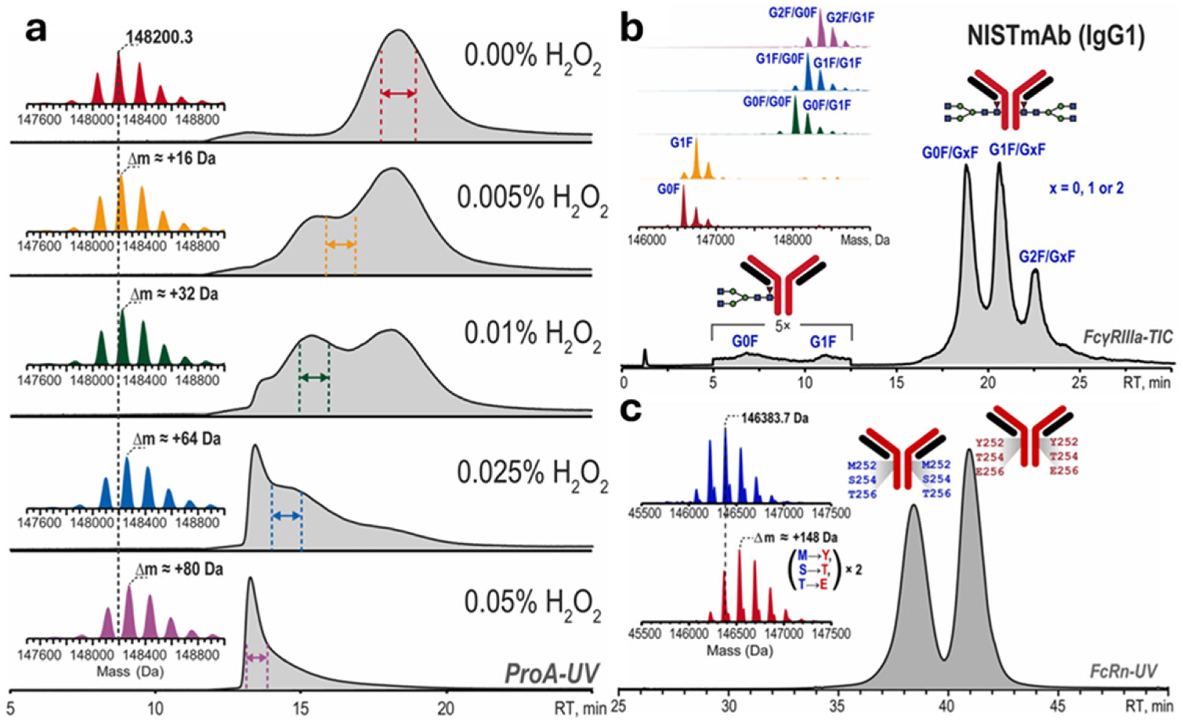
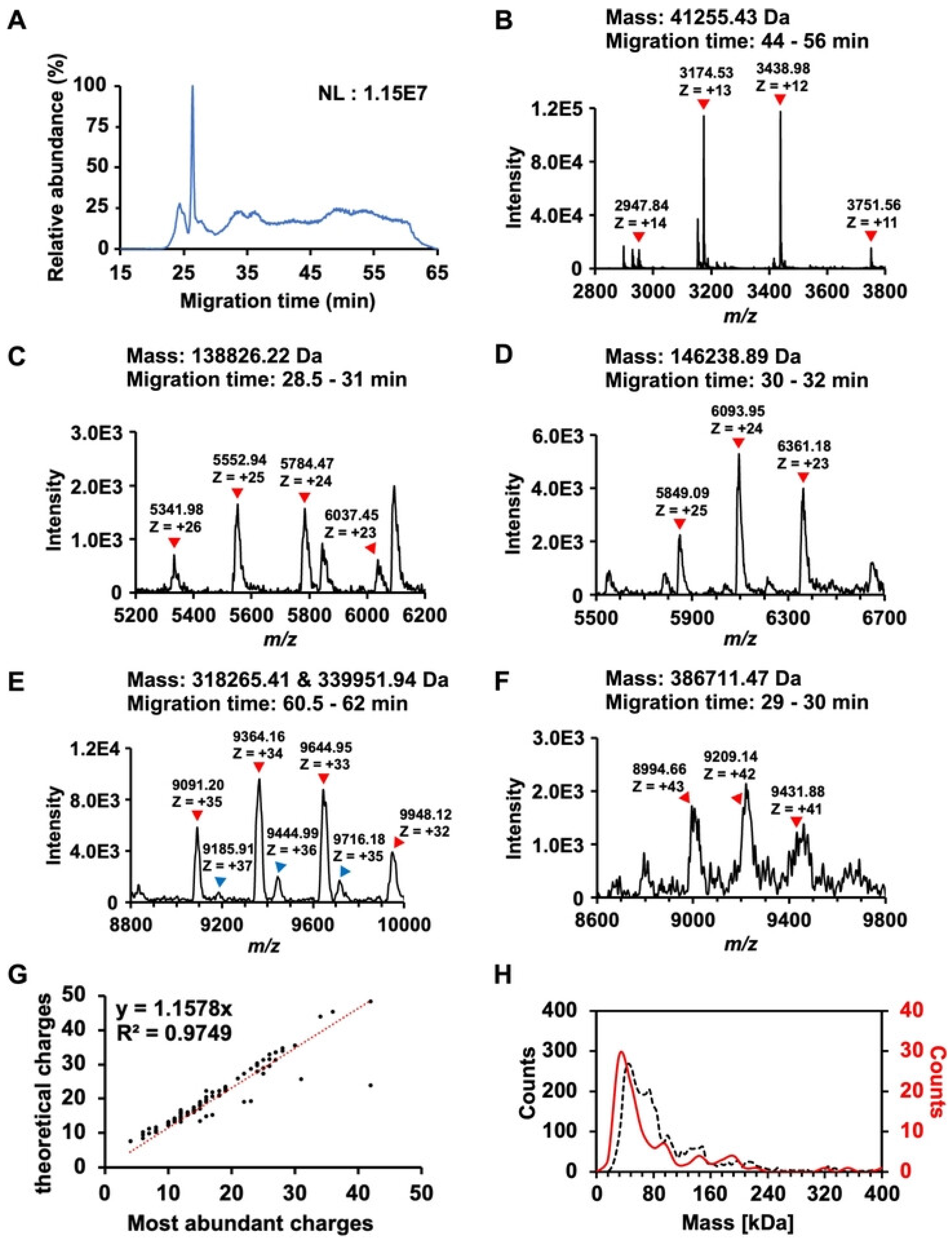
Appliedchem 05 00035 g007
Figure 8. Summary of detected proteoforms or protein complexes from an E. coli cell lysate using nCZE-ESI-UHMR. (A) Representative electropherogram of nCZE-ESI-UHMR analyses of the E. coli cell lysate. (BF) Mass spectra of five examples of large proteoforms/protein complexes detected. The charge states and deconvolved mass of each proteoform/protein complex are labeled. (G) Linear correlation between the most abundant charges and theoretical Rayleigh charges (ZR) of all proteoforms/protein complexes detected in single-shot nCZE-UHMR. (H) Alignment of the mass distribution of proteoforms/protein complexes in the E. coli cell lysate from mass photometry (black dashed line) and nCZE-UHMR (red line) analyses. © Wiley, published under CC-BY [223].
Appliedchem 05 00035 g008
Figure 9. Analysis of a three-protein mixture, RNase A, cytochrome c, and lysozyme. (a) Native mass spectrum. (b) MoCE separation of these three proteins. (c) Ellipsoid shapes of these three proteins obtained by the proposed method (top), and comparison with their corresponding crystal structure in the PDB database (bottom) (the black curves are the corresponding contour profiles of each ellipsoid). © The Royal Society of Chemistry, published under Creative Commons Attribution-NonCommercial 3.0 [227].
Appliedchem 05 00035 g009
Figure 10. (A) Base peak electropherogram of streptavidin (SA) analyzed by ncIEF-MS; (B) deconvoluted mass spectra of seven SA charge variants separated from ncIEF-MS; (C) averaged mass spectra of SA 1, SA 2, SA 3, and SA 4; and (D) averaged mass spectra of SA 5, SA 6, and SA 7. The mass spectra of SA charge variants were overlaid in (C,D) to compare the subtle differences in peaks at the same charge state. The inserted figures represent the zoomed-in peaks of SA charge variants at a charge state of 16+. (* = SA homotetramer, ! = SA homotetramer + Met, !! = SA homotetramer + 2 × Met, # = SA homotetramer + 3 × Met) © American Chemical Society, reprinted with permission [245].
Appliedchem 05 00035 g010
Figure 11. (A) Base peak electropherogram of native cIEF-assisted CZE-MS for the NISTmAb. (B) Mass spectrum averaged across peak 2 in (A). (C,D) Zoomed-in mass spectra of +24 and +38 charge states and (E,F) Deconvolution of NISTmAb proteoforms in the main peak (peak 2). Herein, the LCP-coated capillary was used. © American Chemical Society, reprinted with permission [220].
Appliedchem 05 00035 g011
Figure 12. Schematic of Auto-Ig-MS, a full automation workflow that joins immunoprecipitation with the KingFisher, sample preparation and injection with the SampleStream platform, ion attenuation with AIC, and subsequent charge detection analysis of individual ions via I2MS into a single fully automated pipeline. This technique was applied to a cohort of 58 possible COVID-19-positive subjects. © American Chemical Society, reprinted with permission [270].
Appliedchem 05 00035 g012
Figure 13. Deconvolution of mass spectra of ex vivo alpha-1-antitrypsin (AAT) from the liver tissue of a Z AAT homozygote. (A) Heat map of data acquired on a CIM-QTOF instrument showing mobility versus m/z, with monomeric to pentameric species labeled; data were plotted in Driftscope, with intensity on a log scale. (B) Mass spectra of ex vivo liver samples acquired on a CIM-QTOF, with identified masses labeled. (CF) Heat maps showing drift time in the mobility cell against the m/z values, with detected monomers, low-molecular-weight polymers, and an extended monomeric species highlighted for (C) AATWT, (D) AATHEAT, (E) AATZ, and (F) AATpH. The plots were generated using Driftscope (Waters Corp.) with a logarithmic intensity scale to highlight the presence of low-intensity polymers. © American Chemical Society, published under CC-BY 4.0 [284]. AATWT = wild type alpha-1-antitrypsin, AATHEAT = AATWT after heating at 55 °C for 48 h, AATZ = and AATpH = AATWT after incubating at pH 4.00 and 25 °C for 48 h.
Appliedchem 05 00035 g013
Table 1. Parameters for different HRMS instruments for intact protein species analyses.
InstrumentParameter SettingsAnalyzed Protein Species, Their Concentration, and Sample Introduction ApproachReference
Bruker 15-Tesla SolarisX FT-ICR MSCapillary voltage: 0.7–0.95 kVMembrane protein complex, E. coli AquaporinZ homotetramer (97 kDa) (15–30 µM), acquired via direct nanospray-ESI[96]
Dry gas temperature: 100 °C
Dry gas flow rate: 3.0 L/min
Ion Funnel RF amplitude: 300 Vapp
Ion Funnel 1 voltage: 150 V
Ion Funnel 2 voltage: 6 V
Skimmer 1: 50–125 V
Skimmer 2: 5 V
Multipole 1 RF Frequency: 2 MHz
Quadrupole RF frequency: 1.4 MHz
Transfer Hexapole RF Frequency: 1 MHz
Ion accumulation time: 500 ms
Waters Synapt G2-HDMS Q-TOF MSCapillary voltage: 0.5–1 kV 1, 0.8–1.2 kV 21 Membrane protein complex, E. coli AquaporinZ homotetramer (97 kDa) (15–30 µM), acquired via direct nanospray-ESI
2 Noncovalent protein–ligand complex: Lysozyme and tri-N-acetylchitotriose (NAG3), Trypsin/Pefabloc, Carbonic Anhydrase II/Chlorothiazide, β-Lactoglobulin A/Lauric Acid (5 µM concentration for each protein with 5–25 µM of ligand), acquired via direct nano-ESI
1 [96]
2 [65]
Sample cone: 40 V
Source temperature: 30 °C
Trap CE: 4–110 V 1 (High trap CE used for unfolding), 4–102
Transfer CE: 3 V 1, 2 V 2
Trap pressure: 3 × 10⁻³ mbar 1, 7 × 10⁻³ mbar 2
Transfer pressure: 3 × 10⁻³ mbar 1, 6.7 × 10⁻³ mbar 2
Trap direct current (DC) bias: −2 V 1, 3 V 2
Thermo Fisher Scientific Q Exactive HF Plus Orbitrap EMR MSSource voltage: 1.5 kVProteolysis-targeting chimeras (PROTACs)-ternary complex (5 µM), acquired via direct nanospray-ESI[66]
Capillary temperature: 100 °C and 50 °C
FT resolution: 140,000 (at 200 m/z)
In-source CID voltage: 10 V
HCD CE: 10 V
Automatic gain control (AGC) target: 5 × 10⁶
SCIEX ZenoTOF 7600Spray voltage: 3500 VNIST mAb (0.7–7 µM, converted from published concentration), ADC (Enhertu) (0.6–6 µM, converted from published concentration), acquired via microflow SEC-ESI-MS[97]
Curtain gas: 40 psi
CAD gas: 9
Ion source gas 1: 60 psi
Ion source gas 2: 60 psi
Source temperature: 250–300 °C
Declustering potential (DP): 120 V
CE: 12 V
Accumulation time: 0.25 s
Time bins to sum: 120
Agilent 6545XT AdvanceBio LC/Q-TOFDry gas temperature: 365 °C 3, 150 °C 43 Intact protein complex: Pyruvate kinase tetramer (232 kDa), glutamate dehydrogenase hexamer (335 kDa), and β-galactosidase tetramer (466 kDa) (2–20 µM), acquired via microflow SEC-ESI-MS
4 Intact protein: Myoglobin (Concentration/amount injected not reported), acquired via microflow SEC-ESI-MS
[94]
Dry gas flow: 12 L/min 3, 10 L/min 4
Nebulizer: 35 psig 3, 30 psig 4
Sheath gas temperature: 300 °C 3, 150 °C 4
Sheath gas flow: 12 L/min 3, 10 L/min 4
Capillary voltage: 5500 V 3, 5000 V 4
Nozzle voltage: 2000 V
Fragmentor: 300 V 3, 250 V 4
Skimmer: 220 V 3, 100 V 4
Acquisition rate: 1 spectrum/s
Superscripts are used to identify the instrument settings in column 2 which are used for the analyzed protein species in column 3.
Disclaimer/Publisher’s Note: The statements, opinions and data contained in all publications are solely those of the individual author(s) and contributor(s) and not of MDPI and/or the editor(s). MDPI and/or the editor(s) disclaim responsibility for any injury to people or property resulting from any ideas, methods, instructions or products referred to in the content.