Brief Report

Tears and Saliva as Biological Matrices for Vitamin D and Glucose Assessment: A Pilot Study

by
Pedro Henrique A. Reis
1,†,
Giovanna K. Jorge
2,†,
Edimar C. Pereira
2,
Lai Yu Tsun
2,
Thais M. Gascón
1,
Beatriz da C. A. Alves
1,
Glaucia L. da Veiga
1,
Samantha S. de Carvalho
1,
Renato G. Cerquinho Leça
3,
Vagner L. Lima
3 and
Fernando L. A. Fonseca
1,2,*
1
Laboratório de Análises Clínicas do Centro Universitário FMABC, Santo André 09060-870, SP, Brazil
2
Instituto de Ciências Farmacêuticas da Universidade Federal de São Paulo (UNIFESP), Diadema 04021-001, SP, Brazil
3
Disciplina de Oftalmologia do Centro Universitário FMABC, Santo André 09060-870, SP, Brazil
*
Author to whom correspondence should be addressed.
These authors contributed equally to this work.
Physiologia 2025, 5(3), 28; https://doi.org/10.3390/physiologia5030028
Submission received: 24 July 2025 / Revised: 20 August 2025 / Accepted: 26 August 2025 / Published: 29 August 2025

Abstract

Background: Several studies have established correlations between low serum levels of vitamin D and various pathologies, such as diabetes mellitus and its complications. However, few studies analyze its levels in matrices other than blood plasma, such as tears and saliva. In this study, we aimed to demonstrate the feasibility of using tears and saliva as alternative biological matrices for laboratory assessment of vitamin D and glucose concentration in individuals with type II diabetes mellitus and healthy individuals, using the electrochemiluminescence method. Methods: This study included volunteers with type II diabetes and healthy controls, excluding those with certain comorbidities or a BMI ≥ 40. Blood, tear, and saliva samples were taken after 3 h of fasting for biochemical analysis of fasting glucose and vitamin D. Statistical analysis was conducted using GraphPad Prism® 8.0—with Pearson and other tests to evaluate correlations—at a significance level of 5% and test power > 95%. Results: A negative correlation between serum vitamin D values and those found in saliva (p = 0.041) was found, as well as a positive correlation between serum glucose values and those found in tears (p = 0.0254). Conclusions: Tears and saliva samples can be used as proxies for venous blood samples in specific situations, such as studying blood glucose levels and vitamin D levels. However, expanding the sample size is essential to confirm the correlation and develop an accurate equation for estimating serum levels of these markers using these alternative matrices.

1. Introduction

Vitamin D, or calciferol, is a class of steroids with two main representatives: vitamin D2 (ergocalciferol) and D3 (cholecalciferol). Both share an identical metabolism and are considered prohormones as they need to be activated through hydroxylations at carbon 25 and calciferol 1 (in the liver and kidneys, respectively) to acquire biological activity (with the active form being calcitriol or 1,25-dihydroxyvitamin D) [1,2,3,4].
Vitamin D is unique among vitamins because, in addition to being acquired through the diet, humans can synthesize it in the skin through the irradiation of 7-dehydrocholesterol (7-DHC) with UVB, followed by temperature-dependent isomerization, forming calciferol [1,2,3,4]. Calcitriol primarily acts on calcium and phosphate homeostasis mechanisms, stimulating the active absorption of calcium by the small intestine. Additionally, it facilitates the action of parathyroid hormone (PTH) on the skeleton and increases the expression of calcium transporters in the kidneys. Its known mechanisms of action involve the modulation of gene expression through binding to vitamin D-responsive elements (VDRE or nVDRE), as well as the activation of pre-calbindin into calbindin [1,3,4].
Vitamin D also has anti-inflammatory and immunomodulatory effects [1,5], participating in the regulation of pro-inflammatory cytokine production and in the differentiation of immune system cells, especially T lymphocytes [5], exhibiting synergistic action with IL2 [6]. Monocytes and macrophages exposed to lipopolysaccharides (LPS) or mycobacterium tuberculosis activate the vitamin D receptor gene and the 1α-hydroxylase gene. Increased calcitriol production results in the synthesis of cathelicidin, a peptide capable of destroying M. tuberculosis and other infectious agents. Calcidiol levels below 20 ng/mL (50 nmol/L) prevent macrophages and monocytes from initiating their innate immune response mechanism [7,8,9].
Observational studies suggest an association between low blood concentrations of calcidiol and increased risk of cardiovascular diseases [1,10,11], neoplasms [1,12], autoimmune diseases (including type 1 diabetes) [1,13], metabolic syndrome [5,14,15,16], and infections (including COVID-19) [1,5,17]. Currently, the benefits of vitamin D supplementation on bone mineral density, secondary hyperparathyroidism, and neuromuscular function are widely accepted [8,9]. Additionally, several studies indicate an inverse relationship between serum vitamin D levels and the incidence of chronic diseases associated with chronic inflammation, including diabetes mellitus [1,11,18,19,20].
Serum levels of calcidiol can also be associated with ophthalmological pathologies such as myopia progression, age-related macular degeneration (AMD), diabetic retinopathy, uveitis, and dry eye syndrome [21,22,23,24,25]. However, few studies analyze the concentration of vitamin D in biological matrices beyond blood plasma, such as tears and saliva. In this study, using the electrochemiluminescence method, we aimed to demonstrate the feasibility of using tears and saliva as alternative biological matrices for the laboratory assessment of vitamin D and glucose concentration in individuals with type II diabetes mellitus and healthy individuals.

2. Results

This study involved 14 volunteers, including 9 women and 5 men, with an average age of 45.35 years (±11.57). Of these, 6 individuals had diabetes diagnosed for more than 5 years (13.50 ± 8.0 years), and 8 were healthy (Table 1). The body mass index (BMI) of diabetic volunteers was 29.20 (±4.70) kg/m2, while that of healthy volunteers was 27.01 (±3.82) kg/m2. Among the diabetic participants, 3 were taking vitamin D supplements, while none of the healthy control subjects supplemented their diet.
In this study, the concentrations of vitamin D (calcidiol) and glucose were evaluated in serum, tears, and saliva (Table 2). Our results show that there are no differences in serum vitamin D concentrations between healthy volunteers (27.92 ± 9.85 ng/mL) and diabetics (32.93 ± 15.60 ng/mL). The average value of vitamin D in diabetics is slightly increased in this group, but this increase is because 3 participants are supplementing with this vitamin (1.000 UI/day). If we disregard the measurements of the volunteers taking supplements, the average concentration of vitamin D in diabetics would be 24.8 ng/mL (±8.28). However, this supplementation-induced increase is not observed in either the saliva or tears of these individuals.
To assess the presence of a correlation between serum levels of vitamin D and glucose, as well as their concentrations in the other matrices (tears and saliva), Pearson correlation tests and simple linear regressions were performed between the collected parameters (Figure 1). A negative correlation was found between serum and salivary levels of vitamin D (R = −0.652, R2 = 0.4253, p = 0.041). Our results have also shown a tendency for a negative correlation between serum vitamin D levels and those found in tears (R = −0.6104, R2 = 0.3726, p = 0.0609). Considering glucose, a positive correlation was found between blood and tear levels (R = 0.8668, R2 = 0.7514, p = 0.0254). Correlations between serum glucose levels and those found in saliva, as well as between glucose levels in tears and saliva, were not significant (p = 0.9401 and p = 0.6726, respectively).

3. Discussion

This study sought to evaluate the feasibility of utilizing tears and saliva as alternative biological samples for measuring vitamin D and glucose levels in both individuals with type II diabetes mellitus and healthy controls, employing the electrochemiluminescence technique.
In this study, a negative correlation between serum and salivary levels of vitamin D was found. Several studies provide insights into the relationship between these two matrices. Bahramian et al. [26], for instance, found a significant positive correlation between serum and salivary levels of vitamin D in patients with recurrent aphthous stomatitis compared to healthy subjects. The potential of using saliva to assess vitamin D levels in oral health conditions was also highlighted in patients with oral lichen planus [27]. Concerning diabetic patients, Abdolsamadi et al. [28] have also explored the relationship between serum and salivary levels of vitamin D and found that low concentrations of this vitamin were found in both serum and saliva of newly diagnosed diabetes patients. According to the authors, saliva could serve as a valuable diagnostic tool for early detection, monitoring, and progression of diabetes, although more studies are recommended on this topic. Contrary to what Bahramian et al. [26] described, in our study, a negative correlation was found between these two matrices in diabetes patients, suggesting that salivary levels may reflect serum levels of vitamin D only in certain conditions: the patients included in this study had already been diagnosed with diabetes for at least 5 years, rather than being recently diagnosed as described by Abdolsamadi et al. [28]. Furthermore, the quantification of vitamin D levels in our study was performed via electrochemiluminescence method rather than by ELISA [28], suggesting that the laboratory method can influence the results.
When comparing the detection of vitamin D in serum and tears, we found a tendency for a negative correlation between these two matrices; studies have shown interesting findings, such as significantly higher levels of 25-hydroxyvitamin D in tears compared to serum [29,30]. Our results also showed higher levels of vitamin D in tears than in serum in both groups analyzed, suggesting that tear fluid may be a rich source for detecting vitamin D levels. However, as seen in saliva, our results contrast with those described by these authors, who found a positive correlation between the measurements taken in serum and in the tears of the patients. This difference may be due to the fact that the authors included only healthy volunteers, while our study also included volunteers with diabetes. It is known that patients with diabetes tend to present ocular complications associated with the disease [31], and these alterations may be associated with low levels of vitamin D [32].
A positive correlation was found between blood and tear levels of glucose. Aihara et al. [33] also described a positive correlation between glucose levels measured in the blood and tears. The authors suggest that monitoring tear glucose could be a dependable and non-invasive alternative for tracking blood glucose concentrations in diabetic patients, regardless of their glycated hemoglobin levels and the timing of sample collection.
Although significant correlations were observed between venous blood and saliva/tear levels, the absolute concentrations are not interchangeable. Indeed, differences in protein content, pH, enzymatic activity, and viscosity between these matrices are known to affect analyte distribution and assay performance, which explains the lower quantitative values in saliva and tears compared with serum. Therefore, as suggested, specific reference intervals will need to be established for each biofluid before clinical application, as highlighted in previous studies [34,35,36].
The limited sample size represents a limitation of the present study and may have influenced the strength of the observed correlations. However, it was possible to verify the feasibility of using these alternative matrices for screening serum levels of biochemical markers, such as vitamin D and glucose, through an accessible method like electrochemiluminescence. Additionally, there is clear evidence of a correlation between blood and tear glucose levels, as well as between blood and saliva vitamin D levels. Although there appears to be a correlation between blood and tear vitamin D levels, further expansion of this study would be necessary to confirm it.

4. Materials and Methods

This is a cross-sectional study that uses convenience sampling. This study was approved by the Research Ethics Committee of the Universidade Federal de São Paulo (protocol number 0015.0015.01/2019) and was conducted in accordance with the Declaration of Helsinki. Informed consent was obtained from all subjects involved in the study. Inclusion criteria selected volunteers with type II diabetes mellitus, aged between 20 and 65 years. Volunteers with serological comorbidities (such as HIV, Hepatitis C, Cytomegalovirus), postmenopausal women, smokers, alcoholics, drug dependents, or individuals with a BMI ≥ 40 were excluded.
After obtaining informed consent, 6 volunteers with type II diabetes and 8 healthy volunteers were included as the control group. Data regarding age, weight, height, duration of the disease (type II diabetes), presence of other pathologies, use of vitamin D supplements, and other relevant information were collected. The collection of samples from different biological matrices from each volunteer was performed on the same day, simultaneously, after a minimum fasting period of 3 h.
Venous blood was collected using Vacutainer tubes, both with and without EDTA (BD Vacutainer® K2EDTA tubes (Cat. No. 367525; Becton Dickinson, Franklin Lakes, NJ, USA and BD Vacutainer® Serum Tube, Cat. No. 367614, Becton, Dickinson and Company, Franklin Lakes, NJ, USA). Tear samples were collected using test strips (Schirmer Tear Test; Ophthalmos, Porto Alegre, RS, Brazil) without topical anesthesia. Participants avoided eye drops, ocular lubricants, and cosmetics for 12 h prior to collection, as well as contact lens use for 24 h, and attended a quiet room with controlled temperature (20–24 °C) and reduced illumination. After 30 s of spontaneous blinking, each Schirmer strip was folded at the notch and positioned in the lateral third of the inferior conjunctival fornix, avoiding contact with the cornea and eyelid margin. The eye remained in the primary position, without compression, for 5 min. The strip was then removed and transferred into a microtube containing 100 µL of saline solution. The tubes were vortexed for 5 s and kept under rotational agitation for 30 min at 4 °C to maximize recovery. Subsequently, centrifugation was performed at 10,000× g for 10 min at 4 °C to clarify the eluate and retain paper fibers; the supernatant (tear eluate) was carefully transferred to a new microtube, in which vitamin D and glucose were analyzed.
Saliva samples were collected using Salivette (Sarstedt, Germany, cat no. 51.1534), a method of saliva stimulation and collection. For saliva collection, volunteers fasted for 30–60 min. The device was placed in the volunteer’s mouth for 2 min without chewing. After this period, the sample was centrifuged at 3000 rpm for 2 min at 4 °C, and the supernatant, free of debris, was transferred to a microtube and then analyzed. All samples were collected in the morning.
Biochemical parameters such as fasting glucose and vitamin D (calcidiol) in blood, saliva, and tears were analyzed using an automated biochemical analyzer, Cobas® e411 model, using the electrochemiluminescence method.
To obtain means, standard deviations, and medians, as well as for correlation tests (Pearson, Lin’s Concordance Correlation Coefficient, Bland–Altman Analysis) to evaluate the correlation between the biochemical parameters analyzed in serum/plasma, tears, and saliva, GraphPad Prism® 8.0 software was used, considering a significance level of 5% and a test power greater than 95%.

5. Conclusions

Tear secretion can be used for the measuring of glucose, while saliva can be used to analyze vitamin D levels in patients, as there is a correlation between serum markers and those found in these matrices when analyzed by the electrochemiluminescence method. A limitation of this study is the sample size, which may have affected statistical analyses. Thus, it is evident that expanding the sample size is essential to confirm this correlation and even develop an equation that allows for the estimation of the serum levels of various markers based on the findings from these alternative matrices.

Author Contributions

Conceptualization, F.L.A.F., R.G.C.L. and V.L.L.; methodology, P.H.A.R., G.K.J. and L.Y.T.; validation, T.M.G., S.S.d.C. and E.C.P.; formal analysis, G.L.d.V. and B.d.C.A.A.; resources, F.L.A.F.; writing—original draft preparation, P.H.A.R.; writing—review and editing, B.d.C.A.A.; visualization, G.L.d.V.; supervision, F.L.A.F. All authors have read and agreed to the published version of the manuscript.

Funding

This research received no external funding.

Institutional Review Board Statement

The study was conducted in accordance with the Declaration of Helsinki and approved by the Ethics Committee of Universidade Federal de São Paulo (protocol number 0015.0015.01, approval in September 2019).

Informed Consent Statement

Informed consent was obtained from all subjects involved in the study.

Data Availability Statement

Data supporting this study can be found in Figshare.com (https://doi.org/10.6084/m9.figshare.29640605).

Conflicts of Interest

The authors declare no conflicts of interest.

References

  1. Institute of Medicine (US). Committee to Review Dietary Reference Intakes for Vitamin D and Calcium. In Dietary Reference Intakes for Calcium and Vitamin D; Ross, A.C., Taylor, C.L., Yaktine, A.L., Del Valle, H.B., Eds.; National Academies Press: Washington, DC, USA, 2011. [Google Scholar] [CrossRef]
  2. Grant, W.B.; Strange, R.C.; Garland, C.F. Sunshine is good medicine. The health benefits of ultraviolet-B induced vitamin D production. J. Cosmet. Dermatol. 2003, 2, 86–98. [Google Scholar] [CrossRef]
  3. Lu, X.; Elizondo, R.A.; Nielsen, R.; Christensen, E.I.; Yang, J.; Hammock, B.D.; Watsky, M.A. Vitamin D in tear fluid. Investig. Ophthalmol. Vis. Sci. 2015, 56, 5880–5887. [Google Scholar] [CrossRef]
  4. Bouillon, R. Comparative analysis of nutritional guidelines for vitamin D. Nat. Rev. Endocrinol. 2017, 13, 466–479. [Google Scholar] [CrossRef] [PubMed]
  5. Singh, S.K.; Jain, R.; Singh, S. Vitamin D deficiency in patients with diabetes and COVID-19 infection. Diabetes Metab. Syndr. 2020, 14, 1033–1035. [Google Scholar] [CrossRef] [PubMed]
  6. Jeffery, L.E.; Burke, F.; Mura, M.; Zheng, Y.; Qureshi, O.S.; Hewison, M.; Walker, L.S.K.; Lammas, D.A.; Raza, K.; Sansom, D.M. 1,25-Dihydroxyvitamin D3 and IL-2 combine to inhibit T cell production of inflammatory cytokines and promote development of regulatory T cells. J. Immunol. 2009, 183, 5458–5467. [Google Scholar] [CrossRef] [PubMed]
  7. Liu, P.T.; Stenger, S.; Li, H.; Wenzel, L.; Tan, B.H.; Krutzik, S.R.; Ochoa, M.T.; Schauber, J.; Wu, K.; Meinken, C. Toll-like receptor triggering of a vitamin D-mediated human antimicrobial response. Science 2006, 311, 1770–1773. [Google Scholar] [CrossRef]
  8. Holick, M.F. Vitamin D deficiency. N. Engl. J. Med. 2007, 357, 266–281. [Google Scholar] [CrossRef]
  9. Holick, M.F.; Binkley, N.C.; Bischoff-Ferrari, H.A.; Gordon, C.M.; Hanley, D.A.; Heaney, R.P.; Murad, M.H.; Weaver, C.M. Evaluation, treatment, and prevention of vitamin D deficiency: An Endocrine Society clinical practice guideline. J. Clin. Endocrinol. Metab. 2011, 96, 1911–1930. [Google Scholar] [CrossRef]
  10. Bahrami, L.S.; Ranjbar, G.; Norouzy, A.; Arabi, S.M. Vitamin D supplementation effects on the clinical outcomes of patients with coronary artery disease: A systematic review and meta-analysis. Sci. Rep. 2020, 10, 12923. [Google Scholar] [CrossRef]
  11. Kienreich, K.; Tomaschitz, A.; Verheyen, N.; Pieber, T.; Gaksch, M.; Grübler, M.R.; Pilz, S. Vitamin D and cardiovascular disease. Nutrients 2013, 5, 3005–3021. [Google Scholar] [CrossRef]
  12. Garland, C.F.; Garland, F.C. Do sunlight and vitamin D reduce the likelihood of colon cancer? Int. J. Epidemiol. 1980, 9, 227–231. [Google Scholar] [CrossRef]
  13. Hayes, C.E.; Cantorna, M.T.; DeLuca, H.F. Vitamin D and multiple sclerosis. Proc. Soc. Exp. Biol. Med. 1997, 216, 21–27. [Google Scholar] [CrossRef]
  14. Kayaniyil, S.; Vieth, R.; Retnakaran, R.; Knight, J.A.; Qi, Y.; Gerstein, H.C.; Perkins, B.A.; Harris, S.B.; Zinman, B.; Hanley, A.J. Association of vitamin D with insulin resistance and beta-cell dysfunction in subjects at risk for type 2 diabetes. Diabetes Care 2010, 33, 1379–1381. [Google Scholar] [CrossRef] [PubMed]
  15. Strange, R.C.; Shipman, K.E.; Ramachandran, S. Metabolic syndrome: A review of the role of vitamin D in mediating susceptibility and outcome. World J. Diabetes 2015, 6, 896–911. [Google Scholar] [CrossRef]
  16. Machado, M.R.C.; Gomes Junior, S.C.; Marinheiro, L.P.F. Vitamina D e diabetes mellitus, suas epidemias e o envelhecimento. O que há de novo? Reprod. Clim. 2014, 29, 54–59. [Google Scholar] [CrossRef]
  17. Razdan, K.; Singh, K.; Singh, D. Vitamin D levels and COVID-19 susceptibility: Is there any correlation? Med. Drug Discov. 2020, 7, 100051. [Google Scholar] [CrossRef] [PubMed]
  18. Pines, A. Vitamin D and health issues—Questioned benefits. Climacteric 2014, 17, 657–659. [Google Scholar] [CrossRef]
  19. Mitri, J.; Pittas, A.G. Vitamin D and diabetes. Endocrinol. Metab. Clin. N. Am. 2014, 43, 205–232. [Google Scholar] [CrossRef]
  20. Valipour, G.; Saneei, P.; Esmaillzadeh, A. Serum vitamin D levels in relation to schizophrenia: A systematic review and meta-analysis. J. Clin. Endocrinol. Metab. 2014, 99, 3863–3872. [Google Scholar] [CrossRef]
  21. Yazar, S.; Hewitt, A.W.; Black, L.J.; McKnight, C.M.; Mountain, J.A.; Sherwin, J.C.; Oddy, W.H.; Coroneo, M.T.; Lucas, R.M.; Mackey, D.A. Myopia is associated with lower vitamin D status in young adults. Investig. Ophthalmol. Vis. Sci. 2014, 55, 4552–4559. [Google Scholar] [CrossRef] [PubMed]
  22. Jee, D.; Kang, S.; Yuan, C.; Cho, E.; Arroyo, J.G.; Epidemiologic Survey Committee of the Korean Ophthalmologic Society. Serum 25-Hydroxyvitamin D levels and dry eye syndrome: Differential effects of vitamin D on ocular diseases. PLoS ONE 2016, 11, e0149294. [Google Scholar] [CrossRef]
  23. Yildirim, P.; Garip, Y.; Karci, A.A.; Guler, T. Dry eye in vitamin D deficiency: More than an incidental association. Int. J. Rheum. Dis. 2016, 19, 49–54. [Google Scholar] [CrossRef] [PubMed]
  24. Payne, J.F.; Ray, R.; Watson, D.G.; Delille, C.; Rimler, E.; Cleveland, J.; Lynn, M.J.; Tangpricha, V.; Srivastava, S.K. Vitamin D insufficiency in diabetic retinopathy. Endocr. Pract. 2012, 18, 185–193. [Google Scholar] [CrossRef] [PubMed]
  25. Lu, X.; Vick, S.; Chen, Z.; Chen, J.; Watsky, M.A. Effects of vitamin D receptor knockout and vitamin D deficiency on corneal epithelial wound healing and nerve density in diabetic mice. Diabetes 2020, 69, 1042–1051. [Google Scholar] [CrossRef] [PubMed]
  26. Bahramian, A.; Falsafi, P.; Abbasi, T.; Ghanizadeh, M.; Abedini, M.; Kavoosi, F.; Kouhsoltani, M.; Noorbakhsh, F.; Dabbaghi Tabriz, F.; Rajaeih, S.; et al. Comparing serum and salivary levels of vitamin D in patients with recurrent aphthous stomatitis and healthy individuals. J. Dent. 2018, 19, 295–300. [Google Scholar]
  27. Gholizadeh, N.; Pirzadeh, F.; Mirzaii-Dizgah, I.; Sheykhbahaei, N. Relationship between salivary vitamin D deficiency and oral lichen planus. Photodermatol. Photoimmunol. Photomed. 2020, 36, 384–386. [Google Scholar] [CrossRef]
  28. Abdolsamadi, H.; Vahedi, M.; Borzouei, S.; Soltanian, A.; Hosseini, A.; Avval, M.Z. Evaluation the relationship between serum and salivary levels of 25(OH)Vit.D with type II diabetes in newly diagnosed diabetics. J. Mol. Biol. Res. 2018, 8, 108–113. [Google Scholar] [CrossRef]
  29. Sethu, S.; Shetty, R.; Deshpande, K.; Pahuja, N.; Chinnappaiah, N.; Agarwal, A.; Sharma, A.; Ghosh, A. Correlation between tear fluid and serum vitamin D levels. Eye Vis. 2016, 3, 22. [Google Scholar] [CrossRef]
  30. Lai, Y.T.; Cerquinho, R.G.; Perez, M.M.; Alves, B.D.C.A.; Pereira, E.C.; Azzalis, L.A.; Junqueira, V.B.C.; Soares, L.R.; Fonseca, F.L.A. Determination of vitamin D in tears of healthy individuals by the electrochemiluminescence method. J. Clin. Lab. Anal. 2019, 33, e22830. [Google Scholar] [CrossRef]
  31. Ting, D.S.; Cheung, G.C.; Wong, T.Y. Diabetic retinopathy: Global prevalence, major risk factors, screening practices and public health challenges: A review. Clin. Exp. Ophthalmol. 2016, 44, 260–277. [Google Scholar] [CrossRef]
  32. Usluogullari, C.A.; Balkan, F.; Caner, S.; Ucler, R.; Kaya, C.; Ersoy, R.; Cakir, B. The relationship between microvascular complications and vitamin D deficiency in type 2 diabetes mellitus. BMC Endocr. Disord. 2015, 15, 33. [Google Scholar] [CrossRef] [PubMed]
  33. Aihara, M.; Kubota, N.; Minami, T.; Shirakawa, R.; Sakurai, Y.; Hayashi, T.; Iwamoto, M.; Takamoto, I.; Kubota, T.; Suzuki, R.; et al. Association between tear and blood glucose concentrations: Random intercept model adjusted with confounders in tear samples negative for occult blood. J. Diabetes Investig. 2021, 12, 266–276. [Google Scholar] [CrossRef]
  34. Chiappin, S.; Antonelli, G.; Gatti, R.; De Palo, E.F. Saliva specimen: A new laboratory tool for diagnostic and basic investigation. Clin. Chim. Acta 2007, 383, 30–40. [Google Scholar] [CrossRef]
  35. Kaufman, E.; Lamster, I.B. The diagnostic applications of saliva—A review. Crit. Rev. Oral Biol. Med. 2002, 13, 197–212. [Google Scholar] [CrossRef] [PubMed]
  36. Singh, D.K.; Arya, A.; Seth, A. Salivary glucose levels: Can they be a reliable marker in monitoring diabetes mellitus? J. Oral Maxillofac. Pathol. 2014, 18, 214–219. [Google Scholar] [CrossRef]
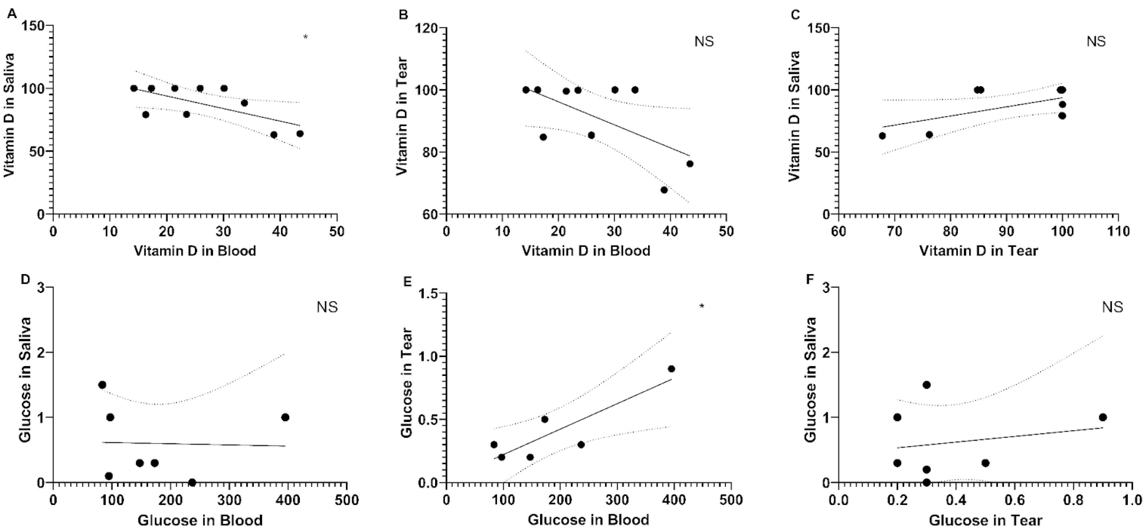
Figure 1. Linear Regression graphs of the parameters analyzed in different matrices. (A) correlation between vitamin D levels in blood and in saliva; (B) correlation between vitamin D levels in blood and in tears; (C) correlation between vitamin D levels in tears and in saliva; (D) correlation between glucose levels in blood and in saliva; (E) correlation between glucose levels in blood and in tears; (F) correlation between glucose levels in tears and in saliva. * Dotted lines indicate the 95% confidence intervals; Values of vitamin D are represented in U/L; Values of Glucose are represented in mg/dL. NS stands for Not Significant.
Physiologia 05 00028 g001
Table 1. Descriptive characteristics of the study volunteers (n = 16).
n%
Total volunteers14100
Female volunteers964.3
Male volunteers535.7
Age (years)
≤30214.3
31–40428.6
41–49214.3
50–61642.8
Mean age (years)45.35
Diabetes status
Type II diabetes642.8
Non-diabetic857.2
Vitamin D supplementation
Diabetic volunteers342.8 *
Non-diabetic volunteers00
* Percentage calculated among diabetic volunteers (n = 7).
Table 2. Description of the biochemical parameters, discriminated by group.
GroupSerum VitD (ng/mL)Tear VitD (ng/mL)Saliva VitD (ng/mL)Serum Glucose (mg/dL)Tear Glucose (mg/dL)Saliva Glucose (mg/dL)
DM32.93 ± 15.6092.10 ± 11.0186.32 ± 15.21189.83 ± 114.450.475 ± 0.310.40 ± 0.424
Control27.92 ± 9.8590.64 ± 14.2488.44 ± 16.8189.95 ± 4.750.267 ± 0.0580.70 ± 0.668
Values expressed in Mean ± Standard Deviation; DM = Volunteers with Type 2 Diabetes Mellitus.
Disclaimer/Publisher’s Note: The statements, opinions and data contained in all publications are solely those of the individual author(s) and contributor(s) and not of MDPI and/or the editor(s). MDPI and/or the editor(s) disclaim responsibility for any injury to people or property resulting from any ideas, methods, instructions or products referred to in the content.