Brief Report

The Effects of Creatine Monohydrate and/or Whey Protein on the Muscle Protein Synthesis and Anabolic Signaling Responses in Non-Stressed C2C12 Murine Myotubes

by
Nicholas J. Kontos
1,
Joshua S. Godwin
1,
Anthony Agyin-Birikorang
1,
Darren G. Candow
2,
Christopher M. Lockwood
3,
Michael D. Roberts
1,* and
Christopher B. Mobley
1,*
1
Nutrabolt Applied and Molecular Physiology Laboratory, School of Kinesiology, Auburn University, Auburn, AL 36849, USA
2
Faculty of Kinesiology and Health Studies, University of Regina, Regina, SK S7N 5A2, Canada
3
Nutrabolt, Austin, TX 78749, USA
*
Authors to whom correspondence should be addressed.
Physiologia 2025, 5(2), 17; https://doi.org/10.3390/physiologia5020017
Submission received: 28 February 2025 / Revised: 7 April 2025 / Accepted: 1 May 2025 / Published: 14 May 2025
(This article belongs to the Special Issue Feature Papers in Human Physiology—3rd Edition)

Abstract

:
Background/Objectives: Creatine monohydrate (CRE) is a popular nutritional supplement that increases lean/muscle mass accretion. Although data regarding CRE and its effects on muscle protein synthesis are mixed, we hypothesized that CRE may potentiate/extend the anabolic response to essential amino acids given that CRE acts as a high-energy phosphate buffer to potentially amplify anabolic signaling. Therefore, we used an in vitro model to determine whether CRE synergistically enhances myotube protein synthesis and the anabolic signaling responses to EAA-rich whey protein (WP). Methods: C2C12 murine myotubes were treated with control media containing PBS (CTL), WP serum (5 mg/mL), CRE (10 mM), or WP + CRE. Myotubes were collected following 1, 4, and 24 h treatments (n = 6 replicates per treatment and time point) and assayed for relative creatine levels, myotube protein synthesis levels, and phosphorylation markers. Results: Cellular creatine levels were greater in CRE and WP + CRE versus CTL and WP at all treatment time points (p < 0.05). The protein synthesis levels with 4 hr treatments with WP and WP + CRE were greater compared to the CTL (p = 0.036 and p < 0.001, respectively), and 24 h levels were greater with WP versus other treatments (p < 0.05). p-p70S6K (Ser389) and p-rpS6 (Ser235/236) were greater with WP at 1 h compared to all other treatments (p < 0.05). No effects across time points were observed for p-mTOR (Ser2448), p-4E-BP1 (Thr37/46), or p-AMPKα (Thr172). Conclusions: WP increases protein synthesis and anabolic signaling with no additive effect from CRE. However, given that myotubes were not stressed nor stimulated to contract, such models are needed with the current treatment schematic to examine potential interactions.

1. Introduction

The preservation of muscle mass relies on the balance between muscle protein synthesis (MPS) and muscle protein breakdown (MPB). Muscle mass increases are observed when MPS exceeds MPB, and MPS is upregulated through the mechanistic target of rapamycin complex 1 (mTORC1), which, when activated, leads to the phosphorylation of several downstream targets that directly regulate the ribosome assembly and the translation of new proteins [1,2]. Two notable activators of mTORC1 include mechanical overload [3] and amino acid sensing [4,5]. Dietary protein supplements have been used to better maintain a net positive protein balance through the bolstering of MPS. In this regard, several studies have shown that protein supplementation enhances the MPS response in the basal and post-exercise states [6,7,8,9]. Critically, whey protein (WP) is superior to other dietary protein supplements in this regard due in part to its rich essential animo acid profile [10,11,12]. The combination of creatine monohydrate (CRE) supplementation and resistance training has been shown on numerous occasions to increase measures of lean tissue and/or muscle accretion compared to resistance training alone [13,14]. However, the mechanisms explaining these greater anabolic responses from CRE remain to be determined.
There is some evidence that CRE reduces leucine oxidation and the plasma leucine rate of appearance (indicator of muscle protein catabolism) in young healthy males [15]. CRE also increases the expression of myogenic transcription factors [16], satellite cell proliferation and activity [17], and basal and post-exercise myosin heavy chain mRNA expression in young males [16,18]. In vitro data also indicate that creatine has the potential to induce favorable effects on muscle protein kinetics. For instance, Deldicque et al. [19] reported that 24 h treatments with 5 mM of CRE increased the rates of sarcoplasmic and myofibrillar protein synthesis in C2C12 myotubes, which was accompanied by enhanced mTORC1 and MAPK signaling. Furthermore, Louis et al. [20] reported that 10 mM of CRE increased IGF-1 and myogenic regulatory factor mRNA levels in C2C12 myotubes. Our laboratory has reported that CRE elicits C2C12 myotube anabolism while also mitigating myostatin-induced myotube atrophy, potentially through increased Akirin1 gene expression [21]. However, unlike WP, human data do not support that CRE directly increases MPS [15] .
The abovementioned findings from the literature support that CRE and WP may exhibit synergistic anabolic effects given that both ingredients target mTORC1 as well as different cellular processes (i.e., MPS versus satellite cell dynamics and gene expression). CRE could also prolong the MPS response induced by WP since CRE acts to buffer cellular ATP concentrations. The latter point is notable for a few reasons. First, it has been posited that nutrient-induced increases in MPS consumes high levels of cellular ATP (thus causing cellular energy stress and a potential refractory response) [22,23]. Additionally, O’Conner and colleagues have reported that phosphocreatine (PCr) acts as a spatiotemporal energy buffer during myoblast fusion and myotube growth in vitro [24]. To this end, the authors suggest that these are high-energy processes and that ATP buffering through PCr may enhance the cellular growth of serval cell types.
Although the synergistic effects of WP and CRE on myofiber hypertrophy are compelling, no in vitro study to date has sought to extensively examine this possibility. Therefore, we performed a series of shorter-term (1 and 4 h) in vitro C2C12 myotube experiments to determine if any synergistic anabolic effects exist through co-treatments of CRE and WP compared to each ingredient alone. Moreover, we performed longer-term 24 h treatments to determine whether WP + CRE exhibited prolonged anabolic signaling and MPS responses compared to either ingredient alone. We hypothesized that shorter- and longer-term WP + CRE treatments would enhance the anabolic signaling and MPS responses and that this would be reflected through a reduction in cellular energetic stress, as determined via phosphorylated AMPKα (Thr172).

2. Materials and Methods

2.1. Cell Culture

C2C12 immortalized murine myoblasts (ATCC, Manassas, VA, USA), with a passage number of 5, were cultured at 37 °C in a 5% CO2 atmosphere. Myoblasts were seeded at a density of ~3 × 10⁵ in six-well plates containing 3 mL/well of growth media (GM) consisting of Dulbecco’s modified eagle’s medium (DMEM; Corning, Corning, NY, USA), 10% Fetal Bovine Serum (Corning), 1% penicillin/streptomycin (VWR, Radnor, PA, USA), and 0.1% gentamycin (VWR). Upon reaching confluency (~80–90%), myoblasts were differentiated into myotubes by switching to differentiation media (DM) containing DMEM, 2% horse serum (VWR), 1% penicillin/streptomycin (VWR), and 0.1% gentamycin (VWR). DM was replaced every 24–48 h until confluency of mature myotubes was achieved (~7 days). Following 7 days of differentiation, myotubes were treated for 1, 4, and 24 h with control media (CTL = DM only), DM with 5 mg/mL WP serum generated from centrifuging out solids (Hilmar Whey Protein Isolate 9400; Hilmar Ingredients, Hilmar, CA, USA), DM with 10 mM CRE (BodyBuilding.com, Boise, ID, USA), or DM with a combination of WP and CRE. These dosages were based upon previous in vitro studies in the literature showing that 5 mg/mL WP serum increases MPS in C2C12 myotubes [25] and that 10 mM CRE induces myotube hypertrophy [20].
Following treatment (30 min prior to collection), cells were pulse labeled with 3.3 µg/mL puromycin dihydrochloride (VWR; Cat. No. 97064-280) to assess relative myotube protein synthesis responses. Cells were then collected using ice-cold lysis buffer (Celling Signaling Technology, Danvers, MA, USA; Cat. No. 9803). Subsequent lysates were processed for total protein using a commercially available BCA protein assay kit (Thermo Scientific, Waltham, MA, USA; Cat. No. A55864) and spectrophotometer (Agilent Biotek Synergy H1 hybrid reader; Agilent, Santa Clara, CA, USA). Lysates were then prepared for Western blotting as described below.

2.2. Western Blotting

Cell lysates were prepared using 4× Laemmli buffer in distilled water at a concentration of 0.5 μg/μL and denatured for 5 min at 100 °C. Prepared samples (15 μL) were loaded onto 4–15% gradient SDS-polyacrylamide gels (Criterion TGX stain-free gels; Bio-Rad Laboratories, Hercules, CA, USA) and subjected to electrophoresis at 180 volts for 50 min. Proteins were then transferred to PVDF membranes at 200 mA for 2 h. Membranes were then stained with ponceau and imaged on a gel documentation system (ChemiDoc Touch; Bio-Rad Laboratories, Hercules, CA, USA). Membranes were reactivated in 100% methanol and subsequently blocked for 1 h in nonfat milk (5% wt/vol in tris-buffered saline and 0.1% tween 20, TBST). Following blocking, membranes were incubated for 24–48 h with the following antibodies (1:1000 in 5% wt/vol BSA): rabbit anti-phosphorylated AMPKα (Thr172) (Cell Signaling Technology, Danvers, MA, USA; Cat. No. 2535), rabbit anti-AMPKα (Cell Signaling Technology; Cat. No. 5831), rabbit anti-phosphorylated mTOR (Ser2448) (Cell Signaling Technology; Cat. No. 5536), rabbit anti-mTOR (Cell Signaling Technology; Cat. No. 2983), rabbit anti-phosphorylated p70s6k (Thr389) (Cell Signaling Technology; Cat. No. 2983), rabbit anti-p70s6k (Cell Signaling Technology; Cat. No. 9202), rabbit anti-phosphorylated rps6 (Ser235/236) (Cell Signaling Technology; Cat. No. 4858), rabbit anti-rpS6 (Cell Signaling Technology; Cat. No. 2217), rabbit anti-phosphorylated 4E-BP1 (Thr37/46) (Cell Signaling Technology; Cat. No. 2855), rabbit anti-4E-BP1 (Cell Signaling Technology; Cat. No. 9644), and mouse anti-puromycin (1:10,000, Millipore Sigma, Burlington, MA, USA; Cat. No. MABE342). Membranes were then washed 3 × 5 min with TBST and incubated for 1 h with horseradish peroxidase-conjugated anti-rabbit or anti-mouse IgG (Cell Signaling Technology; Cat. No. 7074) diluted 1:2000 in 5% BSA. Following secondary antibody incubation, membranes were washed 3 × 5 min with TBST and then developed using chemiluminescent substrate (Millipore Sigma; Cat. No. WBLUF0500) and imaged using a gel documentation system (Bio-Rad Laboratories). Raw band densities were obtained using associated software (Image Lab v6.0.1; Bio-Rad Laboratories). For non-phosphorylated targets, band densities were then normalized to Ponceau lane densities (Target/Ponceau). Target/Ponceau density ratios were then normalized to the aggregate mean of CTL values to obtain relative protein expression values. For phosphorylated targets, phospho-band densities were divided by pan densities and, again, ratios were normalized to the aggregate mean of CTL values to obtain relative phosphorylation values.

2.3. Creatine Assays

Lysates were assayed for cellular creatine levels using a commercially available fluorometric assay (Abcam, Danvers, MA, USA; Cat. No. Ab65339). Briefly, lysates were spun at 14,000 g through 30 kD spin columns (Pall Laboratory, Port Washington, NY, USA). Following centrifugation, deproteinized filtrates were diluted 10× using sample buffer provided in the kit. Creatine assays were then performed on a white 96-well plate according to the manufacturer’s instructions, and fluorometric readings were performed using a microplate fluorometer (Agilent Biotek Synergy H1 hybrid reader). Fluorometric reads were normalized to protein content yielded from the BCA assay and presented as relative fluorometric units (RFUs)/µg protein.

2.4. Cytology of 24-h Treatments

Cells from each 24 h treatment group (n = 6 replicates) were stained for sarcomeric myosin heavy chain to assess morphology. Briefly, cells were fixed with 10% formalin for 15 min at room temperature and then washed 3 × 3 min with PBS containing 0.2% Triton X-100 (PBS/Triton). Cells were then blocked with PBS/Triton containing 1% BSA for 1 h at room temperature, followed by incubation with a primary antibody solution containing anti-myosin heavy chain (1:100) (DSHB, Iowa City, IA, USA; Cat. No. A4.1025) in PBS/Triton/BSA for 3 h at room temperature. Following 3 × 3 min washes with PBS/Triton, cells were incubated with a secondary antibody solution containing goat anti-mouse IgG2a AF488 (Thermo Scientific; Cat. No. A-21131) in PBS/0.2% Triton-X for 2 h at room temperature. Afterward, cells were washed 3 × 3 min with PBS/Triton and incubated with DAPI (Thermo Scientific; Cat. No. D3571) for 10 min. After the last wash, multiple images were obtained using a fluorescent microscope using a 10× objective (Zeiss Axio imager.M2; Zeiss). Subsequent images were analyzed for myotube diameter in ImageJ v1.8.0 software using similar methods previously published by our lab [21,25,26,27].

2.5. Statistics

Statistical analyses were performed using GraphPad Prizm (Version 10.2; San Diego, CA, USA). All data were first checked for normality using Shapiro–Wilk tests. Normally distributed data were compared using one-way ANOVAs with Tukey’s post hoc tests for each treatment time (1 h, 4 h, and 24 h). Non-normally distributed data were compared using Kruskal–Wallis tests with Dunn’s multiple comparison tests. All data herein are presented as mean ± standard deviation values.

3. Results

3.1. Cellular Creatine Levels

To verify that the CRE dosage used was sufficient for increasing intracellular creatine levels, creatine assays on cell lysates were performed as described above (Figure 1). At the 1 h treatment time point, creatine levels reached model significance (p < 0.001), and a post hoc analysis revealed significantly higher levels in the CRE and WP + CRE treatments compared to the CTL and WP treatments (p < 0.001 for both). No difference was detected between the CRE and WP + CRE treatments (p = 0.985). At the 4 h treatment time point, creatine levels reached model significance (p < 0.001), and a post hoc analysis revealed significantly higher levels in the CRE and WP + CRE treatments compared to the CTL and WP treatment (p < 0.001 for both). Again, no difference was detected between the CRE and WP + CRE treatments (p = 0.221). At the 24 h treatment time point, the creatine levels reached model significance (p < 0.001), and a post hoc analysis revealed significantly higher levels in the CRE and WP + CRE treatments compared to the CTL and WP treatment (p < 0.001 for both). Again, no difference was detected between the CRE and WP + CRE treatments (p = 0.550).

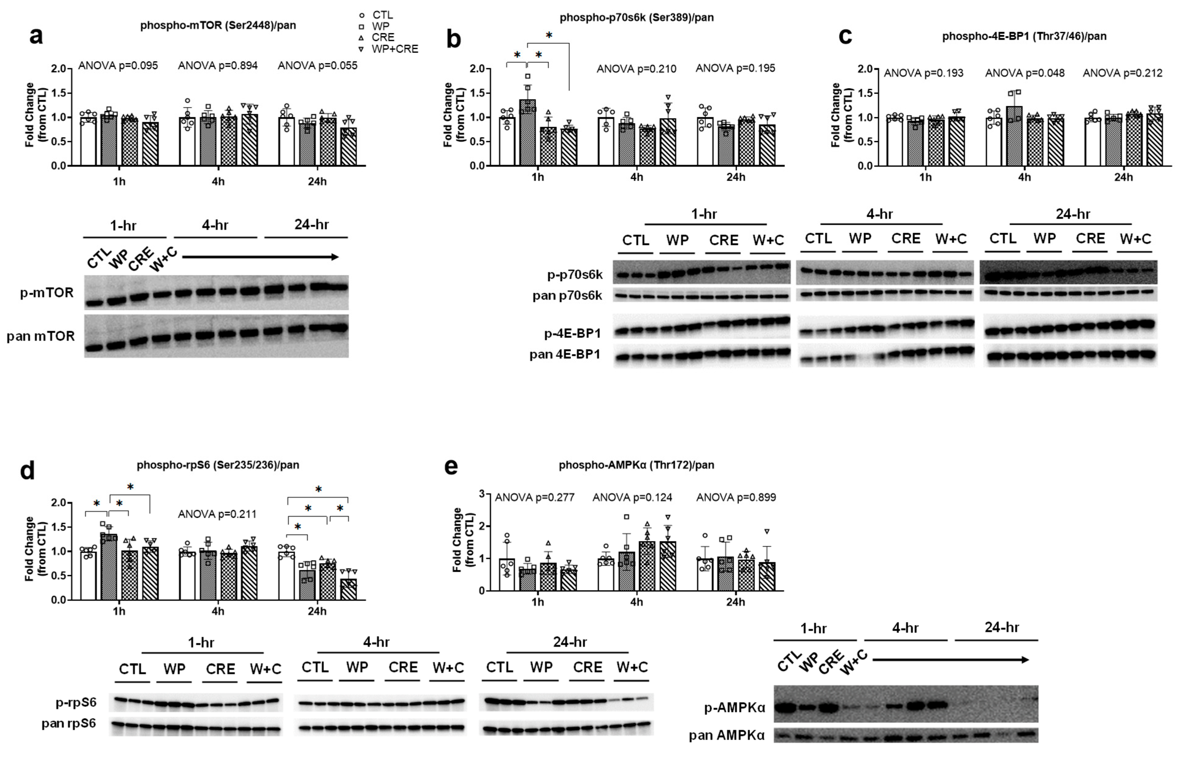
3.2. mTORC1 Pathway and AMPKα Phosphorylation Status

At all treatment time points, phospho-mTOR (Ser2448)/pan did not reach model significance (1 h p = 0.095, 4 h p = 0.894, and 24 h p = 0.055; Figure 2a). At the 1 h treatment time point, phospho-p70S6k (Ser389)/pan reached model significance (p < 0.001; Figure 2b), and post hoc testing revealed that WP was significantly greater than all other treatments (vs. CTL p < 0.001, vs. CRE p < 0.001, vs. WP + CRE; p < 0.001). However, phospho-p70S6k (Ser389)/pan model significance was not observed at the 4 h and 24 h treatment time points (p = 0.210 and p = 0.195, respectively). At the 1 h and 24 h treatment time points, phospho-4E-BP1 (Thr37/46)/pan did not reach model significance (p = 0.193 and p = 0.212, respectively; Figure 2c). Model significance was observed at the 4 h treatment time point (p = 0.048), although post hoc testing did not reveal significant differences between treatments (p > 0.05 for all comparisons), suggesting a subtle overall effect that was not strong enough to be detected between comparisons after multiple testing correction.
Phospho-rpS6 (Ser235/236)/pan displayed model significance at the 1 h treatment time point (p = 0.002; Figure 2d), and further analysis revealed that WP was significantly greater than all other treatments (vs. CTL p = 0.003, vs. CRE p = 0.005, vs. WP + CRE; p = 0.035). Although phospho-rpS6 (Ser235/236)/pan did not reach model significance (p = 0.211) at the 4 h time point, significance was observed at the 24 h treatment time point (p < 0.001), and further analysis revealed that CTL was greater than all other treatments (vs. WP p < 0.001, vs. CRE p = 0.030, vs. WP + CRE; p = 0.003). Phosphorylated-AMPKα (Thr172)/pan did not reach model significance at any treatment time point (1 h, p = 0.277; 4 h, p = 0.124; and 24 h, p = 0.899; Figure 2e).

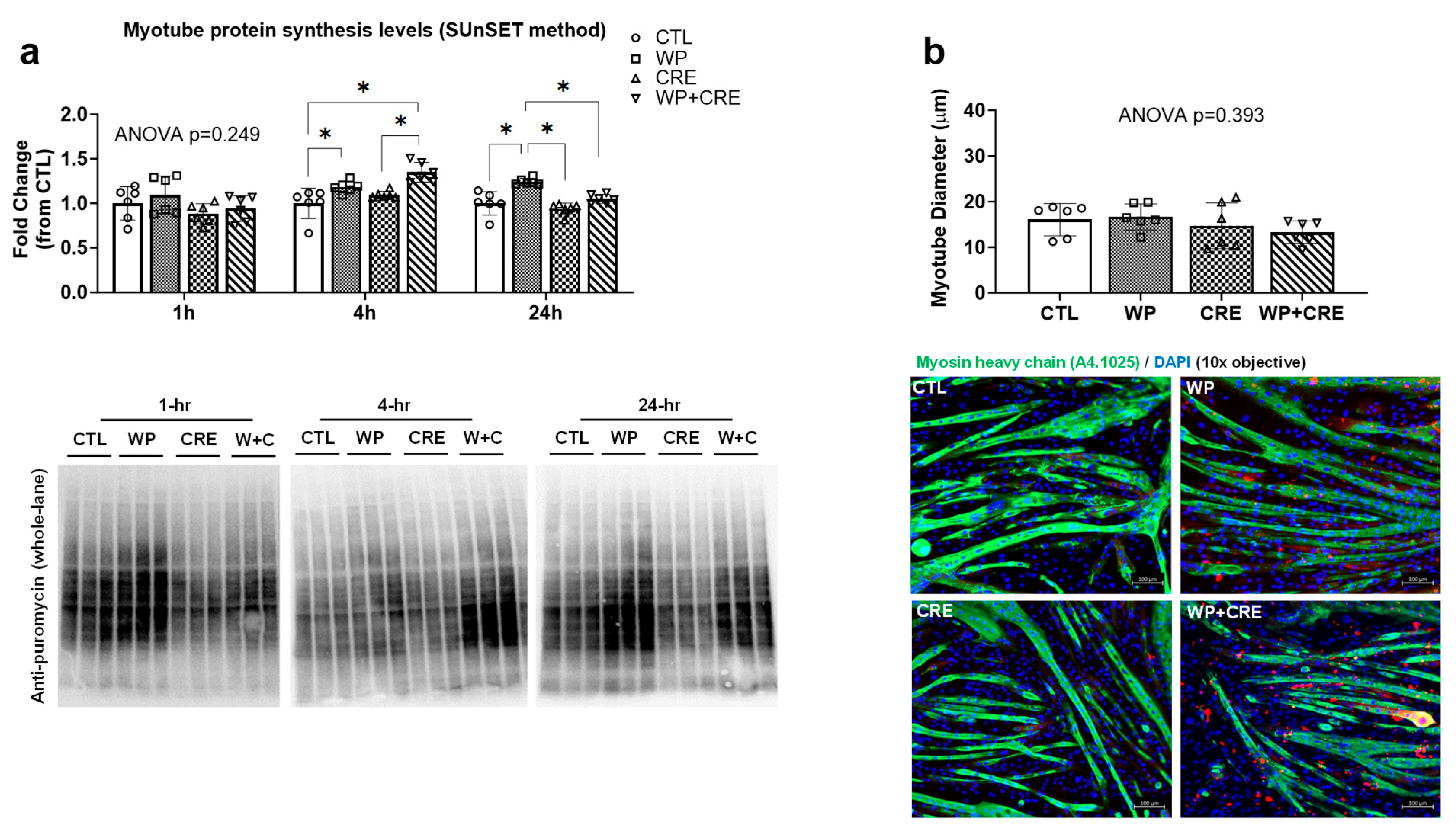
3.3. Myotube Protein Synthesis and Myotube Diameter

Myotube protein synthesis levels are presented in Figure 3a. At the 1 h treatment time point, the response did not reach model significance (p = 0.249). However, at the 4 h treatment time point, the protein synthesis response reached model significance (p < 0.001). Further analysis revealed that WP was greater than the CTL (p < 0.001) and WP + CRE was greater than the CTL and CRE (p < 0.001 and p = 0.003, respectively). No significant difference between WP and WP + CRE was observed (p = 0.068). At the 24 h treatment time point, protein synthesis also reached model significance (p < 0.001). Further analysis revealed that WP was greater compared to all other treatments (vs. CTL p < 0.001, vs. CRE p < 0.001, vs. WP + CRE; p = 0.003). Despite these between-treatment differences in protein synthesis levels, the myotube diameter values following 24 h treatments did not reach model significance (p = 0.393; Figure 3b).

4. Discussion

We used non-stressed/non-stimulated C2C12 myotubes to examine the possible synergistic effects of WP and CRE on MPS and anabolic signaling responses. Although WP and WP + CRE were greater than the CTL at 4 h, prolonged (24 h) myotube protein synthesis was greater in WP versus all other treatments. Thus, the driving stimulus behind the increases in MPS over 24 h was clearly WP. Additionally, p70S6k and rpS6 phosphorylation was greater in WP compared to all other groups at the 1 h treatment time point. At the 24 h time point, phospho-rpS6 expression was significantly lower in the WP and WP + CRE groups compared to the CTL. AMPKα phosphorylation (a surrogate marker of cellular stress) did not differ between treatment groups; note, we hypothesized that CRE treatments would attenuate this marker. Again, this underscores the lack of energetic stress in these cells. Lastly, myotube diameters at 24 h were not different between treatments; however, this is likely due to longer treatment times being needed to observe differences. Contrary to our hypothesis, we interpret these collective findings to suggest that shorter- and longer-term WP + CRE treatments do not synergistically enhance anabolic signaling and MPS responses within a 24 h treatment window.
Certain human and in vitro studies agree in principle with our findings of WP-induced anabolism. For instance, WP supplementation acutely increases MPS and p70S6k phosphorylation in humans in a dose-dependent fashion [28]. Moreover, previous in vitro work, like the current study, supports the finding that WP stimulates anabolic signaling [25,29]. Although the lack of WP + CRE synergism counters our hypothesis, there are also human data that support these observations. For instance, several studies have utilized WP + CRE regimes, mostly in combination with resistance training, and these data suggest that co-supplementation has no added or synergistic effect above whey-protein-only supplementation in relation to various markers of whole-body muscle mass and muscular strength [13,30,31,32]. What is interesting, however, is that although the 10 mM creatine treatments conferred increases in cellular creatine levels, this did not translate to an altered phosphorylation status of mTORC1 proteins or AMPKα and/or myotube protein synthesis. These findings are somewhat at odds with the previously mentioned study by Deldicque et al. [19], which showed that creatine treatments in C2C12 myotubes increases Akt and p38 pathway activation, resulting in increased differentiation and myogenesis. Indeed, the durations of creatine treatments used by these authors were longer than those used in the current study, which could have led to discordant results. Moreover, in vitro studies have consistently indicated that CRE acts to increase myoblast proliferation and fusion-related indices [19,33,34]. Hence, our data and these prior studies support future studies examining CRE in conjunction with a physiological muscle damage response (e.g., mechanical overload) to examine whether supplementation enhances myofiber recovery as it relates to satellite cell proliferation and fusion.

Limitations and Practical Significance

Unfamiliar readers should appreciate that our in vitro model mainly provides insights into nutritional factors that affect cellular signaling. However, this model lacks certain stressors related to exercise (e.g., stimulated contractions) and aging (e.g., oxidative stress and inflammation). Hence, replicating our treatment scheme with these additional conditions may add further insights. What should also be noted is that the use of only an in vitro system limits our extrapolation to human skeletal muscle outcomes, albeit it is also notable that the C2C12 in vitro model has been incredibly insightful for examining the potential anabolic signaling effects of growth factors, amino acids, associated metabolites, and creatine [19,35,36,37,38,39].
Regarding the practical significance of these findings, our data do not discount the utilization of creatine supplementation to aid in muscle growth, as recent comprehensive reviews and meta-analyses support this strategy [40,41]. Rather, our data imply that creatine supplementation likely affects other aspects of muscle growth other than enhancing the MPS and anabolic signaling responses to protein feeding.

5. Conclusions

Taken together, these data indicate that CRE does not synergistically enhance the protein synthesis and anabolic signaling responses to WP in vitro within a 24 h treatment window. However, these data do not discount prior findings indicating that CRE positively affects satellite cell proliferation and resistance training outcomes in vivo. Moreover, these data were obtained using unstressed myotubes, and it is notable that CRE effectively mitigates atrophic stimuli in vitro (e.g., hydrogen peroxide and glucocorticoids) [42,43]. Thus, further insights are needed to determine whether CRE can synergistically enhance contraction-mediated signaling and/or ameliorate catabolic signaling induced via energetic stress in vitro.

Author Contributions

Conceptualization, N.J.K., J.S.G., C.B.M. and M.D.R.; funding acquisition, C.M.L. and M.D.R.; investigation, all co-authors; methodology, N.J.K., J.S.G. and C.B.M.; formal analysis, N.J.K., J.S.G. and M.D.R.; supervision, M.D.R., writing—original draft, N.J.K., J.S.G. and M.D.R.; review and editing, all co-authors. All authors have read and agreed to the published version of the manuscript.

Funding

Funding for creatine assay development and study reagents was provided through a laboratory gift from Nutrabolt (Austin, TX, USA).

Institutional Review Board Statement

Not applicable.

Informed Consent Statement

Not applicable.

Data Availability Statement

Raw data related to the current study outcomes will be provided upon reasonable request by emailing the corresponding author (mdr0024@auburn.edu).

Conflicts of Interest

C.M.L. is the Senior VP for Scientific Affairs at Nutrabolt (Austin, TX, USA). He had a minimal role in designing the study aside from an interest in developing the culture model and assay experiments. Nutrabolt has also committed to a 3-year agreement (2023–2025) to provide gift funds for the naming of the M.D.R. laboratory at Auburn University. M.D.R. has performed industry- and commodity-based contract work. M.D.R. also performs consulting for personal fees with industry partners in accordance with Auburn University’s faculty consulting and annual disclosure policies. D.G.C. has conducted industry-sponsored research involving whey protein and creatine supplementation and received creatine donations for scientific studies and travel support for presentations involving creatine supplementation at scientific conferences. In addition, D.G.C. serves on the Scientific Advisory Board for Alzchem and Create (companies that manufacture creatine products) and as an expert witness/consultant in legal cases involving creatine supplementation. None of the other co-authors have apparent conflicts of interest to report.

References

  1. Saxton, R.A.; Sabatini, D.M. mTOR Signaling in Growth, Metabolism, and Disease. Cell 2017, 168, 960–976. [Google Scholar] [CrossRef] [PubMed]
  2. Wang, X.; Proud, C.G. The mTOR pathway in the control of protein synthesis. Physiology 2006, 21, 362–369. [Google Scholar] [CrossRef] [PubMed]
  3. Roberts, M.D.; McCarthy, J.J.; Hornberger, T.A.; Phillips, S.M.; Mackey, A.L.; Nader, G.A.; Boppart, M.D.; Kavazis, A.N.; Reidy, P.T.; Ogasawara, R.; et al. Mechanisms of mechanical overload-induced skeletal muscle hypertrophy: Current understanding and future directions. Physiol. Rev. 2023, 103, 2679–2757. [Google Scholar] [CrossRef] [PubMed]
  4. Millward, D.J. Interactions between Growth of Muscle and Stature: Mechanisms Involved and Their Nutritional Sensitivity to Dietary Protein: The Protein-Stat Revisited. Nutrients 2021, 13, 729. [Google Scholar] [CrossRef] [PubMed]
  5. White, J.P. Amino Acid Trafficking and Skeletal Muscle Protein Synthesis: A Case of Supply and Demand. Front. Cell Dev. Biol. 2021, 9, 656604. [Google Scholar] [CrossRef]
  6. Michel, J.M.; Lievense, K.K.; Norton, S.C.; Costa, J.V.; Alphin, K.H.; Bailey, L.A.; Miller, G.D. The Effects of Graded Protein Intake in Conjunction with Progressive Resistance Training on Skeletal Muscle Outcomes in Older Adults: A Preliminary Trial. Nutrients 2022, 14, 2739. [Google Scholar] [CrossRef]
  7. Luiking, Y.C.; Deutz, N.E.; Memelink, R.G.; Verlaan, S.; Wolfe, R.R. Postprandial muscle protein synthesis is higher after a high whey protein, leucine-enriched supplement than after a dairy-like product in healthy older people: A randomized controlled trial. Nutr. J. 2014, 13, 9. [Google Scholar] [CrossRef]
  8. Yang, Y.; Breen, L.; Burd, N.A.; Hector, A.J.; Churchward-Venne, T.A.; Josse, A.R.; Tarnopolsky, M.A.; Phillips, S.M. Resistance exercise enhances myofibrillar protein synthesis with graded intakes of whey protein in older men. Br. J. Nutr. 2012, 108, 1780–1788. [Google Scholar] [CrossRef]
  9. Tang, J.E.; Moore, D.R.; Kujbida, G.W.; Tarnopolsky, M.A.; Phillips, S.M. Ingestion of whey hydrolysate, casein, or soy protein isolate: Effects on mixed muscle protein synthesis at rest and following resistance exercise in young men. J. Appl. Physiol. 2009, 107, 987–992. [Google Scholar] [CrossRef]
  10. Kerksick, C.M.; Arent, S.; Schoenfeld, B.J.; Stout, J.R.; Campbell, B.; Wilborn, C.D.; Taylor, L.; Kalman, D.; Smith-Ryan, A.E.; Kreider, R.B.; et al. International society of sports nutrition position stand: Nutrient timing. J. Int. Soc. Sports Nutr. 2017, 14, 33. [Google Scholar] [CrossRef]
  11. Hulmi, J.J.; Lockwood, C.M.; Stout, J.R. Effect of protein/essential amino acids and resistance training on skeletal muscle hypertrophy: A case for whey protein. Nutr. Metab. 2010, 7, 51. [Google Scholar] [CrossRef] [PubMed]
  12. Phillips, S.M.; Tang, J.E.; Moore, D.R. The role of milk- and soy-based protein in support of muscle protein synthesis and muscle protein accretion in young and elderly persons. J. Am. Coll. Nutr. 2009, 28, 343–354. [Google Scholar] [CrossRef] [PubMed]
  13. Cribb, P.J.; Williams, A.D.; Stathis, C.G.; Carey, M.F.; Hayes, A. Effects of whey isolate, creatine, and resistance training on muscle hypertrophy. Med. Sci. Sports Exerc. 2007, 39, 298–307. [Google Scholar] [CrossRef] [PubMed]
  14. Devries, M.C.; Phillips, S.M. Creatine supplementation during resistance training in older adults-a meta-analysis. Med. Sci. Sports Exerc. 2014, 46, 1194–1203. [Google Scholar] [CrossRef]
  15. Parise, G.; Mihic, S.; MacLennan, D.; Yarasheski, K.E.; Tarnopolsky, M.A. Effects of acute creatine monohydrate supplementation on leucine kinetics and mixed-muscle protein synthesis. J. Appl. Physiol. 2001, 91, 1041–1047. [Google Scholar] [CrossRef]
  16. Willoughby, D.S.; Rosene, J. Effects of oral creatine and resistance training on myosin heavy chain expression. Med. Sci. Sports Exerc. 2001, 33, 1674–1681. [Google Scholar] [CrossRef]
  17. Olsen, S.; Aagaard, P.; Kadi, F.; Tufekovic, G.; Verney, J.; Olesen, J.L.; Suetta, C.; Kjaer, M. Creatine supplementation augments the increase in satellite cell and myonuclei number in human skeletal muscle induced by strength training. J. Physiol. 2006, 573, 525–534. [Google Scholar] [CrossRef]
  18. Deldicque, L.; Atherton, P.; Patel, R.; Theisen, D.; Nielens, H.; Rennie, M.J.; Francaux, M. Effects of resistance exercise with and without creatine supplementation on gene expression and cell signaling in human skeletal muscle. J. Appl. Physiol. 2008, 104, 371–378. [Google Scholar] [CrossRef]
  19. Deldicque, L.; Theisen, D.; Bertrand, L.; Hespel, P.; Hue, L.; Francaux, M. Creatine enhances differentiation of myogenic C2C12 cells by activating both p38 and Akt/PKB pathways. Am. J. Physiol. Cell Physiol. 2007, 293, C1263–C1271. [Google Scholar] [CrossRef]
  20. Louis, M.; Van Beneden, R.; Dehoux, M.; Thissen, J.P.; Francaux, M. Creatine increases IGF-I and myogenic regulatory factor mRNA in C(2)C(12) cells. FEBS Lett. 2004, 557, 243–247. [Google Scholar] [CrossRef]
  21. Mobley, C.B.; Fox, C.D.; Ferguson, B.S.; Amin, R.H.; Dalbo, V.J.; Baier, S.; Rathmacher, J.A.; Wilson, J.M.; Roberts, M.D. L-leucine, beta-hydroxy-beta-methylbutyric acid (HMB) and creatine monohydrate prevent myostatin-induced Akirin-1/Mighty mRNA down-regulation and myotube atrophy. J. Int. Soc. Sports Nutr. 2014, 11, 38. [Google Scholar] [CrossRef] [PubMed]
  22. Mitchell, W.K.; Phillips, B.E.; Hill, I.; Greenhaff, P.; Lund, J.N.; Williams, J.P.; Rankin, D.; Wilkinson, D.J.; Smith, K.; Atherton, P.J. Human skeletal muscle is refractory to the anabolic effects of leucine during the postprandial muscle-full period in older men. Clin. Sci. 2017, 131, 2643–2653. [Google Scholar] [CrossRef] [PubMed]
  23. Witard, O.C.; Jackman, S.R.; Breen, L.; Smith, K.; Selby, A.; Tipton, K.D. Myofibrillar muscle protein synthesis rates subsequent to a meal in response to increasing doses of whey protein at rest and after resistance exercise. Am. J. Clin. Nutr. 2014, 99, 86–95. [Google Scholar] [CrossRef]
  24. O’Connor, R.S.; Steeds, C.M.; Wiseman, R.W.; Pavlath, G.K. Phosphocreatine as an energy source for actin cytoskeletal rearrangements during myoblast fusion. J. Physiol. 2008, 586, 2841–2853. [Google Scholar] [CrossRef]
  25. Mobley, C.B.; Mumford, P.W.; McCarthy, J.J.; Miller, M.E.; Young, K.C.; Martin, J.S.; Beck, D.T.; Lockwood, C.M.; Roberts, M.D. Whey protein-derived exosomes increase protein synthesis and hypertrophy in C(2-)C(12) myotubes. J. Dairy Sci. 2017, 100, 48–64. [Google Scholar] [CrossRef]
  26. Michel, J.M.; Godwin, J.S.; Plotkin, D.L.; Mesquita, P.H.C.; McIntosh, M.C.; Ruple, B.A.; Libardi, C.A.; Mobley, C.B.; Kavazis, A.N.; Roberts, M.D. Proteolytic markers associated with a gain and loss of leg muscle mass with resistance training followed by high-intensity interval training. Exp. Physiol. 2023, 108, 1268–1281. [Google Scholar] [CrossRef]
  27. Osburn, S.C.; Vann, C.G.; Church, D.D.; Ferrando, A.A.; Roberts, M.D. Proteasome- and Calpain-Mediated Proteolysis, but Not Autophagy, Is Required for Leucine-Induced Protein Synthesis in C2C12 Myotubes. Physiologia 2021, 1, 22–33. [Google Scholar] [CrossRef]
  28. D’Souza, R.F.; Marworth, J.F.; Figueiredo, V.C.; Della Gatta, P.A.; Petersen, A.C.; Mitchell, C.J.; Cameron-Smith, D. Dose-dependent increases in p70S6K phosphorylation and intramuscular branched-chain amino acids in older men following resistance exercise and protein intake. Physiol. Rep. 2014, 2, e12112. [Google Scholar] [CrossRef]
  29. Mobley, C.B.; Fox, C.D.; Thompson, R.M.; Healy, J.C.; Santucci, V.; Kephart, W.C.; McCloskey, A.E.; Kim, M.; Pascoe, D.D.; Martin, J.S.; et al. Comparative effects of whey protein versus L-leucine on skeletal muscle protein synthesis and markers of ribosome biogenesis following resistance exercise. Amino Acids 2016, 48, 733–750. [Google Scholar] [CrossRef]
  30. Wilborn, C.D.; Outlaw, J.J.; Mumford, P.W.; Urbina, S.L.; Hayward, S.; Roberts, M.D.; Taylor, L.W.; Foster, C.A. A Pilot Study Examining the Effects of 8-Week Whey Protein versus Whey Protein Plus Creatine Supplementation on Body Composition and Performance Variables in Resistance-Trained Women. Ann. Nutr. Metab. 2016, 69, 190–199. [Google Scholar] [CrossRef]
  31. Bemben, M.G.; Witten, M.S.; Carter, J.M.; Eliot, K.A.; Knehans, A.W.; Bemben, D.A. The effects of supplementation with creatine and protein on muscle strength following a traditional resistance training program in middle-aged and older men. J. Nutr. Health Aging 2010, 14, 155–159. [Google Scholar] [CrossRef] [PubMed]
  32. Eliot, K.A.; Knehans, A.W.; Bemben, D.A.; Witten, M.S.; Carter, J.; Bemben, M.G. The effects of creatine and whey protein supplementation on body composition in men aged 48 to 72 years during resistance training. J. Nutr. Health Aging 2008, 12, 208–212. [Google Scholar] [CrossRef] [PubMed]
  33. Vierck, J.L.; Icenoggle, D.L.; Bucci, L.; Dodson, M.V. The effects of ergogenic compounds on myogenic satellite cells. Med. Sci. Sports Exerc. 2003, 35, 769–776. [Google Scholar] [CrossRef] [PubMed]
  34. Xu, W.; Lin, D.; Huang, C. NMR-based metabolomic analysis for the effects of creatine supplementation on mouse myoblast cell line C2C12. Acta Biochim. Biophys. Sin. 2017, 49, 617–627. [Google Scholar] [CrossRef]
  35. Duan, Y.; Zhong, Y.; Song, B.; Zheng, C.; Xu, K.; Kong, X.; Li, F. Suppression of protein degradation by leucine requires its conversion to beta-hydroxy-beta-methyl butyrate in C2C12 myotubes. Aging 2019, 11, 11922–11936. [Google Scholar] [CrossRef]
  36. Areta, J.L.; Hawley, J.A.; Ye, J.M.; Chan, M.H.; Coffey, V.G. Increasing leucine concentration stimulates mechanistic target of rapamycin signaling and cell growth in C2C12 skeletal muscle cells. Nutr. Res. 2014, 34, 1000–1007. [Google Scholar] [CrossRef]
  37. Atherton, P.J.; Smith, K.; Etheridge, T.; Rankin, D.; Rennie, M.J. Distinct anabolic signalling responses to amino acids in C2C12 skeletal muscle cells. Amino Acids 2010, 38, 1533–1539. [Google Scholar] [CrossRef]
  38. Deldicque, L.; Sanchez Canedo, C.; Horman, S.; De Potter, I.; Bertrand, L.; Hue, L.; Francaux, M. Antagonistic effects of leucine and glutamine on the mTOR pathway in myogenic C2C12 cells. Amino Acids 2008, 35, 147–155. [Google Scholar] [CrossRef]
  39. Williamson, D.L.; Kimball, S.R.; Jefferson, L.S. Acute treatment with TNF-alpha attenuates insulin-stimulated protein synthesis in cultures of C2C12 myotubes through a MEK1-sensitive mechanism. Am. J. Physiol. Endocrinol. Metab. 2005, 289, E95–E104. [Google Scholar] [CrossRef]
  40. Kreider, R.B.; Kalman, D.S.; Antonio, J.; Ziegenfuss, T.N.; Wildman, R.; Collins, R.; Candow, D.G.; Kleiner, S.M.; Almada, A.L.; Lopez, H.L. International Society of Sports Nutrition position stand: Safety and efficacy of creatine supplementation in exercise, sport, and medicine. J. Int. Soc. Sports Nutr. 2017, 14, 18. [Google Scholar] [CrossRef]
  41. Wu, S.H.; Chen, K.L.; Hsu, C.; Chen, H.C.; Chen, J.Y.; Yu, S.Y.; Shiu, Y.J. Creatine Supplementation for Muscle Growth: A Scoping Review of Randomized Clinical Trials from 2012 to 2021. Nutrients 2022, 14, 1255. [Google Scholar] [CrossRef] [PubMed]
  42. Sun, M.; Jiao, H.; Zhao, J.; Wang, X.; Li, H.; Zhou, Y.; Lin, H. Research Note: Creatine monohydrate alleviates protein breakdown induced by corticosterone via inhibiting ubiquitin proteasome pathway in chicken myotubes. Poult. Sci. 2022, 101, 102177. [Google Scholar] [CrossRef] [PubMed]
  43. Sestili, P.; Barbieri, E.; Martinelli, C.; Battistelli, M.; Guescini, M.; Vallorani, L.; Casadei, L.; D’Emilio, A.; Falcieri, E.; Piccoli, G.; et al. Creatine supplementation prevents the inhibition of myogenic differentiation in oxidatively injured C2C12 murine myoblasts. Mol. Nutr. Food Res. 2009, 53, 1187–1204. [Google Scholar] [CrossRef] [PubMed]
Figure 1. Relative cellular creatine levels. Data show that CRE and WP + CRE increase cellular creatine levels following 1 h, 4 h, and 24 h treatments. Each treatment contains 6 replicates. Data are presented as bar graphs with means ± standard deviation values as well as individual values. RFU, relative fluorescence unit provided by creatine assay.
Physiologia 05 00017 g001
Figure 2. mTORC1 protein and AMPKα phosphorylation status. Data show phosphorylation status of mTOR (a), p70S6k (b), 4E-BP1 (c), rpS6 (d), and AMPKα (e). Each treatment contains 6 replicates. Data are presented as bar graphs with means ± standard deviation values as well as individual values. Significant differences between treatments are noted by ‘*’, and exact p-values can be found in text.
Physiologia 05 00017 g002
Figure 3. Myotube protein synthesis responses and 24 h treatment myotube diameters. Data show the 1, 4, and 24 h myotube protein synthesis responses (a) and 24 h treatment myotube diameter values (b). Each treatment contains 6 replicates. Data are presented as bar graphs with means ± standard deviation values as well as individual values. Significant differences between treatments are noted by ‘*’, and exact p-values can be found in text.
Physiologia 05 00017 g003
Disclaimer/Publisher’s Note: The statements, opinions and data contained in all publications are solely those of the individual author(s) and contributor(s) and not of MDPI and/or the editor(s). MDPI and/or the editor(s) disclaim responsibility for any injury to people or property resulting from any ideas, methods, instructions or products referred to in the content.

