Article

Assessment of Bacterial Presence Among New and “In Use” Resealable Biomaterials Within the Pediatric Dental Clinic

1
Department of Advanced Education in Pediatric Dentistry, School of Dental Medicine, University of Nevada, Las Vegas, NV 89106, USA
2
Department of Clinical Sciences, School of Dental Medicine, University of Nevada, Las Vegas, NV 89106, USA
3
Department of Biomedical Sciences, School of Dental Medicine, University of Nevada, Las Vegas, NV 89106, USA
*
Author to whom correspondence should be addressed.
Hygiene 2025, 5(3), 42; https://doi.org/10.3390/hygiene5030042
Submission received: 29 June 2025 / Revised: 13 August 2025 / Accepted: 2 September 2025 / Published: 9 September 2025

Abstract

Background: Dental offices and clinics utilize a variety of dental materials that are delivered in reusable containers and dispensers. However, many of these materials, including NeoPutty, BC Putty, Flowable, and Diapex, may be subject to bacterial contamination and microbial exposures from the surrounding dental office environment. Objectives: The aim of this study was to quantify and identify microbial contamination, specifically in regard to these reusable dental materials. Methods: Surfaces of new and used reusable and resealable tubes where the material dispenses and the interior surfaces of the cap were swabbed and cultured. DNA was isolated from each sample and quantitative polymerase chain reaction (qPCR) was performed to determine the presence or absence of microbial contamination, as well as the relative abundance. Results: Microbial contamination was observed among all of the “in use” samples from both the dispensing end and the interior surfaces of the cap and was strongly associated with the amount of usage. Conclusions: These data suggest that environmental contamination may be present in measurable and quantifiable amounts on reusable and resealable dental materials, which suggest the need to create protocols for sanitizing the surfaces of reusable materials to reduce the presence of microbial contamination identified in similar clinical settings.

1. Introduction

Dental offices and clinics utilize a variety of dental materials that are delivered in reusable containers and dispensers [1,2]. For example, a variety of multiple usage materials are commonly used to modify and improve the adhesion of alginate to dental trays (otherwise known as dental tray adhesives) during the process of taking dental impressions [3,4]. However, evidence has demonstrated that reusable and resealable dental materials such as dental tray adhesives may harbor the potential for microbial contamination and possible transmission between dental patients [5,6].
Many additional reusable and resealable dental biomaterials, including flowable composites and root canal fillers, may also be used in the dental office and have been evaluated for their antimicrobial properties [7,8]. However, the containers and dispensers for these biomaterials may also be subject to microbial contamination and microbial exposures from the surrounding dental office environment, although few studies have evaluated this potential [9,10]. In fact, recent reviews have demonstrated that droplet and aerosol generation may have the potential for cross contamination of dental office surfaces and items exposed on nearby dental trays [11,12,13].
Several types of these biomaterials may have extended use in the pediatric dental office, which may increase the potential for cross contamination during repeated use [14,15]. These include the aforementioned flowable composites, which have been extensively tested for their antimicrobial properties both in vitro and in vivo but have not been routinely evaluated for potential cross contamination of the dispenser neck or cap [16,17]. As the SARS-CoV-2 (COVID19) pandemic clearly demonstrated, more rigorous and routine screening for and prevention of microbial cross contamination within the dental office setting is an effective and meaningful objective of dental practitioners worldwide [18,19,20].
To determine the scope and severity of this potential contamination, the aim of this study is to assess and quantify microbial contamination (if any) in dispensers of reusable and resealable dental biomaterials commonly used in a public dental school clinic. More specifically, comparisons of microbial contamination between new and “in use” items will be evaluated from the neck and cap to determine any potential evidence for environmental cross contamination.

2. Materials and Methods

2.1. Environmental Sampling

Four different resealable dental biomaterials were sampled within the pediatric dental clinic on three separate dates. These included NeoPutty, BC Putty, Flowable, and Diapex, which are premixed bioactive and bioceramic root and pulp treatments that are prepackaged for multiple use in resealable dispensers. Surfaces of the tubes where the material dispenses and the interior surfaces of the cap of the material were swabbed using autoclaved (sterile) specimen collection cotton applicators (swabs) from Fisher Scientific (#22-363-160; Fair Lawn, NJ, USA) for subsequent culturing. At least two operators participated in each sample collection, with one operator that was selected to be present at all collection times to ensure methodological quality and consistency. Collections from the neck and cap were taken using one full rotation of the swab by each operator to ensure coverage from the entire circumference of the surface. Both new and “in use” items were sampled on each of the collection dates.

2.2. Estimated Usage

Estimates of “in use” items were made using standardized measurements of the plunger. The plunger of each “new” item was measured using a metric ruler and photographed against a standard graph paper background (5 mm). Estimates of “in use” items were made by measuring the plunger length and dividing by the “new” plunger length to determine the approximate percentage of the item that has been used at the time of sampling (Estimated usage = plunger length in mm/new plunger length in mm). Percentages were calculated and verified by at least two operators.

2.3. Bacterial Culture

Following each site collection, individual sterile swabs were placed into sterile 15 mL microcentrifuge tubes containing 5 mL of Luria–Bertani (LB) broth. LB broth was made using tryptone (10 g), sodium chloride (10 g) and yeast extract (5 g), all obtained from Fisher Scientific (Fair Lawn, NJ, USA). These reagents were dissolved in distilled water 1000 mL and autoclaved in the liquid cycle prior to use. Each swab and corresponding culture were transferred to a biomedical laboratory and placed into a 4C refrigerator overnight. Negative controls created using the sterile swabs that were not used for sample collection but were placed into culturing media and refrigerated overnight with the collected samples. The positive control was created using an aliquot of Mixed Bacteria #55644 from American Type Culture Collection (ATCC; Manassas, VA, USA), which contains multiple aerobic BSL-1 bacteria that includes Aeromonas sp. (ATCC 55641), Corynebacterium sp. (ATCC 55643), Pseudomonas sp. (ATCC 55645), and Zoogloea sp. (ATCC 55649), as previously described [21,22,23]. Cultures Turbidity was subsequently measured at 600 nm for each control and experimental condition following overnight incubation at 37 °C, as previously described [22,23].

2.4. DNA Isolation

Following overnight storage, each sample tube was vortexed for 30 s to dislodge any bacteria from the swab before removal. Aliquots of 500 uL were removed from each tube and DNA was isolated using the FastDNA Bacterial and Fungal DNA isolation Kit from MP Biochemicals (Santa Ana, CA, USA) using the recommended manufacturer protocol, as previously described [21,22]. Qualitative and quantitative analysis of DNA was performed using the NanoDrop 2000 Spectrophotometer from Fisher Scientific (Fair Lawn, NJ, USA) and absorbances of A260 and A280 nm. The ratio of A260:A280 was used to assess sample quality using a minimum standard of 1.70, which is sufficient for quantitative polymerase chain reaction (qPCR) screening [21,22]. Due to the negligible amount of DNA present among the negative control samples, no subtraction of background DNA was necessary, as performed with other similar studies [21,22,23].

2.5. qPCR Screening

DNA isolated from each sample was then screened using the QuantStudio Real-Time Polymerase Chain Reaction (RT-PCR) system from Applied Biosciences (Waltham, MA, USA). Each sample was screened using the Applied Biosystems Fast SYBR master mix and reagent system from ThermoFisher Scientific (Waltham, MA, USA) using the protocol recommended by the manufacturer and previously validated organism-specific primer sets, which included the following [21,22]:
Positive control, bacterial 16S rRNA primers
Forward 16S rRNA primer: 5′-ACG CGT CGA CAG AGT TTG ATC CTG GCT-3′
Reverse 16S rRNA primer: 5′-GGG ACT ACC AGG GTA TCT AAT-3′
Forward Pseudomonas aeruginosa primer; 5′–TCG GCA CCG GAC TTC TTT C–3′
Reverse Pseudomonas aeruginosa primer; 5′–GGT GAC GAT GCG CGG GGA TTC TT–3′
Oral bacteria primers
Forward Streptococcus mutans primer; 5′-GCC TAC AGC TCA GAG ATG CTA TTC T-3′
Reverse Streptococcus mutans primer; 5′-GCC ATA CAC CAC TCA TGA ATT GA-3′
Forward Streptococcus gordonii primer; 5′-TGT ACC CCG TAT CGT TCC TGT G-3′
Reverse Streptococcus gordonii primer; 5′-AAA GAC TGG AGT TGC AAT GTG AAT A-3′
Forward Actinomyces naeslundii primer; 5′-GTC TCA GTT CGG ATC GGT GT-3′
Reverse Actinomyces naeslundii primer; 5′-CCG GTA CGG CTA CCT TGT TA-3′
Forward Lactobacillus acidophilus primer; 5′- AAT TCT CTT CTC GGT CGC TCT A-3′
Reverse Lactobacillus acidophilus primer; 5′-CCT TTC TAA GGA AGC GAA GGA T-3′
Forward Porphyromonas gingivalis primer; 5′-TAC CCA TCG TCG CCT TGG T-3′
Reverse Porphyromonas gingivalis primer; 5′-CGG ACT AAA ACC GCA TAC ACT TG-3′
Forward Fusobacterium nucleatum primer; 5′-CGC AGA AGG TGA AAG TCC TGT AT-3′
Reverse Fusobacterium nucleatum primer; 5′-TGG TCC TCA CTG ATT CAC ACA GA-3′
Forward Veillonella parvula primer; 5′-GGA CAA CGC TTG CCA CCT A-3′
Reverse Veillonella parvula primer; 5′-GGT TAC CTT GTT ACG ACT T-3′

2.6. Statistical Analysis

Calculations were made to determine the minimum sampling or number of replicates required for each group in this repeated measures study using Graph Pad Prism Version 9 software (San Diego, CA, USA). The sample number per group n = 3 was determined using parameters including large effect size (0.8), power of 90% (beta = 1–0.1), and a confidence interval (alpha) of 95%. Data regarding the DNA concentration and qPCR cycle thresholds (CT counts) were imported into Microsoft Excel (Redmond, WA, USA) for comparisons between control (positive, negative) and experimental (new, in use) groups using two-tailed Student’s t-tests, which may be utilized for parametric data analysis. Mean (average) was calculated and presented with standard deviation (STD). The Shapiro–Wilk test was used to determine normality of the data from each set and all statistical comparisons were confirmed using one-way analysis of variance (ANOVA) with post hoc Tukey analysis using the Graph Pad Prism Version 9 software (San Diego, CA, USA), as previously described [23,24].

3. Results

The analysis of data derived from the DNA isolation from each of the sample collections, including the positive and negative controls, were collected and averaged (Table 1). These data demonstrated that a negligible amount of DNA was recovered from the negative controls performed during each of the three sample collections, averaging 0.56 ng/uL (±0.43) with the DNA purity from the A260:A280 ratio estimated at 1.58 (±0.04). Analysis of the positive controls created during each of the sample collections revealed DNA that averaged 1262.70 ng/uL (±25.49), with DNA purity estimated at 1.65 (±0.10).
Analysis of the DNA isolated from the NEW Diapex collections, including both the neck and the cap, demonstrated very small amounts of DNA, averaging 0.75 ng/uL (±0.18) with an average DNA purity of 1.68 (±0.09). Similarly, results of the DNA screening from the NEW Flowable collections also revealed negligible amounts of DNA with an average of 0.53 ng/uL (±0.20)and an average DNA purity of 1.67 (±0.06). Analysis of the NEW sample collections from BC Putty and NeoPutty were found to have similarly low levels of DNA present, averaging 1.76 ng/uL (±1.52) and 1.42 ng/uL (±0.86), respectively, with slightly lower purity observed (1.54 ± 0.09) and 1.60 ± 0.07).
However, analysis of the IN USE samples revealed much more DNA was present in each of the types of reusable materials assessed. For example, the Diapex IN USE samples averaged 336.76 ng/uL (±61.85) with DNA purity estimated at 1.62 (±0.04). The DNA found among the Flowable IN USE samples averaged 552.17 ng/uL (±235.47) and DNA purity averaged 1.73 (±0.13). Similar levels of DNA were found among the BC Putty sample collections with an average of 511.25 ng/uL (±217.11) and DNA purity average of 1.57 (±0.09). Finally, analysis of NeoPutty IN USE samples demonstrated an average DNA of 350.64 ng/uL (±103.47) with average DNA purity of 1.79 (±0.11). The full data set can be found in Appendix Table A1.
These results of the DNA isolation data were plotted to provide a graphic representation of the study samples (Figure 1). This analysis demonstrated that the amounts of DNA isolated from the negative controls (0.56 ng/uL ± 0.43) and NEW clinic samples from Diapex (0.75 ng/uL ± 0.18), Flowable (average 0.53 ng/uL ± 0.20), BC Putty (average 1.76 ng/uL ± 1.52), and NeoPutty (average 1.42 ng/uL ± 0.07) were similar and comparable. However, the results from the IN USE clinic samples demonstrated more significant DNA levels among all reusable items screening, including Diapex (average 336.76 ng/uL ± 61.85) Flowable (average 552.17 ng/uL ± 235.47) BC Putty (average 511.25 ng/uL ± 217.11) and NeoPutty (average 350.64 ng/uL ± 103.47). However, this analysis also revealed small differences among the same clinic items at the same sampling collection (Diapex Cap IN USE Collection 1 versus Diapex Neck IN USE Collection 1). Moreover, more significant differences were between the same clinic items sampled at different collection points over time (Diapex Cap IN USE Collection 1 versus Diapex Cap IN USE Collection 2).
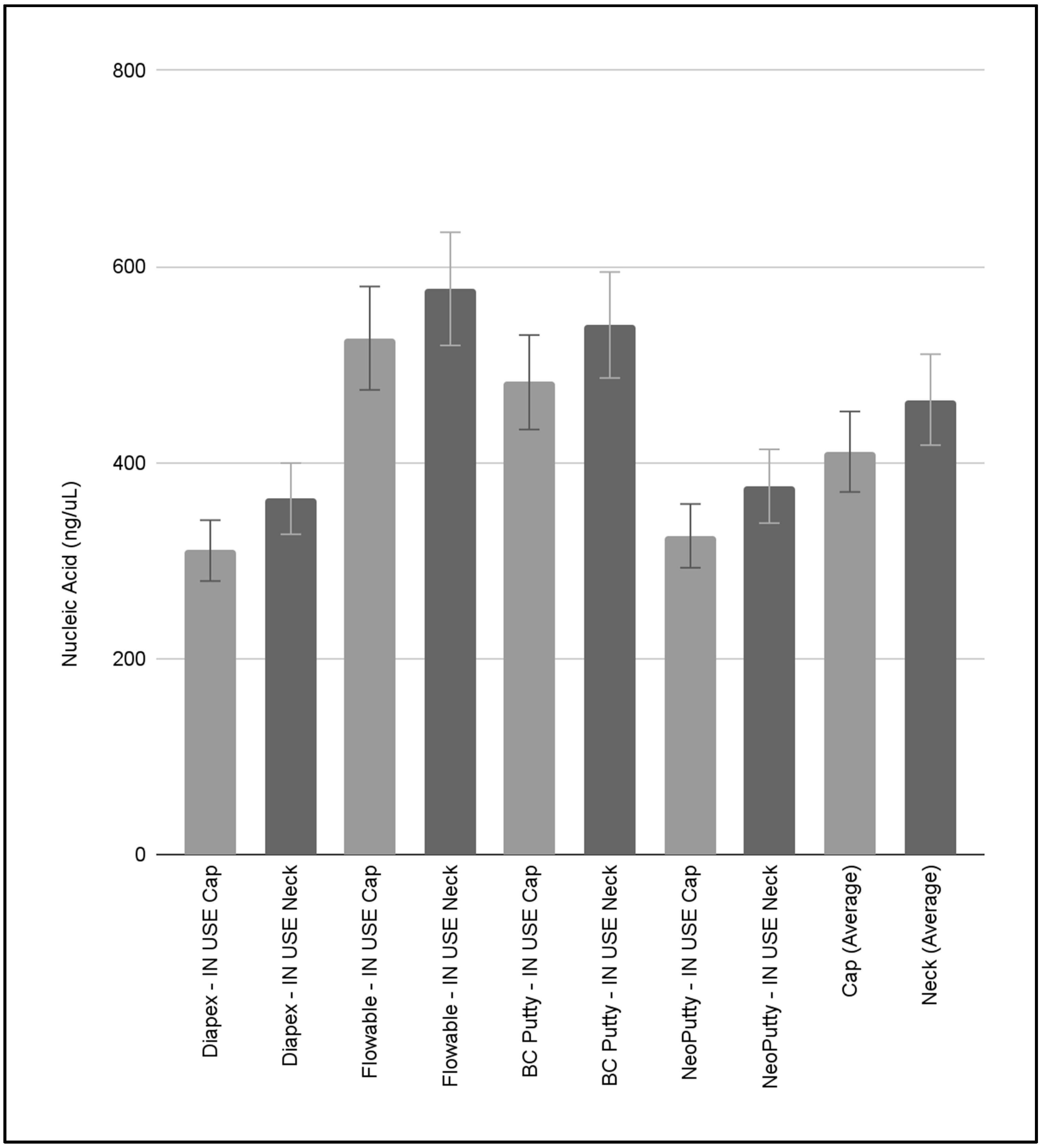
To evaluate the potential differences between the DNA isolated from the Cap and Neck of the same clinic items at the same sampling collection (Diapex Cap IN USE Collection 1 versus Diapex Neck IN USE Collection 1), these data were analyzed and graphed (Figure 2). This analysis revealed that the DNA isolated from the swabs from the neck of each item was higher than the corresponding cap—but those differences were not significant. For example, the average DNA isolated from the Diapex IN USE Cap swabs (average 310.27 ng/uL ± 62.07) was lower when compared with the Diapex IN USE Neck swabs (average 363.26 ng/uL ± 60.03), but this was not statistically significant, p = 0.174. Similarly, the amount of DNA from the Flowable IN USE Cap swabs (average 527.01 ng/uL ± 242.68) was also lower than the Flowable IN USE Neck swabs (average 577.32 ng/uL ± 278.97), but was not statistically significant, p = 0.391. Similar trends were observed with BC Putty IN USE Caps (average 482.07 ng/uL ± 219.68) and NeoPutty IN USE Caps (average 325.31 ng/uL ± 102.16) exhibiting lower amounts of DNA compared with the corresponding samples from the Necks (average 540.44 ng/uL ± 259.12 and 375.97 ± 120.01, respectively), which were also not statistically significant (p = 0.391, p = 0.304, respectively).
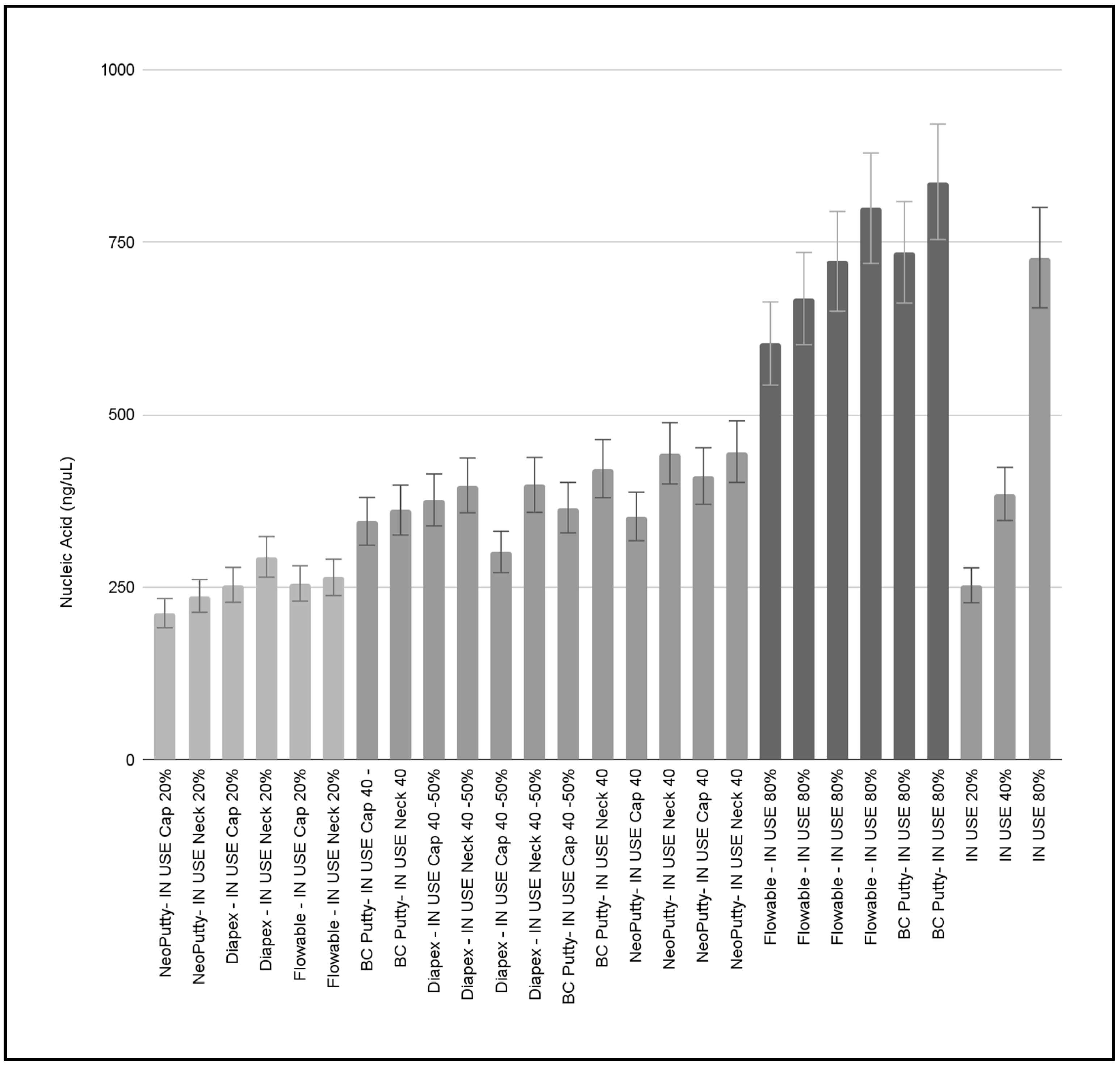
To evaluate the potential differences between the DNA isolated from all clinic items at different estimated stages of use, these data were analyzed and graphed (Figure 3). The data from this analysis revealed that the lowest levels of DNA were found among the items sampled at an estimated use of approximately 20% (average 252.76 ng/uL ± 27.22). More moderate levels of DNA were observed from items sampled at an estimated use of approximately 40–50% (average 385.15 ng/uL ± 42.97). The highest levels of DNA were found among items sampled at an estimated use of approximately 80% (average 727.76 ng/uL ± 85.24). The average DNA between samples of the different estimated usages (20%, 40–50%, 80%) were significantly different from one another, p < 0.0001 and exhibited a strong, linear correlation between estimated usage and the amount of DNA present R2 = 0.939.
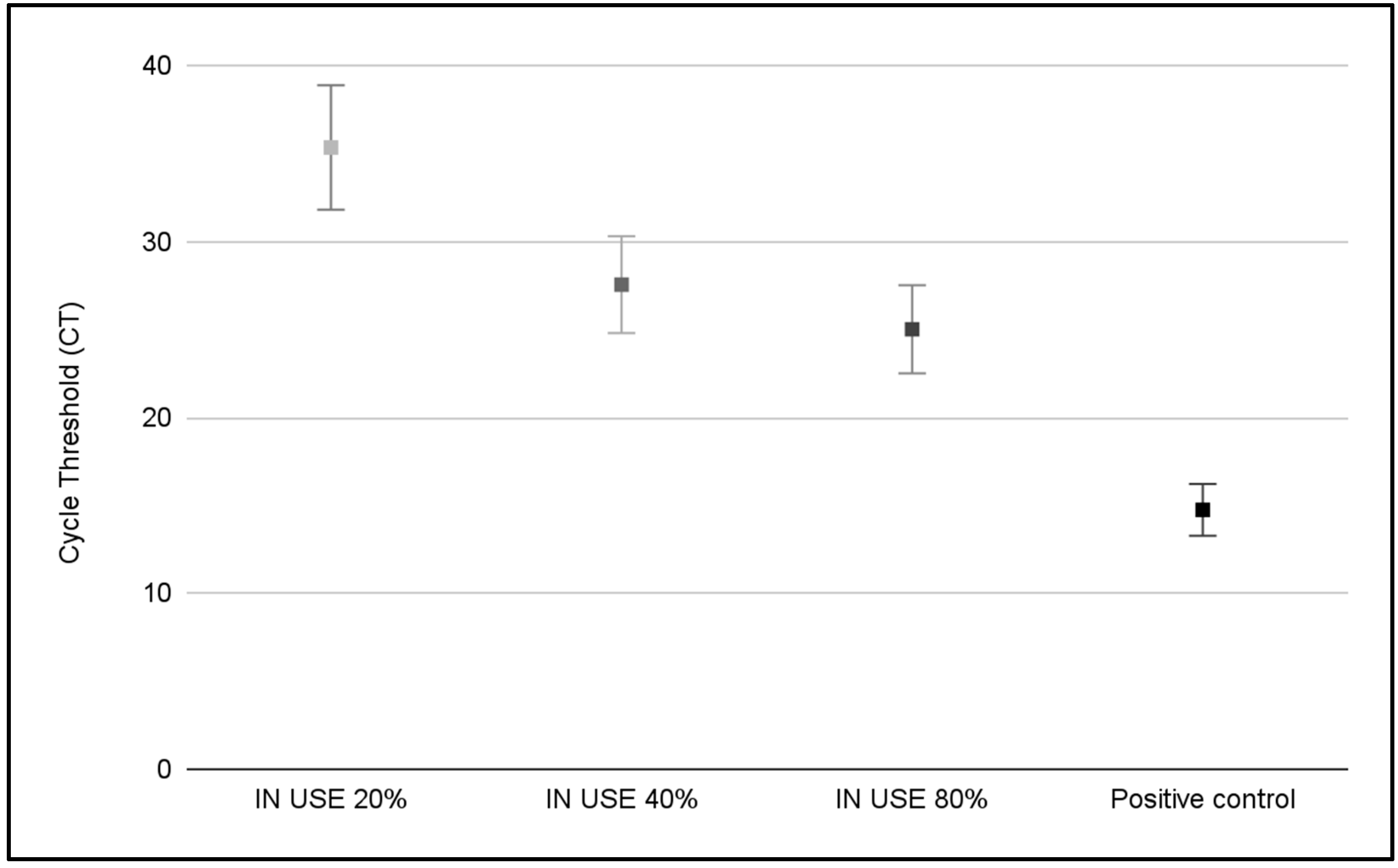
To determine if the DNA isolated from the sample swabs was bacterial, quantitative polymerase chain reaction (qPCR) screening for bacterial 16S rRNA was performed (Figure 4). These data revealed that the presence of bacteria was confirmed for each of the IN USE samples screened. More specifically, the IN USE samples with an estimated usage of 20% were found to have an average cycle threshold (CT) count of 35.35 ± 0.45 (range 34.63 to 35.97), while those samples with an estimated usage of 40–50% had a much lower CT count of 27.55 ± 0.75 (range 26.20 to 28.48). The lowest CT count was observed with the 80% IN USE samples with an estimated CT count of 25.01 ± 0.61 (range 23.97 to 25.48), which was much higher than the positive control standards that exhibited an average CT count of 14.93 ± 0.17 (range 14.76 to 15.05).
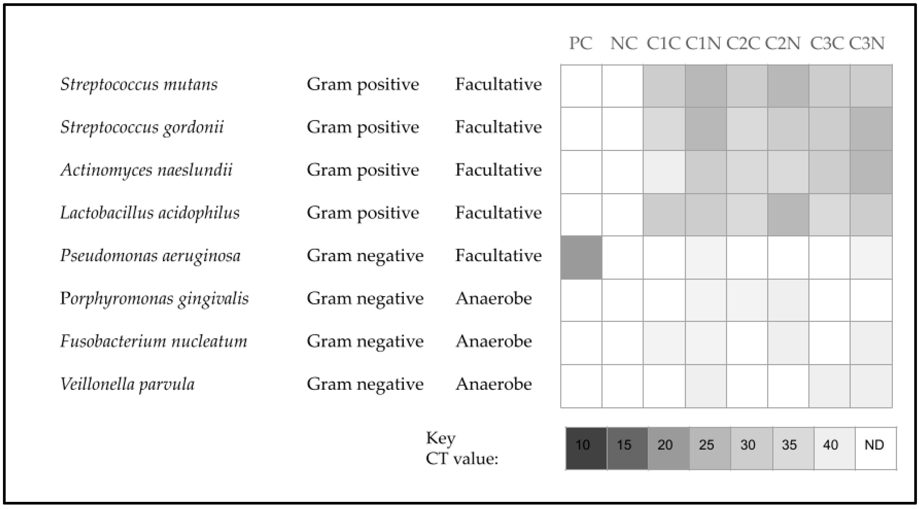
To identify oral bacteria present in the DNA isolated from each sample, qPCR) screening was performed using organism-specific primers (Figure 5). This analysis revealed the presence of each of the Gram-positive oral bacteria among all samples, including the commensal organism Streptococcus gordonii and the cariogenic pathogens Streptococcus mutans, Actinomyces naeslundii, and Lactobacillus acidophilus. The cycle threshold (CT) values obtained from this screening averaged 29.11 ± 0.51 (range 25.52 to 33.31). However, the Gram-negative bacteria were only found in subsets from each screening, such as Pseudomonas aeruginosa (C1 and C3 neck), Porphyromonas gingivalis (C1 neck, C2 cap and neck), Fusobacterium nucleatum (C2 neck, C3 neck), and Veillonella parvula (C1 neck, C3 cap and neck). In addition, the average CT count was much higher among the Gram-negatives with an average of 37.41 ± 0.62 (range 36.33 to 39.87).
Finally, swab cultures were assessed following 24 h of incubation to determine whether any of the bacteria identified in this screening were viable (Table 2). This analysis revealed that the turbidity readings for the negative control (0.21 ± 0.01) and NEW samples (0.23 ± 0.01) were similar and not statistically significant, p = 0.411. However, the IN USE samples exhibited significantly higher turbidity readings (0.32 ± 0.05) than either the negative control or the NEW samples, p = 0.012—although these were lower than the measurements observed with the positive controls (0.49 ± 0.01).

4. Discussion

The goal of this project was to assess and quantify microbial contamination (if any) in dispensers of reusable and resealable dental biomaterials commonly used in a public dental school clinic. These data revealed that all of the new or unused items from the clinic were found to have negligible amounts of DNA that were comparable to the negative controls (sterile swabs placed into sterile, autoclaved bacterial culturing medium), which may confirm that proper sterilization and disinfection protocols were followed in the administration of this investigation [25,26]. However, these results also revealed significant amounts of DNA were found among each of the “in use” or reusable items screened in this study.
More specifically, this study found that the neck and cap of all “in use” reusable items harbored detectable amounts of DNA, which corresponded with the subsequent 16S bacterial DNA screening. These data may suggest that at least some reusable dental clinic items may acquire environmental contamination during the short exposure that is needed to uncap and dispense each of the biomaterials evaluated [27,28]. Moreover, the fact that the neck had slightly higher levels of DNA contamination but was comparable to each of the corresponding caps may suggest that the environmental exposure could be facilitated by the contact of aerosolized bacteria with the dispenser while on the nearby instrument tray—types of contamination that have been observed with other types of dispensers and closures among other reusable biomaterials [29,30,31].
In addition, this study also revealed that the potential amount of exposure, based upon the estimated amount of use, correlated significantly with the amount of DNA contamination and the corresponding level of bacteria present from each sample. This information strongly suggests that bacterial contamination may be highly associated with repeated short-term exposure to environmental aerosols, which has been observed in other studies of bacterial contamination during routine dental procedures [32,33]. This information may suggest that immediate capping or alternative disinfection protocols may be needed to reduce the amount of potential bacterial contamination found among the dispensers of commonly used dental clinic biomaterials [34,35].
Other studies have highlighted potential sources of bacterial contamination and risks of biofilm formation and residues on other reusable dental materials [36,37]. For example, studies of dental implant abutments have demonstrated that bacterial biofilms and load may be related to surface morphology (roughness or protective grooves for bacteria) and biocompatibility (non-toxicity) of these reusable materials, which may help to explain why the findings of this study demonstrated bacterial presence within protective and sequestered areas of the screw cap and neck that are the areas exposed during utilization of these reusable dental materials [38,39]. In fact, additional research has found other dental office materials, such as toys and pediatric anxiety-reducing items, that were not traditionally associated with bacterial contamination but have been identified as potential sources of contamination and exposure risk [40,41]. These studies highlight the need to develop new protocols to limit the potential spread of oral organisms into the dental office environment by reducing the oral bacterial load prior to the start of any potential aerosol generating procedures, such as the implementation of extended mouth washing during the SARS-CoV-2 (COVID-19) pandemic that greatly reduced detectable viral load and also concomitantly reduced the presence of oral microbial pathogens [25,26,27,28,42,43,44]. Other effective strategies have also been implemented for disinfection and decontamination of additional reusable dental office materials, such as the use of disinfectant wipes [36,37,38,39,45].
Although these results may be among the first to demonstrate this type of contamination among reusable dental clinic biomaterials, there are several limitations implicit in this type of study that should be considered. First, this study was limited in scope and was not designed to be an exhaustive screening of all reusable items within the dental clinic. Therefore, there may be other dental office items that may harbor significant contamination that were not screened in this study. In addition, although only empty (no patient) operatories were used for the sampling protocol, there was no assessment of the types of procedures that were performed immediately prior to the use of each clinic item, which may have influenced the amount of environmental aerosols and contamination that were present [46,47]. Moreover, this initial pilot study was aimed at discovering whether any bacterial contamination might be present on these surfaces and assessed only a specific subset of oral bacterial species that could be present on these surfaces [48,49,50]. Future studies may wish to incorporate this type of screening along with a more comprehensive list of reusable dental materials to further investigate the extent of potential bacterial contamination among commonly used dental items, as well as a more comprehensive list of high-risk bacterial or fungal organisms.
The incorporation of these data into the planning of future studies would be of great benefit to the pediatric population, which may be at higher risk due to their young age and immature immune system responses [51,52]. Moreover, children with other health risks, including compromised immune systems and other chronic health conditions, may be particularly vulnerable to additional bacterial exposures that may be introduced through contaminated reusable dental materials [53,54,55]. For example, once information regarding potential pediatric bacterial exposures from impression materials and dental impression guns was published, protocols and guidelines for using steam-sterilization were recommended and implemented that have greatly reduced the potential for cross contamination and bacterial exposures among this patient population [56,57].

5. Conclusions

This study revealed potential sites for bacterial contamination among common reusable dental biomaterials, which also revealed strong associations between the amount of bacterial DNA and the amount of clinical usage. These data strongly suggest that methods for reducing bacterial contamination may need to be expanded to include these surfaces, which may reduce the potential for cross contamination between patients and ensure the highest levels of disinfection for the most effective prevention of patient exposures and highest standards of clinic office hygiene.

Author Contributions

Conceptualization, K.K. and V.S.; methodology, K.K. and S.J.W.; formal analysis, K.K. and G.B.; investigation, G.B., C.K. and C.W.; resources, S.J.W., K.K. and V.S.; data curation, G.B. and K.K.; writing—original draft preparation, G.B. and K.K.; writing—review and editing, G.B., C.K., C.W., S.J.W., K.K. and V.S. All authors have read and agreed to the published version of the manuscript.

Funding

This research received no external funding.

Institutional Review Board Statement

Not applicable—This study did not require ethical approval.

Informed Consent Statement

Not applicable.

Data Availability Statement

Data is contained within the article.

Acknowledgments

The authors wish to acknowledge the Department of Advanced Education in Pediatric Dentistry for their support of this project.

Conflicts of Interest

The authors declare no conflicts of interest.

Abbreviations

The following abbreviations are used in this manuscript:
qPCRQuantitative polymerase chain reaction
LBLuria–Bertani
ATCCAmerican Type Culture Collection
RT-PCRReal-Time Polymerase Chain Reaction

Appendix A

Table A1. DNA isolation raw data from sample collections and controls.
Sample, CollectionLocationEstimated UseNucleic Acid (ng/uL)A260/A280 Ratio
Negative controls
(C1, C2, C3)
N/ANot applicable0.568, 0.981, 0.1261.62, 1.54, 1.56
ave = 0.558 ± 0.43 ave = 1.58 ± 0.04
Positive controls
(C1, C2, C3)
N/ANot applicable1251.077, 1244.016, 1293.019 1.73, 1.53, 1.69
ave = 1262.704 ± 25.49ave = 1.65 ± 0.10
Diapex—NEW C1Cap, NeckEstimated use: 0%0.716, 0.9931.60, 1.67
Diapex—NEW C2Cap, NeckEstimated use: 0%0.615, 0.9391.85, 1.65
Diapex—NEW C3Cap, NeckEstimated use: 0%0.558, 0.6991.71, 1.61
ave = 0.753 ± 0.18ave = 1.68 ± 0.09
Diapex—IN USE C1Cap, NeckEstimated use: 20%253.36, 293.941.58, 1.57
Diapex—IN USE C2Cap, NeckEstimated use: 45%376.45, 397.561.66, 1.65
Diapex—IN USE C3Cap, NeckEstimated use: 50%300.99, 398.271.62, 1.62
ave = 336.76 ± 61.85ave = 1.62 ± 0.04
Flowable—NEW C1Cap, NeckEstimated use: 0%0.851, 0.5211.62, 1.64
Flowable—NEW C2Cap, NeckEstimated use: 0%0.313, 0.4211.75, 1.76
Flowable—NEW C3Cap, NeckEstimated use: 0%0.385, 0.6751.64, 1.65
ave = 0.5276 ± 0.20ave = 1.67 ± 0.06
Flowable—IN USE C1Cap, NeckEstimated use: 20%255.358, 264.221.94, 1.76
Flowable—IN USE C2Cap, NeckEstimated use: 80%603.275, 668.3041.69, 1.59
Flowable—IN USE C3Cap, NeckEstimated use: 80%722.407, 799.4451.76, 1.63
ave = 552.168 ± 235.47ave = 1.73 ± 0.13
BC Putty—NEW C1Cap, NeckEstimated use: 0%0.246, 0.4311.47, 1.48
BC Putty—NEW C2Cap, NeckEstimated use: 0%3.454, 3.7641.65, 1.49
BC Putty—NEW C3Cap, NeckEstimated use: 0%1.029, 1.6561.48, 1.66
ave = 1.763 ± 1.52ave = 1.54 ± 0.09
BC Putty—IN USE C1Cap, NeckEstimated use: 50%365.218, 421.811.70, 1.56
BC Putty—IN USE C2Cap, NeckEstimated use: 50%345.515, 361.8631.64, 1.44
BC Putty—IN USE C3Cap, NeckEstimated use: 80%735.484, 837.6331.57, 1.55
ave = 511.253 ± 217.11 ave = 1.58 ± 0.09
NeoPutty—NEW C1Cap, NeckEstimated use: 0%0.908, 0.5651.57, 1.54
NeoPutty—NEW C2Cap, NeckEstimated use: 0%1.516, 2.9681.50, 1.79
NeoPutty—NEW C3Cap, NeckEstimated use: 0%0.921, 1.6381.59, 1.62
ave = 1.419 ± 0.86ave = 1.60 ± 0.07
NeoPutty—IN USE C1Cap, NeckEstimated use: 40%352.523, 444.0191.75, 1.81
NeoPutty—IN USE C2Cap, NeckEstimated use: 20%212.296, 237.4041.69, 1.99
NeoPutty—IN USE C3Cap, NeckEstimated use: 50%411.098, 446.4991.68, 1.84
ave = 350.639 ± 103.47ave = 1.79 ± 0.11

References

  1. Byrne, D.; Saget, S.; Davidson, A.; Haneef, H.; Abdeldaim, T.; Almudahkah, A.; Basquille, N.; Bergin, A.M.; Prida, J.; Lyne, A.; et al. Comparing the environmental impact of reusable and disposable dental examination kits: A life cycle assessment approach. Br. Dent. J. 2022, 233, 317–325. [Google Scholar] [CrossRef]
  2. Paczkowski, I.; Stingu, C.S.; Hahnel, S.; Rauch, A.; Schierz, O. Cross-Contamination Risk of Dental Tray Adhesives: An In Vitro Study. Materials 2021, 14, 6138. [Google Scholar] [CrossRef]
  3. Naidu, S.; Dhawan, P.; Raman, S. Improving the Accuracy of Dental Impressions: A Study of Tray Adjustments and Materials. Eur. J. Prosthodont. Restor. Dent. 2025, 33, 49–56. [Google Scholar] [CrossRef]
  4. Patil, R.V.; Vijayraghavan, V.; Jadhav, M.; Jajoo, S.; Desai, S.; Jagtap, C. Comparison of Tensile Bond Strength of Addition Silicone with Different Custom Tray Materials Using Different Retentive Methods. J. Contemp. Dent. Pract. 2021, 22, 278–283. [Google Scholar]
  5. Schierz, O.; Müller, H.; Stingu, C.S.; Hahnel, S.; Rauch, A. Dental tray adhesives and their role as potential transmission medium for microorganisms. Clin. Exp. Dent. Res. 2021, 7, 829–832. [Google Scholar] [CrossRef]
  6. Chukwu, S.; Munn, A.; Wilson, J.C.; Ibrahim, H.; Gosling, D.; Love, R.M.; Bakr, M.M. Efficacy of an impression disinfectant solution after repeated use: An in vitro study. Heliyon 2023, 10, e23792. [Google Scholar] [CrossRef] [PubMed]
  7. Cruz Hondares, T.; Hao, X.; Zhao, Y.; Lin, Y.; Napierala, D.; Jackson, J.G.; Zhang, P. Antibacterial, biocompatible, and mineralization-inducing properties of calcium silicate-based cements. Int. J. Paediatr. Dent. 2024, 34, 843–852. [Google Scholar] [CrossRef] [PubMed]
  8. Pelepenko, L.E.; Saavedra, F.; Antunes, T.B.M.; Bombarda, G.F.; Gomes, B.P.F.A.; Zaia, A.A.; Camilleri, J.; Marciano, M.A. Physicochemical, antimicrobial, and biological properties of White-MTAFlow. Clin. Oral Investig. 2021, 25, 663–672. [Google Scholar] [CrossRef] [PubMed]
  9. Hamouda, I.M.; Ahmed, S.A. Effect of microwave disinfection on mechanical properties of denture base acrylic resin. J. Mech. Behav. Biomed. Mater. 2010, 3, 480–487. [Google Scholar] [CrossRef]
  10. AlSaiari, A.K.A.; Alonazi, M.S.; Alotaibi, N.M.; AlQahtani, H.; Alotaibi, W.M.; Yhyha, A.S.S.A.; Aldarorah, A.; Alaklabi, N.M.; Al-Obayli, T.A.S.; Alsaud, O.A.S.; et al. Bioengineering Innovations in Global Dental Infection Control: Applications and Adaptations in Clinical Settings. Int. Dent. J. 2025, 75, 2222–2238. [Google Scholar] [CrossRef]
  11. Nóbrega, M.T.C.; Bastos, R.T.D.R.M.; Mecenas, P.; de Toledo, I.P.; Richardson-Lozano, R.; Altabtbaei, K.; Flores-Mir, C. Aerosol generated by dental procedures: A scoping review. J. Evid. Based Med. 2021, 14, 303–312. [Google Scholar] [CrossRef]
  12. Innes, N.; Johnson, I.G.; Al-Yaseen, W.; Harris, R.; Jones, R.; Kc, S.; McGregor, S.; Robertson, M.; Wade, W.G.; Gallagher, J.E. A systematic review of droplet and aerosol generation in dentistry. J. Dent. 2021, 105, 103556. [Google Scholar] [CrossRef]
  13. Kumar, P.S.; Subramanian, K. Demystifying the mist: Sources of microbial bioload in dental aerosols. J. Periodontol. 2020, 91, 1113–1122. [Google Scholar] [CrossRef]
  14. Zemouri, C.; de Soet, H.; Crielaard, W.; Laheij, A. A scoping review on bio-aerosols in healthcare and the dental environment. PLoS ONE 2017, 12, e0178007. [Google Scholar] [CrossRef] [PubMed]
  15. Ghoneim, A.; Proaño, D.; Kaur, H.; Singhal, S. Aerosol-generating procedures and associated control/mitigation measures: Position paper from the Canadian Dental Hygienists Association and the American Dental Hygienists’ Association. Can. J. Dent. Hyg. 2024, 58, 48–63. [Google Scholar] [PubMed]
  16. Hung, C.C.U.; Costa, R.C.; Pereira, G.; Abdo, V.L.; Noronha, M.D.S.; Retamal-Valdes, B.; Bertolini, M.; Feres, M.; Shibli, J.A.; Barão, V.A.R.; et al. Oral microbial colonization on titanium and polyetheretherketone dental implant healing abutments: An in vitro and in vivo study. J. Prosthet. Dent. 2023, 133, 1545–1552. [Google Scholar] [CrossRef]
  17. Frenzel, N.; Maenz, S.; Sanz Beltrán, V.; Völpel, A.; Heyder, M.; Sigusch, B.W.; Lüdecke, C.; Jandt, K.D. Template assisted surface microstructuring of flowable dental composites and its effect on microbial adhesion properties. Dent. Mater. 2016, 32, 476–487. [Google Scholar] [CrossRef] [PubMed]
  18. Melo, P.; Barbosa, J.M.; Jardim, L.; Carrilho, E.; Portugal, J. COVID-19 Management in Clinical Dental Care. Part I: Epidemiology, Public Health Implications, and Risk Assessment. Int. Dent. J. 2021, 71, 251–262. [Google Scholar] [CrossRef]
  19. Dolev, E.; Eli, I.; Mashkit, E.; Grinberg, N.; Emodi-Perlman, A. Fluorescent Marker as a Tool to Improve Strategies to Control Contaminated Surfaces and Decrease Danger of Cross-Contamination in Dental Clinics, during and beyond the COVID-19 Pandemic. Int. J. Environ. Res. Public Health 2023, 20, 5229. [Google Scholar] [CrossRef]
  20. Miguita, L.; Martins-Chaves, R.R.; Geddes, V.E.V.; Mendes, S.D.R.; Costa, S.F.D.S.; Fonseca, P.L.C.; Menezes, D.; de Souza, R.M.; Queiroz, D.C.; Alves, H.J.; et al. Biosafety in Dental Health Care During the COVID-19 Pandemic: A Longitudinal Study. Front. Oral Health 2022, 3, 871107. [Google Scholar] [CrossRef]
  21. Emett, J.; David, R.; McDaniel, J.; McDaniel, S.; Kingsley, K. Comparison of DNA Extracted from Pediatric Saliva, Gingival Crevicular Fluid and Site-Specific Biofilm Samples. Methods Protoc. 2020, 3, 48. [Google Scholar] [CrossRef]
  22. Pearson, M.; Stewart, S.; Ma, L.; Kingsley, K.; Sullivan, V. Differential Antimicrobial Effects of Endodontic Irrigant Endocyn on Oral Bacteria. Hygiene 2025, 5, 11. [Google Scholar] [CrossRef]
  23. Wilson, J.; Swanbeck, S.; Banning, G.; Alhwayek, T.; Sullivan, V.; Howard, K.M.; Kingsley, K. Assessment of Sodium Diamine Fluoride (SDF) with Light Curing Technique: A Pilot Study of Antimicrobial Effects. Methods Protoc. 2022, 5, 31. [Google Scholar] [CrossRef] [PubMed]
  24. Gamble, T.; Wilkerson, C.; Kim, C.; Kingsley, K.; Sullivan, V. Assessment of Trends in Non-Restorative and Preventative Dental Treatment Pre- and Post-COVID-19: A Health Informatics Pilot Study. Children 2025, 12, 357. [Google Scholar] [CrossRef] [PubMed]
  25. Patiño-Marín, N.; Villa García, L.D.; Aguirre López, E.C.; Medina-Solís, C.E.; Martínez Zumarán, A.; Martínez Rider, R.; Márquez Preciado, R.; Rosales García, P.; Salas Orozco, M.F. Sterilization and Disinfection: Ensuring Infection Control in Dental Practices. Cureus 2025, 17, e79041. [Google Scholar] [CrossRef] [PubMed]
  26. Bharti, B.; Li, H.; Ren, Z.; Zhu, R.; Zhu, Z. Recent advances in sterilization and disinfection technology: A review. Chemosphere 2022, 308 Pt 3, 136404. [Google Scholar] [CrossRef]
  27. Klumdeth, J.; Jantaratnotai, N.; Thaweboon, S.; Pachimsawat, P. Sterility maintenance of reused disposable paper/plastic sterilization pouches in actual clinical practice. Heliyon 2020, 6, e03672. [Google Scholar] [CrossRef]
  28. Venkatasubramanian, R.; Jayanthi; Das, U.M.; Bhatnagar, S. Comparison of the effectiveness of sterilizing endodontic files by 4 different methods: An in vitro study. J. Indian Soc. Pedod. Prev. Dent. 2010, 28, 2–5. [Google Scholar] [CrossRef]
  29. Brannan, D.K.; Dille, J.C. Type of closure prevents microbial contamination of cosmetics during consumer use. Appl. Environ. Microbiol. 1990, 56, 1476–1479. [Google Scholar] [CrossRef]
  30. Coad, C.T.; Osato, M.S.; Wilhelmus, K.R. Bacterial contamination of eyedrop dispensers. Am. J. Ophthalmol. 1984, 98, 548–551. [Google Scholar] [CrossRef]
  31. Høvding, G.; Sjursen, H. Bacterial contamination of drops and dropper tips of in-use multidose eye drop bottles. Acta Ophthalmol. 1982, 60, 213–222. [Google Scholar] [CrossRef]
  32. Franz, J.; Scheier, T.C.; Aerni, M.; Gubler, A.; Schreiber, P.W.; Brugger, S.D.; Schmidlin, P.R. Bacterial contamination of air and surfaces during dental procedures-An experimental pilot study using Staphylococcus aureus. Infect. Control Hosp. Epidemiol. 2024, 45, 658–663. [Google Scholar] [CrossRef]
  33. Wang, B.Y.; Henrichs, L.E.; Arricale, K.; Lien, W.; Savett, D.A.; Vandewalle, K.S. Efficacy of a chairside extraoral suction system in the reduction of aerosol contamination. Gen. Dent. 2023, 71, 16–21. [Google Scholar]
  34. Kumbargere Nagraj, S.; Eachempati, P.; Paisi, M.; Nasser, M.; Sivaramakrishnan, G.; Verbeek, J.H. Interventions to reduce contaminated aerosols produced during dental procedures for preventing infectious diseases. Cochrane Database Syst. Rev. 2020, 10, CD013686. [Google Scholar] [CrossRef] [PubMed]
  35. Boccia, G.; Di Spirito, F.; D’Ambrosio, F.; De Caro, F.; Pecora, D.; Giorgio, R.; Fortino, L.; Longanella, W.; Franci, G.; Santella, B.; et al. Microbial Air Contamination in a Dental Setting Environment and Ultrasonic Scaling in Periodontally Healthy Subjects: An Observational Study. Int. J. Environ. Res. Public Health 2023, 20, 2710. [Google Scholar] [CrossRef] [PubMed]
  36. Triggiano, F.; Veschetti, E.; Veneri, F.; Montagna, M.T.; De Giglio, O. Best practices for disinfection in dental settings: Insights from Italian and European regulations. J. Ann. Ig. 2025, 37, 292–301. [Google Scholar] [CrossRef]
  37. Barreiros, P.; Braga, J.; Faria-Almeida, R.; Coelho, C.; Teughels, W.; Souza, J.C.M. Remnant oral biofilm and microorganisms after autoclaving sterilization of retrieved healing abutments. J. Periodontal Res. 2021, 56, 415–422. [Google Scholar] [CrossRef]
  38. Sharon, E.; Pietrokovski, Y.; Engel, I.; Assali, R.; Houri-Haddad, Y.; Beyth, N. Biocompatibility, Surface Morphology, and Bacterial Load of Dental Implant Abutments following Decontamination Protocols: An In-Vitro Study. Materials 2023, 16, 4080. [Google Scholar] [CrossRef]
  39. Gul, M.; Zafar, K.; Ghafoor, R.; Khan, F.R. Frequency of Contamination on Used Healing Abutments after Sterilization: An In Vitro Study. Int. J. Prosthodont. 2024, 37, 109. [Google Scholar] [CrossRef]
  40. Hardy, A.; Sabatier, V.; Rosello, O.; Salauze, B.; Barbut, F.; Vialle, R. More than just teddy bears: Unconventional transmission agents in the operating room. Arch. Pediatr. 2018, 25, 416–420. [Google Scholar] [CrossRef]
  41. Held, M.; Mignemi, M.; O’Rear, L.; Wise, M.; Zane, G.; Murphy Zane, M.S.; Schoenecker, J.G. Stuffed Animals in the Operating Room: A Reservoir of Bacteria with a Simple Solution. J. Pediatr. Orthop. 2015, 35, e110–e112. [Google Scholar] [CrossRef]
  42. Marui, V.C.; Souto, M.L.S.; Rovai, E.S.; Romito, G.A.; Chambrone, L.; Pannuti, C.M. Efficacy of preprocedural mouthrinses in the reduction of microorganisms in aerosol: A systematic review. J. Am. Dent. Assoc. 2019, 150, 1015–1026.e1. [Google Scholar] [CrossRef] [PubMed]
  43. Shayegh, M.; Sorenson, C.; Downey, J.; Lin, S.; Jiang, Y.; Sodhi, P.; Sullivan, V.; Howard, K.M.; Kingsley, K. Assessment of SARS-CoV-2 (COVID-19) Clinical Mouthwash Protocol and Prevalence of the Oral Pathogen Scardovia wiggsiae: A Pilot Study of Antibacterial Effects. Methods Protoc. 2023, 6, 65. [Google Scholar] [CrossRef] [PubMed]
  44. Sodhi, P.; Jiang, Y.; Lin, S.; Downey, J.; Sorenson, C.; Shayegh, M.; Sullivan, V.; Kingsley, K.; Howard, K.M. Administration of Clinical COVID-19 Mouthwashing Protocol and Potential Modulation of Pediatric Oral Bacterial Prevalence of Selenomonas noxia: A Pilot Study. Pediatr. Rep. 2023, 15, 414–425. [Google Scholar] [CrossRef] [PubMed]
  45. Saini, R.S.; Binduhayyim, R.I.H.; Mosaddad, S.A.; Heboyan, A. Strategies for preventing aerosol-generated microbial contamination in dental procedures: A systematic review and meta-analysis. Am. J. Infect. Control. 2025, 53, 638–647. [Google Scholar] [CrossRef] [PubMed]
  46. Engsomboon, N.; Pachimsawat, P.; Thanathornwong, B. Comparative Dissemination of Aerosol and Splatter Using Suction Device during Ultrasonic Scaling: A Pilot Study. Dent. J. 2022, 10, 142. [Google Scholar] [CrossRef]
  47. Puljich, A.; Jiao, K.; Lee, R.S.B.; Walsh, L.J.; Ivanovski, S.; Han, P. Simulated and clinical aerosol spread in common periodontal aerosol-generating procedures. Clin. Oral Investig. 2022, 26, 5751–5762. [Google Scholar] [CrossRef]
  48. Melzow, F.; Mertens, S.; Todorov, H.; Groneberg, D.A.; Paris, S.; Gerber, A. Aerosol exposure of staff during dental treatments: A model study. BMC Oral Health 2022, 22, 128. [Google Scholar] [CrossRef]
  49. Yang, M.; Chaghtai, A.; Melendez, M.; Hasson, H.; Whitaker, E.; Badi, M.; Sperrazza, L.; Godel, J.; Yesilsoy, C.; Tellez, M.; et al. Mitigating saliva aerosol contamination in a dental school clinic. BMC Oral Health 2021, 21, 52. [Google Scholar] [CrossRef]
  50. Gonçalves Lomardo, P.; Nunes, M.C.; Arriaga, P.; Antunes, L.A.; Machado, A.; Quinelato, V.; Aguiar, T.R.D.S.; Casado, P.L. Concern about the risk of aerosol contamination from ultrasonic scaler: A systematic review and meta-analysis. BMC Oral Health 2024, 24, 417. [Google Scholar] [CrossRef]
  51. Hajishengallis, G.; Lamont, R.J.; Koo, H. Oral polymicrobial communities: Assembly, function, and impact on diseases. Cell Host Microbe 2023, 31, 528–538. [Google Scholar] [CrossRef]
  52. Lamont, R.J.; Hajishengallis, G.; Koo, H. Social networking at the microbiome-host interface. Infect. Immun. 2023, 91, e0012423. [Google Scholar] [CrossRef]
  53. Pittet, L.F.; Posfay-Barbe, K.M. Vaccination of immune compromised children-an overview for physicians. Eur. J. Pediatr. 2021, 180, 2035–2047. [Google Scholar] [CrossRef]
  54. Aranda, C.S.; Gouveia-Pereira, M.P.; da Silva, C.J.M.; Rizzo, M.C.F.V.; Ishizuka, E.; de Oliveira, E.B.; Condino-Neto, A. Severe combined immunodeficiency diagnosis and genetic defects. Immunol. Rev. 2024, 322, 138–147. [Google Scholar] [CrossRef]
  55. Le, T.T.; Hoang, T.N.; Do, D.H.; Nguyen, X.H.; Huynh, C.; Viet, H.D.; Dat, V.Q.; Zengler, K.; Gilbert, J.A.; Avedissian, S.N.; et al. Current state of microbiota clinical applications in neonatal and pediatric bacterial infections. Gut Microbes 2025, 17, 2529400. [Google Scholar] [CrossRef]
  56. Westergard, E.J.; Romito, L.M.; Kowolik, M.J.; Palenik, C.J. Controlling bacterial contamination of dental impression guns. J. Am. Dent. Assoc. 2011, 142, 1269–1274. [Google Scholar] [CrossRef]
  57. Rice, C.D.; Dykstra, M.A.; Gier, R.E. Bacterial contamination in irreversible hydrocolloid impression material and gingival retraction cord. J. Prosthet. Dent. 1991, 65, 496–499. [Google Scholar] [CrossRef] [PubMed]
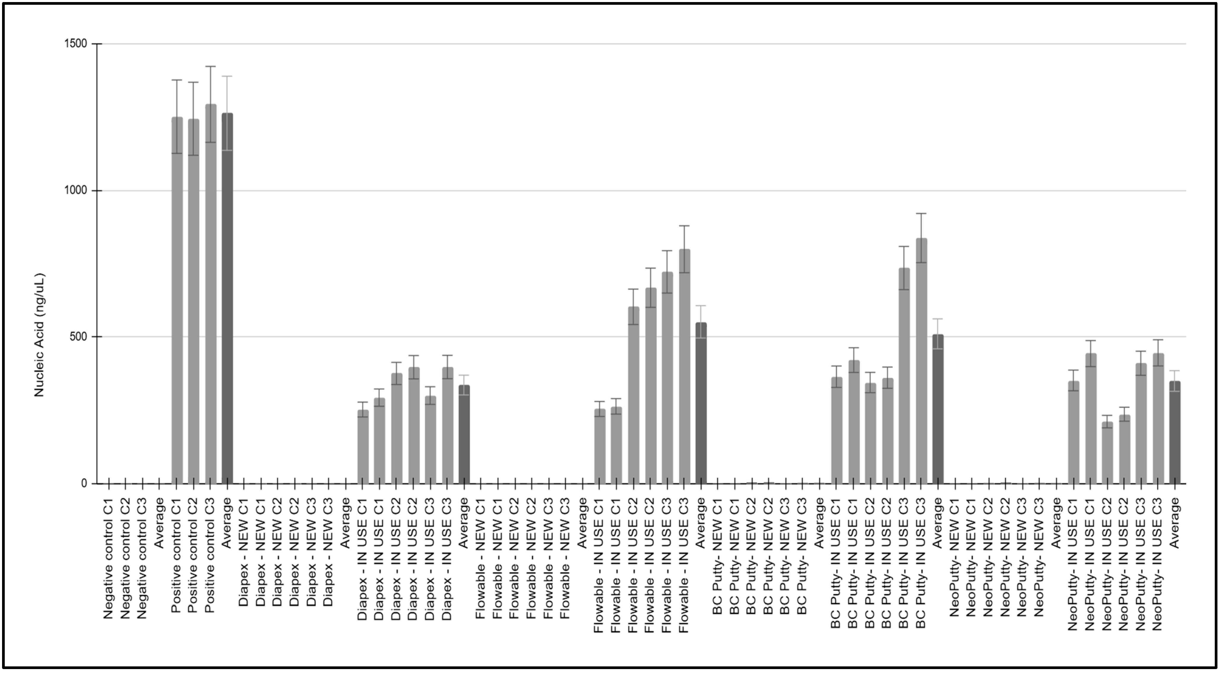
Figure 1. DNA isolation data from all study samples. Negligible amounts of DNA were detected from the negative controls (average 0.56 ng/uL ± 0.43) and NEW clinic samples, including Diapex (average 0.75 ng/uL ± 0.18), Flowable (average 0.53 ng/uL ± 0.20), BC Putty (average 1.76 ng/uL ± 1.52), and NeoPutty (average 1.42 ng/uL ± 0.07). Higher DNA levels among all IN USE items, including Diapex (average 336.76 ng/uL ± 61.85) Flowable (average 552.17 ng/uL ± 235.47) BC Putty (average 511.25 ng/uL ± 217.11) and NeoPutty (average 350.64 ng/uL ± 103.47).
Hygiene 05 00042 g001
Figure 2. Analysis of differences between the DNA isolated from the Cap and Neck. Average DNA isolated from the Cap (average 411.16 ng/uL ± 178.60) was lower than the Neck (average 464.25 ng/uL ± 190.62), although this was not significant, p = 0.260. Similar trends were observed with Diapex IN USE Cap versus Neck (average 310.27 ng/uL ± 62.07 and 363.26 ng/uL ± 60.03 ng/uL, p = 0.174) Flowable IN USE Cap versus Neck (average 527.01 ng/uL ± 242.68 and 577.32 ng/uL ± 278.97, p = 0.391), BC Putty IN USE Cap versus Neck (average 482.07 ng/uL ± 219.68 and 540.44 ng/uL ± 259.12, p = 0.391) and NeoPutty IN USE Cap versus Neck (average 325.31 ng/uL ± 102.16 and 375.97 ng/uL ± 120.01, p = 0.304).
Hygiene 05 00042 g002
Figure 3. Analysis of DNA from clinic items at different estimated stages of use. The levels of DNA found among the items with an estimated use of approximately 20% (average 252.76 ng/uL ± 27.22) were significantly lower than those with an estimated use of approximately 40–50% (average 385.15 ng/uL ± 42.97, p = 0.000001), as well as those at an estimated use of approximately 80% (average 727.76 ng/uL ± 85.24, p = 0.000001).
Hygiene 05 00042 g003
Figure 4. Quantitative polymerase chain reaction (qPCR) screening for bacterial 16S rRNA. IN USE samples (estimated 20%) exhibited the highest average cycle threshold (CT) count of 35.35 ± 0.45, compared with samples of estimated usage of 40–50% (average CT 27.55 ± 0.75, 80% (average CT 25.01 ± 0.61), and the positive control standards (average CT 14.93 ± 0.17).
Hygiene 05 00042 g004
Figure 5. Oral bacteria specific qPCR screening heatmap. Gram-positive oral species, including Streptococcus gordonii, Streptococcus mutans, Actinomyces naeslundii, and Lactobacillus acidophilus were present among all samples with cycle threshold (CT) values averaging 29.11 ± 0.51. Gram-negative bacteria were present only among subsets from each screening, including Pseudomonas aeruginosa (C1 C3), Porphyromonas gingivalis (C1, C2), Fusobacterium nucleatum (C2, C3), and Veillonella parvula (C1, C3) with average CT values much higher 37.41 ± 0.62. PC = positive control, NC = negative control, C1C (collection one cap), C1N (collection one neck), C2C (collection two cap), C2N (collection two neck), C3C (collection three cap), C3N (collection three neck).
Hygiene 05 00042 g005
Table 1. Analysis and summary of DNA isolation from sample collections and controls.
Sample, CollectionLocationEstimated UseNucleic Acid (ng/uL)A260/A280 Ratio
Negative controls
(C1, C2, C3)
N/ANot applicableave = 0.56 ± 0.43ave = 1.58 ± 0.04
Positive controls
(C1, C2, C3)
N/ANot applicableave = 1262.70 ± 25.49ave = 1.65 ± 0.10
Diapex—NEW C1,C2,C3Cap, NeckEstimated use: 0%ave = 0.75 ± 0.18ave = 1.68± 0.09
Diapex—IN USE C1,C2,C3Cap, NeckEstimated use: 20%, 45%, 50%ave = 336.76 ± 61.85ave = 1.62 ± 0.04
Flowable—NEW C1,C2,C3Cap, NeckEstimated use: 0%ave = 0.53 ± 0.20ave = 1.67 ± 0.06
Flowable—IN USE C1,C2,C3Cap, NeckEstimated use: 20%, 80%, 80%ave = 552.17 ± 235.47ave = 1.73 ± 0.13
BC Putty- NEW C1,C2,C3Cap, NeckEstimated use: 0%ave = 1.76 ± 1.52ave = 1.54 ± 0.09
BC Putty- IN USE C1,C2,C3Cap, NeckEstimated use: 50%, 50%, 80%ave = 511.25 ± 217.11ave = 1.58 ± 0.09
NeoPutty- NEW C1,C2,C3Cap, NeckEstimated use: 0%ave = 1.42 ± 0.86ave = 1.60 ± 0.07
NeoPutty- IN USE C1,C2,C3Cap, NeckEstimated use: 40%, 20%, 50%ave = 350.64 ± 103.47ave = 1.79 ± 0.11
Table 2. Turbidity readings for sample collections and controls.
SampleTurbidityRangeStatistical Analysis
Negative controls 0.21 ± 0.010.20 to 0.23Two-tailed t-test
NEW samples0.23 ± 0.010.21 to 0.23p = 0.411
IN USE samples0.32 ± 0.050.26 to 0.37p = 0.012
Positive controls0.49 ± 0.010.49 to 0.51
Disclaimer/Publisher’s Note: The statements, opinions and data contained in all publications are solely those of the individual author(s) and contributor(s) and not of MDPI and/or the editor(s). MDPI and/or the editor(s) disclaim responsibility for any injury to people or property resulting from any ideas, methods, instructions or products referred to in the content.